Polycyclic Aromatic Hydrocarbons a Constituent of Petroleum: Presence and Influence in the Aquatic Environment
Daniela M. Pampanin and Magne O. Sydnes
Additional information is available at the end of the chapter
http://dx.doi.org/10.5772/48176
1. Introduction
Crude oil is a complex mixture of hydrocarbons containing more than 17000 compounds [1]. Among the constituents of crude oil there is a group of substances called polycyclic aromatic hydrocarbons (PAHs). PAHs are aromatic compounds containing from two to eight conjugated ring systems. They can have a range of substituents such as alkyl, nitro, and amino groups in their structure [2]. Nitrogen, sulfur, and oxygen atoms can also be incorporated into their ring system [2,3]. The precursors for PAHs found in crude oil are natural products, such as steroids, that have been chemically converted to aromatic hydrocarbons over time [4].
The PAHs that are present in the marine environment in relevant concentrations are divided into two groups depending on their origin, namely pyrogenic and petrogenic [5]. Pyrogenic PAHs are formed by incomplete combustion of organic material while the petrogenic PAHs are present in oil and some oil products [4,6,7]. In general the pyrogenic PAHs are composed of larger ring systems then the petrogenic PAHs. Sources for pyrogenic PAHs are forest fires [6,7,8], incomplete combustion of fossil fuels [6,7,8], and tobacco smoke [6,7]. A range of PAHs are naturally present in crude oil [4,9,10] and coal [10,11] and these compounds are referred to as petrogenic PAHs. In the costal zones PAHs enters the water primarily from sewage, runoff from roads [12], the smelter industry [13,14,15] and oil spills [16,17], while offshore PAHs chiefly enter the water through oil seeps [18], oil spills [16], and produced water discharge from offshore oil installations [19].
Hydrocarbon 84
2. Oil as a source of polycyclic aromatic hydrocarbons to the aquatic environment
Hydrocarbons in the form of crude oil, and therefore also PAHs, have and are entering the environment naturally through oil seeps, which is oil leaking naturally from oil reservoirs. Oil seeps are found scattered all over the globe with a higher concentration in certain regions of the world [18]. Numerous times the presence of a natural oil seep has resulted in the discovery of oil reservoirs that are large enough for commercial oil production [20], however, the presence of an oil seep does not guarantee the discovery of a production worthy oil reservoir [21]. Oil seeps vary in size with macroseeps resulting in visible oil slicks on the water surface, when the oil seep is situated on the seafloor, and microseeps that are invisible at the surface [22]. Estimates for the world-wide seepage rate vary between 0.02-2.0 x 10⁶ tons per year with the most realistic estimate being 0.2 x 10⁶ tons per year [23]. The presence of natural oil seeps also results in local presence of hydrocarbon-eating microorganisms [24,25], a fact that gives these regions an advantage in the case of an accidental oil spill [24,26]. For example resent research showed that the presence of oil eating microorganisms due to natural oil seeps in the Gulf of Mexico in addition to favorable water currents resulted in a quicker degradation of oil in the region after the Deepwater Horizon accident than otherwise would be expected [26,27]. As expected, it is the lighter fractions, viz. short chain alkanes, of the oil that are first degraded by microorganisms [28]. The easily accessible energy source, short chain alkanes, results in an explosion like increase in the number of the oil degrading microbes. After some time these microorganisms also start degrading the more complex molecules such as long chain alkanes and aromatic compounds like PAHs [29]. A range of PAH degrading bugs has been found naturally in the environment [30-34]. These microorganisms have been isolated, sequenced and studied extensively in the laboratory, and their mechanism of degrading PAHs is fairly well understood [29].
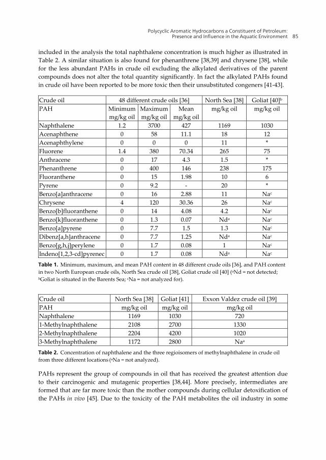
For monitoring purposes The US Environmental Protection Agency has made a list of 16 unsubstituted PAHs that are on a priority pollutant list [35]. These PAHs are usually referred to as the EPA 16 PAHs (Figure 1) and are the PAHs most commonly analyzed for. The concentration of these 16 PAHs and other PAHs, and the ratios between the various compounds differ from oil to oil [36,37]. This fact is quite clearly presented in the work of Kerr et al. where they have analyzed the concentration of the prioritized 16 PAHs in 48 crude oils from around the globe (North America, South America, Africa, and Asia) [36]. Their results are summarized in Table 1 and show an enormous variation of the content of the various PAHs in crude oil from different sites. Crude oil from the North Sea is reported to have a PAH concentration of 0.83% [38] while for example crude oil that leaked out of Exxon Valdez (referred to as Exxon Valdez crude oil hereafter) had a PAH content of 1.47% [39].
Naphthalene, one of the 16 EPA PAHs, is present in the highest concentration in crude oil, nevertheless this quantity does not give the full details of the naphthalene content of the oil. In fact, if the content of methylated naphthalene, a normal constituents of crude oil, is also
Polycyclic Aromatic Hydrocarbons a Constituent of Petroleum: Presence and Influence in the Aquatic Environment 85
included in the analysis the total naphthalene concentration is much higher as illustrated in Table 2. A similar situation is also found for phenanthrene [38,39] and chrysene [38], while for the less abundant PAHs in crude oil excluding the alkylated derivatives of the parent compounds does not alter the total quantity significantly. In fact the alkylated PAHs found in crude oil have been reported to be more toxic then their unsubstituted congeners [41-43].
Crude oil 48 different crude oils [36] North Sea [38] Goliat [40]b
Table 1. Minimum, maximum, and mean PAH content in 48 different crude oils [36], and PAH content in two North European crude oils, North Sea crude oil [38], Goliat crude oil [40] (aNd = not detected; bGoliat is situated in the Barents Sea; cNa = not analyzed for).
Table 2. Concentration of naphthalene and the three regioisomers of methylnaphthalene in crude oil from three different locations (aNa = not analyzed).
PAHs represent the group of compounds in oil that has received the greatest attention due to their carcinogenic and mutagenic properties [38,44]. More precisely, intermediates are formed that are far more toxic than the mother compounds during cellular detoxification of the PAHs in vivo [45]. Due to the toxicity of the PAH metabolites the oil industry in some
Hydrocarbon 86
areas of the world (e.g. North Sea, Mediterranean Sea, Australian Northwest Shelf, Gulf of Mexico) are required to monitor their discharges to the aquatic environment [46-49].In the North Sea this is taken care of through the Water Column Monitorin programs [48]. For monitoring purposes the 16 EPA compounds has been chosen as the most important PAHs to analyze for.
Figure 1. Chemical structure of the EPA selected 16 PAHs.
Unfortunately, accidental oil spills take place from time to time, most recently the Deapwater Horizon accident in the Mexican Gulf in 2010 (April 20th). This incident resulted in the release of 779 million liters of crude oil to the Gulf [50]. The Exxon Valdez oil spill in Prince William Sound, Alaska, USA in 1989 (March 24th), which took place after the oil tanker ran ashore, resulted in the release of 42 million liters of crude oil [16]. In European waters the Erika accident in 1999 (December 12th) released 18000 tons of crude oil into French coastal waters and in Spain Prestige spilt 60000 tons of heavy fuel oil into the waters outside of Galicia in 2002 (November 13th) [17]. These are unfortunately only a few examples of accidental release of large quantities of oil to the aquatic environment. The environmental consequences of the Exxon Valdez spill is the most studied oil spill ever [51,52], however, the influence of the Deapwater Horizon oil release will probably be just as well studied, or even more studied [53].
3. Produced water as a source of polycyclic aromatic hydrocarbons to the aquatic environment
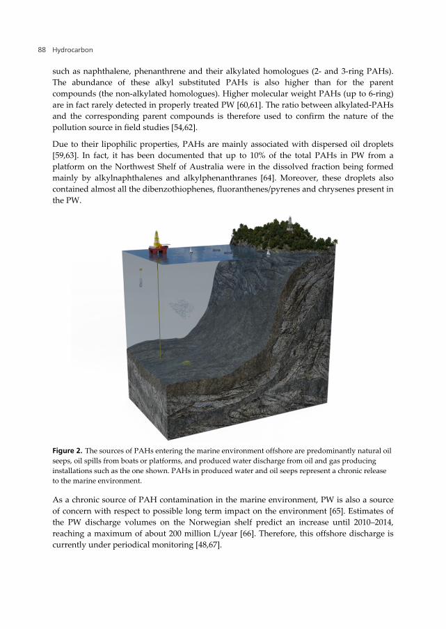
Oil production offshore (Figure 2) (and onshore) also results in the production of large volumes of water, so called produced water (PW), in addition to crude oil. PAHs contained in
Polycyclic Aromatic Hydrocarbons a Constituent of Petroleum: Presence and Influence in the Aquatic Environment 87
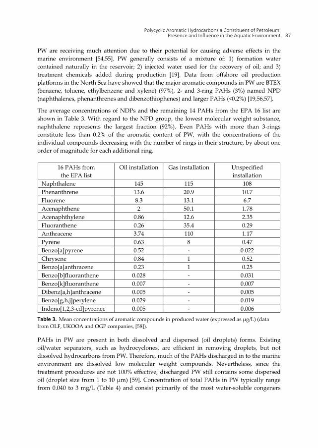
PW are receiving much attention due to their potential for causing adverse effects in the marine environment [54,55]. PW generally consists of a mixture of: 1) formation water contained naturally in the reservoir; 2) injected water used for the recovery of oil; and 3) treatment chemicals added during production [19]. Data from offshore oil production platforms in the North Sea have showed that the major aromatic compounds in PW are BTEX (benzene, toluene, ethylbenzene and xylene) (97%), 2- and 3-ring PAHs (3%) named NPD (naphthalenes, phenanthrenes and dibenzothiophenes) and larger PAHs (<0.2%) [19,56,57].
The average concentrations of NDPs and the remaining 14 PAHs from the EPA 16 list are shown in Table 3. With regard to the NPD group, the lowest molecular weight substance, naphthalene represents the largest fraction (92%). Even PAHs with more than 3-rings constitute less than 0.2% of the aromatic content of PW, with the concentrations of the individual compounds decreasing with the number of rings in their structure, by about one order of magnitude for each additional ring.
16 PAHs from the EPA list
Oil installation Gas installation Unspecified installation
Table 3. Mean concentrations of aromatic compounds in produced water (expressed as µg/L) (data from OLF, UKOOA and OGP companies, [58]).
PAHs in PW are present in both dissolved and dispersed (oil droplets) forms. Existing oil/water separators, such as hydrocyclones, are efficient in removing droplets, but not dissolved hydrocarbons from PW. Therefore, much of the PAHs discharged in to the marine environment are dissolved low molecular weight compounds. Nevertheless, since the treatment procedures are not 100% effective, discharged PW still contains some dispersed oil (droplet size from 1 to 10 µm) [59]. Concentration of total PAHs in PW typically range from 0.040 to 3 mg/L (Table 4) and consist primarily of the most water-soluble congeners
Hydrocarbon 88
such as naphthalene, phenanthrene and their alkylated homologues (2- and 3-ring PAHs). The abundance of these alkyl substituted PAHs is also higher than for the parent compounds (the non-alkylated homologues). Higher molecular weight PAHs (up to 6-ring) are in fact rarely detected in properly treated PW [60,61]. The ratio between alkylated-PAHs and the corresponding parent compounds is therefore used to confirm the nature of the pollution source in field studies [54,62].
Due to their lipophilic properties, PAHs are mainly associated with dispersed oil droplets [59,63]. In fact, it has been documented that up to 10% of the total PAHs in PW from a platform on the Northwest Shelf of Australia were in the dissolved fraction being formed mainly by alkylnaphthalenes and alkylphenanthranes [64]. Moreover, these droplets also contained almost all the dibenzothiophenes, fluoranthenes/pyrenes and chrysenes present in the PW.
Figure 2. The sources of PAHs entering the marine environment offshore are predominantly natural oil seeps, oil spills from boats or platforms, and produced water discharge from oil and gas producing installations such as the one shown. PAHs in produced water and oil seeps represent a chronic release to the marine environment.
As a chronic source of PAH contamination in the marine environment, PW is also a source of concern with respect to possible long term impact on the environment [65]. Estimates of the PW discharge volumes on the Norwegian shelf predict an increase until 2010–2014, reaching a maximum of about 200 million L/year [66]. Therefore, this offshore discharge is currently under periodical monitoring [48,67].
Polycyclic Aromatic Hydrocarbons a Constituent of Petroleum: Presence and Influence in the Aquatic Environment 89
Table 4. Concentrations of individual PAHs or alky congener groups in produced water from various areas (expressed as µg/L) (ND = not detected) (from [61]).
Hydrocarbon 90
4. Environmental monitoring of polycyclic aromatic hydrocarbons
There are various environmental monitoring methods which may be performed in order to assess risks of PAH contamination for organisms and to classify the environmental quality of ecosystems. Five approaches are hereby reported: 1) chemical monitoring: exposure assessment by measuring levels of a selected set of compounds in abiotic environmental compartments; 2) bioaccumulation monitoring: exposure assessment by measuring PAH levels in biota or determining the critical dose at a critical site (bioaccumulation); 3) biological effect monitoring: exposure and effect assessment by determining the early adverse alterations that are partly or fully reversible (biomarkers); 4) health monitoring: effect assessment by examining the occurrence of irreversible diseases or tissue damage in organisms; 5) ecosystem monitoring: assessment of the integrity of an ecosystem by making an inventory of, for instance, species composition, density and diversity [68-70]. All these methods are currently in use to monitor the aquatic environment contamination from PAH compounds.
Since the occurrence and abundance of PAHs in aquatic environments represent a risk to aquatic organisms and ultimately to humans (through fish and shellfish consumption), there is a constant need for their determination and quantification around the world [71]. The monitoring of PAH presence in the aquatic environment is therefore a world-wide activity. Since some of these compounds are well known carcinogens and mutagens [44,72], this contaminant class has been generally regarded as high priority for environmental pollution monitoring. In fact, the European Union included these pollutants in the list of priority hazardous substances for surface waters in the Water Framework Directive 2000/60/EC [73]. Moreover, for PAH content in biota several guidelines exist: the commission regulation (EC 2005) which stipulates a maximum concentration of 10 µg/kg (w/w) of benzo[a]pyrene in edible molluscs (this regulation also limits the benzo[a]pyrene concentration in other alimentary products), the OSPAR Commission which has developed eco-toxicological assessment criteria (i.e., concentrations levels above which concern is indicated) for different PAHs in fish and mussels [74], and the Oregon Health Division which has derived risk-based criteria for PAHs [75] assessing the risk in terms of benzo[a]pyrene equivalents.
Many studies have been carried out to determine the PAH distribution in marine organisms in different geographical areas using different approaches. Different authors have reported the presence of PAHs in waters, marine organisms, and sediments using chemical and biological markers [76-78]. Current monitoring techniques employed to determine environmental quality include the chemical analyses of sediment and water samples for determining the concentration of PAH parent compounds. PAHs are sparingly soluble in water and are difficult to detect although they show a much greater association with sediments. In fact, due to their hydrophobic character, these compounds rapidly tend to become associated with particles and end up in the sediments which act as a sink for them [71,79,80]. High concentrations of pyrogenic PAH mixtures in sediment samples have been found in several freshwater, estuarine and marine regions with heavy vessel traffic or at locations with PAH-containing effluents from industrial areas [81]. Finally, chemical
Polycyclic Aromatic Hydrocarbons a Constituent of Petroleum: Presence and Influence in the Aquatic Environment 91
analyses of these materials are laborious and costly due to the lengthy extraction methodology needed. Since monitoring of a large number of PAHs is expensive, time consuming and analytically demanding, a selection of compounds could be monitored to give an indication of the overall contamination.
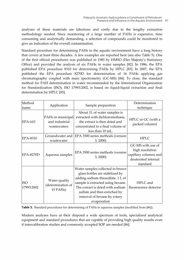
Standard procedure for determining PAHs in the aquatic environment have a long history that covers at least three decades. A few examples are reported here (see also Table 5). One of the first official procedures was published in 1985 by HMSO (Her Majesty’s Stationery Office) and provided the analysis of six PAHs in water samples [82]. In 1986, the EPA published EPA procedure 8310 for determining PAHs by HPLC [83]. In 2007, the EPA published the EPA procedure 8270D for determination of 16 PAHs applying gas chromatography coupled with mass spectrometry (GC-MS) [84]. To close, the standard method for PAH determination in water recommended by the International Organization for Standardization (ISO), ISO 17993:2002, is based on liquid-liquid extraction and final determination by HPLC [85].
Method name
Application Sample preparation Determination
technique
EPA-610 PAHs in municipal
and industrial wastewaters
About 1L of water samples is extracted with dichloromethane,
the extract is then dried and concentrated to a final volume of
less than 10 mL.
HPLC or GC (with a packed column)
EPA-8310 Groundwater and
wastewater EPA 3500 series methods (version
3, 2000) HPLC
EPA-8270D Aqueous samplesEPA 3500 series methods (version
3, 2000)
GC-MS with use of high resolution
capillary columns and deuterated internal
standard
ISO 17993:2002
Water quality (determination of
15 PAHs)
Water samples collected in brown glass bottles are stabilized by
adding sodium thiosulfate. 1 L of sample is extracted using hexane. The extract is dried with sodium
sulfate and then enriched by removal of hexane by rotary
evaporation
HPLC and fluorescence detector
Table 5. Standard procedures for determining of PAHs in aqueous samples (modified from [86]).
Modern analyses have at their disposal a wide spectrum of tools, specialized analytical equipment and standard procedures that are capable of providing high quality results even if intercalibration studies and commonly accepted SOP are needed [86].
Hydrocarbon 92
Chemical fingerprinting of a PAH mixture can lead to the identification of the contamination source, for example an oil spill accident [87]. PAH compounds in a petrogenic exposure often contain one or more methyl- ethyl- or butyl-(and sometimes higher alkyl-) groups on one or more of the aromatic carbons [88]. For example, the ratio between alkyl-PAH compounds and the corresponding parent compounds is commonly used to confirm the nature of the pollution source in field studies from PW discharges (i.e. the abundance of alkyl substituted PAHs is higher than for the parent compounds (the non-alkylated homologues)). It has been shown that mussels caged down-stream of PW discharges from oil platforms accumulate higher concentrations of alkylnaphthalenes, alkylphenanthrenes and alkyldibenzothiophenes, than their respective parent compounds [62]. In a recent study, the ratio of alkylated over non-alkylated PAHs indicated a diffuse petrogenic contamination in the Ekofisk area (up to 2000 m from PW fallout) [55].
PAHs co-occur in various amounts and the composition of the mixture differs depending upon the source from which they are derived and their subsequent degradation. For example a recent study clearly demonstrated the correlation between PAH contamination in river water and the nearby activity of textile factories [89]. A widespread PAH contamination in the San Francisco Bay has been reported after a 10 year monitoring of water, sediment and biota samples [90]. Since the process of industrialization and urbanization is growing rapidly in South America, the potential increase in PAH contamination is under evaluation through a number of surveys in the coastal areas, collecting information about the PAH concentration in water, sediment and biota [91-93]. Of course, oil spills are well-known examples of PAH contamination in the aquatic environment. Unfortunately, many cases of studies are reported in the literature from different parts of the world, such as the Gulf of Mexico in [94], the Mediterranean Sea [95], the Spanish coasts [96], and the Philippines islands [97].
PAH contamination is also constantly under the attention of different countries in relation to oil and gas explorations [98]. For example the Water Column Monitoring program financed by Oljeindustriens Landsforbund (OLF), has provided information about PAH contamination from platform discharges in the North Sea since 2001, almost yearly based [48]. This monitoring program is a good example of integration of chemical monitoring methods with biological effect monitoring approaches in PAH monitoring.
Passive sampling devices, such as semipermeable membrane devices (SPMDs) and polar organic chemical integrative samplers (POCIS) also provide a useful contribution to the monitoring of PAH contaminants in the aquatic environment [99-101]. The principle of the passive sampling technique is the placement of a device in the environment for a fixed period of time, where it is left unattended to accumulate contaminants by diffusive and/or sorptive processes. The main advantages of using passive sampling devices over traditional discreet spot water samples are: 1) concentrations are time-integrative during exposure, compensating for fluctuations in discharges; 2) lower detection limits are normally achievable as a larger sample has been taken and; 3) only the freely dissolved and thus more readily bioavailable fraction is measured. Furthermore, there is a reduction in the need for the use of animals in scientific experiments. However, relating passive sampling device
Polycyclic Aromatic Hydrocarbons a Constituent of Petroleum: Presence and Influence in the Aquatic Environment 93
accumulations to the overall ecological relevance of contaminants is complicated and effects can only be inferred.
Nevertheless, the chemical approach does not provide information about PAH bioavailability, and the toxic potential and the risk posed by the potentially much more toxic daughter compounds produced by many organisms as a result of metabolism and biotransformation of the parent chemicals is not taken into account [102]. Therefore, a biological effect monitoring approach using living organisms for PAH environmental monitoring has been developed during the last couple of decades. Metabolites arising from biotransformation processes maybe concentrated in body fluids, tissues or excreta and the analysis of such biological compartments provides an opportunity to detect and measure exposure of organisms to bioavailable contaminants [103]. On the other hand, PAHs are known to induce toxic effects at the individual level [104,105], and integration of chemical analyses with biomarker responses in organisms has been recommended for monitoring of PAH contamination, in particular in oil related activities (e.g. offshore exploitation activities) [48]. The feasibility of using tissue concentrations of PAH compounds in marine species as a marker of environmental contamination depends on the relative rates of uptake, biotransformation and excretion of the organism. Invertebrate filter feeders, such as Mytilus spp., are highly efficient accumulators and bioconcentrators of PAHs and therefore commonly used [54,106-108]. Total amounts of the EPA 16 PAHs between 10 µg/kg and 20 µg/kg, have been found in mussels caged in the vicinity of Norwegian platforms [48,54,109]. Fish and other invertebrates, rapidly biotransform PAHs and their presence in tissues is low and no representative of the overall contamination. PAHs related to offshore operational discharges in fact are generally not found in muscle of wild specimens of fish collected in regions with oil and gas activity [110].
Some biomarkers are widely used as sensitive and early warning signals of exposure to PAHs. For example, many studies indicated that PAH compounds were detectable several kilometers away from North Sea oil production platforms using in vitro bioassays and biomarkers [111]. Currently the induction of ethoxyresorufin-O-deethylase (EROD) activity, the production of bile metabolites and the formation of DNA adducts have shown the greatest potential for identifying level of exposure to PAHs following contamination of the aquatic environment [87,112-114]. The historically commonly used marker is the induction of Cytochrome P450 1A (CYP1A) in fish measured by the catalyzed O-deethyulation of ethoxyresorufin in hepatic microsomes [104,115-118]. The induction of CYP1A in fish following exposure to certain classes of organic contaminants has been the basis of the use of the cytochrome P450 system as a biomarker in pollution monitoring since the 70’s [104]. Many studies reported the used of this biomarker in various monitoring surveys since the ‘80s [104,119,120]. In many cases, EROD activities or CYP1A protein levels were correlated with environmental levels of CYP1A-inducing chemicals such as PAHs. Nevertheless, a linear dose–response relationship cannot always be found between the PAH concentration and the CYP1A content and/or activity in the natural environment, where a mixture of both inducers and inhibitors of CYP1A may act simultaneously [121]. Moreover, other factors (e.g. temperature, season or sexual hormones) can also modulate the responsiveness of the
Hydrocarbon 94
CYP1A system in fish [122]. Since PAH compounds are absorbed via gills and may be metabolized before reaching the liver, hepatic EROD activity may not be the only and most sensitive organ to reflect the presence of CYP1A inducing agents (such as PAHs) in water. Therefore, a sensitive method to determine EROD activity in gill filaments has also been developed [123].
A very efficient tool to assess PAH exposure in fish is the determination of PAH metabolites in fish bile. They can be measured using several analytical methods from the simple and fast fluorescence assay (fixed fluorescence detection or synchronous fluorescence spectrometry) to the HPLC with fluorescence detection (HPLC-F) after deconjugation, extraction and derivatization of the bile samples, to the extremely sensitive and advanced LC-MS/MS and GC-MS/MS methods. These methods are very different both in regard to their analytical performances towards different PAH metabolites as well as in technical demands and monitoring strategies. A recent review reported the state of the art for the different methods for determining metabolites of PAH pollutants in fish bile [87]. This approach has also been developed for crustaceans. Metabolites in crabs urine have been analyzed to monitor environmental contamination from PAH with success [103,124]. Regarding DNA adduct analysis as a biomarker of exposure to PAHs, a description of methods is reported in the section DNA adducts from polycyclic aromatic hydrocarbons.
5. Polycyclic aromatic hydrocarbon metabolites
The strongly hydrophobic PAHs accumulate in fatty tissue such as liver, where they penetrate the cells by means of passive diffusion. Inside the hepatocytes, PAHs are oxidized and hence made more water-soluble and more reactive by enzymes with aryl hydrocarbon hydroxylase activity to form epoxides and diols according to the general route outlined in Figure 3 [125]. CYP1A is the most important and best described aryl hydrocarbon hydroxylase enzyme participating in the first step (Phase I) of xenobiotic detoxification [126,127]. CYP1A-derived metabolites generally have a high affinity to nucleic acids and proteins, which may result in adduct formation and possible impaired function of these biomolecules.
Figure 3. General outline of the metabolic degradation of PAHs.
PAH PAH[O]-enzyme
[O]-enzyme
H2O-enzyme
H+ H+
OPAH
OHOH
PAH
OHOH
O
PAHPAH
OH OH
Polycyclic Aromatic Hydrocarbons a Constituent of Petroleum: Presence and Influence in the Aquatic Environment 95
The vast majority of Phase I-derived PAH metabolites are passed on to Phase II (most notably glutathione transferase) and Phase III (most notably the transmembrane ATP-binding cassette exporter proteins) to eventually become excreted with the bile fluid [126]. In this context, the presence of PAH metabolites in the bile of fish is a highly regarded biomarker of recent PAH exposure (exposure that has taken place within a few days prior to sampling and analysis) (vide supra) [37,87].
The various PAHs form a range of metabolites in vivo. Most of the studies mapping these metabolites have been performed on humans, rats, mice, and hamsters [128]. However, some studies have been conducted on fish and/or fish cells. The results from these studies show that the point of oxidation varies from species to species for the same PAH due to the presence of various cytochrome P450 (CYP) isoforms [128]. For fish it is predominantly CYP1A’s role in PAH metabolism that has been studied, however, for humans, in particular, other isoforms of CYP have also been well investigated. For a general discussion regarding the various CYP families found in fish, see Uno et al. [129]. Figure 4 summarizes the data reported for oxidation site for a range of PAHs, phenanthrene [130,131], chrysene [131,132], pyrene [130,133], benz[a]pyrene [131,134], benzo[c]phenanthrene [135,136], and dibenzo[a,l]pyrene [135-137], based on in vitro tests with fish CYP1A. Most of the PAHs shown in Figure 4 have predominantly one major site where oxidation takes place, thus, indicating a high regioselectivity in the enzymatic oxidation by CYP.
Figure 4. Point of oxidation with distribution intervals.
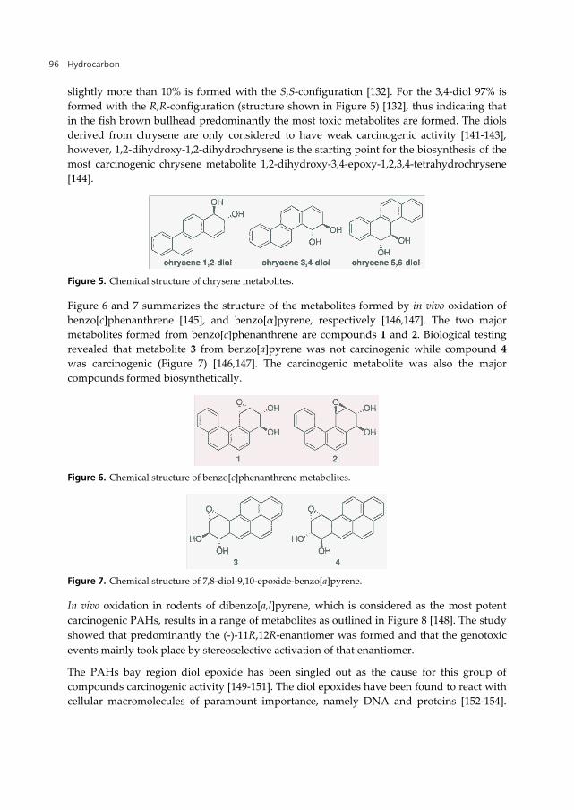
The absolute stereochemistry of PAH metabolites, derived from metabolism of PAHs in the liver, influences the toxicity of the metabolite [138,139]. In particular the diols with R,R-configuration and the R,S-diol-S,R-epoxides show high carcinogenic activity [139]. As shown in Figure 4 for chrysene the predominant diol formed is the 1,2-diol (formed in 58%), with 3,4- and 5,6-diol being formed in 24% and >1%, respectively (structures for the diols are shown in Figure 5) [132]. The data for chrysene depicted in Figure 4 were based on in vitro tests utilizing liver microsomes from brown bullhead, however, these findings were later confirmed by Jonsson et al. in in vivo tests with Atlantic cod [140]. Close to 90% of the chrysene 1,2-diol is formed with the R,R-configuration (structure shown in Figure 5) and
Hydrocarbon 96
slightly more than 10% is formed with the S,S-configuration [132]. For the 3,4-diol 97% is formed with the R,R-configuration (structure shown in Figure 5) [132], thus indicating that in the fish brown bullhead predominantly the most toxic metabolites are formed. The diols derived from chrysene are only considered to have weak carcinogenic activity [141-143], however, 1,2-dihydroxy-1,2-dihydrochrysene is the starting point for the biosynthesis of the most carcinogenic chrysene metabolite 1,2-dihydroxy-3,4-epoxy-1,2,3,4-tetrahydrochrysene [144].
Figure 5. Chemical structure of chrysene metabolites.
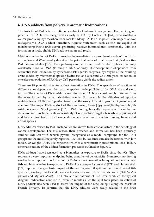
Figure 6 and 7 summarizes the structure of the metabolites formed by in vivo oxidation of benzo[c]phenanthrene [145], and benzo[α]pyrene, respectively [146,147]. The two major metabolites formed from benzo[c]phenanthrene are compounds 1 and 2. Biological testing revealed that metabolite 3 from benzo[a]pyrene was not carcinogenic while compound 4 was carcinogenic (Figure 7) [146,147]. The carcinogenic metabolite was also the major compounds formed biosynthetically.
Figure 6. Chemical structure of benzo[c]phenanthrene metabolites.
Figure 7. Chemical structure of 7,8-diol-9,10-epoxide-benzo[a]pyrene.
In vivo oxidation in rodents of dibenzo[a,l]pyrene, which is considered as the most potent carcinogenic PAHs, results in a range of metabolites as outlined in Figure 8 [148]. The study showed that predominantly the (-)-11R,12R-enantiomer was formed and that the genotoxic events mainly took place by stereoselective activation of that enantiomer.
The PAHs bay region diol epoxide has been singled out as the cause for this group of compounds carcinogenic activity [149-151]. The diol epoxides have been found to react with cellular macromolecules of paramount importance, namely DNA and proteins [152-154].
Polycyclic Aromatic Hydrocarbons a Constituent of Petroleum: Presence and Influence in the Aquatic Environment 97
Miller proposed in 1970 that PAH metabolites are electrophiles that react with nucleophiles in vivo, thus delivering their biological effect [155]. Later it has been proposed that mechanistically the diol epoxides are electrophiles that alkylate the purine bases in DNA via an SN1-like epoxide ring opening process [156,157].
Figure 8. Chemical structure of bibenzo[a,l]pyrene metabolites formed in vivo [148].
In the worst scenario, however, adducts are formed between a PAH metabolite and an oncogene in the genomic DNA, hence switching off the cell’s ability to enter into programmed cell death (apoptosis) and provoking the onset of cancerogenesis. The best described example is DNA adduct formation between the pro-apoptotic p53 gene and benzo[a]pyrene-diol-epoxide after metabolic transformation of benzo[a]pyrene by the combined actions of CYP1A and epoxide hydrolase, another Phase I enzyme (this is also the mechanism by which cigarette smokers develop lung cancer). The presence of DNA adducts in liver tissue is, unlike CYP1A-induction or accumulation of PAH metabolites in bile, the result of cumulative exposure over weeks or even months [158,159]. Hence, hepatocytic CYP1A induction, accumulation of PAH metabolites in bile and elevated liver DNA adducts represent a chain of events that, although partly separated in time, are tightly interrelated from a mechanistic point of view. These three biomarkers have, therefore, received much attention and represent valuable biomarkers of PAH-exposure and effect in fish [159-162]. Analogous with several studies on mammals, responses of these core biomarkers of oil exposure have been associated with genotoxic effects such as liver neoplasia [163].
Hydrocarbon 98
6. DNA adducts from polycyclic aromatic hydrocarbons
The toxicity of PAHs is a continuous subject of intense investigation. The carcinogenic potential of PAHs was recognized as early as 1933 by Cook et al. [164], who isolated a cancer-producing hydrocarbon from coal tar. Many PAHs act as potent carcinogens and/or mutagens via DNA adduct formation. Aquatic vertebrates such as fish are capable of metabolizing PAHs (vide supra), producing reactive intermediates, occasionally with the formation of hydrophobic DNA adducts as an end result.
Metabolic activation of PAHs to reactive intermediates is a prominent mode of their toxic action. Xue and Warshawsky described the principal metabolic pathways that yield reactive PAH intermediates [165]. Two pathways in particular produce electrophiles that may covalently bind to DNA (forming a DNA adduct): 1) electrophilic diol-epoxides from sequential PAH oxidation by cytochrome P450 (CYP) enzymes, hydrolysis of the resulting arene oxides by microsomal epoxide hydrolase, and a second CYP-catalyzed oxidation; 2) one electron oxidation of PAHs by CYP peroxidase yields the radical anion.
There are 18 potential sites for adduct formation in DNA. The specificity of reactions at different sites depends on the reactive species, nucleophilicity of the DNA site and steric factors. The spectra of DNA adducts resulting from PAHs are considerably different from the ones formed by small alkylating agents. For example the dihydrodiol epoxide metabolites of PAHs react predominantly at the exocyclic amino groups of guanine and adenine. The major DNA adduct of the carcinogen, benzo[a]pyrene-7,8-dihydrodiol-9,10-oxide, occurs at N2 of guanine [166]. DNA binding basically depends on its molecular structure and functional state (accessibility of nucleophilic target sites) while physiological and biochemical features determine differences in adduct formation among tissues and across species.
DNA adducts caused by PAH metabolites are known to be crucial factors in the aetiology of cancer development. For this reason their presence and formation has been profusely studied. Adducts with benzo[a]pyrene (recognized as a model compound for the PAH group) are the most frequently reported [167,168], but adducts can also be formed with low molecular weight PAHs, like chrysene, which is a constituent in most mineral oils [169]. A schematic outline of the adduct formation process is outlined in Figure 9.
DNA adducts have been used as a biomarker of exposure to PAHs since the ‘90s. They represent a very important endpoint, being a marker of genotoxicity. Numerous monitoring studies have reported the formation of DNA adduct formation in aquatic organisms (e.g. fish and bivalves) due to exposure to PAHs. For example, Lyons et al [171] and Harvey et al. [172] reported the genotoxic impact of the Sea Empress oil spill accident on different fish species (Lipophorys pholis and Limanda limanda) as well as on invertebrates (Halichondira panicea and Mytilus edulis). The DNA adduct patterns of fish liver exhibited the typical diagonal radioactive zone (DRZ) even 17 months after the spill took place. Detection of DNA adducts has been used to assess the impact of the Erika oil spill along the coasts of French Brittany. To confirm that the DNA adducts were really related to the Erika
Polycyclic Aromatic Hydrocarbons a Constituent of Petroleum: Presence and Influence in the Aquatic Environment 99
petroleum, human hepatocyte (HePG2 cells) were exposed to an Erika fuel extract. Incubation of HePG2 cells lead to the formation of DNA adducts with similar patterns to the ones observed in the monitoring study using fish (Solea solea). These data indicates that human hepatocytes biotransform Erika fuel into genotoxic metabolites similarly to hepatic cells of fish and confirmed that the adducts observed in the monitoring study were related to the contamination of the sediment by the oil spill [173].
Figure 9. Metabolism of PAH leading to protein and DNA adducts. Figure adapted from reference [170].
It has been shown that DNA adducts persisted in vertebrates species due to the low efficiency of repair systems, representing a parameter for long term exposure [174]. These adducts are very persistent in fish liver [175-178]. French et al. [174] observed a steady increase in DNA adduct levels during a chronic exposure of sole (Pleuronectes vetulus) to PAH contaminated sediment for 5 weeks, which were persistent even after a depuration period. The persistency of DNA adducts has been demonstrated also in Atlantic cod (Gadus morhua) [38]. In this study, hepatic DNA adducts appeared after 3 days of exposure to low concentration of crude oil (0.06 ppm) and increased steadily during the entire exposure period of 30 days.
Several techniques (e.g. immunoassay, fluorescence assay, gas chromatography-mass spectroscopy (GC-MS), 32P-postlabelling and mass spectrometry (MS) analysis) have been developed for the analysis of PAH derived DNA adducts. At present, the most sensitive and frequently applied technique in aquatic organisms is the 32P-postlabelling assay [179]. Its high sensitivity is unique and achueves the determination of 1-100 adducts in 109 nucleotides [180]
Hydrocarbon 100
The 32P-postlabelling assay appeared in the early ‘80s and has been applied with a range of protocols in order to detect DNA adducts produced by known carcinogens and complex mixtures [181]. Briefly, the assay involves DNA purification, digestion to normal and adduct-modified 3’-mononucleotides, removal of normal nucleotides (via enzymatic digestion, solvent extraction or chromatographic methods), 32P-postlabelling at the 5’ position of adducted nucleotides followed by chromatographic separation, detection and quantification (via autoradiography, scintillation counting or phosphor screen imaging analysis) [182]. Following this assay, PAHs cause the appearance of the diagonal radioactive zone (DRZ) (Figure 10) [173,178,183,184].
Figure 10. Example of the bulky DNA adduct zone (DRZ) typical of a contamination by PAHs detected by the 32P-postlabelling method (liver sample of fish collected in a PAH contaminated coastal area) (Pampanin unpublished data).
MS/MS analysis has recently emerged as a powerful tool in the detection and structure elucidation of DNA adducts as well as for their quantification at very low concentrations (as often present in biological samples). In fact, electrospray ionization tandem mass spectrometric (ESI-MS/MS) analysis was capable of revealing DNA adducts in different aquatic organisms (e.g. fish (Oreochromis mossambicus) and mussel (Perna perna) soft tissue) [185,186]. This MS/MS approach provided a rapid determination and discrimination of structurally different phenanthrene derived DNA adducts in fish bile samples [186]. This technique is able to detect one modified base in 106-1012 unmodified bases [187].
Development of methodologies to detect DNA damages induced by PAHs is of constant concern in aquatic ecotoxicology. Direct chemical methods, such as high performance liquid chromatography with electro-chemical detection (HPLC-EDC), GC-MS and the 32P-postlabelling, are highly sensitive and specific [180,188], however, they are very time and money consuming. A number of antibodies have been generated against carcinogenic products of DNA modifications, including those generated by PAHs. Immunoassays (immunohistochemistry or ELISA) are routinely employed to detect DNA adducts in humans, while the use of such approaches is more limited in aquatic species [189]. An immunoperoxidase method for revealing 7,8-dihydro-8-oxodeoxyguanosine (8-oxo-dG) in marine organisms has been described [189]. This work was also followed by the use of immunofluorescence and antibodies for DNA adduct detection in both vertebrates (fish,
Polycyclic Aromatic Hydrocarbons a Constituent of Petroleum: Presence and Influence in the Aquatic Environment 101
Anguilla anguilla) and invertebrated (mussel, Mytilus galloprovincialis). The immunohistochemical approach demonstrated a good selectivity, low cost, is easy to perform and readily allowed the analysis of a large number of samples. Nevertheless, it does not reach the high sensitivity of other methods [190].
7. Protein adducts from polycyclic aromatic hydrocarbons
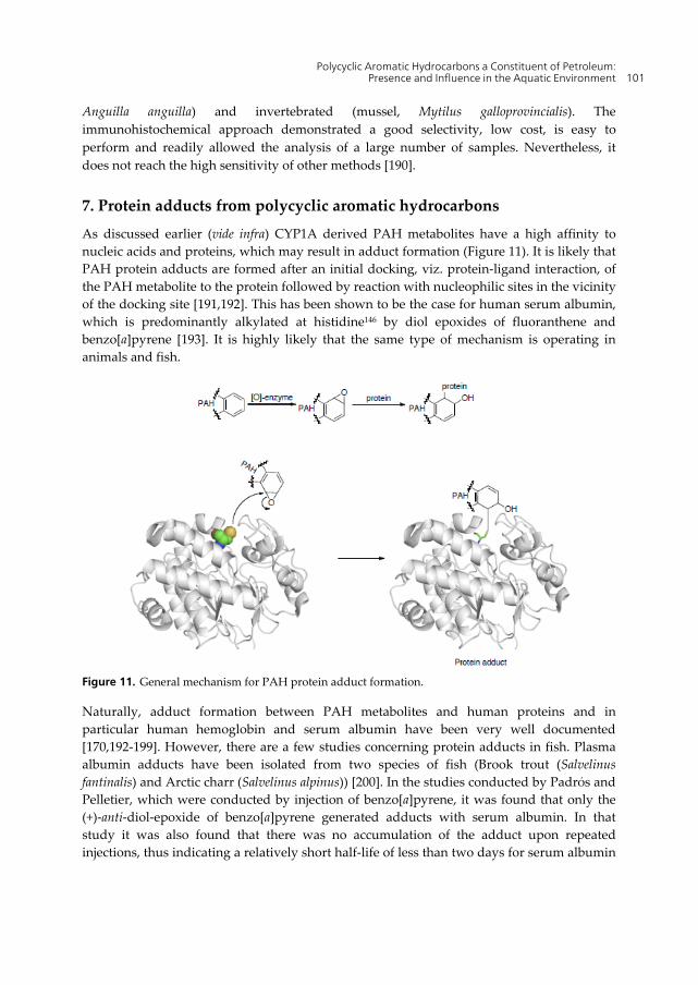
As discussed earlier (vide infra) CYP1A derived PAH metabolites have a high affinity to nucleic acids and proteins, which may result in adduct formation (Figure 11). It is likely that PAH protein adducts are formed after an initial docking, viz. protein-ligand interaction, of the PAH metabolite to the protein followed by reaction with nucleophilic sites in the vicinity of the docking site [191,192]. This has been shown to be the case for human serum albumin, which is predominantly alkylated at histidine146 by diol epoxides of fluoranthene and benzo[a]pyrene [193]. It is highly likely that the same type of mechanism is operating in animals and fish.
Figure 11. General mechanism for PAH protein adduct formation.
Naturally, adduct formation between PAH metabolites and human proteins and in particular human hemoglobin and serum albumin have been very well documented [170,192-199]. However, there are a few studies concerning protein adducts in fish. Plasma albumin adducts have been isolated from two species of fish (Brook trout (Salvelinus fantinalis) and Arctic charr (Salvelinus alpinus)) [200]. In the studies conducted by Padrόs and Pelletier, which were conducted by injection of benzo[a]pyrene, it was found that only the (+)-anti-diol-epoxide of benzo[a]pyrene generated adducts with serum albumin. In that study it was also found that there was no accumulation of the adduct upon repeated injections, thus indicating a relatively short half-life of less than two days for serum albumin
Hydrocarbon 102
in fish. In humans the half-life of serum albumin has been reported to 20 days [201]. In fish the presence of this adduct would be an indication of a very recent exposure to benzo[a]pyrene, while in humans this would also function as a marker of longer term exposure to the PAH. It has been found that the point of adduct formation between human serum albumin and benzo[a]pyrene anti-diol epoxide is dependent on the stereochemistry of the PAH metabolite [194]. The (+)-enantiomer generates a carboxylic ester adduct with Asp187 or Glu188 and that the (-)-enantiomer forms an adduct with His146.
The different isoforms of hemoglobin present in different species results in the formation of different adducts. For example, rat hemoglobin possesses a reactive β-cysteine in position 125 not present in human hemoglobin [202]. syn And anti fluoranthene diol-epoxides form adducts with this particular cysteine in rats. The presence of different isoforms of the same protein in different species results in the possibility of generating different adducts for the same PAH in the different species. The point of adductation most likely reflects on the proteins ability to function. Thus, the adduct formation of a specific protein might affect one species more severely than another.
8. Future perspective
Environmental research related to PAHs has to date, with a few exceptions, predominantly been concerned with finding metabolites of the compounds and detecting the presence of PAH DNA adducts. However, based on the discussion herein it is clear that the next step has to be towards analysis that can provide clear answers regarding the stereochemical outcome of the oxidation processes taking place in vivo. By such means it is easier to evaluate the toxicity of the various PAHs. This is a rather large task since different species metabolize PAHs differently resulting in dissimilar distributions between the stereoisomers.
PAH protein adducts have been studied extensively for humans (vide supra) and rodents, however, for fish and other aquatic animals this topic is barely touched upon. Studies of adduct formation in fish will further aid the evaluation of the toxicity of the different PAHs. In the PAH protein adduct studies that have been conducted on other species we have seen that the point where the adduct is formed in a specific protein varies from species to species. Generating new knowledge as to where adducts are formed with the same PAH in the same protein in other species might in addition to providing increased knowledge as to the impact of adduct formation also possibly generate new interesting research questions. Hemoglobin in fish has a very short half-life so adduct formation on hemoglobin might not have such a great health impact on the fish. However, other less abundant proteins in the blood are also most likely susceptible to adduct formation with PAH metabolites. Detecting the proteins affected and determining the site of adduct formation will aid in the overall judgment of the toxicity of the PAH responsible for the adduct formed. Adducts with proteins present in the bile may also be of value in assessing the toxicity of PAHs.
In human health care proteomics has for some time been extensively used for diagnostics [203,204] and these techniques are also slowly making their way into ecotoxicology [205,206]. It has been found in human amniotic epithelial cells exposed to anti-7,8-
Polycyclic Aromatic Hydrocarbons a Constituent of Petroleum: Presence and Influence in the Aquatic Environment 103
dihydroxy-9,10-epoxy-7,8,9,10-tetrahydrobenzo[a]pyrene, a compound that causes adduct formation on DNA and oxidative damage on DNA, resulting in alternations of the expression of three proteins [207]. This result highlights that proteomics and the study of expression rates of particular proteins can be a powerful method in the future in order to determine if marine animals have been exposed to PAHs present in oil.
Author details
Daniela M. Pampanin Biomiljø, International Research Institute of Stavanger, Mekjarvik, Randaberg, Norway
Magne O. Sydnes Faculty of Science and Technology, University of Stavanger, Stavanger, Norway
Acknowledgement
Funding from the University of Stavanger is gratefully acknowledged. Andrea Bagi, University of Stavanger, is thanked for valuable literature input for the introduction part of the chapter. Emily Lyng, International Research Institute of Stavanger, is acknowledged for her careful proof reading of the manuscript.
9. References
[1] Marshall AG, Rodgers RP (2004) Petroleomics: the next grand challenge for chemical analysis. Acc. Chem. Res. 37:53-59.
[2] Fieser LF, Fieser M (1956) Organic Chemistry, 3rd edition. Boston: D. C. Heath and Co. Chapter 21.
[3] McElroy AE, Bates S, Rice SD, Korn S (1985) Bioavailability of polycyclic aromatic hydrocarbons in the aquatic environment. In: Varanasi U, editor. Metabolism of polycyclic aromatic hydrocarbons in the aquatic environment. Boca Raton: CRC Press. pp. 1-39.
[4] Feng X, Pisula W, Müllen K (2009) Large polycyclic aromatic hydrocarbons: Synthesis and discotic organization. Pure Appl. Chem. 81:2203-2224.
[5] Hylland K (2006) Polycyclic aromatic hydrocarbon (PAH) ecotoxicology in marine ecosystems. J. Toxicol. Environ. Health, Part A 69:109-123.
[6] Lang KF, Buffleb H, Kalowy J (1962) 2-Phenyl-phenanthren und binaphthyl-(2,2') aus steinkohlenteer. Chem. Ber. 95:1052-1053.
[7] Lang KF, Buffleb H, Kalowy J (1964) Fulminen (1,2-benzo-picen) im steinkohlenteer. Chem. Ber. 97:494-497.
[8] Wakeham SG, Schaffner C, Giger W (1980) Polycyclic aromatic hydrocarbons in recent lake sediments – I. Compounds having anthropogenic origins. Geochim. Cosmo. Acta. 44:403-413.
Hydrocarbon 104
[9] Laughlin RB, Neff JM (1979) Interactive effects of salinity, temperature and polycyclic aromatic hydrocarbons on the survival and development rate of larvae of the mud crab Rhithropanopeus harrisii. Marine Biology 53:281-291.
[10] Harvey RG (1996) Polycyclic aromatic hydrocarbons. New York: Wiley-VCH. pp. 1-20.
[11]Achten C, Hofmann T (2010) Umweltrelevanz von natülichen polyzyklischen aromatischen kohlenwassertoffen aus steinkohlen – eine übersicht. Grundwasser 15:5-18.
[12] Durand C, Ruban V, Amblès A, Oudot J (2004) Characterization of the organic matter of sludge: Determination of lipids, hydrocarbons and PAHs from road retention/infiltration ponds in France. Environ. Pollut. 132:375-384.
[13] Beyer J, Aas E, Borgenvik HK, Ravn P (1998) Bioavailability of PAH in effluent water from an aluminium works evaluated by transplant caging and biliary fluorescence measurements of Atlantic cod (Gadus morhua L.). Mar. Environ. Res. 46:233-236.
[14] Næs K, Oug E (1998) The distribution and environmental relationships of polycyclic aromatic hydrocarbons (PAHs) in sediments from Norwegian smelter-affected fjords. Chemosphere 36:561-576.
[15] Smith JN, Levy EM (1990) Geochronology for polycyclic aromatic hydrocarbon contamination in sediments of the Saguenay Fjord. Environ. Sci. Technol. 24:874- 879.
[16] Mascarelli A (2010) After the oil. Nature 467:22-24. [17] Redondo J, Platonov AK (2009) Self-similar distribution of oil spills in European coastal
waters. Environ. Res. Lett. 4:014008. [18] Tedesco SA (1985) Surface geochemistry in petroleum exploration. Chapman & Hall,
New York. [19] Røe Utvik T (1999) Chemical characterization of produced water from four offshore oil
production platforms in the North Sea. Chemosphere 39:2593-2606. [20] Hunt JM (1979) Petroleum geochemistry and geology. San Francisco: W. H. Freeman
and Co. p 617. [21] Bakke T, Hameedi J, Kimstach V, Macdonald R, Melnikov S, Robertson A, Shearer R,
Thomas D (1998) Petroleum hydrocarbons. In: Roberts A, editor. AMAP Assessment Report: Arctic pollution issues. pp 667-668.
[22] van der Meer F, van Dijk P, van der Werff H, Yang H (2002) Remote sensing and petroleum seepage: a review and case study. Terra Nova 14:1-17.
[23] NRC (National Research Council) (1985) Oil in the sea: Inputs, fates, and effects. Washington: National Academy Press. p. 501.
[24] Kemsley J (2012) Water eased oil removal in Gulf. Chemical and Engineering News, February 6:32-33.
[25] Head IM, Jones DM, Röling WFM (2006) Marine microorganisms make a meal of oil. Nat. Rev. Microbiol. 4:173-182.
Polycyclic Aromatic Hydrocarbons a Constituent of Petroleum: Presence and Influence in the Aquatic Environment 105
[26] Valentine DL, Mezić I, Maćešić S, Crnjarić-Žic N, Ivić S, Hogan PJ, Fonoberov VA, Loire S (2012) Dynamic autoinoculation and the microbial ecology of a deep water hydrocarbon irruption. Proc. Natl. Acad. Sci. USA, doi: 10.1073/pnas.1108820109.
[27] Redmond MC, Valentine DL (2012) Natural gas and temperature structured a microbial community response to the Deepwater Horizon oil spill. Proc. Natl. Acad. Sci. USA, doi: 10.1073/pnas.1108756108.
[28] Atlas RM (2011) Oil biodegradation and bioremediation: A tale of the two worst spills in U.S. history. Environ. Sci. Technol. 45:6709-6715.
[29] Pothuluri JV, Cerniglia CE (1994) Microbial metabolism of polycyclic aromatic hydrocarbons. In: Chaudhry GR, editor. Biological degradation and bioremediation of toxic chemicals. London: Chapman & Hall. pp. 92-124.
[30] Volkering F, Breure, AM, Sterkenburg A, van Andel JG (1992) Microbial degradation of polycyclic aromatic hydrocarbons: effect of substrate availability on bacterial growth kinetics. Appl. Microbiol. Biotechnol. 36:548-552.
[31] Geiselbrecht AD, Herwig RP, Deming JW, Staley JT (1996) Enumeration and phylogenetic analysis of polycyclic aromatic hydrocarbon-degrading marine bacteria from Puget Sound sediments. Appl. Environ. Microbiol. 62:3344-3349.
[32] Geiselbrecht AD, Hedlund BP, Tichi MA, Staley JT (1998) Isolation of marine polycyclic aromatic hydrocarbon (PAH)-degrading Cycloclasticus strains from the Gulf of Mexico and comparison of their PAH degradation ability with that of Puget Sound. Appl. Environ. Microbiol. 64:4703-4710.
[33] Cao B, Nagarajan K, Loh K-C (2009) Biodegradation of aromatic compounds: current status and opportunities for biomolecular approaches. Appl. Microbiol. Biotechnol. 85:207-228.
[34] Haritash AK, Kaushik CP (2009) Biodegradation aspects of polycyclic aromatic hydrocarbons (PAHs): A review. J. Hazard. Mat. 169:1-15
[35] Keith LH, Telliard WA (1979) Priority pollutants: I. A perspective view. Environ. Sci. Technol. 13:416-423.
[36] Kerr JM, Melton HR, McMillen SJ, Magaw RI, Naughton G, Little GN (1999) Polyaromatic hydrocarbon content in crude oils around the world. Conference paper from the 1999 SPE/EPA Exploration and production environmental conference held in Austin, Texas, USA, 28 February-3 March.
[37] Faksness L-G, Brandvik PJ, Sydnes LK (2008) Composition of the water accommodated fractions as a function of exposure times and temperatures. Mar. Pollut. Bull. 56:1746-1754.
[38] Aas E, Baussant T, Balk L, Liewenborg B, Andersen OK (2000) PAH metabolites in bile, cytchrome P4501A and DNA adducts as environmental risk parameters for chronic oil exposure: a laboratory experiment with Atlantic cod. Aquat. Toxicol. 51:241-258.
[39] Deepthike HU, Tecon R, van Kooten G, von der Meer JR, Harms H, Wells M, Short J (2009) Unlike PAHs from Exxon Valdez crude oil, PAHs from Gulf of Alaska coals are not readily bioavailable. Environ. Sci. Technol. 43:5864-5870.
Hydrocarbon 106
[40] Sundt RC, Beyer J, Vingen S, Sydnes MO (2011) High matrix interference affecting detection of PAH metabolites in bile of Atlantic hagfish (Myxine glutinosa) used for biomonitoring of deep-water oil production. Mar. Environ. Res. 71:369-374.
[41] Rhodes S, Farwell A, Hewitt LM, MacKinnon M, Dixon DG (2005) The effects of dimethylated and alkylated polycyclic aromatic hydrocarbons on the embryonic development of the Japanese medaka. Ecotox. Environ. Safety 60:247-258.
[42] Carls MG, Holland L, Larsen M, Collier TK, Scholz NL, Incardona JP (2008) Fish embryos are damaged by dissolved PAHs, not oil particles. Aquat. Toxicol 88:121- 127.
[43] Billiard SM, Querbach K, Hodson PV (1999) toxicity of retene to early life stages of two freshwater fish species. Environ. Toxicol. Chem. 18:2070-2077.
[44] White KL (1986) An overview of immunotoxicology and carcinogenic polycyclic aromatic hydrocarbons. J Environ. Sci. Health Part C: Environ. Carcino. Ecotox. Rev. 2:163-202.
[45] Conney AH (1982) Induction of microsomal enzymes by foreign chemicals and carcinogenesis by polycyclic aromatic hydrocarbons. Cancer Res. 42:4875-4917.
[46] Canestro D, Raimondi PT, Reed DC, Schrnitt RJ, Holbrook SJ (1996) A study of methods and techniques for detecting ecological impacts. American Academy of Underwater Sciences (AAUS).
[47] Zhu SQ, King SC, Haasch ML (2008) Biomarker induction in tropical fish species on the Northwest Shelf of Australia by produced formation water. Mar. Environ. Res. 65:315-324.
[48] Hylland K, Tollefsen KE, Ruus A, Jonsson G, Sundt RC, Sanni S, Røe Utvik TI, Johnsen S, Nilssen I, Pinturier L, Balk L, Baršienė J, Marigòmez I, Feist SW, Børseth JF (2008) Water column monitoring near oil installations in the North Sea 2001–2004. Mar. Poll. Bull. 56:414–429.
[49] Gorbi S, Virno Lamberti C, Notti A, Benedetti M, Fattorini D, Moltedo G, Regoli F (2008) An ecotoxicological protocol with caged mussels, Mytilus galloprovincialis, for monitoring the impact of an offshore platform in the Adriatic Sea. Mar. Environ. Res. 65:34-49.
[50] Crone TJ, Tolstoy M (2010) Magnitude of the 2010 Gulf of Mexico oil leak. Science 330:634.
[51] Peterson CH, Rice SD, Short JW, Esler D, Bodkin JL, Ballachey BE, Irons DB (2003) Long-term ecosystem response to the Exxon Valdez oil spill. Science 302:2082-2086.
[52] Short J, Rice SD, Heintz RA, Carls MG, Moles A (2003) Long-term effects of crude oil on developing fish: Lessons from the Exxon Valdez oil spill. Energy Sources 25:509-517.
[53] Lavrova OY, Kostianoy AG (2011) Catastrophic oil spill in the Gulf of Mexico in April-May 2010. Atmospheric and Oceanic Physics 47:1114-1118.
[54] Sundt RC, Pampanin DM, Grung M, Baršienė, Ruus A (2011) PAH body burden and biomarker responses in mussel (Mytilus edulis) exposed to produced water from a
Polycyclic Aromatic Hydrocarbons a Constituent of Petroleum: Presence and Influence in the Aquatic Environment 107
North Sea oil field: Laboratory and field assessments. Mar. Poll. Bull. 62:1498- 1505
[55] Sundt RC, Ruus A, Jonsson H, Skarphéðinsdóttir H, Meier S, Grung M, Beyer J, Pampanin DM (2012) Biomarker responses in Atlantic cod (Gadus morhua) exposed to produced water from a North Sea oil field: Laboratory and field assessments Original Research Article. Mar. Poll. Bull. 64:144-152.
[56] Røe TI, Johnsen S (1996) Discharges of produced water to the North Sea; Effects in the water column. Produced water 2. Environmental Issues and Mitigation Technologies. S. Johnsen. New York: Plenum Press pp. 13-25.
[57] Durell G, Utvik TR, Johnsen S, Frost T, Neff J (2006) Oil well produced water discharges to the North Sea. Part I: Comparison of deployed mussels (Mytilus edulis), semi-permeable membrane devices, and the DREAM model predictions to estimate the dispersion of polycyclic aromatic hydrocarbons. Mar. Environ. Res. 62:194-223.
[58] OGP (2005) Fate and effects of naturally occurring substances in produced water on the marine environment. International Association of Oil & Gas Producers, London, UK, Report No 364. 36 p.
[59] Johnsen S, Røe Utvik TI, Garland E, de Vals B, Campbell J (2004) Environmental fate and effects of contaminants in produced water. SPE 86708. Paper presented at the Seventh SPE international conference on health, safety and environment in oil and gas exploration and production. Society of Petroleum Engineers, Richardson, TX, 9 p.
[60] Latimer JS, Zheng J (2003) The sources, transport and fate of PAHs in the marine environment. In: Doube PET editor. PAHs: an ecotoxicological perspective. Wiley, West Sussex, pp. 9-34.
[61] Neff J, Lee K, De Blois EM (2011) Produced water: overview of composition, fate, and effects. In: Lee K, Neff J editors. Produced Water. Environmental risks and advances in mitigation technologies. Springer, London, UK. pp 3-54.
[62] Ruus A, Tollefsen KE, Grung M, Klungsøyr J, Hylland K (2006) Accumulation of contaminations in pelagic organisms, caged blue mussels, caged cod and semi-permeable membrane device (SPMDs). In: Hylland K, Vethaak AD, Lang T, editors. Biological effects of contaminants in marine pelagic ecosystems (ICES). SETAC publications pp 51-74.
[63] Faksness LG, Grini PG, Daling PS (2004) Partitioning of semi-soluble organic compounds between the water phase and oil droplets in produced water. Mar. Poll. Bull. 48:731-742.
[64] Burns KA, Codi S (1999) Non-volatile hydrocarbon chemistry studies around a production platform on Australia’s northwest shelf. Estur. Cstl. Shelf Sci. 49:853- 876.
[65] Neff JM, Johnsen S, Frost TK, Røe Utvik TI, Durell GS (2006) Oil well produced water discharges to the North Sea. Part II: comparison of deployed mussels (Mytilus edulis) and the DREAM model to predict risk assessment. Mar. Environ. Res. 62:224- 246.
Hydrocarbon 108
[66] OLF (2007) Environmental report 2007. The Norwegian Oil Industry Association (OLF). Stavanger, Norway 63 p.
[67] Farmen E, Harman C, Hylland K, Tollefsen KE (2010). Produced water extracts from North Sea oil production platforms result in cellular oxidative stress in a rainbow trout in vitro bioassay. Mar. Poll. Bull. 60:1092-1098.
[68] Suter GW (1993) Ecological Risk Assessment. Lewis Publishers, Boca Raton, FL, USA 538 p.
[69] Henderson F, Bechtold WE, Bond JA, Sun JD (1989) The use of biological markers in toxicology. Crit. Rev. Toxicol. 20:65–82.
[70] De Zwart D (1995) Monitoring water quality in the future, Volume 3: Biomonitoring. National Institute of Public Health and Environmental Protection (RIVM), Bilthoven, The Netherlands, 81 p.
[71] Antizar-Ladislao B (2009) Polycyclic aromatic hydrocarbons, polycholirnated biphenyls, phthalates and organotins in northern Atlantic Spain’s coastal marine sediments. J. Environ. Monitor. 11:85-91.
[72] Binelli A, Provini A (2003) POPs in edible clams from different Italian and European markets and possible human health risk. Mar. Poll. Bull. 46:879-886.
[73] Water Framework Directive (2000) Directive 2000/60 EU of the European Parliament and of the Council of 23 October 2000 establishing a framework for Community action in the field of water policy.
[74] OSPAR Commission (2000) Quality Status Report 2000. London: OSPAR. [75] Gilroy (2000) Derivation of shellfish harvest reopening criteria following the new
Carissa oil spill in Coos Bay, Oregon. J. Toxicol. Environ. Health 60:317-329. [76] Senthil Kumar K, Sajwan KS, Richardson JP, Kannan K (2008) Contamination profiles of
heavy metals, organochlorine pesticides, polycyclic aromatic hydrocarbons and alkylphenols in sediment and oyster collected from marsh/estuarine Savannah GA, USA. Mar. Poll. Bull. 56:136-162.
[77] Massara Paletto V, Commendatore MG, Esteves JL (2008) Hydrocarbon levels in sediments and bivalve mollusks from Bahía Nueva (Patagonia, Argentina): an assessment of probable origin and bioaccumulation factors. Mar. Poll. Bull. 56:2082-2105.
[78] Francioni EL, Wagener A de LR, Scofield A, Depledge M, Sette CB, Carvalhosa L (2007) Polycyclic aromatic hydrocarbon in mussel Perna perna from Guanabara Bay, Brazil: space-time observations, source investigation and genotoxicity. Sci. Tot. Environ. 372:515-531.
[79] Baumard P, Budzinski H, Garrigues P, Narbonne JF, Burgeot T, Miche X et al. (1999) Polycyclic aromatic hydrocarbon (PAH) burden of mussels (Mytilus sp.) in different marine environments in relation with sediment PAH contamination and bioavailability. Mar. Environ. Res. 47:415-439.
[80] David A, Fenet H, Gomez E (2009) Alkylphenols in marine environments: distribution monitoring strategies and detection considerations. Mar. Poll. Bull. 58:953-960.
[82] HMSO (1985) The determination of 6 specific PAHs. Materials for examination of waters and associated materials. Her Majesty’s Stationery Office, London.
[83] EPA Procedure 8310 (1986) Polycyclic aromatic hydrocarbons. [84] EPA Method 8270D (1998) (SW-846) Semivolatile organic compounds by gas
chromatography/mass spectrometry (GC/MS), Revision 4. [85] ISO Procedure 1799 (2002) Water quality-determination of 15 PAHs in water by HPLC
with fluorescence detection. [86] Wolska L (2008) Determination (monitoring) of PAHs in surface waters: why
an operationally defined procedure is needed. Anal. Bioanal. Chem. 391:2647- 2652.
[87] Beyer J, Jonsson G, Porte C, Krahn MM, Ariese F (2010) Analytical methods for determining metabolites of polycyclic aromatic hydrocarbon (PAH) pollutants in fish bile: a review. Environ. Toxicol. Pharmacol. 30:224-244.
[88] Neff JM (2002) Bioaccumulation in marine organisms. Effect of contaminants from oil well produced water. Oxford, Elsevier Science Ltd., 468 p.
[89] Ardgar H, Ozel MZ, Sen Z (2011) Polycyclic aromatic hydrocarbons in water from the Menderes river, turkey. Bull. Environ. Contam. Toxicol. 86:221-225.
[90] Oros DR, Ross JRM, Spies RB, Mumley T (2007) Polycyclic aromatic hydrocarbon (PAH) contamination in San Francisco Bay: a 10-year retrospective of monitoring in an urbanized estuary. Environ. Res. 105:101-118.
[91] Cataldo D, Colombo JC, Boltovskoy D, Bilos C, Landoni P (2001) Environmental toxicity assessment in the Paraná river delta (Argentina): simultaneous evaluation od selected pollutants and mortality rates of Curbilcula fluminea (bivalvia) early juveniles. Environ. Poll. 112:379-389.
[92] Colombo JC, Barreda C, Bilos NC, migota MC, Skorupka C (2005) Oil spill in the Rio de la Plata estuary, Argentina: 2-hydrocarbon disappearance rates in sediments and soils. Environ. Poll. 134:267-276.
[93] Arias AH, Spetter CV, Freije RH, Marcovecchio JE (2009) Polycyclic aromatic hydrocarbons in water, mussel (Brachidontes sp., Tagelus sp.) and fish (Odontesthes sp.) from Bahía Blanca Estuary, Argentina. Estuar. Coastal Shelf Sci. 85:67-81.
[94] Allan SE, Smith BW, Anderson KA (2012) Impact of the Deepwater Horizon Oil Spill on Bioavailable Polycyclic Aromatic Hydrocarbons in Gulf of Mexico Coastal Waters. Environ. Sci. Technol. 46:2033-2039.
[95] Viarengo A, Dondero F, Pampanin DM, Fabbri R, Poggi E, Malizia M, Bolognesi C, Perrone E, Gollo E, Cossa GP (2007) A biomonitoring study assessing the residual biological effects of pollution caused by the HAVEN wreck on marine organisms in the Ligurian sea (Italy). Arch. Environ. Contam. Toxicol. 53:607-616.
Hydrocarbon 110
[96] Martinez-Gomez C, Fernandez B, Valdes J, Campillo JA, Benedicto J, Sanchez F, Vethaak AD (2009) Evaluation of three-year monitoring with biomarkers in fish following the Prestige oil spill (N Spain). Chemosphere 74:613-620.
[97] Uno S, Koyama J, Kokushi E, Monteclaro H, Santander S, Cheikyula JO, Miki S, Anasco N, Pahila IG, Taberna Jr HS, Matsuoka T (2010) Monitoring of PAHs and alkylated PAHs in aquatic organisms after 1 month from the Solar I oil spill off the coast of Guimaras Island, Philippines. Environ. Monit. Assess. 165:501-515.
[98] Lee K, Neff J (2011) Produced Water. Environmental risks and advances in mitigation technologies. Springer, New York, 608 p.
[99] Harman C, Thomas K, Tollefsen KE, Meier S, Bøyum O, Grung M (2009) Monitoring the freely dissolved concentrations of polycyclic aromatic hydrocarbons (PAH) and alkylphenols (AP) around a Norwegian oil platform by holistic passive sampling. Mar. Poll. Bull. 58:1671-1679.
[100] Harman C, Tollefsen KE, Bøyum O, Thomas K, Grung M (2008) Uptake rates of alkylphenols, PAHs and carbazoles in semipermeable membrane devices (SPMDs) and polar organic chemical integrative samplers (POCIS). Chemosphere 72:1510- 1516.
[101] Sundt RC, Brooks S, Grøsvik BE, Pampanin DM, Farmen E, Harman C, Meier S (2010) Water column monitoring of offshore produced water discharges. Compilation of previous experience and suggestions for future survey design. OLF 2010.
[102] Livingstone DR (1998) The fate of organic xenobiotics in aquatic ecosystems: quantitative and qualitative differences in biotransformation by invertebrates and fish. Comp. Biochem. Physiol. A 120:43-49.
[103] Fillmann G, Watson GM, Howsam M, Francioni E, Depledge MH, Readman JW (2004) Urinary PAH metabolites as biomarkers of exposure in aquatic environments. Environ. Sci. Technol. 38:2649-2656.
[104] van der Oost R, Beyer J, Vermeulen NPE (2003) Fish bioaccumulation and biomarkers in environmental risk assessment: a review. Environ. Toxicol. Pharmacol. 13:57- 149.
[105] Bellas J, Saco-Álvarez L, Nieto Ó, Beiras R (2008) Ecotoxicological evaluation of polycyclic aromatic hydrocarbons using marine invertebrate embryo–larval bioassays. Mar. Poll. Bull. 57:493-502.
[106] Aas E, Beyer J, Jonsson G, Reichert WL, Andersen OK (2001) Evidence of uptake, biotransformation and DNA binding of polycyclic aromatic hydrocarbons in Atlantic cod and corkwing wrasse caught in the vicinity of an aluminium works. Mar. Environ. Res. 52:213-229.
[107] Oros DR, Ross JRM (2005) Polycyclic aromatic hydrocarbons in bivalves from the San Francisco estuary: Spatial distributions, temporal trends, and sources (1993-2001). Mar. Environ. Res. 60:466-488.
Polycyclic Aromatic Hydrocarbons a Constituent of Petroleum: Presence and Influence in the Aquatic Environment 111
[108] Sole M, Buet A, Ortiz L, Maynou F, Bayona JM, Albaiges J (2007) Bioaccumulation and biochemical responses in mussels exposed to the water-accommodated fraction of the Prestige fuel oil. Sci. Mar. 71:373-382.
[109] Røe Utvik TI, Durell GS, Johnsen S (1999) Determining produced water originating polycyclic aromatic hydrocarbons n North Sea waters: comparison of sampling techniques. Mar. Poll. Bull. 38:977-989.
[110] Grøsvik BE, Meier S, Liewenborg B et al. (2009) Condition monitoring in the water column 2008: Oil hydrocarbons in fish from Norwegian waters. IMR report Nr.2- 2009.
[111] Hylland K, Lang T, Vethaak D, editors (2006) Biological Effects of Contaminants in Marine Pelagic Ecosystems. SETAC Press, 475 p.
[112] Shaw GR, Connell DW (2001) DNA adducts as a biomarker of polycyclic aromatic hydrocarbon exposure in aquatic organisms: relationship to carcinogenicity. Biomarkers 6:64-71.
[113] Kammann U, Lang T, Vobach M, Wosniok W (2005) Ethoxyresorufin-O-deethylase (EROD) activity in dab (Limanda limanda) as biomarker for marine monitoring. Environ. Sci. Pollut. Res. 12:140-145.
[114] Balk L, Hylland K, Hansson T, Bertssen MHG, Beyer J, Jonsson G, Melbey A, Grung M, Torstensen BE, Børset JF, Skarphéðinsdóttir H, Klungsøyr J (2011) Biomarkers in natural fish populations indicate adverse biological effects of offshore oil production. PloS ONE 6:1-10.
[115] Maria VL, Correia AC, Santos MA (2002) Benzo[a]pyrene and beta-naphthoflavone mutagenic activation by European eel (Anguilla anguilla L.) S9 liver fraction. Ecotoxicol. Environ. Saf. 53:81-85.
[116] Stephensen E, Adolfsson-Erici M, Celander M, Hulander M, Parkkonen J, Hegelund T, Sturve J, Hasselberg L, Bengtsson M, Forlin L (2003) Biomarker responses and chemical analyses in fish indicate leakage of polycyclic aromatic hydrocarbons and other compounds from car tire rubber. Environ. Toxicol. Chem. 22:2926-2931.
[117] Pathiratne A, Hemachandra CK (2010) Modulation of ethoxyresorufin O-deethylase and glutathione S-transferase activities in Nile tilapia (Oreochromis niloticus) by polycyclic aromatic hydrocarbons containing two to four rings: implications in biomonitoring aquatic pollution. Ecotoxicol. 19:1012–1018.
[118] Bravo, CF, Curtis LR, Myers MS, Meador JP, Johnson LL, Buzitis J, Collier TK, Morrow JD, Laetz CA, Loge FJ, Arkoosh MR (2011) Bioamrker responses and disease susceptibility in juvenile rainbow trout Oncorhynchus mykiss fed a high molecular weight PAH mixture. Environ. Toxicol. Chem. 30:704-714.
[119] Broeg K, Zander S, Diamant A, Korting W, Kruner G, Paperna I, von Westernhagen H (1999) The use of fish metabolic, pathological and parasitological indices in pollution monitoring - 1. North Sea. Helgoland Mar. Res. 53:171-194.
[120] Cajaraville MP, Bebianno MJ, Blasco J, Porte C, Sarasquete C, Viarengo A (2000) The use of biomarkers to assess the impact of pollution in coastal environments of the Iberian Peninsula: a practical approach. Sci. Tot. Environ. 247:295-311.
Hydrocarbon 112
[121] Pluta HJ (1993) Investigations on biotransformation (mixed function oxygenase activities) in fish liver. In: Braunbeck T, Hanke W, Segner H editors. Fish ecotoxicology and ecophysiology. VCH Weinheim pp. 13-33.
[122] Stegeman JJ , Hahn ME (1994) Biochemistry and molecular biology of monooxygenases: current perspectives on forms, functions, and regulation of cytochrome P450 in aquatic species. In: Malins DC, Ostrander GK, editors. Boca Raton: Aquatic Publishers pp. 87–203.
[123] Jőnsson EM, Brandt I, Brunstrom B (2002) Gill filament-based EROD assay for monitoring waterborne dioxin-like pollutants in fish. Environ. Sci. Technol. 36:3340-3344.
[124] Dissanayake A, Bamber SD (2010) Monitoring PAH contamination in the field (South west Iberian Peninsula): biomonitoring using fluorescence spectrophotometry and physiological assessment in the shore crab Carcinus maenas (L.) (Crustacea: Decapoda). Mar. Environ. Res. 70:65-72.
[125] Boyd DR, Kennedy DA, Malone JF, O’Kane GA (1987) Synthesis of triphenylene 1,2-oxide (1,2-epoxy-1,2-dihydrophenylene) and absolute configuration of the trans-1,2-dihydro diol metabolite of triphenylene. Crystal structure of (-)-(1R,2R)-trans-2-bromo-1-menthyloxyacetoxy-1,2,3,4-tetrahydrotiphenylene. J. Chem. Soc. Perkin Trans. 1 369-375.
[126] Rand GM (1995) Fundamentals of aquatic toxicology, second edition: Effects, environmental fate and risk assessment. Washington DC: Taylor and Francis. 128 p.
[127] Nebert DW, Dalton TP, Okey AB, Gonzalez FJ (2004) Role of aryl hydrocarbon receptor-mediated induction of the CYP1 enzymes in environmental toxicity and cancer. J. Biol. Chem. 279:23847-23850.
[128] Jacob J (2008) The significance of polycyclic aromatic hydrocarbons as environmental carcinogens. 35 Years research on PAH-a retrospective. Polycycl. Aromat. Comp. 28:242-272.
[129] Uno T, Ishizuka M, Itakura T (2012) Cytochrome P450 (CYP) in fish. Environ. Toxicol. Pharmacol. 34:1-13.
[130] Jacob J, Raab G, Soballa V, Schmalix WA, Grimmer G, Greim H, Doehmer J, Seidel A (1996) Cytochrom P450-mediated activation of phenanthrene in genetically engineered V79 Chinese hamster cells. Environ. Toxicol. Pharmacol. 1:1-11.
[131] Pangrekar J, Kandaswami C, Kole P, Kumar S, Sikka HC (1995) Comparative metabolism of benzo(a)pyrene, chrysene and phenanthrene by brown bullhead liver microsomes. Mar. Environ. Res. 39:51-55.
[132] Pangrekar J, Kole PL, Honey SA, Kumar S, Sikka HC (2003) Metabolism of chrysene by brown bullhead liver microsomes. Toxicol. Sci. 71:67-73.
[133] Shou M, Korzekwa KR, Krausz KW, Crespi CL, Gonzales FJ, Gelboin HV (1994) Regio- and stereo-selective metabolism of phenanthrene by twelve cDNA-expressed human, rodent, and rabbit cytochrome P-450. Cancer Lett. 83:305-313.
[134] Jacob J, Doehmer J, Grimmer G, Soballa V, Raab G, Seidel A, Greim H (1996) Metabolism of phenanthrene, benz[a]anthracene, benzo[a]pyrene, chrysene and
Polycyclic Aromatic Hydrocarbons a Constituent of Petroleum: Presence and Influence in the Aquatic Environment 113
benzo[c]phenanthrene by eight cDNA-expressed human and rat cytochromes P450. Polycycl. Aromat. Comp. 10:1-9.
[135] Jacob J, Raab G, Soballa VJ, Luch A, Grimmer G, Greim H, Doehmer J, Morrison HL, Stegeman JJ, Seidel A (1999) Species-dependent metabolism and benzo[c]phenanthrene and dibenzo[a,l]pyrene by various CYP450 isoforms. Polycycl. Aromat. Comp. 16:191-203.
[136] Seidel A, Soballa VJ, Raab G, Frank H, Greim H, Grimmer G, Jacob J, Doehmer J (1998) Regio- and stereoselectivity in the metabolism of benzo[c]phenanthrene mediated by genetically engineered V79 Chinese hamster cells expressing rat and human cytochromes P450. Environ. Toxicol. Pharmacol. 5:179-196.
[137] Schober W, Luch A, Soballa VJ, Raab G, Stegeman JJ, Doehmer J, Jacob J, Seidel A (2006) On the species-specific biotransformation of debenzo[a,l]pyrene. Chemico.-Biol. Interact 161:37-48.
[138] Islam NB, Whalen DL, Yagi H, Jerina DM (1987) pH Dependence of the mechanism of hydrolysis of benzo[a]pyrene-cis-7,8-diol 9,10-epoxide catalyzed by DNA, poly(G), and poly(A). J. Am. Chem. Soc. 109:2108-2111.
[139] Thakker DR, Yagi H, Levin W, Wood AW, Conney AH, Jerina DM (1985) Polycyclic aromatic hydrocarbons: Metabolic activation to ultimate carcinogens. In: Ander MW, editor. Bioactivation of Foreign Compounds. Orlando: Academic Press. pp. 177-242.
[140] Jonsson G, Taban IC, Jørgensen KB, Sundt RC (2004) Quantitative determination of de-conjugated chrysene metabolites in fish bile by HPLC-fluorescence and GC-MS. Chemosphere 54:1085-1097.
[141] Wenzel-Hartung R, Brune H, Grimmer G, Germann P, Timm J, Wosniok W (1990) Evaluation of the carcinogenic potency of 4 environmental polycyclic aromatic compounds following intrapulmonary application in rats. Exp. Pathol. 40:221-227.
[142] Harvey RG (1991) Polycyclic aromatic hydrocarbons: chemistry and carcinogenesis. Cambridge: Cambridge University Press. pp. 26-49.
[143] Glatt H, Wameling C, Elsberg S, Thomas H, Marquardt H, Hewer A, Phillips DH, Oesch F, Seidel A (1993) Genotoxicity characteristics of reverse diol-epoxides of chrysene. Carcinogenesis 14:11-19.
[144] Grimmer G, Brune H, Dettbarn G, Heinrich U, Jacob J, Mohtashamipur E, Norpoth K, Pott F, Wenzelhartung R (1988) Urinary and fecal excretion of chrysene and chrysene metabolites by rats after oral, intraperitoneal intratracheal or intrapulmonary application. Arch. Toxicol. 62:401-405.
[145] Bae S, Mah H, Chaturvedi S, Jeknic TM, Baird WM, Katz AK, Carrell HL, Glusker JP, Okazaki T, Laali KK, Zajc B, Lakshman MK (2007) Synthetic, crystallographic, computational, and biological studies of 1,4-difluorobenzo[c]phenanthrene and its metabolites. J. Org. Chem. 72:7625-7633.
[146] Buening MK, Wislocki PG, Levin W, Yagi H, Thakker DR, Akagi H, Koreeda M, Jerina DM, Conney AH (1978) Tumorigenicity of the optical enantiomers of the diastereomeric benzo[a]pyrene 7,8-diol-9,10-epoxides in newborn mice: Exceptional activity of (+)-7beta,8alpha-dihydroxy-9alpha,10alpha-epoxy-7,8,9,10-tetrahydrobenzo[a]pyrene. Proc. Natl. Acad. Sci. USA 75:5358-5361.
Hydrocarbon 114
[147] Slaga TJ, Bracken WB, Gleason C, Levin W, Yagi H, Jerina DM, Conney AH (1979) Marked differences in the skin tumor-initiating activities of the optical enantiomers of the diastereomeric benzo(a)pyrene 7,8-diol-9,10-epoxides. Cancer Res. 39:67-71.
[148] Luch A, Seidel A, Glatt H, Platt KL (1997) Metabolic activation of the (+)-S,S- and (-)-R,R-enantiomers of trans-11,12-dihydroxy-11,12-dihydrodibenzo[a,l]pyrene: Stereoselectivity, DNA adduct formation, and mutagenicity in Chinese hamster V79 cells. Chem. Res. Toxicol. 10:1161-1170.
[149] Miller EC (1978) Some current perspectives on chemical carcinogenesis in humans and experimental animals: Presidential address. Cancer Res. 38:1479-1496.
[150] Jarina DM, Lehr RE, Yagi H, Hermandez O, Dansette PM, Wislocki PG, Wood AW, Chang RL, Levin W, Conney AH (1976) Mutagenicity of benzo[a]pyrene derivatives and the description of a quantum mechanical model which predicts the ease of carbonium ion formation from diol epoxides. In: de Serres FJ, Fouts JR, Bend JR, Philpot RM, editors. In vitro metabolic activation in mutagenesis testing. Amsterdam: Elsevier. pp 159-177.
[151] Dipple A, Moschel RC, Bigger CAH (1984) Polynuclear aromatic carcinogens. In: Searle CE, editor. Chemical carcinogens, 2nd edition. ACS monograph 182; Vol. 1. Washington DC: American Chemical Society. pp 41-163.
[152] Grover PL (1979) Chemical carcinogens and DNA; Vol 1. Boca Raton: CRC Press. 236 p.
[153] Grover PL (1979) Chemical carcinogens and DNA; Vol 2. Boca Raton: CRC Press. 210 p.
[154] Neidle S (1980) Carcinogens and DNA. Nature 283:135. [155] Miller JA (1970) Carcinogenesis by chemicals – an overview (G. H. A. Clowes
Memorial Lecture). Cancer Res. 30:559-576. [156] Szeliga J, Dipple A (1998) DNA adduct formation by polycyclic aromatic hydrocarbon
dihydrodiol epoxides. Chem. Res. Toxicol. 11:1-11. [157] Melendez-Colon VJ, Luch A, Seidel A, Baird WM (1999) cancer initiation by polycyclic
aromatic hydrocarbons results from formation of stable DNA adducts rather than apurinic sites. Carcinogenesis 20:1885-1891.
[158] Stein JE, Reichert WL, French B, Varanasi U (1993) 32P-Postlabeling analysis of DNA adduct formation and persistence in English sole (Pleuroectes vetulus) exposed to benso[a]pyrene and 7H-dibenzo[c,g]carbazole. Chem. –Biol. Interact. 88:55-69.
[159] van der Oost R, Heida H, Satumalay K, van Schooten FJ, Ariese F, Vermeulen NPE (1994) Bioaccumulation, biotransformation and DNA binding of pahs in feral eel (Anguilla anguilla) exposed to polluted sediments: A field survey. Environ. Toxicol. Chem. 13:859-879.
[160] Krahn MM, Rhodes LD, Myers MS, Moore LK, MacLeod WD, Malins DC (1986) Associations between metabolites of aromatic compounds in bile and the accurrence of hepatic lesions in English sole (Pleuroectes vetulus) from Puget Sound, Washington. Arch. Environ. Contam. Toxicol. 15:61-67.
Polycyclic Aromatic Hydrocarbons a Constituent of Petroleum: Presence and Influence in the Aquatic Environment 115
[161] Dunn BP, Black JJ, Maccubbin A (1987) 32P-Postlabeling analysis of aromatic DNA adducts in fish from polluted areas. Cancer Res. 47, 6543-6548.
[162] Stagg RM (1998) the development of an international programme for monitoring the biological effects of contaminants in the OSPAR convention area. Mar. Environ. Res. 46:307-313.
[163] Meyers MS, Johnson LL, Hom T, Collier TK, Stein JE, Varanasi U (1998) Toxiocopathic hepatic lesions in subadult English sole (Pleuronectes vetuls) from Puget Sound, Washington, USA: Relationships with other biomarkers of contaminant exposure. Mar. Environ. Res. 45:47-67.
[164] Cook JW, Hewett CL, Hieger I (1933) The isolation of a cancer producing hydrocarbon from coal tar. Part I, II and III. J. Chem. Soc. 395-405.
[165] Xue W, Warshawsky D (2005) Metabolic activation of polycyclic and heterocyclic aromatic hydrocarbons and DNA damage: a review. Toxicol. Appl. Pharmacol. 206:73-93.
[166] La DK, Swenberg JA (1996) DNA adducts: biological markers of exposure and potential applications to risk assessment. Mutat. Res. 365:129-146.
[167] Hsu GW, Huang X, Luneva N, Geacintov NE, Beese LS (2005) Structure of a high fidelity DNA polymerase bound to a benzo[a]pyrene adduct that blocks replication. J. Biol. Chem. 280:3764-3770.
[168] Christian TD, Romano LJ (2009) Monitoring the conformation of benzo[a]pyrene adducts in the polymerase active site using fluorescence resonance energy transfer. Biochem. 48:5382-5388.
[169] Noaksson E, Tjärnlund U, Ericson G, Balk L (1998) Biological effects on viviparous blenny exposed to chrysene and held in synthetic as well as in natural brackish water. Mar. Environ. Res. 46:81-85.
[170] Skipper PL, Tannenbaum SR (1990) Protein adducts in the molecular dosimetry of chemical carcinogens. Carcinogenesis 11:507-518.
[171] Lyons BP, Harvey JS, Parry JM (1997) An initial assessment of the genotoxic impact of the Sea Empress oil spill by the measurement of DNA adduct levels in the intertidal teleost Lipophorys pholis. Mutat. Res. 263-268.
[172] Harvey JS, Lyons BP, Page TS, Stewart C, Parry JM (1999) An assessment of the genotoxicity impact of the Sea Empress oil spill by measurement of DNA adduct levels in selected invertebrate and vertebrate species. Mutat. Res. 103-114.
[173] Amat A, Burgeot T, Castegnaro M, Pfohl-Leszkowicz A (2006) DNA adducts in fish following an oil spill exposure. Environ. Chem. Lett. 4:93-99.
[174] French B, Reichert WL, Hom HR, Nishimoto HR, Stein JE (1996) Accumulation and dose response of hepatic DNA adducts in English sole (Pleuronectes vetulus) exposed to a gradient of contaminated sediments. Aquat. Toxicol. 36:1-16.
[175] Ericson G, Noaksson E, Balk L (1999) DNA adduct formation and persistence in liver and extrahepatic tissues of northern pike (Exos Lucius) following oral exposure to
Hydrocarbon 116
benzo[a]pyrene, genzo(k)fluoranthrene and 7H-dibenzo(c,g)carbazole. Mutat. Res. 427:135-145.
[176] Stein EJ, Reichert WL, Varanasi U (1994) Molecular epizootiology: assessment of exposure to genotoxic compounds in teleost. Environ. Health Perspect. 102:20- 23.
[177] Varanasi U, Reichert WL, Stein JE (1989) 32P-postlabelling analysis of DNA adducts in liver of wild English sole (Parophrys vetulus) and winter flounder (Pseudopleuronectus americanue). Cancer Res. 49:1171-1177.
[178] Ericson G, Larsson A (2000) DNA adduct in perch (Perca fluviatilis) living in coastal water polluted with bleached pulp mill effluents. Ecotox. Environ. Safety 46: 167- 173.
[179] Reichert WL, Myers MS, Peck-Miller K, French B, Anulacion BF, Collier TK, Stein JE, Varanasi U (1998) Molecular epizootiology of genotoxic events in marine fish: linking contaminant exposure, DNA damage and tissues-level alterations. Mutat. Res. 411:215-225.
[180] Phillips DH (1997) Detection of DNA damage by the 32P-postlabelling assay. Mutat. Res. 378:1-12.
[181] Beach AC, Gupta RC (1992) Human biomonitoring and the 32P-postlabelling assay. Carcinogenesis 13:1053-1074.
[182] Reichert WL, Stein JE, French B, Goodwin P, Varanasi U (1992) Storage phosphor imaging technique for detection and quantitation of DNA adducts measured by the 32P-postlabelling assay. Carcinogenesis 13:1475-1479.
[183] Randerath E, Mittal D, Randerath K (1988) Tissue distribution of covalent DNA damage in mice treated dermally with cigarette “tar”. Preference for lung and heart DNA. Carcinogenesis 9:75-80.
[184] Schilderman PAEL, Moonen AJC, Maas LM, Welle I, Kleinjans JCS (1999) Use of crayfish in biomonitoring studies of environmental pollution of the river Meuse. Ecotox. Env. Saf. 44:241-252.
[185] de Almeida EA, de Almeida Marques S, Klitzke CF, Dias Bainy AC, Gennari de Medeiros MH, Di Mascio P, de Melo Moureiro AP (2003) DNa damage in digestive gland and mantle tissue of the mussel Perna perna. Comp. Biochem. Physiol. C 135:295-303.
[186] Wahidulla S, Rajamanickam YR (2010) Detection of DNA damage in fish Oreochromis mossambicus induced by co-exposure to phenanthrene and nitrite by ESI-MS/MS. Enviorn. Sci. Pollut. Res. 17:441-452.
[187] Sturla JS (2007) DNa adduct profiles: chemical approached to addressing the biological impact of DNA damage from small molecules. Curr. Opin. Chem. Biol. 11:293-299.
[188] Halliwell B (1999) Oxygen and nitrogen are procarcinogens. Damage to DNA by reactive oxygen, chlorine and nitrogen species: measurements, mechanism and the effects of nutrition. Mutat. Res. 443:37-52.
[189] Machella N, Regoli F, Cambria A, Santella RM (2004). Application od an immunoperoxidase staining method for detection of 7,8-dihydro-8-oxodeoxyguanosine
Polycyclic Aromatic Hydrocarbons a Constituent of Petroleum: Presence and Influence in the Aquatic Environment 117
as a biomarker of chemical-induced oxidative stress in marine organisms. Aquat. Toxicol. 67:23-32.
[190] Machella N, Regoli F, Santella RM (2005) Immunofluorescent detection of 8-oxo-dG and PAH bulky adducts in fish liver and mussel digestive gland. Aquat. Toxicol. 71:335-343.
[191] Skipper PL (1996) Influence of tertiary structure on nucleophilic substitution reactions of proteins. Chem. Res. Toxicol. 9:918-923.
[192] Skipper PL (1996) Influence of tertiary structure on nucleophilic substitution reactions of proteins. Chem. Res. Toxicol. 9:918-923.
[193] Brunmark P, Harriman S, Skipper PL, Wishnok JS, Amin S, Tannenbaum SR (1997) Identification of subdomain IB in human serum albumin as a major binding site for polycyclic aromatic hydrocarbon epoxides. Chem. Res. Toxicol. 10:880-886.
[194] Day BW, Skipper PL, Zaia J, Singh K, Tannenbaum SR (1994) Enantiospecificity of covalent adduct formation by benzo[a]pyrene annti-diol epoxide with human serum albumin. Chem. Res. Toxicol. 7:829-835.
[195] Meyer MJ, Bechtold WE (1996) Protein adduct biomarkers: State of the art. Environ. Health Perspect. 104:879-882.
[196] Day BW, Doxtader MM, Rich RH, Skipper PL, Singh K, Dasari RR, Tannenbaum SR (1992) Human serum albumin-benzo[a]pyrene anti-diol epoxide adduct structure elucidation by fluorescence line narrowing spectroscopy. Chem. Res. Toxicol. 5:71-76.
[197] Day BW, Skipper PL, Zaia J, Tannenbaum SR (1991) Benzo[a]pyrene anti-diol epoxide covalently modifies human serum albumin carboxylate side chains and imidazole side chain of histidine(146). J. Am. Chem. Soc. 113:8505-8509.
[198] Zaia J, Biemann K (1994) Proteinase K activity inhibited near amino acid carrying large substituents: Three PAH diolepoxides covalently modify His-146 human serum albumin residue. J. Am. Chem. Soc. 116:7407-7408.
[199] Käfferlein HU, Marczynski B, Mensing T, Brüning T (2010) Albumin and hemoglobin adducts of benzo[a]pyrene in humans-Analytical methods, exposure assessment, and recommendations for future directions. Critical Rev. Toxicol. 40:126-150.
[200] Padrόs J, Pelletier E (2000) In vivo formation of (+)-anti-benzo[a]pyrene diol-epoxide-plasma albumin adducts in fish. Marine Environ. Res. 50:347-351.
[201] Törnqvist M, Fred C, Haglund J, Helleberg H, Paulsson B, Rydberg P (2002) Protein adducts: quantitative and qualitative aspects of their formation, analysis and applications. J. Chromatogr. B 778:279-308.
[202] Hutchins DA, Skipper PL, Naylor S, Tannenbaum SR (1988) Isolation and characterization of major fluoranthene-hemoglobin adducts formed in vivo in the rat. Cancer Res. 48:4756-4761.
[203] Gromov P, Moreira JMA, Gromova I, Celis JE (2008) Proteomic strategies in bladder cancer: From tissue to fluid and back. Proteomics Clin. Appl. 2:974-988.
[204] Yim E-K, Park J-S (2006) Role of proteomics in translational research in cervical cancer. Exp. Rev. Proteomics 3:21-36.
[205] Monsinjon T, Knigge T (2007) Proteomic applications in ecotoxicology. Proteomics 7:2997-3009.
Hydrocarbon 118
[206] Lemos MFL, Soares AMVM, Correia AC, Esteves AC (2010) Proteins in ecotoxicology – How, why and why not? Proteomics 10:873-887.
[207] Liu H, Shen J, Feng L, Yu Y (2009) Low concentration of anti-7,8-dihydroxy-9,10-epoxy-7,8,9,10-tetrahydrobenzo[a]pyrene induces alterations of extracellular protein profile of exposed epithelial cells. Proteomics 9:4259-4264.