polymers Review Polymeric Nanoparticles in Gene Therapy: New Avenues of Design and Optimization for Delivery Applications Raj Rai † , Saniya Alwani † and Ildiko Badea * Drug Design and Discovery Research Group, College of Pharmacy and Nutrition, University of Saskatchewan, Saskatoon, SK S7N 5E5, Canada; [email protected] (R.R.); [email protected] (S.A.) * Correspondence: [email protected]† Authors contributed equally. Received: 15 February 2019; Accepted: 18 April 2019; Published: 25 April 2019 Abstract: The field of polymeric nanoparticles is quickly expanding and playing a pivotal role in a wide spectrum of areas ranging from electronics, photonics, conducting materials, and sensors to medicine, pollution control, and environmental technology. Among the applications of polymers in medicine, gene therapy has emerged as one of the most advanced, with the capability to tackle disorders from the modern era. However, there are several barriers associated with the delivery of genes in the living system that need to be mitigated by polymer engineering. One of the most crucial challenges is the effectiveness of the delivery vehicle or vector. In last few decades, non-viral delivery systems have gained attention because of their low toxicity, potential for targeted delivery, long-term stability, lack of immunogenicity, and relatively low production cost. In 1987, Felgner et al. used the cationic lipid based non-viral gene delivery system for the very first time. This breakthrough opened the opportunity for other non-viral vectors, such as polymers. Cationic polymers have emerged as promising candidates for non-viral gene delivery systems because of their facile synthesis and flexible properties. These polymers can be conjugated with genetic material via electrostatic attraction at physiological pH, thereby facilitating gene delivery. Many factors influence the gene transfection efficiency of cationic polymers, including their structure, molecular weight, and surface charge. Outstanding representatives of polymers that have emerged over the last decade to be used in gene therapy are synthetic polymers such as poly(l-lysine), poly(l-ornithine), linear and branched polyethyleneimine, diethylaminoethyl-dextran, poly(amidoamine) dendrimers, and poly(dimethylaminoethyl methacrylate). Natural polymers, such as chitosan, dextran, gelatin, pullulan, and synthetic analogs, with sophisticated features like guanidinylated bio-reducible polymers were also explored. This review outlines the introduction of polymers in medicine, discusses the methods of polymer synthesis, addressing top down and bottom up techniques. Evaluation of functionalization strategies for therapeutic and formulation stability are also highlighted. The overview of the properties, challenges, and functionalization approaches and, finally, the applications of the polymeric delivery systems in gene therapy marks this review as a unique one-stop summary of developments in this field. Keywords: top down and bottom up synthesis; green chemistry; colloidal stability of polymeric nanoparticles; blood circulation of polymeric nanoparticles; cytotoxicity; cellular internalization of the polymeric nanoparticles; biodistribution 1. Introduction The field of polymeric nanoparticles (PNP) is quickly expanding and playing a pivotal role in a wide spectrum of areas from electronics, photonics, conducting materials, and sensors to pollution Polymers 2019, 11, 745; doi:10.3390/polym11040745 www.mdpi.com/journal/polymers
Transcript
polymers
Review
Polymeric Nanoparticles in Gene Therapy:New Avenues of Design and Optimization forDelivery Applications
Raj Rai † , Saniya Alwani † and Ildiko Badea *Drug Design and Discovery Research Group, College of Pharmacy and Nutrition, University of Saskatchewan,Saskatoon, SK S7N 5E5, Canada; [email protected] (R.R.); [email protected] (S.A.)* Correspondence: [email protected]† Authors contributed equally.
Received: 15 February 2019; Accepted: 18 April 2019; Published: 25 April 2019�����������������
Abstract: The field of polymeric nanoparticles is quickly expanding and playing a pivotal role in awide spectrum of areas ranging from electronics, photonics, conducting materials, and sensors tomedicine, pollution control, and environmental technology. Among the applications of polymersin medicine, gene therapy has emerged as one of the most advanced, with the capability totackle disorders from the modern era. However, there are several barriers associated with thedelivery of genes in the living system that need to be mitigated by polymer engineering. One ofthe most crucial challenges is the effectiveness of the delivery vehicle or vector. In last fewdecades, non-viral delivery systems have gained attention because of their low toxicity, potentialfor targeted delivery, long-term stability, lack of immunogenicity, and relatively low productioncost. In 1987, Felgner et al. used the cationic lipid based non-viral gene delivery system for thevery first time. This breakthrough opened the opportunity for other non-viral vectors, such aspolymers. Cationic polymers have emerged as promising candidates for non-viral gene deliverysystems because of their facile synthesis and flexible properties. These polymers can be conjugatedwith genetic material via electrostatic attraction at physiological pH, thereby facilitating genedelivery. Many factors influence the gene transfection efficiency of cationic polymers, includingtheir structure, molecular weight, and surface charge. Outstanding representatives of polymersthat have emerged over the last decade to be used in gene therapy are synthetic polymers such aspoly(l-lysine), poly(l-ornithine), linear and branched polyethyleneimine, diethylaminoethyl-dextran,poly(amidoamine) dendrimers, and poly(dimethylaminoethyl methacrylate). Natural polymers,such as chitosan, dextran, gelatin, pullulan, and synthetic analogs, with sophisticated features likeguanidinylated bio-reducible polymers were also explored. This review outlines the introduction ofpolymers in medicine, discusses the methods of polymer synthesis, addressing top down and bottomup techniques. Evaluation of functionalization strategies for therapeutic and formulation stability arealso highlighted. The overview of the properties, challenges, and functionalization approaches and,finally, the applications of the polymeric delivery systems in gene therapy marks this review as aunique one-stop summary of developments in this field.
Keywords: top down and bottom up synthesis; green chemistry; colloidal stability of polymericnanoparticles; blood circulation of polymeric nanoparticles; cytotoxicity; cellular internalization ofthe polymeric nanoparticles; biodistribution
1. Introduction
The field of polymeric nanoparticles (PNP) is quickly expanding and playing a pivotal role in awide spectrum of areas from electronics, photonics, conducting materials, and sensors to pollution
control, environmental technology, and medicine [1–3]. Gene therapy is a relatively new area ofmedicine that is able to alleviate and cure many diseases that are unable to be mitigated by traditionalmedicine, and PNPs could play a crucial role in advancing this field. Recently, US Food and DrugAdministration (FDA) approved Alnylam’s siRNA drug Onpattro for hereditary amyloidosis. Onpattroencapsulates the therapeutic siRNA moiety into a lipid nanoparticle (NP), delivering the drug directlyto the liver via an infusion and preventing the body from producing disease-causing proteins [4].However, significant efforts are still needed to overcome barriers for gene delivery. Viral vectors,while efficient, pose safety issues, despite the continuous efforts of virologists to minimize theirimmunogenicity and side effects. Non-viral delivery systems entered the scene because of their lowtoxicity, potential for targeted delivery, long-term stability, lack of immunogenicity, and relatively lowproduction cost [1].
Cationic lipid-based non-viral gene delivery systems were the first type of non-viral systems [5].They interact with the negatively charged phosphate groups present in nucleic acids via electrostaticforces to form nanoparticles (NPs), called lipoplexes. Lipoplexes are able to protect their genetic cargofrom degradation, and deliver inside mammalian cells. This breakthrough opened the opportunityfor other non-viral vectors, such as polymers. Cationic polymers are promising candidates fornon-viral gene delivery due to their facile synthesis and flexible properties [6]. Polymers canbe conjugated with genetic material, forming polyplexes at physiological pH, to facilitate genedelivery [2,7]. There are many factors that influence the gene transfection efficiency of cationicpolymers, i.e., structure, molecular weight, and surface charge. Outstanding representatives ofpolymers to be used in gene therapy are synthetic polymers such as poly(l-lysine), poly(l-ornithine),linear and branched polyethyleneimine, diethylaminoethyl-dextran, poly(amidoamine) dendrimers,and poly(dimethylaminoethyl methacrylate [3,8–12]. Natural polymers, such as chitosan, dextran,and gelatin, and complex synthetic designs like guanidinylated bioreducible polymer have alsobeen explored.
This review presents a comprehensive assessment of polymers used in gene therapy (Figure 1),connecting a range of topics from their synthesis to practical applications. Summaries providedhere regarding synthesis, formulation design, functionalization, and therapeutic applications fornon-invasive gene delivery utilizing PNPs makes this review a unique one-stop summary for manydevelopments emerging in this field.
Polymers 2019, 11, x FOR PEER REVIEW 2 of 34
1. Introduction
The field of polymeric nanoparticles (PNP) is quickly expanding and playing a pivotal role in a wide spectrum of areas from electronics, photonics, conducting materials, and sensors to pollution control, environmental technology, and medicine [1–3]. Gene therapy is a relatively new area of medicine that is able to alleviate and cure many diseases that are unable to be mitigated by traditional medicine, and PNPs could play a crucial role in advancing this field. Recently, US Food and Drug Administration (FDA) approved Alnylam’s siRNA drug Onpattro for hereditary amyloidosis. Onpattro encapsulates the therapeutic siRNA moiety into a lipid nanoparticle (NP), delivering the drug directly to the liver via an infusion and preventing the body from producing disease-causing proteins [4]. However, significant efforts are still needed to overcome barriers for gene delivery. Viral vectors, while efficient, pose safety issues, despite the continuous efforts of virologists to minimize their immunogenicity and side effects. Non-viral delivery systems entered the scene because of their low toxicity, potential for targeted delivery, long-term stability, lack of immunogenicity, and relatively low production cost [1].
Cationic lipid-based non-viral gene delivery systems were the first type of non-viral systems [5]. They interact with the negatively charged phosphate groups present in nucleic acids via electrostatic forces to form nanoparticles (NPs), called lipoplexes. Lipoplexes are able to protect their genetic cargo from degradation, and deliver inside mammalian cells. This breakthrough opened the opportunity for other non-viral vectors, such as polymers. Cationic polymers are promising candidates for non-viral gene delivery due to their facile synthesis and flexible properties [6]. Polymers can be conjugated with genetic material, forming polyplexes at physiological pH, to facilitate gene delivery [2,7]. There are many factors that influence the gene transfection efficiency of cationic polymers, i.e., structure, molecular weight, and surface charge. Outstanding representatives of polymers to be used in gene therapy are synthetic polymers such as poly(L-lysine), poly(L-ornithine), linear and branched polyethyleneimine, diethylaminoethyl-dextran, poly(amidoamine) dendrimers, and poly(dimethylaminoethyl methacrylate [3,8–12]. Natural polymers, such as chitosan, dextran, and gelatin, and complex synthetic designs like guanidinylated bioreducible polymer have also been explored.
This review presents a comprehensive assessment of polymers used in gene therapy (Figure 1), connecting a range of topics from their synthesis to practical applications. Summaries provided here regarding synthesis, formulation design, functionalization, and therapeutic applications for non-invasive gene delivery utilizing PNPs makes this review a unique one-stop summary for many developments emerging in this field.
Figure 1. Polymeric nanoparticles for the intracellular delivery of DNA and siRNA: (1) complexation of anionic DNA and siRNA with cationic polymers to form polyplexes (2) cellular uptake of polyplexes via different endocytic routes, (3) enclosure and subsequent release of polyplexes from endo-lysosomal compartments, (4) release of free DNA and siRNA from polyplexes leaving behind
Figure 1. Polymeric nanoparticles for the intracellular delivery of DNA and siRNA: (1) complexationof anionic DNA and siRNA with cationic polymers to form polyplexes (2) cellular uptake of polyplexesvia different endocytic routes, (3) enclosure and subsequent release of polyplexes from endo-lysosomalcompartments, (4) release of free DNA and siRNA from polyplexes leaving behind polymer remnants,and (5) transfer of DNA to the nucleus for expression by nuclear membrane transport proteins andbinding of siRNA by RNA-induced silencing complex (RISC).
Polymers 2019, 11, 745 3 of 35
2. Properties of Polymeric Nanoparticles Advantageous for Biomedical Use
Polymers are some of the most common materials studied as nanocarriers for drug and genedelivery. This is due to numerous innate properties, such as the versatility of structural conformations,biodegradability, and ease of synthesis, which have served to be beneficial for the design of PNPs.
2.1. Biodegradability
Mankind has been provided with many naturally occurring polymers, which we have utilizedover time in our daily living. Nature has revealed many building blocks laying the foundation forthe synthetic generation of commonly used polymers in nanoscience. Common organic compoundslike cellulose or lignin being the most abundant biopolymers on earth has defined the possibility ofversatile structural and compositional conformations. Due to this diversity, it is easy to modifythe structures of PNPs to package and deliver cargo, genetic material in this review, to thedesired site. There have been many studies over the years that describes tailored modificationof polymers to allow gene binding and protection. Amine-functionalized, diene-based polymers poly[2-(N,N-diethylaminomethyl)-1,3-butadiene] exhibited low cytotoxicity and high transfection efficiencyto deliver plasmid deoxyribonucleic acid (pDNA) [13]. Polymers have also been functionalized withtumor-targeting peptides to direct the delivery of genetic materials specifically to the malignant cells.One such designed technology is the linear-dendritic hybrid polymer with a peptide that targetsglucose-regulated antigens (protein-78 kDa) on human cancer cells [14].
Most of the new technologies in polymeric gene delivery targets two major aspects simultaneously,which are: (1) optimum gene delivery in the targeted cells and (2) minimal retention of the deliveryvehicle in the body to quench toxic effects. Natural or synthetic biodegradable polymers with variousfunctionalization designs perform this function well. The major advantage of biodegradable NPsversus non-biodegradable counterparts is the prevention of toxicity and the ease of elimination, i.e.,no concern regarding the accumulation after repeated administration [15]. This property is uniqueto PNPs and many studies have utilized this property to target complex disease sites including thebrain [16]. The science of biodegradable NPs encompasses the use of proteins, polysaccharides,and synthetic polymers as well. Synthetic polymers like poly-lactic-acid (PLA), poly-d-l-glycolide(PLG), poly-d-l-lactide-co-glycolide (PLGA), and poly-cyanoacrylate (PCA) are commonly used togenerate biodegradable PNPs [16]. For example, PLGA degrades via the hydrolysis of its ester linkagesin the presence of water, generating lactic acid and glycolic acid, both being natural metabolites in thebody [17]. The rate of degradation can be controlled by optimizing the number of lactic and glycolicacid monomers. Due to the presence of a methyl group, the increase in lactic acid content makesPLGA more hydrophobic and slows down its degradation in the water-rich body environment [18].Moreover, molecular weight, polydispersity index (PDI), and type of the encapsulated cargo alsoaffects the biodegradability of PNPs [16,19]. Biodegradability of PLGA NPs has been put into practiceas drug delivery implants for brain tumors, and have been successful in reaching the clinical trialsstage of studies [20].
Another example of biodegradable polymer used in nanomedicine is PLA, in which the carboxylicacid hydrolyzes and contributes to the acid-catalyzed hydrolysis [21]. It hydrolyses in lactic acid,which in turn converts into glucose and is used as a source of energy for metabolic functions. Since PLAhas a minimal risk of accumulation at the target site, it is a good candidate for drug delivery totargets like hair follicles and sebaceous glands [22]. Another interesting candidate in this area isPCA, the polymer that is intrinsically unstable and can be easily degraded by coming in contact withwater, elevated temperature, or even in basic solution [23]. The degradation mechanism involvesbase-catalyzed unzipping from the chain terminus and then following the re-equilibration of the chains.The polymer has a slower rate of degradation as compared to polylactides and are used for generatinglong-term implantable devices [16].
Polymers 2019, 11, 745 4 of 35
2.2. Facile Chemistry
2.2.1. Versatality of Functionalization
Common polymers like PLGA are soluble in a wide range of common solvents includingdichloromethane, acetone, ethyl acetate, or tetrahydrofuran, which makes the functionalization andother chemical modifications fast and easy [24]. The stability of polyethylene glycol (PEG), anotherpolymer used in nanomedicine under variable synthetic conditions, has been employed to performfacile functionalization of soft and hard NPs. PEG remains stable at high temperature and the sonicationperformed during preparation of the particles [25]. Solubility of PEG in different solvents has openednew avenues to target this approach for many types of NPs.
2.2.2. Ease of Synthesis
PNPs of different sizes can be generated for specific applications under mild conditions. One suchexample is the mini-emulsion polymerization technique where PNPs were generated at roomtemperature with minimal chemical and photo-stress to carry out encapsulation of sensitive imagingdyes [26]. These techniques are detailed in the next chapter. In addition, research is expandingregarding creating environmentally friendly PNPs as part of the “green nanomedicine” movement,a recent approach to make use of technologies that are environmentally friendly and has its base innature surrounding us [27,28]. To expand biological drug delivery vehicles based on proteins and lipids,the development of NPs from natural origins or through utilizing environmentally friendly chemicalreactions has opened a new chapter in drug/gene delivery science regarded as “green nanotechnology.”In terms of polymer science, this area mainly encompass two major focuses: (1) the production ofNPs either using natural polymers or green chemistry and (2) the coating of other organic/inorganicNPs with natural polymers to improve their innate biocompatibility [28]. An example of greennanomedicine using PNPs is the creation of a nanocomposite consisting of polyethylenimine-graftedchitosan oligosaccharide with hyaluronic acid and small interfering RNA (siRNA) to target genetherapy for endometriosis. This formulation effectively delivered the therapeutic gene in CD44cells, and significantly reduced the clinical signs of endometriosis in tissues. The system was alsobiocompatible towards reproductive organs [29].
Another aspect of green chemistry applied to polymers is their utilization for coating organic andinorganic NPs to enhance the biocompatibility and targeting, as well as for reducing trafficking anddegradation during circulation. Among such applications, the development of pH sensitive hollowmesoporous silica NPs is noteworthy. The NPs were coated first with cyclodextrins (CDs) followedby conjugation of PEG via a linking moiety called adamantine. This design introduced new insightsinto green chemistry-derived synthesis using simple pH-responsive chemical linkages. The bondsbetween different entities in the NPs were built such that they utilize natural cleavage mechanisms atpH changes in the tumor microenvironment. Moreover, the chemicals and solvents used in fabricationcorrespond to the criteria for green-chemistry [30].
Table 1 summarizes some major aspects of green nanotechnology [31] and their practical implicationwith respect to polymer science.
Polymers 2019, 11, 745 5 of 35
Table 1. Major aspects of green nanotechnology and their practical implications for polymer nanoscience.
Green NanotechnologyAspects Practical Implication of the Aspect
1. Waste prevention
- Prevent intensive purification steps that utilizes harsh solvents and create chemicalwaste; Reference [32] enlists the solvents that meet the criteria for green chemistry
- Use of more sophisticated purification techniques like nanofiltration to minimizesolvent use
- Bottom-up approach of PNP design is favored for fulfilling these aspects
2. Safer solvents andauxiliary reagents
3. Reduce derivatives
4. Atom economy- Compact synthetic procedures with less steps involved- Use of catalysts to achieve selective reaction chemistries- Adjusting process parameters to reduce wastage of starting materials
(atom efficiency)- Reduced production of by-products- Process monitoring at all steps involving complex PNP designs
5. Catalysis
6. Process monitoringand control
7. Design of biologicallysafe nanoplatforms
- Monitoring and adjustment of physical and chemical characteristics of PNPs,minimizing physiological toxicity
- Chemical modification of NPs with polymers to reduce their innate toxicity
8. Use ofnatural/renewableraw materials
- Use of safe naturally occurring polymers for design of PNPs- Use of natural compounds like starch, proteins, sugars, and ascorbic acid, or safe
synthetic compounds like PEG for NP coating to reduce toxicity- Use biodegradable polymers that degrade to harmless subunits easily removed by
body’s defense mechanisms- Avoid accumulation of degradation remnants in the biological chain
9. Designfor self-degradation
10. Safer reaction chemistry - Use of procedures and reagents with pre-defined safety- Identify replacement for highly toxic or pyrophoric reagents- Favor reaction chemistries that can be done at ambient temperatures- Ensure lowest possible reaction times to allow minimal exposure to
chemical conditions- Utilize purest reagents and solvent systems
11. Energy efficient processof synthesis andmaintaining stability
2.3. Scalable Production
Due to extensive research performed on PNPs over time and well-established proof-of-concepts,many new procedures arose for their large-scale industrial production. These innovations focuson reducing the inter-batch variations in the particle properties and making the process cost- andlabor-effective. Two ways are presented in the literature for improving the large-scale productionof PNPs: (1) process automation via developing sophisticated equipment and (2) tailoring syntheticprocedures for easy reproducibility on an industrial scale.
The first approach includes automation units like the Nanoassemblr® platform designed byprecision nanosystems [33]. Automation has been applied in pre-existing technology for producingPNPs [34]. This approach has been applied for designing non-spherical anisotropic PNPs as a deliveryvehicle for genetic materials [35–38]. In many cases, anisotropic particles are designed via an expensivetop-down approach called particle replication in a non-wetting template or PRINT, a technique thatcopies the shape as presented on the photolithographic mask [39]. Another technique producingnon-spherical PNPs, which is relatively cheaper, is a thin-film stretching method where polymersare adhered to the glass surface. The conventional form of this technique utilizes a manual controlthat is time-consuming and is unable to maintain uniform strain across the field leading to manyinconsistencies. Hence, its industrial application is very limited. This study improved the process byintroducing an automated thin-film stretcher, resulting in the ability to control particle size distribution.
Polymers 2019, 11, 745 6 of 35
Moreover, a controlled strain rate in two dimensions yielded ellipsoid particles of various diameters.The technique proved to be successful in designing many types of PNPs including PLGA, PCL,and PLGA/PBAE (poly-beta amino esters) hybrids [34].
Another strategy to improve scalability encompass modifications of chemical methods to producePNPs without compromising the core particle properties. One such technique providing scalableproduction of conjugated PNPs for bioimaging is nanoprecipitation or mini-emulsion method.Nanoprecipitation uses very dilute polymer solutions which results in solid content typically lessthan 500 ppm, whereas mini-emulsion is a multi-step procedure, making it time-consuming anddifficult to maintain consistency. They utilized Suzuki aryl-aryl coupling of conjugated polymerswith the aid of an ionic surfactant to maintain emulsion stability. The resulting conjugated PNPswere fluorescent and easily detected in flow cytometry after just two hours of incubation. Moreover,they were not cytotoxic for an extended incubation time of 24 h. Since the technology is simple andthe resulting particles are pure, it is more scalable than previously employed methods [40]. Earlier,another group suggested a method to produce biodegradable PNPs consisting of copolymers of PLGAand PEG via a scalable emulsification method using low molecular weight emulsifiers. Simply, thereplacement of emulsifier from high molecular weight to low molecular weight enabled these NPs topossess mucus-penetrating properties rather than mucus-adhesive properties [41]. They were found topenetrate in undiluted human mucus, opening avenues for designing treatments to target respiratoryairways, gastrointestinal tract, and female reproductive organs. This technology particularly focusedon preparing NPs to encapsulate proteins, nucleic acids, and peptides that are difficult to formulate viaa nanoprecipitation method [41]. On the other hand, the emulsification method is straightforward andindustrially applicable.
3. Strategies for Designing Polymeric Nanoparticles
The overall efficiency of a gene delivery vehicle depends on many factors like entrapmentefficiency, particle size, and surface chemistry of the NP/gene complexes. The process parametersinvolved in synthesis also play a major role. Each synthetic approach is unique in providing specificentrapment efficiency for water-soluble or water-insoluble therapeutic moieties. Moreover, most PNPsare designed in house for applications including gene therapy. Therefore, it is imperative to discussthese synthetic approaches utilized for designing common polymeric gene carriers.
The following section summarizes some traditional and novel approaches providing reference totheir suitability for water-soluble therapeutic moieties mainly genetic materials. PNPs are preparedeither using a top-down strategy (involves milling of pre-made polymers to appropriately sized particles)or using a bottom up strategy (requires direct polymerization of monomers using conventionalpoly-reactions), summarized in Figure 2 [42]. The review briefly confers the routes to duplicatepolymers of natural origin such as chitin/chitosan, dextran, and their derivatives. Chitin is composedof β (1→4)-linked 2-acetamido-2-deoxy-β-D-glucose (N-acetylglucosamine) making it soluble fora few dilute organic acids and inorganic acids [43]. Chitosan and chitosan derivatives are mostcommonly synthesized via deacetylation of chitin and further derivatization. Zhang et al. synthesizedtwo water-soluble chitin and chitosan derivatives, namely O-(2-hydroxy-3-trimethylammonium)propyl chitin (OHT-chitin) and N-(2-hydroxy-3-trimethylammonium) propyl chitosan (NHT-chitosan).Similarly, another derivative, polyethylenimine-graft-chitosan (PEI-g-chitosan), was synthesized viaperforming the cationic polymerization of aziridine in the presence of water-soluble oligo-chitosan as anovel gene delivery system [44]. Dextran, another polymer, was used to synthesize dextran-sperminecationic polysaccharide via the reductive amination between oxidized dextran and natural oligoaminespermine [45]. Recently published reviews elaborate various aspects of synthesis and the design ofnatural polymers in gene therapy [46,47].
Polymers 2019, 11, 745 7 of 35
Polymers 2019, 11, x FOR PEER REVIEW 6 of 34
designing treatments to target respiratory airways, gastrointestinal tract, and female reproductive organs. This technology particularly focused on preparing NPs to encapsulate proteins, nucleic acids, and peptides that are difficult to formulate via a nanoprecipitation method [41]. On the other hand, the emulsification method is straightforward and industrially applicable.
3. Strategies for Designing Polymeric Nanoparticles
The overall efficiency of a gene delivery vehicle depends on many factors like entrapment efficiency, particle size, and surface chemistry of the NP/gene complexes. The process parameters involved in synthesis also play a major role. Each synthetic approach is unique in providing specific entrapment efficiency for water-soluble or water-insoluble therapeutic moieties. Moreover, most PNPs are designed in house for applications including gene therapy. Therefore, it is imperative to discuss these synthetic approaches utilized for designing common polymeric gene carriers.
The following section summarizes some traditional and novel approaches providing reference to their suitability for water-soluble therapeutic moieties mainly genetic materials. PNPs are prepared either using a top-down strategy (involves milling of pre-made polymers to appropriately sized particles) or using a bottom up strategy (requires direct polymerization of monomers using conventional poly-reactions), summarized in Figure 2 [42]. The review briefly confers the routes to duplicate polymers of natural origin such as chitin/chitosan, dextran, and their derivatives. Chitin is composed of β (1→4)-linked 2-acetamido-2-deoxy-β-D-glucose (N-acetylglucosamine) making it soluble for a few dilute organic acids and inorganic acids [43]. Chitosan and chitosan derivatives are most commonly synthesized via deacetylation of chitin and further derivatization. Zhang et al. synthesized two water-soluble chitin and chitosan derivatives, namely O-(2-hydroxy-3-trimethylammonium) propyl chitin (OHT-chitin) and N-(2-hydroxy-3-trimethylammonium) propyl chitosan (NHT-chitosan). Similarly, another derivative, polyethylenimine-graft-chitosan (PEI-g-chitosan), was synthesized via performing the cationic polymerization of aziridine in the presence of water-soluble oligo-chitosan as a novel gene delivery system [44]. Dextran, another polymer, was used to synthesize dextran-spermine cationic polysaccharide via the reductive amination between oxidized dextran and natural oligoamine spermine [45]. Recently published reviews elaborate various aspects of synthesis and the design of natural polymers in gene therapy [46,47].
Figure 2. Summary of top-down and bottom-up techniques for generating polymeric nanoparticles. Figure 2. Summary of top-down and bottom-up techniques for generating polymeric nanoparticles.
3.1. Top-Down Strategy for Polymer Synthesis
Top down strategies utilize pre-formed polymers to generate PNPs using solvent evaporation,salting-out dialysis, nanoprecipitation, and supercritical fluid technology, singly or in combination.
3.1.1. Solvent Evaporation Method
Geckeler et al. developed the method of PNP preparation from pre-formed polymers using solventevaporation (Figure 3) [42]. The method calls for emulsion formulation using polymer solutions involatile solvents.
The NP dispersion is generated from the emulsion by evaporating the solvent from polymerand diffusing it through the continuous phase [48,49]. Conventionally, there are two approachesto form emulsions: (1) the preparation of single-emulsions, e.g., oil-in-water, or (2) more complexdouble-emulsions such as (water-in-oil)-in-water [50]. Zambaux et al. synthesized poly(lactic acid)(PLA) PNPs with an average size of 200 nm and a low PDI (<0.1) using dichloromethane and polyvinylalcohol (PVA) as the solvent and stabilizing agent, respectively [51]. The particle size in this method isdependent on the type and concentration of stabilizer, homogenizer speed, and polymer concentrationused in the process [52].
Polymers 2019, 11, x FOR PEER REVIEW 7 of 34
3.1. Top-Down Strategy for Polymer Synthesis
Top down strategies utilize pre-formed polymers to generate PNPs using solvent evaporation, salting-out dialysis, nanoprecipitation, and supercritical fluid technology, singly or in combination.
3.1.1. Solvent Evaporation Method
Geckeler et al. developed the method of PNP preparation from pre-formed polymers using solvent evaporation (Figure 3) [42]. The method calls for emulsion formulation using polymer solutions in volatile solvents.
The NP dispersion is generated from the emulsion by evaporating the solvent from polymer and diffusing it through the continuous phase [48,49]. Conventionally, there are two approaches to form emulsions: (1) the preparation of single-emulsions, e.g., oil-in-water, or (2) more complex double-emulsions such as (water-in-oil)-in-water [50]. Zambaux et al. synthesized poly(lactic acid) (PLA) PNPs with an average size of 200 nm and a low PDI (<0.1) using dichloromethane and polyvinyl alcohol (PVA) as the solvent and stabilizing agent, respectively [51]. The particle size in this method is dependent on the type and concentration of stabilizer, homogenizer speed, and polymer concentration used in the process [52].
Figure 3. Schematic representation of the solvent-evaporation technique. Reprinted with permission from Reference [53]. Copyright 2006 Elsevier.
3.1.2. Solvent Displacement Method
This method, also known as nanoprecipitation, consists of precipitating the pre-formed polymer from an organic solution and then diffusing the organic solvent in an aqueous medium with or without a surfactant (Figure 4) [54–57]. In this method, the polymer, such as PLA, is dissolved in a water-miscible solvent of transitional polarity, causing nanosphere precipitation. This occurs due to interactions with surfactants in stirred aqueous solution. The fast diffusion of the solvent forms a polymer deposition on the interface between water and the organic solvent resulting in the formation of colloidal dispersion [58]. A small volume of non-toxic oil can be incorporated in the organic phase for synthesizing nanocapsules, as well through a solvent displacement technique resulting in increased loading efficiency. This simple technique can only be used for water-miscible solvents to achieve a sufficient rate for spontaneous emulsification [58]. It is mostly applicable to lipophilic moieties because entrapment efficiencies as high as 98% can be achieved [59]. While it is relatively simple and cost effective, is not a first choice to encapsulate water-soluble drugs, such as DNA and RNA.
Figure 3. Schematic representation of the solvent-evaporation technique. Reprinted with permissionfrom Reference [53]. Copyright 2006 Elsevier.
Polymers 2019, 11, 745 8 of 35
3.1.2. Solvent Displacement Method
This method, also known as nanoprecipitation, consists of precipitating the pre-formed polymerfrom an organic solution and then diffusing the organic solvent in an aqueous medium with orwithout a surfactant (Figure 4) [54–57]. In this method, the polymer, such as PLA, is dissolved in awater-miscible solvent of transitional polarity, causing nanosphere precipitation. This occurs due tointeractions with surfactants in stirred aqueous solution. The fast diffusion of the solvent forms apolymer deposition on the interface between water and the organic solvent resulting in the formationof colloidal dispersion [58]. A small volume of non-toxic oil can be incorporated in the organic phasefor synthesizing nanocapsules, as well through a solvent displacement technique resulting in increasedloading efficiency. This simple technique can only be used for water-miscible solvents to achieve asufficient rate for spontaneous emulsification [58]. It is mostly applicable to lipophilic moieties becauseentrapment efficiencies as high as 98% can be achieved [59]. While it is relatively simple and costeffective, is not a first choice to encapsulate water-soluble drugs, such as DNA and RNA.Polymers 2019, 11, x FOR PEER REVIEW 8 of 34
Figure 4. Schematic representation of the nanoprecipitation (solvent displacement) technique. Reprinted with permission from Reference [53]. Copyright 2006 Elsevier.
3.1.3. Salting Out
This technique is based on the simple principle of separating a water miscible solvent from aqueous solution via salting out (Figure 5). The procedure is the improved and modified version of the emulsification/solvent diffusion. Here, an organic solvent, such as acetone, is used to dissolve the polymer and the drug, followed by its emulsification in an aqueous gel containing the salting-out agent (such as magnesium chloride, calcium chloride, or magnesium acetate), or non-electrolytes (such as sucrose) and a colloidal stabilizer (such as polyvinylpyrrolidone (PVP) or hydroxyethylcellulose) [60]. The formation of nanospheres is triggered by diluting the oil/water emulsion using an adequate amount of aqueous phase [61]. The salting-out agent plays a vital role in deciding the encapsulation efficiency of the drug; therefore, careful selection should be applied. Like the nanoprecipitation method, salting out also has a greater affinity to encapsulate lipophilic moieties [62].
Figure 5. Schematic representation of the salting out technique. Reprinted with permission from Reference [53]. Copyright 2006 Elsevier.
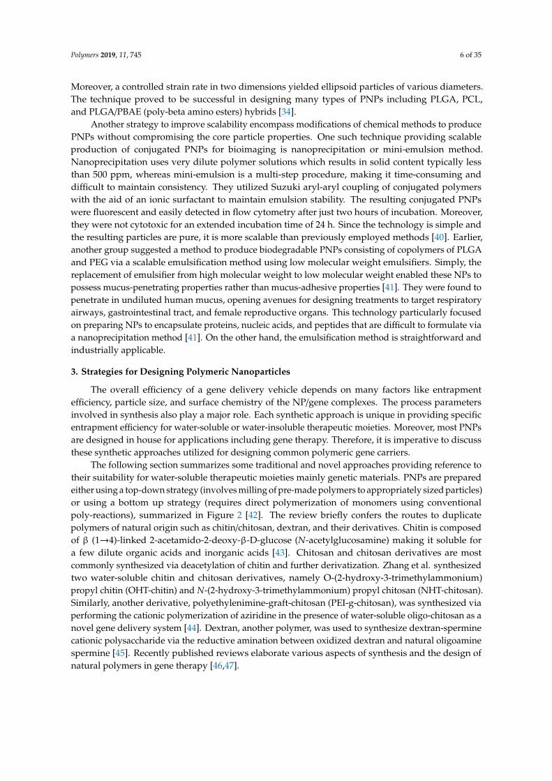
3.1.4. Dialysis
Dialysis is one of the simplest and effective methods to prepare small and uniformly distributed PNPs [54,63–65]. The method involves keeping the polymer (dissolved in an organic solvent) inside a dialysis tube with appropriate molecular weight range and performing dialysis against a non-miscible solvent. The homogenous suspension of NPs is formed resulting from solvent displacement inside the membrane followed by polymer aggregation due to a loss of solubility (Figure 6) [66]. The mechanism of PNP formation via dialysis method is yet to be understood. There are various polymers and co-polymers that have been synthesized using this technique [66–71] such as poly(benzyl-L-glutamate)-b-poly(ethylene oxide) and poly(lactide)-b-poly(ethylene oxide) NPs [72].
Figure 4. Schematic representation of the nanoprecipitation (solvent displacement) technique. Reprintedwith permission from Reference [53]. Copyright 2006 Elsevier.
3.1.3. Salting Out
This technique is based on the simple principle of separating a water miscible solvent fromaqueous solution via salting out (Figure 5). The procedure is the improved and modified version ofthe emulsification/solvent diffusion. Here, an organic solvent, such as acetone, is used to dissolve thepolymer and the drug, followed by its emulsification in an aqueous gel containing the salting-out agent(such as magnesium chloride, calcium chloride, or magnesium acetate), or non-electrolytes (such assucrose) and a colloidal stabilizer (such as polyvinylpyrrolidone (PVP) or hydroxyethylcellulose) [60].The formation of nanospheres is triggered by diluting the oil/water emulsion using an adequate amountof aqueous phase [61]. The salting-out agent plays a vital role in deciding the encapsulation efficiencyof the drug; therefore, careful selection should be applied. Like the nanoprecipitation method, saltingout also has a greater affinity to encapsulate lipophilic moieties [62].
Polymers 2019, 11, x FOR PEER REVIEW 8 of 34
Figure 4. Schematic representation of the nanoprecipitation (solvent displacement) technique. Reprinted with permission from Reference [53]. Copyright 2006 Elsevier.
3.1.3. Salting Out
This technique is based on the simple principle of separating a water miscible solvent from aqueous solution via salting out (Figure 5). The procedure is the improved and modified version of the emulsification/solvent diffusion. Here, an organic solvent, such as acetone, is used to dissolve the polymer and the drug, followed by its emulsification in an aqueous gel containing the salting-out agent (such as magnesium chloride, calcium chloride, or magnesium acetate), or non-electrolytes (such as sucrose) and a colloidal stabilizer (such as polyvinylpyrrolidone (PVP) or hydroxyethylcellulose) [60]. The formation of nanospheres is triggered by diluting the oil/water emulsion using an adequate amount of aqueous phase [61]. The salting-out agent plays a vital role in deciding the encapsulation efficiency of the drug; therefore, careful selection should be applied. Like the nanoprecipitation method, salting out also has a greater affinity to encapsulate lipophilic moieties [62].
Figure 5. Schematic representation of the salting out technique. Reprinted with permission from Reference [53]. Copyright 2006 Elsevier.
3.1.4. Dialysis
Dialysis is one of the simplest and effective methods to prepare small and uniformly distributed PNPs [54,63–65]. The method involves keeping the polymer (dissolved in an organic solvent) inside a dialysis tube with appropriate molecular weight range and performing dialysis against a non-miscible solvent. The homogenous suspension of NPs is formed resulting from solvent displacement inside the membrane followed by polymer aggregation due to a loss of solubility (Figure 6) [66]. The mechanism of PNP formation via dialysis method is yet to be understood. There are various polymers and co-polymers that have been synthesized using this technique [66–71] such as poly(benzyl-L-glutamate)-b-poly(ethylene oxide) and poly(lactide)-b-poly(ethylene oxide) NPs [72].
Figure 5. Schematic representation of the salting out technique. Reprinted with permission fromReference [53]. Copyright 2006 Elsevier.
Polymers 2019, 11, 745 9 of 35
3.1.4. Dialysis
Dialysis is one of the simplest and effective methods to prepare small and uniformly distributedPNPs [54,63–65]. The method involves keeping the polymer (dissolved in an organic solvent)inside a dialysis tube with appropriate molecular weight range and performing dialysis againsta non-miscible solvent. The homogenous suspension of NPs is formed resulting from solventdisplacement inside the membrane followed by polymer aggregation due to a loss of solubility(Figure 6) [66]. The mechanism of PNP formation via dialysis method is yet to be understood. There arevarious polymers and co-polymers that have been synthesized using this technique [66–71] such aspoly(benzyl-l-glutamate)-b-poly(ethylene oxide) and poly(lactide)-b-poly(ethylene oxide) NPs [72].Polymers 2019, 11, x FOR PEER REVIEW 9 of 34
Figure 6. Schematic representation of an osmosis-based method for the preparation of polymer nanoparticles.
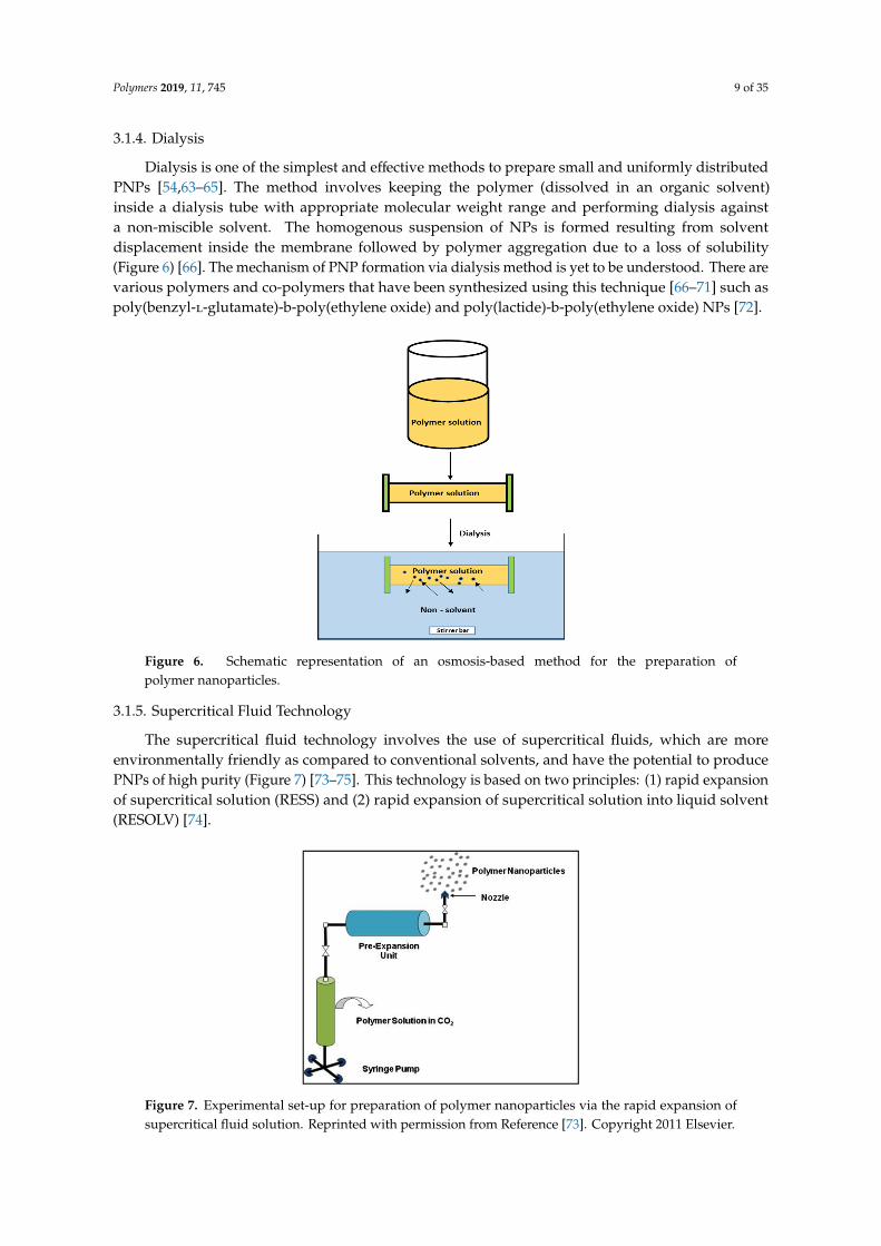
3.1.5. Supercritical Fluid Technology
The supercritical fluid technology involves the use of supercritical fluids, which are more environmentally friendly as compared to conventional solvents, and have the potential to produce PNPs of high purity (Figure 7) [73–75]. This technology is based on two principles: (1) rapid expansion of supercritical solution (RESS) and (2) rapid expansion of supercritical solution into liquid solvent (RESOLV) [74].
Using the conventional RESS principle for supercritical fluid technology, well-dispersed particles are formed by dissolving the solute in a supercritical fluid, followed by the rapid expansion of the solution across an orifice into surrounding air. The high degree of super saturation along with prompt reduction in the pressure for expansion, results in the formation of homogenous particles [50]. Poly (perfluoropolyetherdiamide) droplets are produced using this technique. Keshavarz et al. were able to use this technique to prepare raloxifene NPs with the smallest particle size being 18.93 ± 3.73 nm and having a PDI less than 0.1 [76].
Figure 7. Experimental set-up for preparation of polymer nanoparticles via the rapid expansion of supercritical fluid solution. Reprinted with permission from Reference [73]. Copyright 2011 Elsevier.
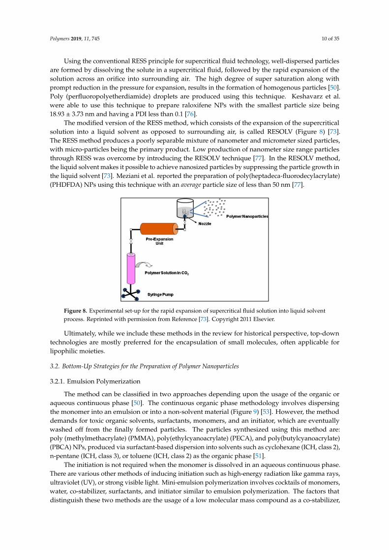
The modified version of the RESS method, which consists of the expansion of the supercritical solution into a liquid solvent as opposed to surrounding air, is called RESOLV (Figure 8) [73]. The RESS method produces a poorly separable mixture of nanometer and micrometer sized particles, with micro-particles being the primary product. Low production of nanometer size range particles
Figure 6. Schematic representation of an osmosis-based method for the preparation ofpolymer nanoparticles.
3.1.5. Supercritical Fluid Technology
The supercritical fluid technology involves the use of supercritical fluids, which are moreenvironmentally friendly as compared to conventional solvents, and have the potential to producePNPs of high purity (Figure 7) [73–75]. This technology is based on two principles: (1) rapid expansionof supercritical solution (RESS) and (2) rapid expansion of supercritical solution into liquid solvent(RESOLV) [74].
Polymers 2019, 11, x FOR PEER REVIEW 9 of 34
Figure 6. Schematic representation of an osmosis-based method for the preparation of polymer nanoparticles.
3.1.5. Supercritical Fluid Technology
The supercritical fluid technology involves the use of supercritical fluids, which are more environmentally friendly as compared to conventional solvents, and have the potential to produce PNPs of high purity (Figure 7) [73–75]. This technology is based on two principles: (1) rapid expansion of supercritical solution (RESS) and (2) rapid expansion of supercritical solution into liquid solvent (RESOLV) [74].
Using the conventional RESS principle for supercritical fluid technology, well-dispersed particles are formed by dissolving the solute in a supercritical fluid, followed by the rapid expansion of the solution across an orifice into surrounding air. The high degree of super saturation along with prompt reduction in the pressure for expansion, results in the formation of homogenous particles [50]. Poly (perfluoropolyetherdiamide) droplets are produced using this technique. Keshavarz et al. were able to use this technique to prepare raloxifene NPs with the smallest particle size being 18.93 ± 3.73 nm and having a PDI less than 0.1 [76].
Figure 7. Experimental set-up for preparation of polymer nanoparticles via the rapid expansion of supercritical fluid solution. Reprinted with permission from Reference [73]. Copyright 2011 Elsevier.
The modified version of the RESS method, which consists of the expansion of the supercritical solution into a liquid solvent as opposed to surrounding air, is called RESOLV (Figure 8) [73]. The RESS method produces a poorly separable mixture of nanometer and micrometer sized particles, with micro-particles being the primary product. Low production of nanometer size range particles
Figure 7. Experimental set-up for preparation of polymer nanoparticles via the rapid expansion ofsupercritical fluid solution. Reprinted with permission from Reference [73]. Copyright 2011 Elsevier.
Polymers 2019, 11, 745 10 of 35
Using the conventional RESS principle for supercritical fluid technology, well-dispersed particlesare formed by dissolving the solute in a supercritical fluid, followed by the rapid expansion of thesolution across an orifice into surrounding air. The high degree of super saturation along withprompt reduction in the pressure for expansion, results in the formation of homogenous particles [50].Poly (perfluoropolyetherdiamide) droplets are produced using this technique. Keshavarz et al.were able to use this technique to prepare raloxifene NPs with the smallest particle size being18.93 ± 3.73 nm and having a PDI less than 0.1 [76].
The modified version of the RESS method, which consists of the expansion of the supercriticalsolution into a liquid solvent as opposed to surrounding air, is called RESOLV (Figure 8) [73].The RESS method produces a poorly separable mixture of nanometer and micrometer sized particles,with micro-particles being the primary product. Low production of nanometer size range particlesthrough RESS was overcome by introducing the RESOLV technique [77]. In the RESOLV method,the liquid solvent makes it possible to achieve nanosized particles by suppressing the particle growth inthe liquid solvent [73]. Meziani et al. reported the preparation of poly(heptadeca-fluorodecylacrylate)(PHDFDA) NPs using this technique with an average particle size of less than 50 nm [77].
Polymers 2019, 11, x FOR PEER REVIEW 10 of 34
through RESS was overcome by introducing the RESOLV technique [77]. In the RESOLV method, the liquid solvent makes it possible to achieve nanosized particles by suppressing the particle growth in the liquid solvent [73]. Meziani et al. reported the preparation of poly(heptadeca-fluorodecylacrylate) (PHDFDA) NPs using this technique with an average particle size of less than 50 nm [77].
Figure 8. Experimental set-up for the rapid expansion of supercritical fluid solution into liquid solvent process. Reprinted with permission from Reference [73]. Copyright 2011 Elsevier.
Ultimately, while we include these methods in the review for historical perspective, top-down technologies are mostly preferred for the encapsulation of small molecules, often applicable for lipophilic moieties.
3.2. Bottom-Up Strategies for the Preparation of Polymer Nanoparticles
3.2.1. Emulsion Polymerization
The method can be classified in two approaches depending upon the usage of the organic or aqueous continuous phase [50]. The continuous organic phase methodology involves dispersing the monomer into an emulsion or into a non-solvent material (Figure 9) [53]. However, the method demands for toxic organic solvents, surfactants, monomers, and an initiator, which are eventually washed off from the finally formed particles. The particles synthesized using this method are: poly (methylmethacrylate) (PMMA), poly(ethylcyanoacrylate) (PECA), and poly(butylcyanoacrylate) (PBCA) NPs, produced via surfactant-based dispersion into solvents such as cyclohexane (ICH, class 2), n-pentane (ICH, class 3), or toluene (ICH, class 2) as the organic phase [51].
The initiation is not required when the monomer is dissolved in an aqueous continuous phase. There are various other methods of inducing initiation such as high-energy radiation like gamma rays, ultraviolet (UV), or strong visible light. Mini-emulsion polymerization involves cocktails of monomers, water, co-stabilizer, surfactants, and initiator similar to emulsion polymerization. The factors that distinguish these two methods are the usage of a low molecular mass compound as a co-stabilizer, and the use of high-shear devices such as ultrasound generators. Mini-emulsions are disparagingly stabilized, calling for high-shear to achieve a steady state and have a high interfacial tension [73]. On the contrary, micro-emulsion polymerization results in having considerably smaller particle size and average number of chains per particle [50]. In micro-emulsion polymerization, a water-soluble agent acting as an initiator is mixed in the aqueous phase of thermodynamically stable micro-emulsion containing swollen micelles. The type and concentration of the initiator, nature of the surfactant and the monomer, and reaction temperature are a few factors influencing micro-emulsion polymerization kinetics and the properties of PNP [54,78].
Figure 8. Experimental set-up for the rapid expansion of supercritical fluid solution into liquid solventprocess. Reprinted with permission from Reference [73]. Copyright 2011 Elsevier.
Ultimately, while we include these methods in the review for historical perspective, top-downtechnologies are mostly preferred for the encapsulation of small molecules, often applicable forlipophilic moieties.
3.2. Bottom-Up Strategies for the Preparation of Polymer Nanoparticles
3.2.1. Emulsion Polymerization
The method can be classified in two approaches depending upon the usage of the organic oraqueous continuous phase [50]. The continuous organic phase methodology involves dispersingthe monomer into an emulsion or into a non-solvent material (Figure 9) [53]. However, the methoddemands for toxic organic solvents, surfactants, monomers, and an initiator, which are eventuallywashed off from the finally formed particles. The particles synthesized using this method are:poly (methylmethacrylate) (PMMA), poly(ethylcyanoacrylate) (PECA), and poly(butylcyanoacrylate)(PBCA) NPs, produced via surfactant-based dispersion into solvents such as cyclohexane (ICH, class 2),n-pentane (ICH, class 3), or toluene (ICH, class 2) as the organic phase [51].
The initiation is not required when the monomer is dissolved in an aqueous continuous phase.There are various other methods of inducing initiation such as high-energy radiation like gamma rays,ultraviolet (UV), or strong visible light. Mini-emulsion polymerization involves cocktails of monomers,water, co-stabilizer, surfactants, and initiator similar to emulsion polymerization. The factors thatdistinguish these two methods are the usage of a low molecular mass compound as a co-stabilizer,
Polymers 2019, 11, 745 11 of 35
and the use of high-shear devices such as ultrasound generators. Mini-emulsions are disparaginglystabilized, calling for high-shear to achieve a steady state and have a high interfacial tension [73].On the contrary, micro-emulsion polymerization results in having considerably smaller particle sizeand average number of chains per particle [50]. In micro-emulsion polymerization, a water-solubleagent acting as an initiator is mixed in the aqueous phase of thermodynamically stable micro-emulsioncontaining swollen micelles. The type and concentration of the initiator, nature of the surfactant andthe monomer, and reaction temperature are a few factors influencing micro-emulsion polymerizationkinetics and the properties of PNP [54,78].Polymers 2019, 11, x FOR PEER REVIEW 11 of 34
Figure 9. Schematic representation of the emulsification/solvent diffusion technique. Reprinted with permission from Reference [53]. Copyright 2006 Elsevier.
3.2.2. Recombinant Technology
Cationic polymers synthesized by utilizing recombinant DNA technology have the potential to address some of the major challenges of gene delivery such as the low ability to target cells, poor intracellular trafficking of the genetic material, and nuclear uptake. Synthetic methods of polymer production involving conventional thermodynamically-driven chemical techniques are inadequate for gene delivery purposes as the resultant products are heterogeneous with regard to composition and molecular weight. In contrast, amino acid-based polymers synthesized via recombinant technology in living systems, such as E. coli, produces homogenous biopolymers with a specific composition where function can be influenced by the amino acid sequence [79]. This paves the path for multiple functionality approaches, allowing for a single biopolymer to have multiple functions merely by changing the protein expression. Aris et al. reported the engineering of a gene delivery system, namely 249AL, composed of a cationic lysine oligomer (K10) conjugated with ß-galactosidase-derived protein displaying an arginylglycylaspartic acid (RGD) cell attachment peptide [80]. The K10
aids in the condensation of plasmid DNA (pDNA), whereas RGD interacts with the αVβ3 integrin present on the cell membrane [81]. The efficiency of the system was determined by complexing 249AL with pDNA encoding a luciferase reporter gene and transfecting CaCo2 cells. Since the 249AL was aimed to be target-specific, the percentage of transfected cells along with the total gene expression could help better understand the efficiency of the system. The transfection efficiency of 249AL was significantly lower compared to commercially available transfecting agents since 249AL was unable to perform an endosomal escape [81]. Similarly, Furgeson’s group reported the development of a recombinant elastin-based cationic di-block biopolymer for gene delivery [82]. The biopolymer consisted of a cationic oligomer block (VGK8G) conjugated with a thermo-responsive elastin-like polymer with 60 repeats of Val–Pro–Gly–Xaa–Gly (VPGXG). This particular approach is pseudo-biosynthetic, utilizing a recursive directional ligation method to synthesize the gene [82,83]. Similar to the 249AL, the biopolymer was not capable of escaping the endosome. To overcome this challenge, another group, Hatefi et al., reported the first recombinant cationic biopolymer with tandem repeating units of basic amino acids such as lysine (K) and histidine (H) residues conjugated with fibroblast growth factor 2 (FGF2) [84]. The biopolymer denoted as dKH-FGF2, with 36 lysine residues and 24 histidine residues, facilitated in condensing the pDNA and performing an endosomal escape via a proton sponge effect respectively [85]. FGF2 conjugation provided specific targeting to fibroblast growth factor receptor (FGFR) on cells such as T47D (breast cancer) and NIH3T3 (fibroblasts). The results showed that the biopolymer was able to condense pDNA into NPs and induced significant cell proliferation. While the result of the transfection efficiency studies suggested targeted gene transfer via FGFR, the biopolymer efficiency was not optimal, but showed potential for optimization [84].
Overall, the preparation of PNPs is yet to be perfected. Lipophilic drug loaded nanospheres or nanocapsules can be synthesized using simple, safe methods with good reproducibility. However, as we move to more complex and sensitive therapeutic cargos like genetic materials, careful selection of the appropriate method is crucial in order to achieve appropriate physicochemical characteristics (Table 2), minimal interference of process parameters and maximum entrapment efficiency.
Figure 9. Schematic representation of the emulsification/solvent diffusion technique. Reprinted withpermission from Reference [53]. Copyright 2006 Elsevier.
3.2.2. Recombinant Technology
Cationic polymers synthesized by utilizing recombinant DNA technology have the potentialto address some of the major challenges of gene delivery such as the low ability to target cells,poor intracellular trafficking of the genetic material, and nuclear uptake. Synthetic methods of polymerproduction involving conventional thermodynamically-driven chemical techniques are inadequate forgene delivery purposes as the resultant products are heterogeneous with regard to composition andmolecular weight. In contrast, amino acid-based polymers synthesized via recombinant technologyin living systems, such as E. coli, produces homogenous biopolymers with a specific compositionwhere function can be influenced by the amino acid sequence [79]. This paves the path for multiplefunctionality approaches, allowing for a single biopolymer to have multiple functions merely bychanging the protein expression. Aris et al. reported the engineering of a gene delivery system,namely 249AL, composed of a cationic lysine oligomer (K10) conjugated with ß-galactosidase-derivedprotein displaying an arginylglycylaspartic acid (RGD) cell attachment peptide [80]. The K10 aids inthe condensation of plasmid DNA (pDNA), whereas RGD interacts with the αVβ3 integrin presenton the cell membrane [81]. The efficiency of the system was determined by complexing 249AL withpDNA encoding a luciferase reporter gene and transfecting CaCo2 cells. Since the 249AL was aimed tobe target-specific, the percentage of transfected cells along with the total gene expression could helpbetter understand the efficiency of the system. The transfection efficiency of 249AL was significantlylower compared to commercially available transfecting agents since 249AL was unable to performan endosomal escape [81]. Similarly, Furgeson’s group reported the development of a recombinantelastin-based cationic di-block biopolymer for gene delivery [82]. The biopolymer consisted of a cationicoligomer block (VGK8G) conjugated with a thermo-responsive elastin-like polymer with 60 repeats ofVal–Pro–Gly–Xaa–Gly (VPGXG). This particular approach is pseudo-biosynthetic, utilizing a recursivedirectional ligation method to synthesize the gene [82,83]. Similar to the 249AL, the biopolymerwas not capable of escaping the endosome. To overcome this challenge, another group, Hatefi et al.,reported the first recombinant cationic biopolymer with tandem repeating units of basic amino acidssuch as lysine (K) and histidine (H) residues conjugated with fibroblast growth factor 2 (FGF2) [84].The biopolymer denoted as dKH-FGF2, with 36 lysine residues and 24 histidine residues, facilitated incondensing the pDNA and performing an endosomal escape via a proton sponge effect respectively [85].FGF2 conjugation provided specific targeting to fibroblast growth factor receptor (FGFR) on cellssuch as T47D (breast cancer) and NIH3T3 (fibroblasts). The results showed that the biopolymer was
Polymers 2019, 11, 745 12 of 35
able to condense pDNA into NPs and induced significant cell proliferation. While the result of thetransfection efficiency studies suggested targeted gene transfer via FGFR, the biopolymer efficiencywas not optimal, but showed potential for optimization [84].
Overall, the preparation of PNPs is yet to be perfected. Lipophilic drug loaded nanospheres ornanocapsules can be synthesized using simple, safe methods with good reproducibility. However,as we move to more complex and sensitive therapeutic cargos like genetic materials, careful selectionof the appropriate method is crucial in order to achieve appropriate physicochemical characteristics(Table 2), minimal interference of process parameters and maximum entrapment efficiency.
Table 2. Comparison of average particle size and polydispersity index (PDI) between varioussynthesis methods.
Method of Polymer Synthesis Example Average ParticleSize (nm) PDI Ref.
3.3. Polymerization Chemistries for Common Synthetic Polymers
3.3.1. Poly(Lactic Acid)
Some synthetic polymers can be designed via multiple types of polymerization chemistry.An example of such a polymer is PLA. It has been researched widely for gene therapy over the yearsdue to its ease of availability, reasonable pricing, and biodegradable nature. The building block of thispolymer is lactic acid, produced naturally via fermentation. The literature identifies two polymerizationmechanisms for synthesizing PLA [86].
1. Direct condensation of lactic acid: It is the conventional method of synthesis utilizing solventsand exhibiting high reaction times [86]. It has been done using diphenyl ether as a solventin the presence of tin (II) chloride as the catalyst. The process is strictly dependent on thepolymerization temperature and pressure. An increase in temperature leads to a high molecularweight PLA [87]. Other solvent systems like p-xylene [88] have also been employed. Solid-statedirect poly-condensation without the use of a solvent was proposed utilizing this reactionchemistry. A pre-polymer product was formed first, using p-toluene sulfonic acid without theaddition of any catalyst. This product was then subjected to solid-state polymerization underhigh temperature and pressure conditions [89].
2. Ring opening polymerization of lactide: This process is completed in two steps. In the firststep, lactic acid cyclizes into lactide (a close chain lactone di-ester) under heat and a vacuum.A nitrogen-controlled inert environment is used to speed up the removal of water vapors,enhancing cyclization. The second step involves disruption of the cyclic ring, followed by theunion of open chains forming the polymer. This step is catalyzed by stannous octoate to promoteformation of ester linkages [86,90]. Process parameters and solvents used in this approachfulfills the requirements of “green chemistry” [90], a novel advancement in polymer nanoscience,which was discussed previously.
Polymers 2019, 11, 745 13 of 35
3.3.2. Poly-l-Lysine
Poly-l-lysine (PLL) has been synthesized in many structural conformations and molecular weights,i.e., linear, dendritic, and hyper-branched, each exhibiting a characteristic safety profile [91]. Linear PLLis synthesized via polymerizing the monomers under heat and vacuum, followed by precipitation usingdiethyl ether. Dendritic PLL is created by conjugating lysine monomers as a branch unit. They haveshown improved gene transfection compared to their linear counterparts [92]. Dendritic conformationutilizes an initiator core like hexa-methylene-diamine, on which lysine monomers are coupledrepeatedly in various conformations [93]. Hyper-branched PLL is a relatively newer conformationstructurally related to dendritic PLLs. Unlike dendritic conformations that exhibits a single core,hyper-branched PLL possesses a randomly branched structure, and it is attractive as it can be producedin a one-step operation [94]. No initiator core is involved and lysine monomers are polymerized usingheat in an inert environment created using a nitrogen gas influx. Once polymerization is completed,the final product is collected via precipitation [94,95].
3.3.3. Poly(Amidoamine)
Poly(amidoamine) (PAMAM) is also an attractive non-viral vector for gene delivery, especiallyfor complex targets like cochlea in the inner ear [96] or glioblastoma in the brain [97]. It consists ofan alkyl-amine core and tertiary amine branches in dendritic conformations. In most cases, the coreutilizes ethylene-diamine-tetra-acetic acid (EDTA) [98].
3.3.4. Poly(Methyl-Methacrylate)
Poly(methyl-methacrylate) PMMA is one of the extensively investigated polymers for itsapplication in electrospinning [99], synthesis of carbon nanotube/PMMA composites, and for highrefractive index thin-film fabrication [100]. The properties of the PMMA depends greatly on theresulting molecular weight. A bottom up approach of its polymerization involves ionic and freeradical polymerization. The anionic polymerization consist of an active anionic center. It is adynamic polymerization technique in which the chain termination does not occur until the additionof a terminating agent [101]. The degree of polymerization is determined by the molar ratio ofmonomer versus initiator, in the absence of a terminating agent. Anionic polymerization is used toproduce PMMA-PS with a low molecular weight. The bottleneck of the anionic polymerization is therequirement for stringent reaction conditions as the anion is sensitive to an environment consisting ofboth oxygen and water. Hence, the process requires purification of all the polymerization reagentsand inert atmospheric conditions [102,103]. The polymerization technique is used to synthesize highmolecular weight PMMA and its related block copolymers. Another polymerization technique isreversible addition fragmentation chain transfer (RAFT). The technique is very similar to other freeradical polymerization. In RAFT, the thermochemical initiator or the interaction of gamma or UVradiation with some reagents gives away free radicals [104]. High molecular weight PMMA can also besuccessfully synthesized by other techniques like activators regenerated by electron transfer (ARGET)or atom transfer radical polymerization (ATRP). These new techniques require a significantly smalleramount of Cu (II) species [105]. Conclusively, a number of bottom-up techniques are available for thesynthesis of high molecular weight PMMA.
3.3.5. Poly(Ethylene-Imine)
Twenty-five kilodalton (kDa) branched poly(ethylene-imine) (PEI) and 22 kDa linear PEI are themost common types employed for gene therapy [106]. The mechanism put forward for the synthesisof linear PEI involves a cationic ring opening polymerization of 2-oxazoline [106,107]. When acylatedoxazoline is used, i.e., methyl or ethyl oxazoline, the reaction is processed via hydrolysis under strongacid and elevated temperature in the aqueous medium [107]. Like linear PEIs, branched PEI is preparedvia a cationic ring opening polymerization of aziridine [106]. This is achieved through an electrophilic
Polymers 2019, 11, 745 14 of 35
attack of protons on the aziridine monomer [106,108]. The overall polymerization reaction can beprocessed via the catalytic activity of acid and input of heat in aqueous or alcoholic solution [108].Detailed reaction chemistry and the control of individual steps is presented by Jager et al [106].
PEI has been successfully developed as a commercial DNA transfection reagent. The system calledjetPEITM produces an efficient gene transfection for up to 4 h with minimal cytotoxicity [109]. It ismade of linear PEI chains and is useful for both adherent and suspension cells. Cell-specific versionsare also designed for this reagent like jetPEI®-Macrophage (primary macrophages, glial, and dendriticcells), jetPEI®-Hepatocyte (liver cells), and jetPEI®-HUVEC (endothelial cells) [109].
3.3.6. Poly(Lactic-co-Glycolide)
Poly(lactic-co-glycolide) (PLGA) is a synthetic copolymer of lactic acid and glycolic acid. PLGA canbe synthesized via direct poly-condensation of lactic and glycolic acid; however, the most efficientand prevalent scheme to obtain high molecular weight copolymers is the ring opening polymerizationof lactide and glycolide. The synthesis of high molecular weight PGLA using poly-condensationdemands for a high degree of dehydration, which is difficult to achieve and is considered an inefficientmethod to obtain a good yield of polymers [110]. To prepare high molecular weight PLGA in a shorterreaction time, it is important to proceed via ring-opening polymerization of cyclic diesters, lactide,and glycolide.
Both PLA and PLGA are FDA-approved polymers used in commercially available drug products.They have been extensively used in drug delivery for reducing the lowest effective dose, targetingdrug activity, and reducing the drug toxicity. To date, FDA has approved 15 drug products utilizingPLA or PLGA [111]. Some examples include:
Since their use is successful in the area of drug delivery, PLA and PLGA-based gene carriers arepromising for achieving similar success for gene therapeutics.
4. Challenges Associated with the Use of Polymers in Nanomedicine
Every new technology is a two-edged sword. Like many other non-viral vectors, PNPs also havesome challenges in terms of safety and stability as nanocarriers. As new generations of PNPs pavetheir way toward clinical trials as vectors, focus is laid on overcoming these challenges. PNPs withoptimum shape and flexibility for the best interaction with the cellular membrane, and which exhibitcompartmentalization for loading genes and targeting moieties, are designed. To reduce the systemic
Polymers 2019, 11, 745 15 of 35
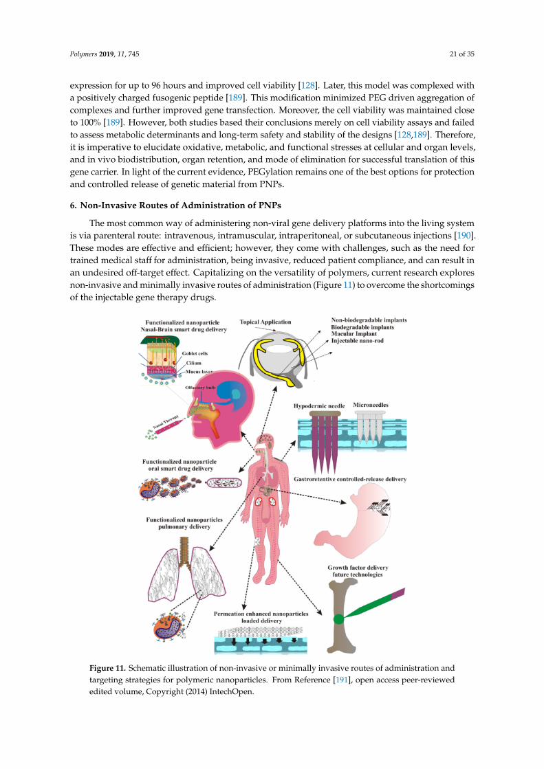
toxicity, PNPs that mimic biological molecules are created. Importance is given to reduce traffickingand off-target accumulation, and finally, effort is made to introduce stimulus-responsive behaviors.The following paragraphs will briefly discuss the limitations of PNPs and advancement in research toovercome them (Figure 10).
Polymers 2019, 11, x FOR PEER REVIEW 17 of 34
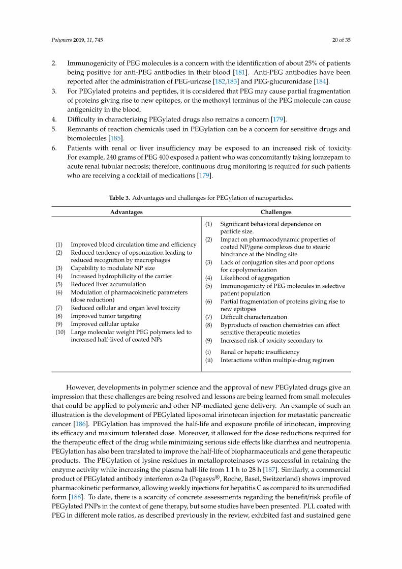
their in vivo metabolism and elimination routes. This further adds to the efforts and resources required for the formulation, development, and metabolite tracking. Therefore, although this area has consistently shown value for drug/gene delivery, many designs do not successfully reach the stage of clinical translation. Moreover, bench-top research now focuses on multifuntional PNPs containing a combination of functional groups to achieve encapsulation, delivery, and targeting. More functionalization means more sophisticated reaction chemistries, a need for elaborative tracking for degradation products, and more stringent process design parameters, which are all associated with higher cost, development times, and regulatory hurdles. Moreover, when these formulations are introduced in the marketplace, they are generally very expensive. Launching generic counterparts quickly after approval and bringing government and private drug plans on board for these technologies would make them more accessible to the general patient population.
Figure 10. Summary of properties and challenges of polymeric nanoparticles for gene delivery and associated factors influencing each of these parameters.
5. Biodistribution and Cellular Interaction of Polymeric Nanoparticles
PNPs are capable of overcoming multiple biological barriers and controlling the release of their therapeutic load. However, preventing a rapid clearance of circulating NPs is an acute issue for their application to full potential; therefore, it is imperative to understand the factors influencing their circulation time and biodistribution. These factors define the ability to overcome body’s defense mechanisms and include tailorable physiochemical properties, such as composition, configuration, size, core properties, and surface functionalization (PEGylation, charged moieties, and targeting ligand functionalization) [157]. The interaction of the NPs with the biological environment is mainly influenced by opsonization, where opsonin proteins found in the blood serum interact and expose the NPs to macrophages in the mononuclear phagocytic system (MPS), triggering their removal from the biological system [120]. After opsonization, phagocytosis occurs, which involves the engulfing and destruction of foreign material from the bloodstream. PNPs that cannot be eliminated by phagocytes are sequestered in the MPS organs (liver and spleen). Here it is important to note that in the case of non-biodegradable PNPs, accumulation in these target organs may result in negative outcomes [158–160]. Overall, the process of in vivo trafficking in the blood stream compromise their designed therapeutic function.
There are no straight-forward strategies to completely avoid opsonization of NPs. However, significant research in this area over the last 35 years have identified patterns and method for
Figure 10. Summary of properties and challenges of polymeric nanoparticles for gene delivery andassociated factors influencing each of these parameters.
4.1. Stability of PNPs in an Electrolyte and Protein-Rich Biological Medium
Polyplexes formed between cationic polymers and genetic materials are based on electrostaticinteractions. Factors including the molecular weight, hydrophilicity, surface charge, and structure of thecationic polymers define the efficacy of the carrier [15]. Among common PNPs, PLA (degradable) andPMMA (non-degradable) are most noteworthy as they are approved for human use by FDA [112,113].Lazzari et al. analyzed the colloidal stability of these polymers in simulated biological media basedon different characteristics. PLA NPs showed a high degree of aggregation and a 20% increasein the mean particle size distribution in synthetic gastric juice, while PMMA NPs remained stable.This was anticipated due to the difference in the surface charge densities (i.e., zeta potentials) of thetwo formulations. If the NPs aggregate during circulation, the risk of clotting increases and overallrelease of the drug is compromised. PMMA NPs showed higher stability in serum and other organhomogenates as compared to PLA NPs due to higher surface charges [114].
Another prominent parameter controlling PNPs stability in biological fluids is the hydrophobicityof the surface. For example, a less hydrophobic version of N-isopropyl-acrylamide and N-tertiary-butyl-acrylamide copolymer particles hindered protein binding on the surface as compared to amore hydrophobic version, which bound series of proteins including apo-lipoproteins, albumin,and fibrinogen, thus increasing the risk of phagocytic destruction [115]. Unlike many other NPs,the challenge for PNPs is that changing the hydrophobicity of polymers is very difficult withoutadversely affecting the size, surface charge, and composition [116].
Despite the instability of some NPs in serum rich media, few polymers when conjugated withother organic and inorganic NPs improves the overall stability of the carrier and reduces opsonizationduring blood circulation. A typical example put into practice for many NP formulations is PEGylation.Attachment of PEG on the surface extends the circulation time of NPs by effectively reducing
Polymers 2019, 11, 745 16 of 35
random interactions with proteins, recognition by immune cells, and clearance through excretoryorgans [117–119].
4.2. Accumulation and Toxicity of Polymeric Nanoparticles
PNPs made of non-degradable polymers tend to accumulate in the organs, mainly the liver andspleen, leading to toxicity [120]. The most studied example for drug delivery purposes is dextran,a glucose-based polymer [121–124]. Serious allergic reactions including anaphylaxis, volume overload,pulmonary and cerebral edema, and platelet dysfunction have been reported via in vivo studies on thiscarrier [123–125]. Dextran also exhibits a strong osmotic effect leading to acute renal failure and henceits use is contraindicated in patients with renal insufficiency and diabetes mellitus [126]. Although,the knowledge gained for this system might not be directly applicable to synthetic polymers used nowfor gene delivery, the overall chemical composition, particle size, shape, and surface properties ofPNPs are contributors to their safety profile.
PLL is one of the primary cationic polymer used as a non-viral gene vector [127]. UnmodifiedPLL shows high in vitro toxicity, reducing the cell viability to as low as 50% [128]. Multiple effortshave been put forward to improve the biocompatibility of this carrier. Coating of PLL with PEGranging from 5–25 mol% improved the transfection and biocompatibility for up to 96 h [128]. In recentyears, more sophisticated coatings have been performed to further improve both in vitro and in vivobiocompatibility. An example of such a design is PEG-PLL-PLGA copolymers, which showed no bloodtoxicity or genotoxicity in vivo and revealed a wide safe scale of dosing [129].
Polyethylenimine (PEI) is another pioneer polymer used for gene delivery. It is the proof-of-conceptfor the design of NPs capable of condensing genetic materials and allowing intracellular endosomalescape to enhance transfection efficiency [130]. This cationic polymer has also shown considerabletoxicity, depending on the structure (either linear or branched) and molecular weight [131,132].Low molecular weight and moderately branched design is generally considered safer as compared tolinear PEI [132]. However, the literature shows evidence that branched PEI has lower IC50 values ascompared to linear PEI (37 mg vs. 74 mg, respectively) [133]. Moreover, the number of primary amineson the surface, and hence the surface cationic character, is directly proportional to hematologicalcompatibility and cytotoxicity [133]. In free form, PEI may interact with the negatively chargedserum proteins and red blood cells, destabilizing the plasma membrane and leading to immediatetoxicity [134]. As complexes with genetic materials, this effect decreases but can be restored oncethe cargo is transported and free PEI appears back in the blood for excretion. Free PEI also interactswith cellular components and interferes with the normal processes [135,136]. PEGylation of PEI NPsis one the approaches presented to improve their safety profile. Conjugation of PEG at 6 and 10%weight ratios significantly reduced the cytotoxicity and aided in the local delivery of genes to themuscles. This study showed that a higher PEG ratio on the surface exhibits better safety [137]. Anotherapproach tried to reduce the toxicity of free PEI is the coating of PEI/DNA polyplexes with lysinylated,histidylated, and arginylated cholesterol. This morphology improved the nuclear delivery of DNAat low PEI/DNA weight ratio of 2, reducing the need of excess free PEI chains in the system [138].Only a few approaches are defined here; however, over time many designs have been put forward tominimize and even mitigate the safety challenges for these two most common PNPs.
Novel type of PNPs are dendrimers, which are repeatedly branched large spherical structureshaving a core, an inner shell, and an outer shell [139]. PAMAM is one of such well-knowndendrimer used as chemotherapeutic and non-viral DNA delivery vehicle [140–142]. Amine-terminatedpositively charged PAMAM dendrimers exhibited hemolysis in rat blood cells by destabilizing thecell membrane [143]. Due to excellent delivery capabilities of this carrier, many approaches wereassessed to overcome this safety challenge. One such approach is to change the type of the terminalamine functionalities. Replacing primary amines with secondary or tertiary amines reduced thecytotoxicity of this carrier [144]. Fatty acid functionalization or PEGylation (discussed at length inthe following chapter) of PAMAM dendrimers reduced the cytotoxicity of the whole conjugate [145].
Polymers 2019, 11, 745 17 of 35
It is also believed that hydroxyl or meth-oxy terminated dendrimers are relatively safer than othercounterparts [125].
Physiological behavior of many PNPs also depends greatly on the encapsulated payload, as thisparameter changes its innate size, charge, stability, and other physicochemical properties. For examplePEG-PLGA NPs encapsulating a trial drug “KU50019” at various ratios yielded different particle sizes.The smaller drug-encapsulated PNPs showed higher small bowel toxicity than larger particles [146].
4.3. Oxidative Degradation of Polymers—Generation of Toxic Metabolites
Common biodegradable polymers are generally regarded as safe, but there is a scarcity ofdata collected on the short-term and long-term toxicity resulting from the accumulation of polymerdegradation products in the body. One elaborative study investigated in vitro toxicity of some polymersincluding PLGA, poly-ε-caprolactone (PCL), and poly(lactide-co-caprolactone) (PLCL), by studyingthe mechanisms of cytotoxicity induced through such metabolites [147]. The study concluded that allthe above polymers exhibited concentration- and time-dependent damage to healthy cells includingmacrophages, hepatocytes, lung and kidney epithelial cells, and neuronal cells [147]. It is debatablewhether these results can be extrapolated to in vivo scenarios as the human body is able to eliminatesuch metabolites through various pathways (e.g., lactate may undergo the Krebs cycle), which in turnretains the biocompatibility of the overall system [148–150]. However, the possibility of toxicity basedon concentrations and time of exposure persists if these degradative products are not readily clearedout from the site of accumulation.
PLGA is the most widely accepted FDA-approved biodegradable polymer [125,151,152]. Althoughdegradation products of PLGA are easily metabolized and eliminated by the body, and therefore thesystemic toxicity is limited, it is highly dependent on the ratio of lactide/glycolide components andthe total molecular weight [125,151,153]. Toxicity assays for PLGA NPs performed in mouse modelsshowed no tissue damage, but exhibited 40% retention in the liver following oral administration [154].Most studies conclude that, although PLGA NPs may tend to accumulate in tissues to a certain degree,they show very limited cytotoxicity or inflammatory responses irrespective of their surface properties.However, many times the use of PLGA in gene therapy requires further functionalization to improvegene transfection. This can lead to changes in their innate safety profile. So far, the functionalizationof PLGA NPs with other active molecules including secondary polymers have shown to either haveno effect or compliment the innate biocompatibility. Thus, no studies in the literature provide anyevidence that functionalization may trigger loss of innate PLGA safety. For instance, PEGylation ofPLGA NPs are shown to improve sub-cellular organelle targeted delivery, minimizing the randomexposure and accumulation of polymer complexes thus complimenting its safety [155]. On theother hand, functionalizing PLGA NPs with poly(ε-carbo-benzoxy-l-lysine) to improve the loadingcapacity of vascular endothelial growth factor did not have any effect on the polymer’s innatebiocompatibility [156].
4.4. High Cost Associated with Biological Analyses and Process Development
In addition to the above-mentioned challenges, another major concern that seems to be commonfor all nanoformulations is the time and cost associated with the assessment of their safety andefficacy. Since newer polymers are self-destructing biodegradable polymers, it is crucial to elucidatetheir in vivo metabolism and elimination routes. This further adds to the efforts and resourcesrequired for the formulation, development, and metabolite tracking. Therefore, although this areahas consistently shown value for drug/gene delivery, many designs do not successfully reach thestage of clinical translation. Moreover, bench-top research now focuses on multifuntional PNPscontaining a combination of functional groups to achieve encapsulation, delivery, and targeting.More functionalization means more sophisticated reaction chemistries, a need for elaborative trackingfor degradation products, and more stringent process design parameters, which are all associatedwith higher cost, development times, and regulatory hurdles. Moreover, when these formulations are
Polymers 2019, 11, 745 18 of 35
introduced in the marketplace, they are generally very expensive. Launching generic counterpartsquickly after approval and bringing government and private drug plans on board for these technologieswould make them more accessible to the general patient population.
5. Biodistribution and Cellular Interaction of Polymeric Nanoparticles
PNPs are capable of overcoming multiple biological barriers and controlling the release of theirtherapeutic load. However, preventing a rapid clearance of circulating NPs is an acute issue for theirapplication to full potential; therefore, it is imperative to understand the factors influencing theircirculation time and biodistribution. These factors define the ability to overcome body’s defensemechanisms and include tailorable physiochemical properties, such as composition, configuration,size, core properties, and surface functionalization (PEGylation, charged moieties, and targeting ligandfunctionalization) [157]. The interaction of the NPs with the biological environment is mainly influencedby opsonization, where opsonin proteins found in the blood serum interact and expose the NPs tomacrophages in the mononuclear phagocytic system (MPS), triggering their removal from the biologicalsystem [120]. After opsonization, phagocytosis occurs, which involves the engulfing and destruction offoreign material from the bloodstream. PNPs that cannot be eliminated by phagocytes are sequesteredin the MPS organs (liver and spleen). Here it is important to note that in the case of non-biodegradablePNPs, accumulation in these target organs may result in negative outcomes [158–160]. Overall,the process of in vivo trafficking in the blood stream compromise their designed therapeutic function.
There are no straight-forward strategies to completely avoid opsonization of NPs. However,significant research in this area over the last 35 years have identified patterns and method forincreasing the overall blood circulation half-life and effectiveness of the delivery system. For example,research provides evidence that opsonization of hydrophobic particles is quicker compared tohydrophilic counterparts due to the enhanced adsorption of serum proteins on those surfaces [161–163].For hydrophilic NPs, in vitro studies have established a correlation between surface charge andopsonization, indicating that neutrally charged particles have a much lower opsonization rate thancharged particles [164]. There are several polymeric systems that have been used as shieldinggroups to reduce protein adsorption, such as polysaccharides, polyacrylamide, poly(vinyl alcohol),poly(Nvinyl-2-pyrrolidone), PEG, and PEG-containing copolymers, such as poloxamers, poloxamines,and polysorbates. Out of all the polymers tested so far, the most effective solution is PEG orPEG-containing copolymers [120], thus we discuss the details of its biological profile and currentdevelopments in the next segment.
5.1. Biodistribution of the PEGylated Polymers
The PEGylated stealth improves the residence times in the blood and ultimately the efficiency ofPNPs. However, the rate of clearance and final biodistribution in the organ is dependent on multiplefactors. First and foremost, the particle size plays a crucial role in defining the final distribution andblood clearance of the PEGylated PNPs. Particles with a hydrodynamic size over 200 nm typicallyexhibit a more rapid rate of clearance than particles under 200 nm, regardless of PEGylation [165].In addition to the blood clearance rate, the final biodistribution is also affected by particle size.For instance, PEGylated NPs sized less than 150 nm was shown to exhibit a higher uptake in thebone marrow of rabbits, whereas particles sized 250 nm mostly sequestered in the spleen and liver,with only a small fraction of uptake by the bone marrow [165]. Moghimi et al. [166] suggested thatthe size-dependent biodistribution might be attributed to a filtering effect, whereby the spleen andliver removes larger particles more rapidly, while the smaller particles are directed to the bone marrow.However, the exact reason for these size dependencies is yet to be understood. Like particle size,molecular weight also plays an important role. Molecules that have a molecular weight less than5000 kDa, or even more conservative for dense polymers like dendrimers, can be removed from thebody via the renal system [167].
Polymers 2019, 11, 745 19 of 35
Due to the hydrophilic nature and capability to modulate NP size, PEGylation has been shownto reduce liver accumulation by half to one-third [167,168]. This feature is attributed to the stealtheffect, i.e., diminished random protein binding during circulation [168]. Moreover, macrophagesand liver Kupffer cells, which are the major limiting factors, hinder the circulation times of NPs.Hydrophilicity of PEG on the NP surface reduces its tendency to attach opsonin proteins, thus reducingtheir recognition and transport to the liver for excretion via these cells [120]. One such example is thePEGylation of gadolinium oxide, a fluorescent NP utilized for both imaging and phototherapy [169].Gadolinium oxide NPs embedded in a polysiloxane shell was functionalized with PEG monomerswith different chain lengths and terminal groups. These include PEG250-COOH, PEG2000-COOH,PEG2000-NH2, and PEG2000-OCH3. Three PEGs containing amino or carboxylic groups at bothends, and one PEG containing methoxy group on one end and carboxylic group on the other end,were covalently grafted on the surface of these fluorescent hybrid NPs. Complexes grafted withPEG2000-COOH and PEG2000-OCH3 prevented the accumulation in the liver, spleen, and lungs.PEG2000-NH2 localized these particles largely in the liver and spleen. Moreover, shorter chained PEGfailed to localize NPs at the tumor site [169].
Jones et al. [170] studied the delivery of siRNA via NPs of triblock copolymers consisting ofhyper-branched PEI grafted-PCL-block-poly(ethylene glycol) (hy-PEI-g-PCL-b-PEG) with and withouta folic acid targeting ligand [171–173]. To mimic the clinical manifestations of ovarian cancer, an animalmodel was created with cells that overexpress folate receptorα to an accuracy degree of 85%. The overalltumor uptake of targeted NPs was 3.4% compared to 2.4% for non-targeted particles. A strong uptakeof the targeted formulations was observed 4 h post-injection in the primary tumor as well as metastases.It decreased over time due to NP washout. At 24 h post-injection, only diffuse uptake and targetingwas observed [170].
There are two general patterns that seem to be consistent throughout most biodistribution studies.First, a larger molecular weight PEG coating on NPs results in longer blood circulation half-lives inmost vivo studies [174]. The second common pattern exhibited is that uncoated NPs accumulatedmore in the liver, whereas PEGylation hindered this shift [159]. For example, 24 h after injection, 40% ofPEGylated particles were found in the liver, while 90% of naked NPs resided in the liver after only3 minutes of injection. On the other hand, PEGylated NPs were found to accumulate more readily inspleen. Over 60 min of blood circulation, the concentration of PEGylated particles in the spleen was12% while it was only 2% for “naked” particles [174].
5.2. Translational Concerns Regarding PEGylation of Gene Complexes
Although PEGylation is a widely researched approach for the efficient delivery of drugmolecules [119,175], only a few formulations have paved their way in clinical trials as marketeddrugs [176]. Two major physicochemical challenges are identified with PEGylation of polymers: (1) thelack of conjugation sites and poor options for copolymerization, allowing only one drug molecule tobe loaded per linear chain [177]; and (2) PEGylation may impact the pharmacodynamic behavior ofdrugs at the site of binding due to steric hindrance at the target binding site [178,179]. Formation ofPEG-based dendrimers and block copolymers can overcome the first challenge and allow multipledrug loadings per chain [180].
Moreover, there is growing evidence challenging the gold standard position of PEGylation innanomedicine, including PEGylation of gene-delivery PNPs [179]. Some translational challengesregarding the use PEGylated NPs in clinical settings are (also summarized in Table 3):
1. As PEG-based dendrimers and block copolymers are designed, it is imperative to control theaggregation and micelle formation that can compromise the biocompatibility and in vivo stabilityof the formulation [180]. For instance, injectable products should have a particle size restricted to<0.5 µm with a maximum aggregation limit of 5 µm to avoid serious embolic episodes.
Polymers 2019, 11, 745 20 of 35
2. Immunogenicity of PEG molecules is a concern with the identification of about 25% of patientsbeing positive for anti-PEG antibodies in their blood [181]. Anti-PEG antibodies have beenreported after the administration of PEG-uricase [182,183] and PEG-glucuronidase [184].
3. For PEGylated proteins and peptides, it is considered that PEG may cause partial fragmentationof proteins giving rise to new epitopes, or the methoxyl terminus of the PEG molecule can causeantigenicity in the blood.
4. Difficulty in characterizing PEGylated drugs also remains a concern [179].5. Remnants of reaction chemicals used in PEGylation can be a concern for sensitive drugs and
biomolecules [185].6. Patients with renal or liver insufficiency may be exposed to an increased risk of toxicity.
For example, 240 grams of PEG 400 exposed a patient who was concomitantly taking lorazepam toacute renal tubular necrosis; therefore, continuous drug monitoring is required for such patientswho are receiving a cocktail of medications [179].
Table 3. Advantages and challenges for PEGylation of nanoparticles.
Advantages Challenges
(1) Improved blood circulation time and efficiency(2) Reduced tendency of opsonization leading to
reduced recognition by macrophages(3) Capability to modulate NP size(4) Increased hydrophilicity of the carrier(5) Reduced liver accumulation(6) Modulation of pharmacokinetic parameters
(dose reduction)(7) Reduced cellular and organ level toxicity(8) Improved tumor targeting(9) Improved cellular uptake(10) Large molecular weight PEG polymers led to
(2) Impact on pharmacodynamic properties ofcoated NP/gene complexes due to stearichindrance at the binding site
(3) Lack of conjugation sites and poor optionsfor copolymerization
(4) Likelihood of aggregation(5) Immunogenicity of PEG molecules in selective
patient population(6) Partial fragmentation of proteins giving rise to
new epitopes(7) Difficult characterization(8) Byproducts of reaction chemistries can affect
sensitive therapeutic moieties(9) Increased risk of toxicity secondary to:
(i) Renal or hepatic insufficiency(ii) Interactions within multiple-drug regimen
However, developments in polymer science and the approval of new PEGylated drugs give animpression that these challenges are being resolved and lessons are being learned from small moleculesthat could be applied to polymeric and other NP-mediated gene delivery. An example of such anillustration is the development of PEGylated liposomal irinotecan injection for metastatic pancreaticcancer [186]. PEGylation has improved the half-life and exposure profile of irinotecan, improvingits efficacy and maximum tolerated dose. Moreover, it allowed for the dose reductions required forthe therapeutic effect of the drug while minimizing serious side effects like diarrhea and neutropenia.PEGylation has also been translated to improve the half-life of biopharmaceuticals and gene therapeuticproducts. The PEGylation of lysine residues in metalloproteinases was successful in retaining theenzyme activity while increasing the plasma half-life from 1.1 h to 28 h [187]. Similarly, a commercialproduct of PEGylated antibody interferon α-2a (Pegasys®, Roche, Basel, Switzerland) shows improvedpharmacokinetic performance, allowing weekly injections for hepatitis C as compared to its unmodifiedform [188]. To date, there is a scarcity of concrete assessments regarding the benefit/risk profile ofPEGylated PNPs in the context of gene therapy, but some studies have been presented. PLL coated withPEG in different mole ratios, as described previously in the review, exhibited fast and sustained gene
Polymers 2019, 11, 745 21 of 35
expression for up to 96 hours and improved cell viability [128]. Later, this model was complexed witha positively charged fusogenic peptide [189]. This modification minimized PEG driven aggregation ofcomplexes and further improved gene transfection. Moreover, the cell viability was maintained closeto 100% [189]. However, both studies based their conclusions merely on cell viability assays and failedto assess metabolic determinants and long-term safety and stability of the designs [128,189]. Therefore,it is imperative to elucidate oxidative, metabolic, and functional stresses at cellular and organ levels,and in vivo biodistribution, organ retention, and mode of elimination for successful translation of thisgene carrier. In light of the current evidence, PEGylation remains one of the best options for protectionand controlled release of genetic material from PNPs.
6. Non-Invasive Routes of Administration of PNPs
The most common way of administering non-viral gene delivery platforms into the living systemis via parenteral route: intravenous, intramuscular, intraperitoneal, or subcutaneous injections [190].These modes are effective and efficient; however, they come with challenges, such as the need fortrained medical staff for administration, being invasive, reduced patient compliance, and can result inan undesired off-target effect. Capitalizing on the versatility of polymers, current research exploresnon-invasive and minimally invasive routes of administration (Figure 11) to overcome the shortcomingsof the injectable gene therapy drugs.Polymers 2019, 11, x FOR PEER REVIEW 21 of 34
Figure 11. Schematic illustration of non-invasive or minimally invasive routes of administration and targeting strategies for polymeric nanoparticles. From Reference [191], open access peer-reviewed edited volume, Copyright (2014) IntechOpen.
6.1. PNPs for Oral Administration
Oral delivery presents a stimulating alternative approach to improve the ease of administration and promote patient compliance. It also provides access to a large cellular surface area (i.e., intestinal epithelium) for transfection. Furthermore, the ability to elicit local and systemic responses is one of the major theoretical advantages [192,193]. However, the route has its own challenges such as overcoming different conditions encountered along the GI tract including the variation in pH from 1 in the stomach to 7 in the small intestine and colon, or preventing the therapeutic agent from enzymatic degradation [194]. Additionally, the 450-µm thick intestinal mucus layer poses another obstacle [194]. Therefore, vigilant composition selection and design is mandatory to make a successful oral gene delivery system. This chapter will discuss the improvement of PNP stability within the GI tract, including enhanced particle uptake and increased cell targeting, to potentiate orally delivered gene therapies. Target specificity induced via functionalization of polyplexes is needed to improve the gastric stability of genetic material. For instance, Chunbai et al. designed mannose-modified tri-methyl-chitosan-cysteine NPs, which successfully encapsulated TNF-α siRNA for oral therapy against inflammation-related actuate hepatic injury [195]. These polyplexes were stable in both gastric and intestinal fluids.
Results from animal models of the above mentioned system used to orally induce gene therapy against acute hepatic injury demonstrated low TNF-α in the serum and low TNF-α mRNAs in spleen, lung, and liver macrophages [195]. Bile conjugates of functionalized NPs demonstrated enhanced gastric stability and particle uptake via intestinal enterocytes [196]. Nurunnabi et al. demonstrated the use of these PNPs as a way to treat type 2 diabetes. NPs of DNA and branched PEI (bPEI) were coated with taurocholic acid-conjugated heparin, improving the oral stability, as well as intestinal
Figure 11. Schematic illustration of non-invasive or minimally invasive routes of administration andtargeting strategies for polymeric nanoparticles. From Reference [191], open access peer-reviewededited volume, Copyright (2014) IntechOpen.
Polymers 2019, 11, 745 22 of 35
6.1. PNPs for Oral Administration
Oral delivery presents a stimulating alternative approach to improve the ease of administrationand promote patient compliance. It also provides access to a large cellular surface area (i.e., intestinalepithelium) for transfection. Furthermore, the ability to elicit local and systemic responses is oneof the major theoretical advantages [192,193]. However, the route has its own challenges such asovercoming different conditions encountered along the GI tract including the variation in pH from1 in the stomach to 7 in the small intestine and colon, or preventing the therapeutic agent fromenzymatic degradation [194]. Additionally, the 450-µm thick intestinal mucus layer poses anotherobstacle [194]. Therefore, vigilant composition selection and design is mandatory to make a successfuloral gene delivery system. This chapter will discuss the improvement of PNP stability within the GItract, including enhanced particle uptake and increased cell targeting, to potentiate orally deliveredgene therapies. Target specificity induced via functionalization of polyplexes is needed to improvethe gastric stability of genetic material. For instance, Chunbai et al. designed mannose-modifiedtri-methyl-chitosan-cysteine NPs, which successfully encapsulated TNF-α siRNA for oral therapyagainst inflammation-related actuate hepatic injury [195]. These polyplexes were stable in both gastricand intestinal fluids.
Results from animal models of the above mentioned system used to orally induce gene therapyagainst acute hepatic injury demonstrated low TNF-α in the serum and low TNF-α mRNAs in spleen,lung, and liver macrophages [195]. Bile conjugates of functionalized NPs demonstrated enhancedgastric stability and particle uptake via intestinal enterocytes [196]. Nurunnabi et al. demonstrated theuse of these PNPs as a way to treat type 2 diabetes. NPs of DNA and branched PEI (bPEI) were coatedwith taurocholic acid-conjugated heparin, improving the oral stability, as well as intestinal enterocyteuptake of these NPs. The treatment encoding glucagon like peptide-1, a stimulator of insulin secretionby pancreatic cells, were delivered orally to potentiate non-invasive treatment for type 2 diabetes.A normal blood glucose level was maintained for 21 days [196], suggesting a potential for translationof these NPs into clinical use.
Another strategy for overcoming gastrointestinal barriers is the use of dual material particulatesystems capable of protecting the therapeutic payload during gastric transit and allowing its release inthe intestine. Bhavsar et al. designed such a system for the treatment of inflammatory bowel disease.It was composed of solid, protective PCL microspheres coated over gelatin NP/DNA complexes.The system upon reaching the intestine gets preferentially degraded via enzymatic activity andreleases the therapeutic DNA encoding anti-inflammatory cytokine interleukin-10 [197]. The PCLmicrosphere matrix provides an additional layer of protection to gelatin/DNA NPs. In a mouse modelof acute colitis, the delivery system significantly reduced pro-inflammatory cytokines and diseaseseverity [197]. A similar system was designed that encapsulated both siRNA and a pharmacologicalagent in a hyaluronic acid (HA)-functionalized, chitosan-coated (CS-coated) PLGA dual-material NPs.It was designed to alleviate inflammation associated with ulcerative colitis [198]. Oral delivery of thePLGA/CS/siRNA system created an anti-inflammatory environment within the gut, protecting themucosal layer in a mouse model of ulcerative colitis [198].
The dual-material particulate system has also been investigated in cancer therapy. SiRNA/goldNPs encapsulated in CS-taurocholic acid were formulated to treat secondary hepatic cancer causedby metastatic colorectal cancer [198]. This dual-coated system, when delivered orally, reduced theexpression of Akt2 proto oncogene in the liver, increased apoptosis of cancer cells, and reduced tumorsize significantly more than the polyplexes made with gold NPs coated only with CS [199].
Based on these studies, PNP mediated DNA and siRNA delivery via the oral route holds apromising potential for local and systemic gene therapy.
6.2. PNPs for Topical Therapeutics
Stratum corneum, the top-most layer of the epidermis, is the rate-limiting step for topicaltherapeutics [200]. In order to transport any type of therapeutic moiety across this layer, it should be
Polymers 2019, 11, 745 23 of 35
made such that it is capable of either penetrating via the intracellular route or cross via extracellularspaces through passive diffusion. However, diffusion is not always feasible for drugs with largemolecular weight or hydrophilic moieties like pDNA, siRNA, or antibodies [201]. PNPs are widelyresearched for cutaneous drug delivery due to their flexible morphology. Most formulations developedin these studies were capable of delivering small molecules or highly lipophilic drugs via this route.Moreover, due to insufficient capabilities for overcoming cutaneous barriers, there is only a limited thenumber of FDA-approved drugs administered as transdermal preparations [202].
For cutaneous gene delivery, PNPs have only recently been considered. One such study employschitosan, whereby multiple compositions of chitosan NP/DNA complexes were prepared via gelationusing sodium tri-poly-phosphate. Particle sizes ranged from 200 to 287 nm, and a positive surfacecharge from 20.8 to 29.2 mV provided more than 90% encapsulation capacity. β-galactosidase expressionwas recorded following pDNA transfection. It was expressed continuously in NP-treated skin during7 days of observation. A 4-fold higher transgene expression was recorded in baby rats as comparedto adult rats. Throughout treatment, no skin irritation or redness was observed [203]. This studydemonstrated the feasibility of cutaneious gene delivery and opens the way for optimization andexpansion of PNPs in the area.
7. Non-Conventional Polymeric Designs for Gene Delivery
Polymeric hydrogels are relatively newer platforms used in gene delivery science. They arescaffolds of cross-linked polymers from natural and synthetic origins [204]. Although there have beenmany types of polymeric hydrogels developed over time [204–207], cyclodextrin (CD)-based hydrogelsare one of such developments designed to improve many aspects of gene delivery like:
- Achieving higher gene transfection as compared to conventional PNPs- Achieving sustained and controllable release of therapeutic genes- Achieving cellular targeting to improve localization of polyplexes within in vivo tumor models.
CD has been conjugated with cationic polymers like PEI and synthesized as a film to achievethe sustained delivery of genetic materials. PEI conjugated with CD formed polyplexes with pDNA,and this complex was embedded in poly-lactic acid films to achieve sustained gene release over thecourse of 28 h [208].
Polyrotaxane is a unique structure developed by combining linear polymers with CD. This structureis created through mechanical interlocking between straight polymeric chains like PEI, PEG,or poly(propylene glycol) and CD via non-covalent interactions, and terminated with bulky moleculesproviding unique properties like targeting [209]. One such design was synthesized using a PEG chainwith alpha-CD and benzyl-oxy-carbonyl tyrosine as terminating groups [210]. The system complexedpDNA at a very low charge ratio of 0.5, yielding polyplexes with an optimum size (178–189 nm) andan overall positive charge (ζ-potential + 4.8 mV). The transfection efficiency improved significantlywhen compared to polyplexes formed with linear PEIs. Figure 12 illustrates the chemical structure ofthe complex and expected condensation and de-condensation for releasing pDNA intracellularly [210].
Polypseudorotaxane combining CD with a cationic block copolymer methoxy-poly(ethyleneglycol)-b-poly(ε-caprolactone)-b-poly(ethylene imine) (MPEG–PCL–PEI) as the linear string is anotherexample showing the application of a polymeric hydrogel for the sustained release of therapeutic genesover the course of 7 days. It was successful in achieving a controllable expression of BCL-2 conversiongenes, downregulating this anti-apoptotic protein in a hepatocellular carcinoma model [211].
A CD-based polyrotaxamer is also used for combining gene delivery with cellular targeting forin vivo administration [212]. Here, PEI was linked to CD and folate molecules were used as terminatingmoieties for target localization of these NPs at the tumor site. These particles were able to formpolyplexes at 2–3 charge ratio with an overall positive charge (+20–30 mV). In vitro assays revealedthat the transfection was specific in the cells expressing a high number of folate receptors, while in vivo
Polymers 2019, 11, 745 24 of 35
analyses also confirmed high tumor suppression in mice with this system when compared to otherdelivery controls.
Polymers 2019, 11, x FOR PEER REVIEW 23 of 34
- Achieving sustained and controllable release of therapeutic genes - Achieving cellular targeting to improve localization of polyplexes within in vivo tumor models.
CD has been conjugated with cationic polymers like PEI and synthesized as a film to achieve the sustained delivery of genetic materials. PEI conjugated with CD formed polyplexes with pDNA, and this complex was embedded in poly-lactic acid films to achieve sustained gene release over the course of 28 h [208].
Polyrotaxane is a unique structure developed by combining linear polymers with CD. This structure is created through mechanical interlocking between straight polymeric chains like PEI, PEG, or poly(propylene glycol) and CD via non-covalent interactions, and terminated with bulky molecules providing unique properties like targeting [209]. One such design was synthesized using a PEG chain with alpha-CD and benzyl-oxy-carbonyl tyrosine as terminating groups [210]. The system complexed pDNA at a very low charge ratio of 0.5, yielding polyplexes with an optimum size (178–189 nm) and an overall positive charge (ζ-potential + 4.8 mV). The transfection efficiency improved significantly when compared to polyplexes formed with linear PEIs. Figure 12 illustrates the chemical structure of the complex and expected condensation and de-condensation for releasing pDNA intracellularly [210].
Figure 12. Chemical structure of biocleavable polyrotaxane: (a) polyplex formation and (b) terminal cleavage-triggered de-condensation of the polyplex. Reprinted with permission from Reference [210]. Copyright 2006 American Chemical Association.
Polypseudorotaxane combining CD with a cationic block copolymer methoxy-poly(ethylene glycol)-b-poly(ε-caprolactone)-b-poly(ethylene imine) (MPEG–PCL–PEI) as the linear string is another example showing the application of a polymeric hydrogel for the sustained release of therapeutic genes over the course of 7 days. It was successful in achieving a controllable expression of BCL-2 conversion genes, downregulating this anti-apoptotic protein in a hepatocellular carcinoma model [211].
A CD-based polyrotaxamer is also used for combining gene delivery with cellular targeting for in vivo administration [212]. Here, PEI was linked to CD and folate molecules were used as terminating moieties for target localization of these NPs at the tumor site. These particles were able to form polyplexes at 2–3 charge ratio with an overall positive charge (+20–30 mV). In vitro assays revealed that the transfection was specific in the cells expressing a high number of folate receptors, while in vivo analyses also confirmed high tumor suppression in mice with this system when compared to other delivery controls.
8. Conclusions
This review summarized the current standing of polymers in nanomedicine and highlighted the diversity of approaches for its design and utilization. Current evidence depicts that preparing PNPs is a dynamic state-of-the-art technology requiring thorough research for selecting a tailored synthetic approach. Moreover, there is still a need for analyses focused on improving the design for optimum biodistribution and therapeutic efficacy. Many factors regarding synthesis and functionalization,
Figure 12. Chemical structure of biocleavable polyrotaxane: (a) polyplex formation and (b) terminalcleavage-triggered de-condensation of the polyplex. Reprinted with permission from Reference [210].Copyright 2006 American Chemical Association.
8. Conclusions
This review summarized the current standing of polymers in nanomedicine and highlighted thediversity of approaches for its design and utilization. Current evidence depicts that preparing PNPs isa dynamic state-of-the-art technology requiring thorough research for selecting a tailored syntheticapproach. Moreover, there is still a need for analyses focused on improving the design for optimumbiodistribution and therapeutic efficacy. Many factors regarding synthesis and functionalization,such as the application of green chemistry, reproducibility of process conditions, control of particleparameters like the droplet size, and overall surface charge, are yet to be fully understood. The reviewalso summarizes the current understanding and encountered challenges regarding complex stealthtechnology through the PEGylation of other therapeutic moieties and NPs. With more research in thisarea and proper long-term characterizations, the clinical translation of many new designs is possible inthe future. This work also summarized the applications of PNPs for non-invasive gene delivery overthe years. A relatively simple and novel combinatorial designof both natural and synthetic polymersrendered PNPs suitable to deliver genetic material by many routes of administration. For instance,the oral delivery of polyplexes have not only targeted gene therapy for local issues like IBS but hasbeen useful for dealing with systemic ailments like diabetes and hepatic injury. Development oftopical scaffolds are now showing success in overcoming epidermal barriers to achieve systemicgene expression without being an irritant to the layers of the skin. Over time, more sophisticatedcombinational strategies have been proposed to simultaneously improve multiple aspects of genedelivery. Polymeric hydrogels like CD-based polyrotaxane and polypseudorotaxane have openednew avenues for working with practical in vivo targets for the controllable delivery and expression oftherapeutic genes.
Funding: This research received no external funding. RR and SA were funded by the Natural Sciences andEngineering Research Council of Canada (Grant # RGPIN-2015-03689) and the University of Saskatchewan.The APC was waived.
Conflicts of Interest: The authors declare no conflict of interest.
2. Hosseinkhani, H.; Abedini, F.; Ou, K.-L.; Domb, A. Polymers in gene therapy technology. Polym. Adv. Technol.2015, 26, 198–211. [CrossRef]
3. Coll, J.; Chollet, P.; Brambilla, E.; Desplanques, D.; Behr, J.; Favrot, M. In vivo delivery to tumors of DNAcomplexed with linear polyethylenimine. Hum. Gene 1999, 10, 1659–1666. [CrossRef] [PubMed]
4. Alnylam Pharmaceuticals Inc. Alnylam Announces First-Ever FDA Approval of an RNAi Therapeutic,ONPATTRO™ (patisiran) for the Treatment of the Polyneuropathy of Hereditary Transthyretin-MediatedAmyloidosis in Adults. Available online: http://investors.alnylam.com/news-releases/news-release-details/alnylam-announces-first-ever-fda-approval-rnai-therapeutic (accessed on 23 April 2019).
5. Felgner, P.; Gadek, T.; Holm, M.; Roman, R.; Chan, H.; Wenz, M.; Northrop, J.; Ringold, G.; Danielsen, M.Lipofection: A highly efficient, lipid-mediated DNA-transfection procedure. Proc. Natl. Acad. Sci. USA 1987,84, 7413–7417. [CrossRef] [PubMed]
6. Samal, S.; Dash, M.; Van Vlierberghe, S.; Kaplan, D.; Chiellini, E.; van Blitterswijk, C.; Moroni, L.; Dubruel, P.Cationic polymers and their therapeutic potential. Chem. Soc. Rev. 2012, 41, 7147–7194. [CrossRef]
7. Lechardeur, D.; Verkman, A.; Lukacs, G. Intracellular routing of plasmid DNA during non-viral gene transfer.Adv. Drug. Deliv. Rev. 2005, 57, 755–767. [CrossRef] [PubMed]
8. Bielinska, A.; Shivdasani, R.; Zhang, L.; Nabel, G. Regulation of gene expression with double-strandedphosphorothioate oligonucleotides. Science 1990, 250, 997–1000. [CrossRef] [PubMed]
9. Boussif, O.; Zanta, M.; Behr, J. Optimized galenics improve in vitro gene transfer with cationic molecules upto 1000-fold. Gene 1996, 3, 1074–1080.
10. Erbacher, P.; Zou, S.; Bettinger, T.; Steffan, A.; Remy, J. Chitosan-based vector/DNA complexes for genedelivery: Biophysical characteristics and transfection ability. Pharm. Res. 1998, 15, 1332–1339. [CrossRef]
11. Ferrari, S.; Moro, E.; Pettenazzo, A.; Behr, J.; Zacchello, F.; Scarpa, M. ExGen 500 is an efficient vector for genedelivery to lung epithelial cells in vitro and in vivo. Gene 1997, 4, 1100–1106. [CrossRef] [PubMed]
12. Smith, B.; Baker, H.; Curiel, D.; Jiang, W.; Conry, R. Humoral and cellular immune responses of dogsimmunized with a nucleic acid vaccine encoding human carcinoembryonic antigen. Gene 1998, 5, 865–868.[CrossRef] [PubMed]
13. Yang, Y.; Lee, J.; Cho, M.; Sheares, V. Synthesis of amine-functionalized diene-based polymers as novel genedelivery vectors. Macromolecules 2006, 39, 8625–8631. [CrossRef]
14. Wood, K.; Azarin, S.; Arap, W.; Pasqualini, R.; Langer, R.; Hammond, P. Tumor-targeted gene delivery usingmolecularly engineered hybrid polymers functionalized with a tumor-homing peptide. Bioconjug. Chem.2008, 19, 403–405. [CrossRef]
15. Bolhassani, A.; Javanzad, S.; Saleh, T.; Hashemi, M.; Aghasadeghi, M.; Sadat, S. Polymeric nanoparticlespotent vectors for vaccine delivery targeting cancer and infectious diseases. Hum. Vaccin. Immunother. 2014,10, 321–332. [CrossRef]
16. Mahapatro, A.; Singh, D. Biodegradable nanoparticles are excellent vehicle for site directed in-vivo deliveryof drugs and vaccines. J. Nanobiotechnol. 2011, 9, 55. [CrossRef]
17. Makadia, H.; Siegel, S. Poly Lactic-co-glycolic acid (PLGA) as biodegradable controlled drug delivery carrier.Polymers 2011, 3, 1377–1397. [CrossRef] [PubMed]
18. Houchin, M.; Topp, E. Physical properties of PLGA films during polymer degradation. J. Appl. Polym. Sci.2009, 114, 2848–2854. [CrossRef]
19. Siegel, S.; Kahn, J.; Metzger, K.; Winey, K.; Werner, K.; Dan, N. Effect of drug type on the degradation rate ofPLGA matrices. Eur. J. Pharm. Biopharm. 2006, 64, 287–293. [CrossRef]
20. Brem, H.; Gabikian, P. Biodegradable polymer implants to treat brain tumors. J. Control. Release 2001, 74,63–67. [CrossRef]
21. Siparsky, G.; Voorhees, K.J.; Miao, F. Hydrolysis of polylactic acid (PLA) and polycaprolactone (PCL) inaqueous acetonitrile solutions: Autocatalysis. J. Environ. Polym. Degrad. 1998, 6, 31–41. [CrossRef]
22. Rancan, F.; Papakostas, D.; Hadam, S.; Hackbarth, S.; Delair, T.; Primard, C.; Verrier, B.; Sterry, W.;Blume-Peytavi, U.; Vogt, A. Investigation of polylactic acid (PLA) nanoparticles as drug delivery systems forlocal dermatotherapy. Pharm. Res. 2009, 26, 2027–2036. [CrossRef]
23. Han, M.; Kim, S.; Liu, S. Synthesis and degradation behavior of poly(ethyl cyanoacrylate). Polym. Degrad. Stab.2008, 93, 1243–1251. [CrossRef]
24. Uhrich, K.; Cannizzaro, S.; Langer, R.; Shakesheff, K. Polymeric systems for controlled drug release. Chem. Rev.1999, 99, 3181–3198. [CrossRef]
26. Zubris, K.; Khullar, O.; Griset, A.; Gibbs-Strauss, S.; Frangioni, J.; Colson, Y.; Grinstaff, M. Ease of synthesis,controllable sizes, and in vivo large animal lymph migration of polymeric nanoparticles. ChemMedChem2010, 5, 1435–1438. [CrossRef]
28. Jahangirian, H.; Lemraski, E.; Webster, T.; Rafiee-Moghaddam, R.; Abdollahi, Y. A review of drug deliverysystems based on nanotechnology and green chemistry: Green nanomedicine. Int. J. Nanomed. 2017, 12,2957–2978. [CrossRef]
30. Liu, J.; Luo, Z.; Zhang, J.; Luo, T.; Zhou, J.; Zhao, X.; Cai, K. Hollow mesoporous silica nanoparticles facilitateddrug delivery via cascade pH stimuli in tumor microenvironment for tumor therapy. Biomaterials 2016, 83,51–65. [CrossRef]
31. Maksimovic, M.; Omanovic-Miklicanin, E. Towards green nanotechnology: Maximizing benefits andminimizing harm. In CMBEBIH 2017. IFMBE Proceedings; Springer: Singapore, 2017; Volume 62, pp. 164–170.
32. Prat, D.; Hayler, J.; Wells, A. A survey of solvent selection guides. Green Chem. 2014, 16, 4546–4551. [CrossRef]33. Precision Nanosystems. Product Suite and Solutions Overview. Available online: https://www.
precisionnanosystems.com/our-technology/product-comparison (accessed on 23 April 2019).34. Meyer, R.; Meyer, R.; Green, J. An automated multidimensional thin film stretching device for the generation
of anisotropic polymeric micro- and nanoparticles. J. Biomed. Mater. Res. A 2015, 103, 2747–2757. [CrossRef][PubMed]
35. Xu, J.; Luft, J.; Yi, X.; Tian, S.; Owens, G.; Wang, J.; Johnson, A.; Berglund, P.; Smith, J.; Napier, M.; et al. RNAreplicon delivery via lipid-complexed PRINT protein particles. Mol. Pharm. 2013, 10, 3366–3374. [CrossRef]
36. Jiang, X.; Leong, D.; Ren, Y.; Li, Z.; Torbenson, M.; Mao, H. String-like micellar nanoparticles formed bycomplexation of PEG-b-PPA and plasmid DNA and their transfection efficiency. Pharm. Res. 2011, 28,1317–1327. [CrossRef]
37. Barua, S.; Yoo, J.; Kolhar, P.; Wakankar, A.; Gokarn, Y.; Mitragotri, S. Particle shape enhances specificity ofantibody-displaying nanoparticles. Proc. Natl. Acad. Sci. USA 2013, 110, 3270–3275. [CrossRef]
38. Florez, L.; Herrmann, C.; Cramer, J.; Hauser, C.; Koynov, K.; Landfester, K.; Crespy, D.; Mailänder, V. Howshape influences uptake: Interactions of anisotropic polymer nanoparticles and human mesenchymal stemcells. Small 2012, 8, 2222–2230. [CrossRef]
39. Rolland, J.; Maynor, B.; Euliss, L.; Exner, A.; Denison, G.; DeSimone, J. Direct fabrication and harvesting ofmonodisperse, shape-specific nanobiomaterials. J. Am. Chem. Soc. 2005, 127, 10096–10100. [CrossRef]
40. Behrendt, J.; Guzman, J.; Purdie, L.; Willcock, H.; Morrison, J.; Foster, A.; O’Reilly, R.; McCairn, M.; Turner, M.Scalable synthesis of multicolour conjugated polymer nanoparticles via Suzuki-Miyaura polymerisation in aminiemulsion and application in bioimaging. React. Funct. Polym. 2016, 107, 69–77. [CrossRef]
41. Xu, Q.; Boylan, N.; Cai, S.; Miao, B.; Patel, H.; Hanes, J. Scalable method to produce biodegradablenanoparticles that rapidly penetrate human mucus. J. Control. Release 2013, 170, 279–286. [CrossRef][PubMed]
43. Zhang, X.; Geng, X.; Jiang, H.; Li, J.; Huang, J. Synthesis and characteristics of chitin and chitosan with the(2-hydroxy-3-trimethylammonium)propyl functionality, and evaluation of their antioxidant activity in vitro.Carbohydr. Polym. 2012, 89, 486–491. [CrossRef] [PubMed]
44. Wong, K.; Sun, G.; Zhang, X.D.; Liu, Y.; He, C.; Leong, K. PEI-g-chitosan, a novel gene delivery systemwith transfection efficiency comparable to polyethylenimine in vitro and after liver administration in vivo.Bioconjug. Chem. 2006, 17, 152–158. [CrossRef]
45. Hosseinkhani, H.; Azzam, T.; Tabata, Y.; Domb, A. Dextran-spermine polycation: An efficient nonviral vectorfor in vitro and in vivo gene transfection. Gene 2004, 11, 194–203. [CrossRef]
46. Kritchenkov, A.; Andranovitš, S.; Skorik, Y. Chitosan and its derivatives: Vectors in gene therapy.Russ. Chem. Rev. 2017, 86, 231. [CrossRef]
47. Santos-Carballal, B.; Fernández, E.; Goycoolea, F. Chitosan in non-viral gene delivery: Role of structure,characterization methods, and insights in cancer and rare diseases therapies. Polymers 2018, 10, 444.[CrossRef]
48. Alleman, E.; Gurny, R.; Doelker, E.; Allémann, E.; Deolker, E. Drug-loaded nanoparticles—Preparationmethods and drug targeting issues. Eur. J. Pharm. Biopharm. 1993, 39, 173–191.
49. Anton, N.; Benoit, J.; Saulnier, P. Design and production of nanoparticles formulated from nano-emulsiontemplates—A review. J. Control. Release 2008, 128, 185–199. [CrossRef]
50. Nagavarma, B.; Yadav, H.; Ayaz, A.; Vasudha, L.; Shivakumar, H. Different techniques for preparation ofpolymeric nanoparticles- A review. Asian J. Pharm. Clin. Res. 2012, 5, 16–23.
51. Zambaux, M.; Bonneaux, F.; Gref, R.; Maincent, P.; Dellacherie, E.; Alonso, M.; Labrude, P.; Vigneron, C.Influence of experimental parameters on the characteristics of poly(lactic acid) nanoparticles prepared by adouble emulsion method. J. Control. Release 1998, 50, 31–40. [CrossRef]
52. Lemoine, D.; Préat, V. Polymeric nanoparticles as delivery system for influenza virus glycoproteins.J. Control. Release 1998, 54, 15–27. [CrossRef]
53. Reis, C.; Neufeld, R.; Ribeiro, A.; Veiga, F. Nanoencapsulation I. Methods for preparation of drug-loadedpolymeric nanoparticles. Nanomedicine 2006, 2, 8–21. [CrossRef]
54. Fessi, H.; Puisieux, F.; Devissaguet, J.; Ammoury, N.; Benita, S. Nanocapsule formation by interfacial polymerdeposition following solvent displacement. Int. J. Pharm. 1989, 55, R1–R4. [CrossRef]
55. Barichello, J.; Morishita, M.; Takayama, K.; Nagai, T. Encapsulation of hydrophilic and lipophilic drugs inPLGA nanoparticles by the nanoprecipitation method. Drug Dev. Ind. Pharm. 1999, 25, 471–476. [CrossRef]
56. Galindo-Rodriguez, S.; Allémann, E.; Fessi, H.; Doelker, E. Physicochemical parameters associatedwith nanoparticle formation in the salting-out, emulsification-diffusion, and nanoprecipitation methods.Pharm. Res. 2004, 21, 1428–1439. [CrossRef]
57. François, G.; Katz, J. Nanoparticles and nanocapsules created using the Ouzo effect: Spontaneousemulisification as an alternative to ultrasonic and high-shear devices. Chemphyschem 2005, 6, 209–216.
58. Quintanar-Guerrero, D.; Allémann, E.; Fessi, H.; Doelker, E. Preparation techniques and mechanisms offormation of biodegradable nanoparticles from preformed polymers. Drug Dev. Ind. Pharm. 1998, 24,1113–1128. [CrossRef]
59. Allémann, E.; Leroux, J.; Gurny, R. Polymeric nano- and microparticles for the oral delivery of peptides andpeptidomimetics. Adv. Drug Deliv. Rev. 1998, 34, 171–189. [CrossRef]
60. Zhang, Z.; Grijpma, D.; Feijen, J. Poly(trimethylene carbonate) and monomethoxy poly(ethyleneglycol)-block-poly(trimethylene carbonate) nanoparticles for the controlled release of dexamethasone.J. Control. Release 2006, 111, 263–270. [CrossRef]
61. Song, C.; Labhasetwar, V.; Murphy, H.; Qu, X.; Humphrey, W.; Shebuski, R.; Levy, R. Formulation andcharacterization of biodegradable nanoparticles for intravascular local drug delivery. J. Control. Release 1997,43, 197–212. [CrossRef]
62. Couvreur, P.; Dubernet, C.; Puisieux, F. Controlled drug delivery with nanoparticles: Current possibilitiesand future trends. Eur. J. Pharm. Biopharm. 1995, 41, 2–13.
63. Reis, C.; Neufeld, R.; Veiga, F. Preparation of Drug-Loaded Polymeric Nanoparticles. In Nanomedicine inCancer; Pan Stanford Publishing Pte. Ltd.: Singapore, 2017; pp. 171–198.
64. Jeong, Y.; Cho, C.; Kim, S.; Ko, K.; Kim, S.; Shim, Y.-H.; Nah, J.-W. Preparation of poly(DL-lactide-co-glycolide)nanoparticles without surfactant. J. Appl. Polym. Sci. 2001, 80, 2228–2236. [CrossRef]
65. Jeon, H.; Jeong, Y.; Jang, M.; Park, Y.; Nah, J. Effect of solvent on the preparation of surfactant-freepoly(DL-lactide-co-glycolide) nanoparticles and norfloxacin release characteristics. Int. J. Pharm. 2000, 207,99–108. [CrossRef]
66. Liu, M.; Zhou, Z.; Wang, X.; Xu, J.; Yang, K.; Cui, Q.; Chen, X.; Cao, M.; Weng, J.; Zhang, Q. Formation ofpoly(l,d-lactide) spheres with controlled size by direct dialysis. Polymers 2007, 48, 5767–5779. [CrossRef]
67. Jung, S.; Jeong, Y.; Kim, Y.; Kim, S. Self-assembled polymeric nanoparticles of poly(ethylene glycol) graftedpullulan acetate as a novel drug carrier. Arch. Pharm. Res. 2004, 27, 562–569. [CrossRef]
68. Hornig, S.; Heinze, T. Nanoscale structures of dextran esters. Carbohydr. Polym. 2007, 68, 280–286. [CrossRef]69. Heinze, T.; Michealis, N.; Schubert, S. Reactive polymeric nanoparticles based on unconventional dextran
derivatives. Eur. Polym. J. 2007, 43, 697–703. [CrossRef]70. Park, K.; Song, H.; Na, K.; Bom, H.; Lee, K.; Kim, S.; Kang, D.; Lee, D. Ionic strength-sensitive pullulan acetate
nanoparticles (PAN) for intratumoral administration of radioisotope: Ionic strength-dependent aggregationbehavior and (99m)Technetium retention property. Colloids Surf. B Biointerfaces 2007, 59, 16–23. [CrossRef]
71. Choi, S.; Kim, J. Design of surface-modified poly(D,L-lactide-co-glycolide) nanoparticles for targeted drugdelivery to bone. J. Control. Release 2007, 122, 24–30. [CrossRef]
72. Lee, J.; Cho, E.; Cho, K. Incorporation and release behavior of hydrophobic drug in functionalizedpoly(D,L-lactide)-block-poly(ethylene oxide) micelles. J. Control. Release 2004, 94, 323–335. [CrossRef]
73. Rao, J.; Geckeler, K. Polymer nanoparticles: Preparation techniques and size-control parameters. Prog. Polym. Sci.2011, 36, 887–913. [CrossRef]
74. York, P. Strategies for particle design using supercritical fluid technologies. Pharm. Sci. Technol. Today 1999, 2,430–440. [CrossRef]
75. Kawashima, Y. Nanoparticulate systems for improved drug delivery. Adv. Drug Deliv. Rev. 2001, 47, 1–2.[CrossRef]
76. Keshavarz, A.; Karimi-Sabet, J.; Fattahi, A.; Golzary, A.; Rafiee-Tehrani, M.; Dorkoosh, F.A. Preparationand characterization of raloxifene nanoparticles using rapid expansion of supercritical solution (RESS).J. Supercrit. Fluids 2012, 63, 169–179. [CrossRef]
77. Meziani, M.; Pathak, P.; Hurezeanu, R.; Thies, M.; Enick, R.; Sun, Y. Supercritical-fluid processing techniquefor nanoscale polymer particles. Angew. Chem. Int. Ed. 2004, 43, 704–707. [CrossRef]
78. Puig, J. Microemulsion polymerization. J. Polym. Mat. Encycl. 1996, 6, 4333–4341.79. Canine, B.; Hatefi, A. Development of recombinant cationic polymers for gene therapy research. Adv. Drug
Deliv. Rev. 2010, 62, 1524–1529. [CrossRef]80. Arís, A.; Feliu, J.; Knight, A.; Coutelle, C.; Villaverde, A. Exploiting viral cell-targeting abilities in a single
polypeptide, non-infectious, recombinant vehicle for integrin-mediated DNA delivery and gene expression.Biotechnol. Bioeng. 2000, 68, 689–696. [CrossRef]
81. Villaverde, A.; Feliu, J.; Harbottle, R.; Benito, A.; Coutelle, C. A recombinant, arginine-glycine-aspartic acid(RGD) motif from foot-and-mouth disease virus binds mammalian cells through vitronectin and, to a lowerextent, fibronectin receptors. Gene 1996, 180, 101–106. [CrossRef]
82. Chen, T.; Bae, Y.; Furgeson, D. Intelligent biosynthetic nanobiomaterials (IBNs) for hyperthermic genedelivery. Pharm. Res. 2008, 25, 683–691. [CrossRef]
83. Meyer, D.; Chilkoti, A. Genetically encoded synthesis of protein-based polymers with precisely specifiedmolecular weight and sequence by recursive directional ligation: Examples from the elastin-like polypeptidesystem. Biomacromolecules 2002, 3, 357–367. [CrossRef]
84. Hatefi, A.; Megeed, Z.; Ghandehari, H. Recombinant polymer-protein fusion: A promising approach towardsefficient and targeted gene delivery. J. Gene Med. 2006, 8, 468–476. [CrossRef]
85. Behr, J. The proton sponge: A trick to enter cells the viruses did not exploit. Chim. Int. J. Chem. 1997, 51,34–36.
86. Lasprilla, A.; Martinez, J.; Lunelli, B.; Figueroa, J.; Jardini, A.; Filho, R. Synthesis and characterization of poly(lactic acid) for use in biomedical field. Chem. Eng. Trans. 2011, 24, 985.
87. Laonuad, P.; Chaiyut, N.; Ksapabutr, B. Poly(lactic acid) preparation by polycondensation method.Optoelectron. Adv. Mater. Rapid Commun. 2010, 4, 1200–1202.
88. Zhang, J.; Krishnamachari, P.; Lou, J.; Shahbazi, A. Synthesis of Poly(L(+) Lactic Acid) by PolycondensationMethod in Solution. In Proceedings of the 2007 National Conference on Environmental Science and Technology;Springer-Verlag: New York, NY, USA, 2009; pp. 3–8.
89. Pivsa-Art, S.; Tong-ngok, T.; Junngam, S.; Wongpajan, R.; Pivsa-Art, W. Synthesis of poly(D-lactic acid) usinga 2-steps direct polycondensation process. Energy Procedia 2013, 34, 604–609. [CrossRef]
90. Robert, J.; Aubrecht, K. Ring-opening polymerization of lactide to form a biodegradable polymer.J. Chem. Educ. 2008, 85, 258. [CrossRef]
91. Kadlecova, Z.; Baldi, L.; Hacker, D.; Wurm, F.; Klok, H. Comparative study on the in vitro cytotoxicity oflinear, dendritic, and hyperbranched polylysine analogues. Biomacromolecules 2012, 13, 3127–3137. [CrossRef][PubMed]
92. Yamagata, M.; Kawano, T.; Shiba, K.; Mori, T.; Katayama, Y.; Niidome, T. Structural advantage of dendriticpoly(L-lysine) for gene delivery into cells. Bioorg. Med. Chem. 2007, 15, 526–532. [CrossRef]
93. Ohsaki, M.; Okuda, T.; Wada, A.; Hirayama, T.; Niidome, T.; Aoyagi, H. In vitro gene transfection usingdendritic poly(L-lysine). Bioconjug. Chem. 2002, 13, 510–517. [CrossRef]
94. Kadlecova, Z.; Rajendra, Y.; Matasci, M.; Baldi, L.; Hacker, D.; Wurm, F.; Klok, H. DNA delivery withhyperbranched polylysine: A comparative study with linear and dendritic polylysine. J. Control. Release2013, 169, 276–288. [CrossRef]
95. Zu, G.; Liu, M.; Zhang, K.; Hong, S.; Dong, J.; Cao, Y.; Jiang, B.; Luo, L.; Pei, R. Functional hyperbranchedpolylysine as potential contrast agent probes for magnetic resonance imaging. Biomacromolecules 2016, 17,2302–2308. [CrossRef]
96. Wang, H.; Shi, H.; Yin, S. Polyamidoamine dendrimers as gene delivery carriers in the inner ear: How toimprove transfection efficiency. Exp. Med. 2011, 2, 777–781. [CrossRef] [PubMed]
97. Bae, Y.; Rhim, H.; Lee, S.; Ko, K.; Han, J.; Choi, J. Apoptin gene delivery by the functionalized polyamidoaminedendrimer derivatives induces cell death of U87-MG glioblastoma cells. J. Pharm. Sci. 2017, 106, 1618–1633.[CrossRef] [PubMed]
98. Shadrack, D.M.; Mubofu, E.B.; Nyandoro, S. Synthesis of polyamidoamine dendrimer for encapsulatingtetramethylscutellarein for potential bioactivity enhancement. Int. J. Mol. Sci. 2015, 16, 26363–26377.[CrossRef]
99. Gupta, P.; Elkins, C.; Long, T.; Wilkes, G. Electrospinning of linear homopolymers of poly(methylmethacrylate): Exploring relationships between fiber formation, viscosity, molecular weight and concentrationin a good solvent. Polymer 2005, 46, 4799–4810. [CrossRef]
100. Du, F.; Fischer, J.; Winey, K. Coagulation method for preparing single-walled carbon nanotube/poly(methylmethacrylate) composites and their modulus, electrical conductivity, and thermal stability. J. Polym. Sci. PartB Polym. Phys. 2003, 41, 3333–3338. [CrossRef]
101. Glusker, D.; Stiles, E.; Yoncoskie, B. The mechanism of the anionic polymerization of methyl methacrylate. I.Quantitative determination of active chains using radioactive terminators. J. Polym. Sci. 1961, 49, 297–313.[CrossRef]
102. Hadjichristidis, N.; Iatrou, H.; Pispas, S.; Pitsikalis, M. Anionic polymerization: High vacuum techniques.J. Polym. Sci. Part A Polym. Chem. 2000, 38, 3211–3234. [CrossRef]
103. Uhrig, D.; Mays, J. Experimental techniques in high-vacuum anionic polymerization. J. Polym. Sci. Part APolym. Chem. 2005, 43, 6179–6222. [CrossRef]
104. Kiani, K.; Hill, D.; Rasoul, F.; Whittaker, M.; Rintoul, L. Raft mediated surface grafting of t-butyl acrylateonto an ethylene–propylene copolymer initiated by gamma radiation. J. Polym. Sci. Part A Polym. Chem.2007, 45, 1074–1083. [CrossRef]
105. Nicolay, R.; Kwak, Y.; Matyjaszewski, K. A Green Route to well-defined high-molecular-weight (Co)polymersusing ARGET ATRP with alkyl pseudohalides and copper catalysis. J. Ger. Chem. Soc. 2010, 49, 541–544.[CrossRef]
106. Jäger, M.; Schubert, S.; Ochrimenko, S.; Fischer, D.; Schubert, U. Branched and linear poly(ethyleneimine)-based conjugates: Synthetic modification, characterization, and application. Chem. Soc. Rev. 2012, 41,4755–4767.
107. Brissault, B.; Kichler, A.; Guis, C.; Leborgne, C.; Danos, O.; Cheradame, H. Synthesis of linear polyethyleniminederivatives for DNA transfection. Bioconjug. Chem. 2003, 14, 581–587. [CrossRef]
108. Von Harpe, A.; Petersen, H.; Li, Y.; Kissel, T. Characterization of commercially available and synthesizedpolyethylenimines for gene delivery. J. Control. Release 2000, 69, 309–322. [CrossRef]
109. Polyplus Transfection®, P. jetPEI®DNA transfection, HTS application. Available online: https://www.polyplus-transfection.com/products/jetpei/ (accessed on 23 April 2019).
110. Kitchell, J.; Wise, D. Poly(lactic/glycolic acid) Biodegradable Drug—Polymer Matrix Systems. In Methods inEnzymology; Elsevier Inc.: Amsterdam, The Netherlands, 1985; Volume 112, pp. 436–448.
111. Wang, Y.; Qu, W.; Choi, S. FDA’s regulatory science program for generic PLA/ PLGA-based drug products.Am. Pharm. Rev. 2016, 19, 5–9. Available online: https://www.americanpharmaceuticalreview.com/Featured-Articles/188841-FDA-s-Regulatory-Science-Program-for-Generic-PLA-PLGA-Based-Drug-Products/(accessed on 23 April 2019).
112. Oh, J. Polylactide (PLA)-based amphiphilic block copolymers: Synthesis, self-assembly, and biomedicalapplications. Soft Matter 2011, 7, 5096–5108. [CrossRef]
113. Squire, M.; Ludwig, B.; Thompson, J.; Jagodzinski, J.; Hall, D.; Andes, D. Premixed antibiotic bone cement:An in vitro comparison of antimicrobial efficacy. J. Arthroplast. 2008, 23, 110–114. [CrossRef]
114. Lazzari, S.; Moscatelli, D.; Codari, F.; Salmona, M.; Morbidelli, M.; Diomede, L. Colloidal stability ofpolymeric nanoparticles in biological fluids. J. Nanopart. Res. 2012, 14, 920. [CrossRef]
115. Cedervall, T.; Lynch, I.; Foy, M.; Berggård, T.; Donnelly, S.; Cagney, G.; Linse, S.; Dawson, K. Detailedidentification of plasma proteins adsorbed on copolymer nanoparticles. Angew. Chem. Int. Ed. Engl. 2007,46, 5754–5756. [CrossRef] [PubMed]
116. Aggarwal, P.; Hall, J.; McLeland, C.; Dobrovolskaia, M.; McNeil, S. Nanoparticle interaction with plasmaproteins as it relates to particle biodistribution, biocompatibility and therapeutic efficacy. Adv. Drug Deliv. Rev.2009, 61, 428–437. [CrossRef] [PubMed]
118. Otsuka, H.; Nagasaki, Y.; Kataoka, K. PEGylated nanoparticles for biological and pharmaceutical applications.Adv. Drug Deliv. Rev. 2003, 55, 403–419. [CrossRef]
119. Suk, J.; Xu, Q.; Kim, N.; Hanes, J.; Ensign, L. PEGylation as a strategy for improving nanoparticle-based drugand gene delivery. Adv. Drug Deliv. Rev. 2016, 99, 28–51. [CrossRef]
120. Owens, D.E.; Peppas, N. Opsonization, biodistribution, and pharmacokinetics of polymeric nanoparticles.Int. J. Pharm. 2006, 307, 93–102. [CrossRef]
121. Bisht, S.; Maitra, A. Dextran-doxorubicin/chitosan nanoparticles for solid tumor therapy. Wiley Interdiscip.Rev. Nanomed. Nanobiotechnol. 2009, 1, 415–425. [CrossRef] [PubMed]
122. Ichinose, K.; Tomiyama, N.; Nakashima, M.; Ohya, Y.; Ichikawa, M.; Ouchi, T.; Kanematsu, T. Antitumoractivity of dextran derivatives immobilizing platinum complex (II). Anticancer Drugs 2000, 11, 33–38.[CrossRef]
123. Dhaneshwar, S.; Kandpal, M.; Gairola, N.; Kadam, S. Dextran: A promising macromolecular drug carrier.Indian J. Pharm. Sci. 2006, 68, 705–714. [CrossRef]
124. Lam, W.; Leung, C.; Chan, H.; Fong, W. Toxicity and DNA binding of dextran-doxorubicin conjugates inmultidrug-resistant KB-V1 cells: Optimization of dextran size. Anticancer Drugs 2000, 11, 377–384. [CrossRef][PubMed]
125. Sharma, A.; Madhunapantula, S.; Robertson, G. Toxicological considerations when creatingnanoparticle-based drugs and drug delivery systems. Expert Opin. Drug Metab. Toxicol. 2012, 8, 47–69.[CrossRef]
127. Trubetskoy, V.; Torchilin, V.; Kennel, S.; Huang, L. Use of N-terminal modified poly(L-lysine)-antibodyconjugate as a carrier for targeted gene delivery in mouse lung endothelial cells. Bioconjug. Chem. 1992, 3,323–327. [CrossRef] [PubMed]
128. Choi, Y.; Liu, F.; Kim, J.; Choi, Y.; Park, J.; Kim, S. Polyethylene glycol-grafted poly-L-lysine as polymericgene carrier. J. Control. Release 1998, 54, 39–48. [CrossRef]
129. Guo, L.; Chen, B.; Liu, R.; Xia, G.; Wang, Y.; Li, X.; Wei, C.; Wang, X.; Jiang, H. Biocompatibilityassessment of polyethylene glycol-poly L-lysine-poly lactic-co-glycolic acid nanoparticles in vitro andin vivo. J. Nanosci. Nanotechnol. 2015, 15, 3710–3719. [CrossRef] [PubMed]
130. Lungwitz, U.; Breunig, M.; Blunk, T.; Göpferich, A. Polyethylenimine-based non-viral gene delivery systems.Eur. J. Pharm. Biopharm. 2005, 60, 247–266. [CrossRef] [PubMed]
131. Godbey, W.; Wu, K.; Mikos, A. Size matters: Molecular weight affects the efficiency of poly(ethylenimine) asa gene delivery vehicle. J. Biomed. Mater. Res. 1999, 45, 268–275. [CrossRef]
132. Kunath, K.; von Harpe, A.; Fischer, D.; Petersen, H.; Bickel, U.; Voigt, K.; Kissel, T. Low-molecular-weightpolyethylenimine as a non-viral vector for DNA delivery: Comparison of physicochemical properties, transfectionefficiency and in vivo distribution with high-molecular-weight polyethylenimine. J. Control. Release 2003, 89,113–125. [CrossRef]
133. Kafil, V.; Omidi, Y. Cytotoxic impacts of linear and branched polyethylenimine nanostructures in a431 cells.Bioimpacts 2011, 1, 23–30. [PubMed]
134. Fischer, D.; Bieber, T.; Li, Y.; Elsässer, H.; Kissel, T. A novel non-viral vector for DNA delivery based on lowmolecular weight, branched polyethylenimine: Effect of molecular weight on transfection efficiency andcytotoxicity. Pharm. Res. 1999, 16, 1273–1279. [CrossRef] [PubMed]
136. Lv, H.; Zhang, S.; Wang, B.; Cui, S.; Yan, J. Toxicity of cationic lipids and cationic polymers in gene delivery.J. Control. Release 2006, 114, 100–109. [CrossRef]
137. Hong, J.; Park, J.; Huh, K.; Chung, H.; Kwon, I.; Jeong, S. PEGylated polyethylenimine for in vivo local genedelivery based on lipiodolized emulsion system. J. Control. Release 2004, 99, 167–176. [CrossRef]
138. Song, H.; Wang, G.; He, B.; Li, L.; Li, C.; Lai, Y.; Xu, X.; Gu, Z. Cationic lipid-coated PEI/DNA polyplexes withimproved efficiency and reduced cytotoxicity for gene delivery into mesenchymal stem cells. Int. J. Nanomed.2012, 7, 4637–4648.
139. Mintzer, M.; Grinstaff, M. Biomedical applications of dendrimers: A tutorial. Chem. Soc. Rev. 2011, 40,173–190. [CrossRef] [PubMed]
140. Medina, S.; El-Sayed, M. Dendrimers as carriers for delivery of chemotherapeutic agents. Chem. Rev. 2009,109, 3141–3157. [CrossRef] [PubMed]
141. Svenson, S.; Tomalia, D. Dendrimers in biomedical applications–reflections on the field. Adv. Drug Deliv. Rev.2005, 57, 2106–2129. [CrossRef] [PubMed]
142. Masotti, A.; Miller, M.; Celluzzi, A.; Rose, L.; Micciulla, F.; Hadoke, P.; Bellucci, S.; Caporali, A. Regulation ofangiogenesis through the efficient delivery of microRNAs into endothelial cells using polyamine-coatedcarbon nanotubes. Nanomedicine 2016, 12, 1511–1522. [CrossRef]
143. Jevprasesphant, R.; Penny, J.; Jalal, R.; Attwood, D.; McKeown, N.; D’Emanuele, A. The influence of surfacemodification on the cytotoxicity of PAMAM dendrimers. Int. J. Pharm. 2003, 252, 263–266. [CrossRef]
144. Fischer, D.; Li, Y.; Ahlemeyer, B.; Krieglstein, J.; Kissel, T. In vitro cytotoxicity testing of polycations: Influenceof polymer structure on cell viability and hemolysis. Biomaterials 2003, 24, 1121–1131. [CrossRef]
145. El-Sayed, M.; Ginski, M.; Rhodes, C.; Ghandehari, H. Transepithelial transport of poly(amidoamine)dendrimers across Caco-2 cell monolayers. J. Control. Release 2002, 81, 355–365. [CrossRef]
146. Caster, J.; Yu, S.; Patel, A.; Newman, N.; Lee, Z.; Warner, S.; Wagner, K.; Roche, K.; Tian, X.; Min, Y.; et al.Effect of particle size on the biodistribution, toxicity, and efficacy of drug-loaded polymeric nanoparticles inchemoradiotherapy. Nanomedicine 2017, 13, 1673–1683. [CrossRef]
147. Singh, R.; Ramarao, P. Accumulated polymer degradation products as effector molecules in cytotoxicity ofpolymeric nanoparticles. Toxicol. Sci. 2013, 136, 131–143. [CrossRef]
148. Prencipe, G.; Tabakman, S.; Welsher, K.; Liu, Z.; Goodwin, A.; Zhang, L.; Henry, J.; Dai, H. PEG branchedpolymer for functionalization of nanomaterials with ultralong blood circulation. J. Am. Chem. Soc. 2009, 131,4783–4787. [CrossRef]
149. Forrest, M.; Kwon, G. Clinical developments in drug delivery nanotechnology. Adv. Drug Deliv. Rev. 2008,60, 861–862. [CrossRef]
150. Jain, A.; Swarnakar, N.; Godugu, C.; Singh, R.; Jain, S. The effect of the oral administration of polymericnanoparticles on the efficacy and toxicity of tamoxifen. Biomaterials 2011, 32, 503–515. [CrossRef]
151. Jain, R. The manufacturing techniques of various drug loaded biodegradable poly(lactide-co-glycolide)(PLGA) devices. Biomaterials 2000, 21, 2475–2490. [CrossRef]
152. Panyam, J.; Labhasetwar, V. Biodegradable nanoparticles for drug and gene delivery to cells and tissue.Adv. Drug Deliv. Rev. 2003, 55, 329–347. [CrossRef]
153. Shive, M.; Anderson, J. Biodegradation and biocompatibility of PLA and PLGA microspheres. Adv. DrugDeliv. Rev. 1997, 28, 5–24.
154. Semete, B.; Booysen, L.; Lemmer, Y.; Kalombo, L.; Katata, L.; Verschoor, J.; Swai, H. In vivo evaluation of thebiodistribution and safety of PLGA nanoparticles as drug delivery systems. Nanomedicine 2010, 6, 662–671.[CrossRef]
155. Savic, R.; Luo, L.; Eisenberg, A.; Maysinger, D. Micellar nanocontainers distribute to defined cytoplasmicorganelles. Science 2003, 300, 615–618. [CrossRef]
156. Bertram, J.; Jay, S.; Hynes, S.; Robinson, R.; Criscione, J.; Lavik, E. Functionalized poly(lactic-co-glycolicacid) enhances drug delivery and provides chemical moieties for surface engineering while preservingbiocompatibility. Acta Biomater 2009, 5, 2860–2871. [CrossRef]
157. Alexis, F.; Pridgen, E.; Molnar, L.; Farokhzad, O. Factors affecting the clearance and biodistribution ofpolymeric nanoparticles. Mol. Pharm. 2008, 5, 505–515. [CrossRef]
158. Ilium, L.; Hunneyball, I.; Davis, S. The effect of hydrophilic coatings on the uptake of colloidal particles bythe liver and by peritoneal macrophages. Int. J. Pharm. 1986, 29, 53–65. [CrossRef]
159. Peracchia, M.; Fattal, E.; Desmaële, D.; Besnard, M.; Noël, J.; Gomis, J.; Appel, M.; d’Angelo, J.; Couvreur, P.Stealth PEGylated polycyanoacrylate nanoparticles for intravenous administration and splenic targeting.J. Control. Release 1999, 60, 121–128. [CrossRef]
160. Plard, J.-P.; Bazile, D. Comparison of the safety profiles of PLA50 and Me.PEG-PLA50 nanoparticles aftersingle dose intravenous administration to rat. Colloids Surf. B Biointerfaces 1999, 16, 173–183. [CrossRef]
161. Carrstensen, H.; Müller, R.; Müller, B. Particle size, surface hydrophobicity and interaction with serum ofparenteral fat emulsions and model drug carriers as parameters related to RES uptake. Clin. Nutr. 1992, 11,289–297. [CrossRef]
162. Müllera, R.; Wallis, K.; Tröster, S.; Kreuter, J. In vitro characterization of poly(methyl-methaerylate)nanoparticles and correlation to their in vivo fate. J. Control. Release 1992, 20, 237–246.
163. Norman, M.; Williams, P.; Illum, L. Human serum albumin as a probe for surface conditioning (opsonization)of block copolymer-coated microspheres. Biomaterials 1992, 13, 841–849. [CrossRef]
164. Roser, M.; Fischer, D.; Kissel, T. Surface-modified biodegradable albumin nano- and microspheres. II: Effectof surface charges on in vitro phagocytosis and biodistribution in rats. Eur. J. Pharm. Biopharm. 1998, 46,255–263. [CrossRef]
165. Moghimi, S.; Hedeman, H.; Christy, N.; Illum, L.; Davis, S. Enhanced hepatic clearance of intravenouslyadministered sterically stabilized microspheres in zymosan-stimulated rats. J. Leukoc. Biol. 1993, 54, 513–517.[CrossRef]
166. Moghimi, S.; Hedeman, H.; Muir, I.; Illum, L.; Davis, S. An investigation of the filtration capacity and the fateof large filtered sterically-stabilized microspheres in rat spleen. Biochim. Biophys. Acta 1993, 1157, 233–240.[CrossRef]
167. Kwon, G. Polymeric micelles for delivery of poorly water-soluble compounds. Crit. Rev. Drug Carr. Syst.2003, 20, 357–403. [CrossRef]
168. Jokerst, J.; Lobovkina, T.; Zare, R.; Gambhir, S. Nanoparticle PEGylation for imaging and therapy. Nanomedicine2011, 6, 715–728. [CrossRef]
169. Faure, A.; Dufort, S.; Josserand, V.; Perriat, P.; Coll, J.; Roux, S.; Tillement, O. Control of the in vivobiodistribution of hybrid nanoparticles with different poly(ethylene glycol) coatings. Small 2009, 5, 2565–2575.[CrossRef]
170. Jones, S.; Douglas, K.; Shields, A.; Merkel, O. Correlating quantitative tumor accumulation and geneknockdown using SPECT/CT and bioluminescence imaging within an orthotopic ovarian cancer model.Biomaterials 2018, 178, 183–192. [CrossRef]
171. Wu, M.; Fan, J.; Gunning, W.; Ratnam, M. Clustering of GPI-anchored folate receptor independent of bothcross-linking and association with caveolin. J. Memb. Biol. 1997, 159, 137–147. [CrossRef]
172. Ross, J.; Chaudhuri, P.; Ratnam, M. Differential regulation of folate receptor isoforms in normal and malignanttissues in vivo and in established cell lines. Physiologic and clinical implications. Cancer 1994, 73, 2432–2443.[CrossRef]
173. Elnakat, H.; Ratnam, M. Distribution, functionality and gene regulation of folate receptor isoforms:Implications in targeted therapy. Adv. Drug Deliv. Rev. 2004, 56, 1067–1084. [CrossRef]
174. Gref, R.; Domb, A.; Quellec, P.; Blunk, T.; Müller, R.; Verbavatz, J.; Langer, R. The controlled intravenousdelivery of drugs using PEG-coated sterically stabilized nanospheres. Adv. Drug Deliv. Rev. 1995, 16, 215–233.[CrossRef]
175. Veronese, F.; Pasut, G. PEGylation, successful approach to drug delivery. Drug Discov. Today 2005, 10,1451–1458. [CrossRef]
176. Banerjee, S.; Aher, N.; Patil, R.; Khandare, J. Poly(ethylene glycol)-prodrug conjugates: Concept, design, andapplications. J. Drug Deliv. 2012. [CrossRef]
177. Riber, C.; Zelikin, A. Recent advances in macromolecular prodrugs. Curr. Opin. Colloid Interface Sci. 2017, 31,1–9. [CrossRef]
178. Riley, T. The benefits and challenges of PEGylating small molecules. Pharm. Technol. 2008, 32.179. Zhang, X.; Wang, H.; Ma, Z.; Wu, B. Effects of pharmaceutical PEGylation on drug metabolism and its clinical
concerns. Expert Opin. Drug Metab. Toxicol. 2014, 10, 1691–1702. [CrossRef] [PubMed]180. Pasut, G.; Scaramuzza, S.; Schiavon, O.; Mendichi, R.; Veronese, F. PEG-epirubicin conjugates with high drug
loading. J. Bioact. Comtatible Polym. 2005, 20, 213–230. [CrossRef]181. Garay, R.; El-Gewely, R.; Armstrong, J.; Garratty, G.; Richette, P. Antibodies against polyethylene glycol
in healthy subjects and in patients treated with PEG-conjugated agents. Expert Opin. Drug Deliv. 2012, 9,1319–1323. [CrossRef] [PubMed]
182. Sherman, M.; Saifer, M.; Perez-Ruiz, F. PEG-uricase in the management of treatment-resistant gout andhyperuricemia. Adv. Drug Deliv. Rev. 2008, 60, 59–68. [CrossRef]
183. Ganson, N.; Kelly, S.; Scarlett, E.; Sundy, J.; Hershfield, M. Control of hyperuricemia in subjects with refractorygout, and induction of antibody against poly(ethylene glycol) (PEG), in a phase I trial of subcutaneousPEGylated urate oxidase. Arthritis Res. 2006, 8, R12. [CrossRef]
184. Cheng, T.; Chen, B.; Chern, J.; Wu, M.; Roffler, S. Efficient clearance of poly(ethylene glycol)-modifiedimmunoenzyme with anti-PEG monoclonal antibody for prodrug cancer therapy. Bioconjug. Chem. 2000, 11,258–266. [CrossRef] [PubMed]
185. Zhang, H.; Wilson, J.; Zhang, J.; Luo, Y. Characterization of potential degradation products in a PEGylatingreagent 20 kDa monomethoxy polyethylene glycol propionaldehyde by RP-HPLC, APCI-MS and NMR.J. Pharm. Biomed. Anal. 2014, 89, 221–226. [CrossRef]
circulation half-life while preserving in vitro and in vivo activity of tissue inhibitor of metalloproteinases-1(TIMP-1). PLoS ONE 2012, 7, e50028. [CrossRef]
188. Bailon, P.; Palleroni, A.; Schaffer, C.; Spence, C.; Fung, W.; Porter, J.; Ehrlich, G.; Pan, W.; Xu, Z.; Modi, M.;et al. Rational design of a potent, long-lasting form of interferon: A 40 kDa branched polyethyleneglycol-conjugated interferon alpha-2a for the treatment of hepatitis C. Bioconjug. Chem. 2001, 12, 195–202.[CrossRef]
189. Lee, H.; Jeong, J.; Park, T. PEG grafted polylysine with fusogenic peptide for gene delivery: High transfectionefficiency with low cytotoxicity. J. Control. Release 2002, 79, 283–291. [CrossRef]
190. Foldvari, M.; Chen, D.; Nafissi, N.; Calderon, D.; Narsineni, L.; Rafiee, A. Non-viral gene therapy: Gainsand challenges of non-invasive administration methods. J. Control. Release 2016, 240, 165–190. [CrossRef][PubMed]
191. Bennet, D.; Kim, S. Polymer Nanoparticles for Smart Drug Delivery. In Application of Nanotechnology in DrugDelivery; IntechOpen: London, UK, 2014; pp. 257–310. [CrossRef]
192. Page, D.; Cudmore, S. Innovations in oral gene delivery: Challenges and potentials. Drug Discov. Today 2001,6, 92–101. [CrossRef]
193. Chen, S.; Jones, D.; Fynan, E.; Farrar, G.; Clegg, J.; Greenberg, H.; Herrmann, J. Protective immunity inducedby oral immunization with a rotavirus DNA vaccine encapsulated in microparticles. J. Virol. 1998, 72,5757–5761. [PubMed]
194. Liu, C.; Kou, Y.; Zhang, X.; Cheng, H.; Chen, X.; Mao, S. Strategies and industrial perspectives to improve oralabsorption of biological macromolecules. Expert Opin. Drug Deliv. 2018, 15, 223–233. [CrossRef] [PubMed]
195. He, C.; Yin, L.; Song, Y.; Tang, C.; Yin, C. Optimization of multifunctional chitosan-siRNA nanoparticles fororal delivery applications, targeting TNF-α silencing in rats. Acta Biomater. 2015, 17, 98–106. [CrossRef]
196. Nurunnabi, M.; Lee, S.; Revuri, V.; Hwang, Y.; Kang, S.; Lee, M.; Cho, S.; Cho, K.; Byun, Y.; Bae, Y.; et al.Oral delivery of a therapeutic gene encoding glucagon-like peptide 1 to treat high fat diet-induced diabetes.J. Control. Release 2017, 268, 305–313. [CrossRef] [PubMed]
197. Bhavsar, M.; Amiji, M. Oral IL-10 gene delivery in a microsphere-based formulation for local transfectionand therapeutic efficacy in inflammatory bowel disease. Gene 2008, 15, 1200–1209. [CrossRef] [PubMed]
198. Xiao, B.; Zhang, Z.; Viennois, E.; Kang, Y.; Zhang, M.; Han, M.; Chen, J.; Merlin, D. combination therapyfor ulcerative colitis: Orally targeted nanoparticles prevent mucosal damage and relieve inflammation.Theranostics 2016, 6, 2250–2266. [CrossRef]
199. Kang, S.; Revuri, V.; Lee, S.; Cho, S.; Park, I.; Cho, K.; Bae, W.; Lee, Y. Oral siRNA delivery to treat colorectalliver metastases. ACS Nano 2017, 11, 10417–10429. [CrossRef]
200. Scheuplein, R.; Blank, I. Permeability of the skin. Physiol. Rev. 1971, 51, 702–747. [CrossRef]201. Zhang, Z.; Tsai, P.; Ramezanli, T.; Michniak-Kohn, B. Polymeric nanoparticles-based topical delivery systems
for the treatment of dermatological diseases. Wiley Interdiscip. Rev. Nanomed. Nanobiotechnol. 2013, 5, 205–218.[CrossRef]
202. Samad, A.; Ullah, Z.; Alam, M.; Wais, M.; Shams, M. Transdermal drug delivery system: Patent reviews.Recent Pat. Drug Deliv. 2009, 3, 143–152. [CrossRef]
203. Özbas-Turan, S.; Akbuga, J. Plasmid DNA-loaded chitosan/TPP nanoparticles for topical gene delivery.Drug Deliv. 2011, 18, 215–222. [CrossRef]
204. Youngblood, R.; Truong, N.; Segura, T.; Shea, L. It’s all in the delivery: designing hydrogels for cell andnon-viral gene therapies. Mol. Ther. 2018, 26, 2087–2106. [CrossRef]
205. Yang, Y.; Zhao, H.; Jia, Y.; Guo, Q.; Qu, Y.; Su, J.; Lu, X.; Zhao, Y.; Qian, Z. A novel gene delivery compositesystem based on biodegradable folate-poly (ester amine) polymer and thermosensitive hydrogel for sustainedgene release. Sci. Rep. 2016, 6, 21402. [CrossRef]
206. Keeney, M.; Onyiah, S.; Zhang, Z.; Tong, X.; Han, L.; Yang, F. Modulating polymer chemistry to enhancenon-viral gene delivery inside hydrogels with tunable matrix stiffness. Biomaterials 2013, 34, 9657–9665.[CrossRef] [PubMed]
207. Rafiee, A.; Riazi-Rad, F.; Alimohammadian, M.; Gazori, T. Hydrogel nanoparticle encapsulated plasmid as asuitable gene delivery system. Cytol. Genet. 2015, 49, 90–94. [CrossRef]
208. Hu, Y.; Cai, K.; Luo, Z.; Hu, R. Construction of Polyethyleneimine-β-cyclodextrin/pDNA multilayer structurefor improved in situ gene transfection. Adv. Eng. Mater. 2010, 12, 18–25. [CrossRef]