+ All Categories
Home > Documents > Poor performance of the rapid test for human brucellosis ... · Brucella abortus and B. melitensis...

Poor performance of the rapid test for human brucellosis ... · Brucella abortus and B. melitensis...

Date post: 16-Mar-2019
Category:
Upload: letram
View: 222 times
Download: 0 times
Share this document with a friend
15
RESEARCH ARTICLE Poor performance of the rapid test for human brucellosis in health facilities in Kenya William A. de Glanville 1,2¤ *, Raquel Conde-A ´ lvarez 3 , Ignacio Moriyo ´n 3 , John Njeru 4,5 , Ramo ´ n Dı ´az 3 , Elizabeth A. J. Cook 1,2 , Matilda Morin 6 , Barend M. de C. Bronsvoort 7 , Lian F. Thomas 1,2 , Samuel Kariuki 4 , Eric M. Fèvre 2,8 * 1 Centre for Immunity, Infection and Evolution, Institute for Immunology and Infection Research, School of Biological Sciences, University of Edinburgh, Edinburgh, United Kingdom, 2 International Livestock Research Institute, Nairobi, Kenya, 3 Institute for Tropical Health, Navarra Institute for Sanitary Research and Department of Microbiology and Parasitology, Medical School, University of Navarra, Pamplona, Spain, 4 Centre for Microbiology Research, Kenya Medical Research Institute, Nairobi, Kenya, 5 Institute for Bacterial Infections and Zoonoses, Friedrich Loeffler Institute, Berlin, Germany, 6 Department of Medical Biochemistry and Microbiology, Uppsala University, Uppsala, Sweden, 7 Epidemiology, Economics and Risk Assessment Group, The Roslin Institute, University of Edinburgh, Edinburgh, United Kingdom, 8 Institute of Infection and Global Health, University of Liverpool, Liverpool, United Kingdom ¤ Current address: Institute of Biodiversity, Animal Health and Comparative Medicine, University of Glasgow, Glasgow, United Kingdom * [email protected] (WAdG); [email protected] (EMF) Abstract Human brucellosis is considered to be an important but typically under-diagnosed cause of febrile illness in many low and middle-income countries. In Kenya, and throughout East Africa, laboratory diagnosis for the disease is based primarily on the febrile antigen Brucella agglutination test (FBAT), yet few studies of the diagnostic accuracy of this test exist. As- sessment of the performance of the FBAT is essential for its appropriate clinical use, as well as for evaluating surveillance data reported by public health systems. To assess FBAT per- formance, we collected sera from people with symptoms compatible with brucellosis attend- ing two health facilities in Busia County, Kenya. Sera were tested using the FBAT and results compared with those from the Rose Bengal Test (RBT), an assay with well-known performance characteristics. Positives on either test were confirmed using the classical serum agglutination test (SAT)-Coombs test combination and a rapid IgM/IgG lateral flow immunochromatography assay (LFA). A questionnaire focussing on known risk factors for exposure to Brucella spp. was also conducted, and relationships with FBAT positivity exam- ined using logistic regression. Out of 825 recruited individuals, 162 (19.6%) were classified as positive using the FBAT. In contrast, only eight (1.0%) were positive using the RBT. Of the 162 FBAT positives, one (0.62%) had an atypical agglutination in SAT and three (1.9%) showed low Coombs titres. Out of 148 FBAT positive individuals tested using the LFA, five (3.4%) were IgM positive and none were IgG positive. Poor or no correlation was observed between FBAT results and most established risk factors for Brucella infection. We observed substantial disagreement between the FBAT and a number of well-known serological tests, with the majority of reactive FBAT results appearing to be false positives. Poor FBAT speci- ficity, combined with a lack of confirmatory testing, strongly suggests overdiagnosis of bru- cellosis is common in this low prevalence setting. This is expected to have important PLOS Neglected Tropical Diseases | https://doi.org/10.1371/journal.pntd.0005508 April 7, 2017 1 / 15 a1111111111 a1111111111 a1111111111 a1111111111 a1111111111 OPEN ACCESS Citation: de Glanville WA, Conde-A ´ lvarez R, Moriyo ´n I, Njeru J, Dı ´az R, Cook EAJ, et al. (2017) Poor performance of the rapid test for human brucellosis in health facilities in Kenya. PLoS Negl Trop Dis 11(4): e0005508. https://doi.org/10.1371/ journal.pntd.0005508 Editor: Jakob Zinsstag, Swiss Tropical and Public Health Institute, SWITZERLAND Received: October 5, 2016 Accepted: March 20, 2017 Published: April 7, 2017 Copyright: © 2017 de Glanville et al. This is an open access article distributed under the terms of the Creative Commons Attribution License, which permits unrestricted use, distribution, and reproduction in any medium, provided the original author and source are credited. Data Availability Statement: Data are available from the University of Liverpool open access repository: 10.17638/datacat.liverpool.ac.uk/302 Funding: WAdG and LFT were supported by Biotechnology and Biological Sciences Research Council DTG awards, and EAJC by a Medical Research Council DTG award. This work was conducted as part of the “The epidemiology of zoonoses in livestock and their keepers in western Kenya” study, supported by the Wellcome Trust (085308). WAdG and EMF received additional
Transcript

RESEARCH ARTICLE

Poor performance of the rapid test for human

brucellosis in health facilities in Kenya

William A. de Glanville1,2¤*, Raquel Conde-Alvarez3, Ignacio Moriyon3, John Njeru4,5,

Ramon Dıaz3, Elizabeth A. J. Cook1,2, Matilda Morin6, Barend M. de C. Bronsvoort7, Lian

F. Thomas1,2, Samuel Kariuki4, Eric M. Fèvre2,8*

1 Centre for Immunity, Infection and Evolution, Institute for Immunology and Infection Research, School of

Biological Sciences, University of Edinburgh, Edinburgh, United Kingdom, 2 International Livestock Research

Institute, Nairobi, Kenya, 3 Institute for Tropical Health, Navarra Institute for Sanitary Research and

Department of Microbiology and Parasitology, Medical School, University of Navarra, Pamplona, Spain,

4 Centre for Microbiology Research, Kenya Medical Research Institute, Nairobi, Kenya, 5 Institute for

Bacterial Infections and Zoonoses, Friedrich Loeffler Institute, Berlin, Germany, 6 Department of Medical

Biochemistry and Microbiology, Uppsala University, Uppsala, Sweden, 7 Epidemiology, Economics and Risk

Assessment Group, The Roslin Institute, University of Edinburgh, Edinburgh, United Kingdom, 8 Institute of

Infection and Global Health, University of Liverpool, Liverpool, United Kingdom

¤ Current address: Institute of Biodiversity, Animal Health and Comparative Medicine, University of Glasgow,

Glasgow, United Kingdom

* [email protected] (WAdG); [email protected] (EMF)

Abstract

Human brucellosis is considered to be an important but typically under-diagnosed cause of

febrile illness in many low and middle-income countries. In Kenya, and throughout East

Africa, laboratory diagnosis for the disease is based primarily on the febrile antigen Brucella

agglutination test (FBAT), yet few studies of the diagnostic accuracy of this test exist. As-

sessment of the performance of the FBAT is essential for its appropriate clinical use, as well

as for evaluating surveillance data reported by public health systems. To assess FBAT per-

formance, we collected sera from people with symptoms compatible with brucellosis attend-

ing two health facilities in Busia County, Kenya. Sera were tested using the FBAT and

results compared with those from the Rose Bengal Test (RBT), an assay with well-known

performance characteristics. Positives on either test were confirmed using the classical

serum agglutination test (SAT)-Coombs test combination and a rapid IgM/IgG lateral flow

immunochromatography assay (LFA). A questionnaire focussing on known risk factors for

exposure to Brucella spp. was also conducted, and relationships with FBAT positivity exam-

ined using logistic regression. Out of 825 recruited individuals, 162 (19.6%) were classified

as positive using the FBAT. In contrast, only eight (1.0%) were positive using the RBT. Of

the 162 FBAT positives, one (0.62%) had an atypical agglutination in SAT and three (1.9%)

showed low Coombs titres. Out of 148 FBAT positive individuals tested using the LFA, five

(3.4%) were IgM positive and none were IgG positive. Poor or no correlation was observed

between FBAT results and most established risk factors for Brucella infection. We observed

substantial disagreement between the FBAT and a number of well-known serological tests,

with the majority of reactive FBAT results appearing to be false positives. Poor FBAT speci-

ficity, combined with a lack of confirmatory testing, strongly suggests overdiagnosis of bru-

cellosis is common in this low prevalence setting. This is expected to have important

PLOS Neglected Tropical Diseases | https://doi.org/10.1371/journal.pntd.0005508 April 7, 2017 1 / 15

a1111111111

a1111111111

a1111111111

a1111111111

a1111111111

OPENACCESS

Citation: de Glanville WA, Conde-Alvarez R,

Moriyon I, Njeru J, Dıaz R, Cook EAJ, et al. (2017)

Poor performance of the rapid test for human

brucellosis in health facilities in Kenya. PLoS Negl

Trop Dis 11(4): e0005508. https://doi.org/10.1371/

journal.pntd.0005508

Editor: Jakob Zinsstag, Swiss Tropical and Public

Health Institute, SWITZERLAND

Received: October 5, 2016

Accepted: March 20, 2017

Published: April 7, 2017

Copyright: © 2017 de Glanville et al. This is an

open access article distributed under the terms of

the Creative Commons Attribution License, which

permits unrestricted use, distribution, and

reproduction in any medium, provided the original

author and source are credited.

Data Availability Statement: Data are available

from the University of Liverpool open access

repository: 10.17638/datacat.liverpool.ac.uk/302

Funding: WAdG and LFT were supported by

Biotechnology and Biological Sciences Research

Council DTG awards, and EAJC by a Medical

Research Council DTG award. This work was

conducted as part of the “The epidemiology of

zoonoses in livestock and their keepers in western

Kenya” study, supported by the Wellcome Trust

(085308). WAdG and EMF received additional

economic impacts on affected patients subjected to the long and likely unnecessary courses

of multiple antibiotics required for treatment of the disease.

Author summary

Brucellosis is a debilitating disease of people caused by infection with one of a number of

different Brucella species. In almost all cases, people acquire the infection through expo-

sure to infected animals or contaminated animal products. Human brucellosis is well

known for its wide range of symptoms, and is often clinically indistinguishable from other

infectious diseases, such as malaria or typhoid. Diagnosing the disease therefore typically

relies on laboratory tests. A wide range of tests are available, but little is known about the

accuracy of the principal test used in Government health facilities in Kenya, the febrile

Brucella agglutination test (FBAT). In this study, we identified people with symptoms

compatible with brucellosis attending health centres in Kenya. By comparing results from

the FBAT performed on samples collected from these individuals with the results from a

range of well-established diagnostic tests, we were able to show that the FBAT produces

large numbers of false positive results. We expect that this leads to a high levels of overdi-

agnosis of brucellosis in some parts of Kenya. Treatment of the disease involves multiple

weeks of multiple antibiotics, and these incorrect diagnoses may have important and

unnecessary negative impacts on affected patients.

Introduction

Brucellosis is a zoonotic disease with a global distribution that can cause severe illness in peo-

ple and important economic losses in livestock [1–4]. Brucella abortus and B. melitensis are the

most important zoonotic pathogens, and both species are likely to be endemic in cattle and

small ruminants in Kenya [5–7].

In humans, brucellosis is a debilitating disease that can cause a wide range of symptoms

including fever, arthralgia, myalgia and fatigue [8–10]. Development of focal (e.g. joint, pul-

monary, gastrointestinal, hepatobiliary, genitourinary and neurological) complications is com-

mon and influenced by the length of time before diagnosis and initiation of treatment [11,12].

Treatment of human disease requires long courses of combined antibiotics and whilst rates of

relapse are low with the best regimes [13], compliance in resource-limited countries is often

difficult to achieve. Brucellosis can therefore be a considerable economic burden on affected

individuals, both in terms of the cost of therapy and days of potential work lost [14,15]. Whilst

animal contact is a major risk factor for human infection, Brucella spp. are excreted in milk

and may be present in the offal and meat of infected animals [16]. Hence, individuals living in

both livestock and non-livestock keeping households may be at risk in areas in which Brucellaspp. are endemic in animals. There is no human vaccine, and prevention of infection relies on

reducing people’s exposure to infected animals and animal products [16].

Little is known about the prevalence of human brucellosis in sub-Saharan Africa [1,17].

The disease can mimic a variety of acute febrile illnesses, and laboratory tests are essential for

diagnosis. Identifying cases presents a particular challenge in settings with limited laboratory

capacity and where better-known causes of fever, such as malaria or typhoid, co-occur [1,17–

21]. It is generally considered that human brucellosis is under-diagnosed in many of the areas

in which it is endemic in livestock [9,22,23].

Poor brucellosis test performance

PLOS Neglected Tropical Diseases | https://doi.org/10.1371/journal.pntd.0005508 April 7, 2017 2 / 15

support during writing and analysis from the

Biotechnology and Biological Sciences Research

Council, Department for International

Development, the Economic & Social Research

Council, the Medical Research Council, the Natural

Environment Research Council and the Defence

Science & Technology Laboratory, under the

Zoonoses and Emerging Livestock Systems (ZELS)

programme, grant reference BB/L019019/1 (EMF)

and BB/L018926/1 (WAdG)). This work received

support from the CGIAR Research Program on

Agriculture for Nutrition and Health (A4NH), led by

the International Food Policy Research Institute

(IFPRI). We also acknowledge the CGIAR Fund

Donors (http://www.cgiar.org/who-we-are/cgiar-

fund/fund-donors-2). Research at the Department

of Microbiology and Parasitology of the University

of Navarra is supported by The Institute for

Tropical Health and by MINECO grant AGL2014-

58795-CA. The funders had no role in study

design, data collection and analysis, decision to

publish, or preparation of the manuscript.

Competing interests: The authors have declared

that no competing interests exist.

Culture of clinical specimens has 100% specificity, but often poor sensitivity, particularly in

chronic cases, and also requires the availability of appropriate facilities [24]. Serological tests

are therefore the mainstay of laboratory diagnosis in the majority of endemic areas. Serum

agglutination assays using whole smooth Brucella cell suspensions as antigens are the most

commonly used test and are available in a variety of formats. These include the rapid slide Bru-cella agglutination test performed with plain serum at neutral pH and the buffered plate agglu-

tination tests, a family of rapid tests that use B. abortus suspensions in lactate buffer at low pH.

The Rose Bengal Test (RBT) is the most widely used of the buffered plate agglutination tests

[25]. While these rapid tests may be used to provide a semi-quantitative assessment of anti-

body titre through serial dilutions, the standard tube agglutination test (SAT), performed with

serum dilutions in tubes or microplates at neutral pH using saline as the diluent, enables more

accurate assessment of antibody levels [24]. Infection with Brucella can result in the produc-

tion of antibodies that do not agglutinate at neutral pH, which steadily increase and replace

agglutinating antibodies over the course of the infection [11,25–27]. Thus, the SAT is usually

complemented with the Brucella Coombs test [28], an IgG and IgA anti-immunoglobulin

assay [11,24,25]. All of these tests detect antibodies to the Brucella smooth lipopolysaccharide

[24], and the purified antigen is also used in a variety of non-agglutination based tests, includ-

ing the lateral flow immunochromatographic assay (LFA) [29,30]. The sensitivity and specific-

ity of serological testing for brucellosis varies depending upon the characteristics of the test

and the antigenic suspensions used, as well as the stage of infection [24–27]. Reduced specific-

ity can result from the persistence of anti-lipopolysaccharide antibody titres in a proportion of

recovered patients as well as infections by cross-reacting bacteria [26,27,31–33].

The serological test currently used for the laboratory diagnosis of human brucellosis in gov-

ernment health facilities in Kenya and, to our knowledge, throughout Tanzania and Uganda,

is a variant of the rapid slide Brucella agglutination test. There are several commercial kits cur-

rently available in East Africa, comprising separate ‘melitensis’ and ‘abortus’ antigens, marketed

as part of a "febrile antigen test” kit. Despite their widespread use in East Africa, very little has

been reported regarding the diagnostic accuracy of these febrile antigen Brucella agglutination

tests (FBAT) or their predictive value in brucellosis endemic areas. Clearly, filling this informa-

tion gap has important implications for the clinical application of this test. Furthermore, a

recent review highlighted the paucity of data available to estimate global human brucellosis

burden, and identified the strengthening of public health systems as an important mechanism

to improve the quality of data captured through routine reporting [34]. A major consideration

in interpreting the data generated by public health systems is the performance of tests used to

diagnose brucellosis in endemic areas.

The aim of this study was to assess the performance of the FBAT as currently used in health

facilities in Kenya. We conducted a serological and questionnaire-based survey of individuals

presenting to health facilities in western Kenya with symptoms compatible with brucellosis.

The performance of the FBAT was compared with that of a range of serological assays with

well-established diagnostic performance. To provide epidemiological evidence to support test

performance, logistic regression was used to explore predictors of FBAT seropositivity.

Methods

Ethics statement

Ethical approval for the study was granted by the Kenya Medical Research Institute (KEMRI)

ethical review board (SCC1701). All participants provided written informed consent; minors

between 5 and 17 signed an assent document and their guardians provided consent.

Poor brucellosis test performance

PLOS Neglected Tropical Diseases | https://doi.org/10.1371/journal.pntd.0005508 April 7, 2017 3 / 15

Study area

The study was conducted in Busia county which has a total area of 1637 km2 and a population

of 786,365 people (Open Kenya). Two health facilities were selected for this study. These were

Busia County Referral Hospital (BCH) and a private clinic at the Kenya Medical Research

Institute (KEMRI) field station at Alupe. Livestock production in the County is characterised

by a predominantly small-scale, mixed crop and livestock system [35].

Patient selection and sampling

Sampling took place on weekdays from June to December 2012. A separate focus of the study

was to use epidemiological tools to identify the source of infection in suspect brucellosis cases,

with sampling effort intended to maximise the number of identified cases within the study

period. Patients attending the outpatient clinic of BCH and the KEMRI clinic were recruited

into the study by non-study health centre clinicians. The criterion for inclusion was clinical

suspicion of brucellosis, including exposure history and compatible symptoms (see Table 1).

We therefore aimed to include those patients who would normally present for laboratory-

based brucellosis testing within either health facility. Exclusion criteria were patients less than

5 years of age, minors (5–17 years) not accompanied by a guardian, patients presenting with

severe illness requiring hospitalisation, and those patients with an already established diagnosis

explaining illness. Patients typically pay for brucellosis diagnostics in government facilities in

Kenya (1.50–3.00 USD), but testing was provided free of charge to all participants enrolled in

this study.

Table 1. Presenting symptoms and exposure characteristics of study participants.

Presenting signs % (95% CI) Exposures* % (95% CI)

Malaise 88.5 (85.8–90.7) Male gender 31.3 (27.9–34.9)

Headache 79.2 (76.0–82.1) Travel outside home county 20.4 (17.5–23.6)

Fever 64.6 (60.9–68.1) Previous brucellosis diagnosis 11.6 (9.3–14.2)

Abdominal pain 60.5 (56.7–64.1) Know someone with similar symptoms 7.3 (5.5–9.5)

Anorexia 54.3 (50.6–58.1) Know someone with brucellosis diagnosis 20.3 (17.5–23.5)

Joint pain 51.1 (47.3–54.8) High risk milk consumption 7.6 (5.8–9.9)

Back pain 43.2 (39.6–47.0) High risk yoghurt consumption 15.1 (12.5–18.1)

Sweats 42.2 (38.6–46.0) High risk meat consumption 2.7 (1.6–4.3)

Chills 37.6 (34.0–41.3) Blood consumption 4.1 (2.8–5.9)

Chest pain 34.4 (30.9–38.1) Keep cattle at home 47.5 (43.7–51.3)

Muscle aches 34.2 (30.7–37.8) Keep sheep or goats at home 31.5 (28.1–35.1)

Cough 27.3 (24.1–30.8) Keep pigs at home 20.6 (17.7–23.8)

Constipation 25.3 (22.2–28.7) Cattle born in compound 13.6 (11.2–16.4)

Neck pain 17.2 (14.5–20.3) Sheep/goats born in compound 7.0 (5.3–9.2)

Diarrhoea 12.4 (10.1–15.1) Direct contact with animal birth products 2.7 (1.6–4.3)

Vomiting 11.1 (8.9–13.7) Abortion in compound 1.9 (1.0–3.2)

Sore throat 9.2 (7.3–11.7) Involvement in animal slaughter 1.0 (0.44–2.1)

Breathlessness 6.8 (5.1–9.0)

Weight loss 3.2 (2.0–4.8) Any high risk food consumption 21.4 (18.4–24.6)

Joint swelling 3.0 (1.9–4.6) Any high risk animal contact 5.5 (3.9–7.6)

Rash 2.3 (1.4–3.8) Any high risk activity (food or animal) 25.8 (22.6–29.4)

Orchitis (in males) 0.9 (0.2–3.5)

* In the 180 days prior to onset of clinical symptoms.

https://doi.org/10.1371/journal.pntd.0005508.t001

Poor brucellosis test performance

PLOS Neglected Tropical Diseases | https://doi.org/10.1371/journal.pntd.0005508 April 7, 2017 4 / 15

A study technician collected 6 ml of blood from study participants into a plain tube. Clotted

blood was spun at 3000 rpm for 5 to 10 minutes and serum extracted.

Patient interviews

Each consenting or assenting participant underwent a questionnaire interview with a study cli-

nician who collected information on clinical history, presenting clinical symptoms, and health

seeking behaviour. On the basis of a maximum expected brucellosis incubation period of 6

months [36], participants were given a structured questionnaire in which they were asked to

recall high-risk exposures (i.e. to food products or livestock) in the 180 days prior to the onset

of symptoms.

Serological assays in hospital

Serum was used to perform the FBAT and RBT at BCH and the KEMRI clinic. Antigens for

the FBAT used in these settings were manufactured by Fortress diagnostics, UK (http://www.

fortressdiagnostics.com, "Febrile Antigen Kit") and purchased locally. Standard hospital oper-

ating procedures were used for the FBAT. These were the same in both health centres and

involved mixing a 50μl drop of serum with a drop (approximately 50μl) of ‘abortus’ antigen

and drop of ‘melitensis’ antigen on a dedicated agglutination slide. Serum and antigen solu-

tion were rotated on a mechanical rotator for 2 minutes. As per standard procedures in

both health facilities, tests were reported as ‘reactive’ if any evidence of agglutination was

observed on naked eye examination. While the study health centres did not routinely mea-

sure antibody titres in reactive samples, these were semi-quantitatively assessed as part of

this study using a serial dilution approach as described by the manufacturers. For this, 80,

40, 20, 10 and 5 μl drops of serum were placed on agglutination tiles and a 50 μl drop of

reagent was added to each, followed by mixing and rotation for 2 minutes. Agglutination in

the first, second, third, fourth or fifth well was considered suggestive of a 1:20, 1:40, 1:80,

1:160 or 1:320 titre, respectively.

The RBT performed in each health centre followed standard procedures [25] and involved

mixing 25μl of serum with 25μl of antigen on a white glossy ceramic tile with a wooden tooth-

pick and rotation on a mechanical rotator for 4 minutes. The antigen, produced at the Univer-

sity of Navarra, was a suspension of fully smooth B. abortus 1119 standardized according to

internationally established guidelines [37] and controlled for quality using a panel of brucello-

sis positive and negative serum samples [38]. Samples were confirmed as reactive if any per-

ceptible agglutination, including the formation of a ring around the sample [25], was observed

on naked eye examination.

Sera that were reactive using either the RBT or FBAT was subjected to the rapid LFA for the

presence of IgG and IgM antibodies [30]. This test was marketed by Life Assay (South Africa)

and was performed according to the manufacturer’s instructions.

Remaining serum was stored at -40˚C on the day of collection before being shipped on ice

to the University of Navarra, Spain, for confirmatory serological testing. All samples were

shipped at the end of the sampling period.

Confirmatory testing

At the University of Navarra, samples from the 825 individuals were subjected to a repeat of

the RBT. The second RBT was performed by mixing 30μl of serum with 30μl of the aforemen-

tioned antigen on a white glossy tile, followed by gentle rotation by hand for 4 minutes, and

sera yielding doubtful results rotated for an additional period of 4 minutes. Positive samples

were subjected to semi-quantitative titration using the modified version of the test [25]. For

Poor brucellosis test performance

PLOS Neglected Tropical Diseases | https://doi.org/10.1371/journal.pntd.0005508 April 7, 2017 5 / 15

this, eight 30μl drops of saline were dispensed on the tile and the first mixed with 30 μl serum.

From this, 30μl was transferred to the second drop using a micropipette. This was repeated for

each drop to derive dilutions ranging from 1:2 to 1:64. Each drop was tested with an equal vol-

ume (30 μl) of the RBT reagent, resulting in a range of final dilutions from 1:2 to 1:128. Positive

samples on the FBAT or RBT were also subjected to the SAT and Coombs test. The SAT was

performed in a microplate format using a standardised antigenic suspension of B. abortus1119–3 as previously described [39] and serum dilutions from 1:20 to 1:2,560. The Coombs

test was performed with anti-total human immunoglobulin serum in microplates [40]. A titre

�1:160 was considered suspicious for the SAT. The Coombs test was considered positive for a

titre� 2 times the SAT titre from the same sample [24].

A random selection of FBAT positive samples (n = 24) were retested by technicians at the

University of Navarra using the same FBAT test procedure performed in participating health

centres, but with a different kit (Febrile Serodiagnostics, Biosystems).

Data analysis

Multivariable logistic regression was used to assess the relationship between known risk factors

for brucellosis and the log odds of seropositivity to the FBAT based on a 1:2 dilution (i.e. equal

volumes of serum and each antigen). Risk factors were extracted from the patient interview

and were age in years, sex, consumption of high risk milk and yoghurt products, consumption

of high risk meat or blood products, direct contact with the birth products of livestock (cattle,

sheep, goats or pigs), abortion of livestock in the participant’s compound, involvement in live-

stock slaughter, and knowing someone with a brucellosis diagnosis in the 6 months prior to

the onset of symptoms. Food exposures were considered ‘high risk’ when products were con-

sumed without prior heat treatment, or without adequate heat treatment in the case of blood

and meat. The number of variables included in the final model was determined based on the

number of FBAT positive results. One variable was selected for every 10 positive cases in order

to ensure model complexity did not exceed the available number of degrees of freedom [41].

To meet this criterion, variables for inclusion were either a summarised ‘high risk’ exposure to

any food or livestock, or the component parts (i.e. different food types or different livestock

exposure types) where the sample size of FBAT cases allowed. No further model selection was

performed. Model fit was assessed using the le Cessie-van Houwelingen normal test. Analysis

was conducted using the rms package [41] in R, version 3.1.1. (http://cran.r-project.org/).

Results

Participant enrolment and demographics

A total of 825 individuals attending our study sites for care were recruited into the study, with

691 (84%) coming from BCH and 134 (16%) from the KEMRI clinic. It was possible to con-

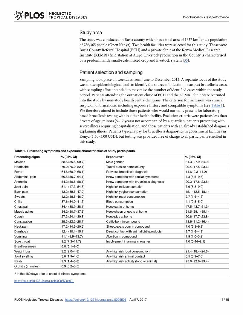
duct the patient survey and collect clinical data from 703 (85%) of these. The presenting symp-

toms and potential Brucella spp. exposure characteristics are summarised in Table 1. The

average age of participants was 37.6 years, with a median of 35 and a range between 5 and 98

years. The mean duration of symptoms was 118 days, but this was skewed by a small number

of individuals (n = 8) who reported the duration of current illness as more than 3 years. The

median value was 14 days. The majority of individuals (426, 60.6%) reported that this was

their first visit to a health care provider for their current illness. Of the remaining 277, 177

(64%) had already visited one health facility without resolution, 61 (22.2%) had visited two, 20

(7.2%) had visited three, 11 (3.9%) had visited four and eight (2.9%) had visited five or more.

The median duration of symptoms in those presenting for the first time was seven days com-

pared to 61 days in those who had previously sought health care for their current condition

Poor brucellosis test performance

PLOS Neglected Tropical Diseases | https://doi.org/10.1371/journal.pntd.0005508 April 7, 2017 6 / 15

(p<0.001). High-risk food consumption was reported in 150 (21.4%) participants, and high-

risk animal contact (defined as direct contact with animal birth products, animal abortion in

the participant’s home compound or involvement in livestock slaughter) in 37 (5.5%) individ-

uals (Table 1).

Brucellosis sero-diagnostics

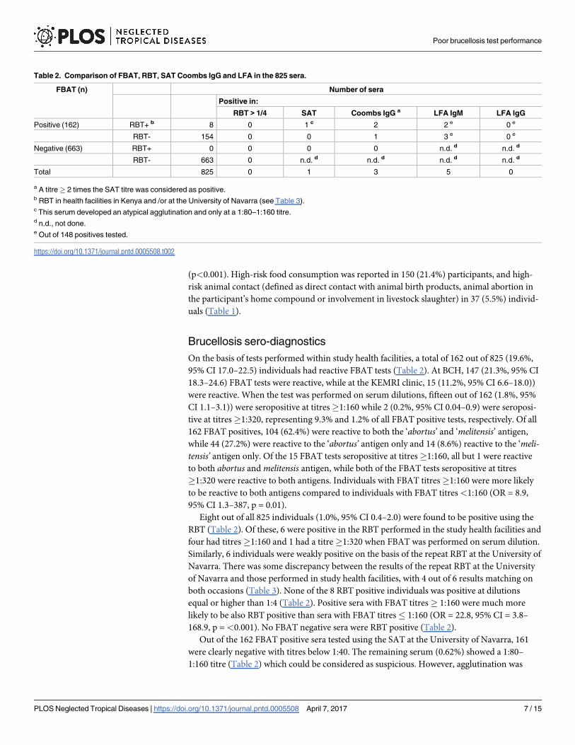
On the basis of tests performed within study health facilities, a total of 162 out of 825 (19.6%,

95% CI 17.0–22.5) individuals had reactive FBAT tests (Table 2). At BCH, 147 (21.3%, 95% CI

18.3–24.6) FBAT tests were reactive, while at the KEMRI clinic, 15 (11.2%, 95% CI 6.6–18.0))

were reactive. When the test was performed on serum dilutions, fifteen out of 162 (1.8%, 95%

CI 1.1–3.1)) were seropositive at titres�1:160 while 2 (0.2%, 95% CI 0.04–0.9) were seroposi-

tive at titres�1:320, representing 9.3% and 1.2% of all FBAT positive tests, respectively. Of all

162 FBAT positives, 104 (62.4%) were reactive to both the ‘abortus’ and ‘melitensis’ antigen,

while 44 (27.2%) were reactive to the ‘abortus’ antigen only and 14 (8.6%) reactive to the ‘meli-tensis’ antigen only. Of the 15 FBAT tests seropositive at titres�1:160, all but 1 were reactive

to both abortus and melitensis antigen, while both of the FBAT tests seropositive at titres

�1:320 were reactive to both antigens. Individuals with FBAT titres�1:160 were more likely

to be reactive to both antigens compared to individuals with FBAT titres <1:160 (OR = 8.9,

95% CI 1.3–387, p = 0.01).

Eight out of all 825 individuals (1.0%, 95% CI 0.4–2.0) were found to be positive using the

RBT (Table 2). Of these, 6 were positive in the RBT performed in the study health facilities and

four had titres�1:160 and 1 had a titre�1:320 when FBAT was performed on serum dilution.

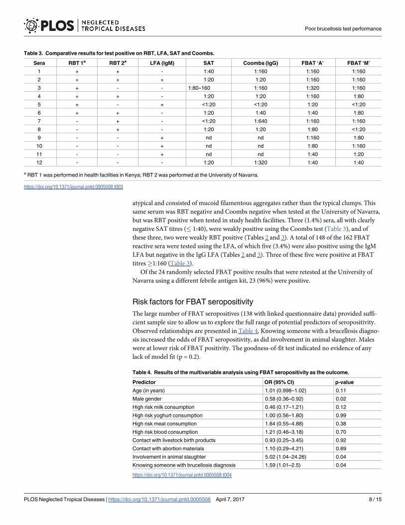
Similarly, 6 individuals were weakly positive on the basis of the repeat RBT at the University of

Navarra. There was some discrepancy between the results of the repeat RBT at the University

of Navarra and those performed in study health facilities, with 4 out of 6 results matching on

both occasions (Table 3). None of the 8 RBT positive individuals was positive at dilutions

equal or higher than 1:4 (Table 2). Positive sera with FBAT titres� 1:160 were much more

likely to be also RBT positive than sera with FBAT titres� 1:160 (OR = 22.8, 95% CI = 3.8–

168.9, p =<0.001). No FBAT negative sera were RBT positive (Table 2).

Out of the 162 FBAT positive sera tested using the SAT at the University of Navarra, 161

were clearly negative with titres below 1:40. The remaining serum (0.62%) showed a 1:80–

1:160 titre (Table 2) which could be considered as suspicious. However, agglutination was

Table 2. Comparison of FBAT, RBT, SAT Coombs IgG and LFA in the 825 sera.

FBAT (n) Number of sera

Positive in:

RBT > 1/4 SAT Coombs IgG a LFA IgM LFA IgG

Positive (162) RBT+ b 8 0 1 c 2 2 e 0 e

RBT- 154 0 0 1 3 e 0 e

Negative (663) RBT+ 0 0 0 0 n.d. d n.d. d

RBT- 663 0 n.d. d n.d. d n.d. d n.d. d

Total 825 0 1 3 5 0

a A titre� 2 times the SAT titre was considered as positive.b RBT in health facilities in Kenya and /or at the University of Navarra (see Table 3).c This serum developed an atypical agglutination and only at a 1:80–1:160 titre.d n.d., not done.e Out of 148 positives tested.

https://doi.org/10.1371/journal.pntd.0005508.t002

Poor brucellosis test performance

PLOS Neglected Tropical Diseases | https://doi.org/10.1371/journal.pntd.0005508 April 7, 2017 7 / 15

atypical and consisted of mucoid filamentous aggregates rather than the typical clumps. This

same serum was RBT negative and Coombs negative when tested at the University of Navarra,

but was RBT positive when tested in study health facilities. Three (1.4%) sera, all with clearly

negative SAT titres (� 1:40), were weakly positive using the Coombs test (Table 3), and of

these three, two were weakly RBT positive (Tables 2 and 3). A total of 148 of the 162 FBAT

reactive sera were tested using the LFA, of which five (3.4%) were also positive using the IgM

LFA but negative in the IgG LFA (Tables 2 and 3). Three of these five were positive at FBAT

titres�1:160 (Table 3).

Of the 24 randomly selected FBAT positive results that were retested at the University of

Navarra using a different febrile antigen kit, 23 (96%) were positive.

Risk factors for FBAT seropositivity

The large number of FBAT seropositives (138 with linked questionnaire data) provided suffi-

cient sample size to allow us to explore the full range of potential predictors of seropositivity.

Observed relationships are presented in Table 4. Knowing someone with a brucellosis diagno-

sis increased the odds of FBAT seropositivity, as did involvement in animal slaughter. Males

were at lower risk of FBAT positivity. The goodness-of-fit test indicated no evidence of any

lack of model fit (p = 0.2).

Table 3. Comparative results for test positive on RBT, LFA, SAT and Coombs.

Sera RBT 1a RBT 2a LFA (IgM) SAT Coombs (IgG) FBAT ‘A’ FBAT ‘M’

1 + + - 1:40 1:160 1:160 1:160

2 + + + 1:20 1:20 1:160 1:160

3 + - - 1:80–160 1:160 1:320 1:160

4 + + - 1:20 1:20 1:160 1:80

5 + - + <1:20 <1:20 1:20 <1:20

6 + + - 1:20 1:40 1:40 1:80

7 - + - <1:20 1:640 1:160 1:160

8 - + - 1:20 1:20 1:80 <1:20

9 - - + nd nd 1:160 1:80

10 - - + nd nd 1:80 1:160

11 - - + nd nd 1:40 1:20

12 - - - 1:20 1:320 1:40 1:40

a RBT 1 was performed in health facilities in Kenya; RBT 2 was performed at the University of Navarra.

https://doi.org/10.1371/journal.pntd.0005508.t003

Table 4. Results of the multivariable analysis using FBAT seropositivity as the outcome.

Predictor OR (95% CI) p-value

Age (in years) 1.01 (0.998–1.02) 0.11

Male gender 0.58 (0.36–0.92) 0.02

High risk milk consumption 0.46 (0.17–1.21) 0.12

High risk yoghurt consumption 1.00 (0.56–1.80) 0.99

High risk meat consumption 1.64 (0.55–4.88) 0.38

High risk blood consumption 1.21 (0.46–3.18) 0.70

Contact with livestock birth products 0.93 (0.25–3.45) 0.92

Contact with abortion materials 1.10 (0.29–4.21) 0.89

Involvement in animal slaughter 5.02 (1.04–24.26) 0.04

Knowing someone with brucellosis diagnosis 1.59 (1.01–2.5) 0.04

https://doi.org/10.1371/journal.pntd.0005508.t004

Poor brucellosis test performance

PLOS Neglected Tropical Diseases | https://doi.org/10.1371/journal.pntd.0005508 April 7, 2017 8 / 15

Discussion

We have demonstrated substantial disagreement between results from the FBAT and results

from a range of serological tests that have been repeatedly shown to have high diagnostic accu-

racy for human brucellosis [11,24,25,27,30,42]. Whilst brucellosis should not be excluded as a

possible cause of febrile illness in the population under study, our results suggest it is an

uncommon cause, and certainly much rarer than results from the FBAT would suggest. It is

important to stress that the small number of positive results obtained using the RBT, SAT--

Coombs combination and LFA are not by themselves indicative of active brucellosis. There is

no established diagnostic titre for a single SAT, which varies from 1:80 to 320 depending on

levels of endemicity [24], and any SAT result should be considered along with the Coomb’s

result. In this study, all but one FBAT positive individual had SAT titres < 1:160, and only

three had weak Coombs titres. In addition, none of the sera were positive on the RBT at dilu-

tions higher than 1:2, which may suggest previous exposure rather than current infection [25].

Five FBAT positive sera were IgM positive in the LFA. The presence of the rheumatoid factor

(a group of auto-antibodies that recognize Fc regions of IgG [43]) is a known cause of false

positives in this test [42]. Since IgM is readily detected using the SAT [24], this is a possible

explanation for the observed discord between LFA and SAT results. In the absence of bacterio-

logical evidence or the availability of convalescent antibody titres, the few positive sera identi-

fied using SAT-Coombs, RBT and LFA can therefore only be considered as "suspicious" for

brucellosis. This fact, combined with the low number of “suspicious” cases identified, limits

our ability to derive formal estimates of test performance for the FBAT. However, results from

this study strongly suggest that this test generates large numbers of false positives and has poor

diagnostic specificity. This interpretation is further supported by the fact that we found no evi-

dence of an association between FBAT seropositivity and most of the anticipated high-risk

food or animal exposures. The only exception was involvement with animal slaughter, which

has been shown to be a risk factor for human brucellosis in other studies [44].

The poor performance of the FBAT cannot be attributed to the particular kit used in health

facilities involved in this study, since a high rate of false positive results were also observed at

the University of Navarra with a different kit. Neither health facility involved in this study rou-

tinely performed serial dilutions for positive cases and, based on discussion with laboratory

managers at other health facilities in western Kenya, the semi-quantitative estimation of anti-

body titres using the FBAT appears to be rare practice in the region. Our ability to evaluate

appropriate cut-offs is limited in this study by the absence of confirmed cases and small num-

ber of suspicious cases, but there was evidence of a strong association between RBT positivity

and titres�1:160 on the FBAT, suggesting the use of a higher cut-off may improve the diag-

nostic specificity of the test.

The low prevalence of brucellosis in the study population combined with the apparently

poor specificity of FBAT suggests considerable overdiagnosis of human brucellosis. An impor-

tant consideration, however, is that the provision of free brucellosis testing to participants may

have contributed to the capture of more patients than would typically be tested for the disease

in the study health facilities. It is therefore possible that levels of overdiagnosis indicated by

this study are exaggerated. In the period from June 2010 to June 2011, BCH (then named

Busia District Hospital) recorded 129 cases of brucellosis, whilst we identified 162 individuals

as reactive on the FBAT over an approximately 6 month period between June and December

2012. However, given the small number of suspicious cases we observed over the six months of

data collection, the number of cases detected in 2010 and 2011 also point to high levels of over-

diagnosis for this disease. Typical treatment for brucellosis in health facilities in the study area

is 6 weeks of oral doxycycline combined with either 2 weeks of intramuscular streptomycin or

Poor brucellosis test performance

PLOS Neglected Tropical Diseases | https://doi.org/10.1371/journal.pntd.0005508 April 7, 2017 9 / 15

6 weeks of oral rifampicin. Whilst the clinical management of recruited patients was not

recorded as part of this study, treatment is routinely recommended in both health facilities to

any individual with a reactive FBAT test. We therefore expect that a substantial proportion of

brucellosis treatments in western Kenya are inappropriate. This will result in unnecessary eco-

nomic costs for patients, and may represent important misuse of antibiotics. This is of particu-

lar concern for the selection of resistance to rifampicin, a first line treatment for TB [45].

In 2012, 75,256 brucellosis cases in individuals over five years of age were reported to the

Kenya Health Information System (www.hiskenya.org), giving an annual incidence of 230 per

100,000 people [46]. We suspect that the vast majority of these cases were diagnosed on the

basis of the FBAT. Whilst brucellosis is very likely to be an important disease in Kenya, partic-

ularly amongst pastoral communities [5,47], poor performance of the diagnostic test used to

derive surveillance data means there is considerable uncertainty regarding the true burden of

human brucellosis in the country.

An internet search revealed that all twelve of the FBAT providers identified include both B.

abortus and B. melitensis suspensions in their kits. The purpose of this is unclear since it was

well established in the first half of the 20th century that suspensions of B. abortus and B. meli-tensis can be exchanged for diagnostic purposes [18]. The target for Brucella agglutination tests

is the O-polysaccharide of the smooth lipopolysaccharide of the cell surface of smooth Brucellaspecies, which has a structure dominated by a highly repeated C epitope common to all smooth

brucellae [48] (excluding the recently described B. inopinata BO1 [49]). Hence, there is no

serological test that can distinguish infection with B. abortus versus B. melitensis [11,18,24,48].

Despite this, we noted a not unsurprising belief amongst laboratory technicians and clinicians

working in both study health facilities that the FBAT, marketed with separate ‘abortus’ and

‘melitensis’ antigens, can distinguish between infections with these different Brucella species.

This misconception is likely to be strengthened by the inconsistent results obtained with the

‘abortus’ and ‘melitensis’ suspensions of FBAT, as illustrated by our work and recently pub-

lished work from Tanzania [50]. Long and repeated experience [11,18,24,51] shows that the

most plausible reasons for these inconsistencies are deficient standardisation of antigen sus-

pensions and poor antigen quality. Smooth brucellae are notorious for their ability to dissoci-

ate in vitro, a genetic drift that results in large proportions of rough mutants that lack the O-

polysaccharide of the lipopolysaccharide and have profoundly altered cell surface properties

[48]. The consequence of this drift is an absence of diagnostically valid epitopes and a strong ten-

dency to autoagglutinate. Such deficient antigenic suspensions are an important cause of false

positivity and the likely explanation for the high numbers of false positives obtained with FBAT

in this study, as well as for the inconsistency between reactions to ‘abortus’ and ‘melitensis’ anti-

gens. We consider antigen quality to be a more likely explanation for poor FBAT specificity than

cross-reaction with antibodies to other gram negative bacteria, since such cross-reactions are

well known to occur for all serological assays that rely on detection of antibodies to the Brucellasmooth lipopolysaccharide, including RBT, SAT and Coombs [31], and are likely also with the

LFA. Whilst the clinical management of infection with B. melitensis or B. abortus is the same, an

indirect consequence of the inconsistency between reactions to different FBAT antigens is that

results may be misleading with respect to the source of infection, since B. melitensis is typically

associated with small ruminants while B. abortus is more typically associated with cattle [16].

The catchment area of study health facilities incorporate a mixed farming area with a very

high livestock density. Despite this, a substantial proportion of patients presenting for brucel-

losis diagnostic testing did not report what we considered high risk contacts with livestock or

their products over the six months prior to the onset of clinical symptoms. Studies from Tan-

zania have reported limited awareness of zoonotic disease among health workers [52], includ-

ing the range of pathways through which Brucella species may be transmitted to people [53].

Poor brucellosis test performance

PLOS Neglected Tropical Diseases | https://doi.org/10.1371/journal.pntd.0005508 April 7, 2017 10 / 15

Detailed clinical history taking and the targeting of testing to individuals considered to be at

higher risk of brucellosis, is likely to improve the predictive value of serological testing, partic-

ularly in low prevalence populations. An important consideration, however, is that whilst we

have a broad understanding of the epidemiology of brucellosis in sub-Saharan Africa [1,17],

there is generally a lack of information on the specific transmission routes by which people in

specific settings acquire their infections. Population-based studies in endemic areas should

seek to fill this information gap. Further studies could also seek to derive diagnostic algorithms

for human brucellosis that allow for the formalisation of who to test, and who should be con-

sidered positive based on a range of criteria, including the results of imperfect serological

assays, exposure history and presenting signs [54,55].

Future studies should seek to derive estimates of sensitivity and specificity for the FBAT,

including assessment of the value of deriving semi-quantitative estimates of antibody titres.

However, given the likely poor specificity we have demonstrated, future work should also exam-

ine alternative diagnostic tests that can be used to replace the FBAT in health facilities in west-

ern Kenya and the region more widely. The standard RBT is inexpensive and straightforward to

perform, follows the same general procedure as the FBAT and thus requires the same basic labo-

ratory equipment and expertise. Several authors have advocated for the routine use of this test

in the diagnosis of human brucellosis in health facilities in resource limited settings [25,56,57].

This test showed considerably better agreement with the results of the SAT-Coombs and LFA

than the FBAT in this setting, and is likely to represent a better alternative. A potential limita-

tion of the RBT, although one that applies to all rapid agglutination tests, including the FBAT,

are inconsistencies between technicians in reading agglutination results [58]. In our study, there

was some discrepancy between results of the RBTs performed in Kenya and those performed by

different operators in Spain. One of the two weak RBT positives that were missed in Kenya was

Coombs positive, whilst one of the two that was missed in Spain was SAT positive. Problems

around repeatability are likely to be minimised through the use of a glossy white tile, testing

only a limited number of sera each time, adequate training of technicians to recognise all agglu-

tination patterns, and the use of good quality controls [25]. The measurement of RBT titres

using serial dilution, and particularly evidence of rising titres on repeat testing, can provide bet-

ter evidence of infection when combined with clinical presentation and adequate history taking

[25,57]. Confirmation of agglutination results with a second test would also contribute to

improved diagnostic accuracy. The LFA used in this study provides a simple and inexpensive

option, and was found to have good test performance when manufactured by the Royal Tropical

Institute, Netherlands [30]. These tests can distinguish between IgM and IgG antibodies, and

can therefore also contribute to the clinical assessment of the stage of infection [59,60].

We do not have additional aetiological information for those patients enrolled in this study

who appear to have had false positive brucellosis diagnoses, and an important question

remains regarding the cause of their illnesses. In addition to HIV, malaria and typhoid, for

which many recruited participants were also tested by participating health centres, a wide

range of infectious causes of febrile illness are known to be prevalent in the study area. These

include Coxiella burnetii [61,62], Leptospira spp. [63], Rickettsia spp. [64], and Chikungunya

virus [65]. To our knowledge, neither health facility routinely tested for any of these pathogens

at the time of the study, and while rapid tests are available for some, these often have poor diag-

nostic performance [66] and have not been widely adopted [67]. The demonstration that Bru-cella may be less common than anticipated in this setting therefore increases the diagnostic

challenge for clinicians. Improved diagnostic services, including the development and valida-

tion of point of care tests and testing strategies, and the development of diagnostic and man-

agement algorithms for patients presenting with febrile illness, are urgently needed in this

setting, as in many others in the region [19,54].

Poor brucellosis test performance

PLOS Neglected Tropical Diseases | https://doi.org/10.1371/journal.pntd.0005508 April 7, 2017 11 / 15

Summary

The findings from this study strongly suggest that human brucellosis is being over-diagnosed

in a mixed farming area of western Kenya. We expect that this is contributing to the over use

of antibiotics, and has important economic impacts on affected patients. The FBAT used in

government facilities throughout Kenya appears to have very poor diagnostic specificity and

should be phased out. Further studies are needed to assess alternative diagnostic tests and test-

ing strategies that can replace the FBAT in health facilities in the region. However, in the short

term, and while awaiting the development of these alternatives, published recommendations

and the results from this study suggest that the standard RBT, with antigen sourced from

established sources with high standards of quality control, would provide a better alternative

than the FBAT for the laboratory diagnosis of human brucellosis.

Supporting information

S1 Checklist. STROBE checklist

(PDF)

Acknowledgments

We thank our field and lab team, Lorren Alumasa, Daniel Cheruiyot, Eunice Wauye, Enock

Musumba, as well as the management and staff at participating health facilities. The skilful

assistance of Rosario Urdaci, Alberto Delgado and Begoña Alonso-Urmeneta in the serological

analysis performed at UNAV is also gratefully acknowledged. Thanks to Dr. Shama Cash-

Goldwasser for useful comments on the manuscript and Professor Heinrich Neubauer and Dr

Falk Melzer on the study.

Author Contributions

Conceptualization: WAdG EAC BMdCB LFT EF.

Data curation: WAdG.

Formal analysis: WAdG.

Funding acquisition: EMF IM.

Investigation: WAdG RCA IM JN RD.

Methodology: WAdG JN BMdCB EMF.

Resources: MM.

Supervision: SK.

Validation: RCA IM RD.

Writing – original draft: WAdG RCA IM EMF.

Writing – review & editing: WAdG RCA IM JN EAJC MM BMdCB LFT SK EMF.

References

1. McDermott JJ, Arimi SM. Brucellosis in sub-Saharan Africa: epidemiology, control and impact. Vet

Microbiol. 2002; 90: 111–134. PMID: 12414138

2. McDermott J, Grace D, Zinsstag J. Economics of brucellosis impact and control in low-income coun-

tries. Rev Sci Tech. 2013; 32: 249–261. PMID: 23837382

Poor brucellosis test performance

PLOS Neglected Tropical Diseases | https://doi.org/10.1371/journal.pntd.0005508 April 7, 2017 12 / 15

3. Godfroid J, Cloeckaert A, Liautard J-P, Kohler S, Fretin D, Walravens K, et al. From the discovery of the

Malta fever’s agent to the discovery of a marine mammal reservoir, brucellosis has continuously been a

re-emerging zoonosis. Vet Res. 2005; 36: 313–326. https://doi.org/10.1051/vetres:2005003 PMID:

15845228

4. Corbel MJ. Brucellosis: an overview. Emerging Infect Dis. 1997; 3: 213–221. https://doi.org/10.3201/

eid0302.970219 PMID: 9204307

5. Kadohira M, McDermott JJ, Shoukri MM, Kyule MN. Variations in the prevalence of antibody to brucella

infection in cattle by farm, area and district in Kenya. Epidemiol Infect. Cambridge University Press;

1997; 118: 35–41. PMID: 9042033

6. Muendo EN, Mbatha PM, Macharia J, Abdoel TH, Janszen PV, Pastoor R, et al. Infection of cattle in

Kenya with Brucella abortus biovar 3 and Brucella melitensis biovar 1 genotypes. Trop Anim Health

Prod. Springer Netherlands; 2012; 44: 17–20. https://doi.org/10.1007/s11250-011-9899-9 PMID:

21660647

7. Philpott M, Auko O. Caprine brucellosis in Kenya. Br Vet J. 1972; 128: 642–651. PMID: 4647703

8. Young EJ. An overview of human brucellosis. Clin Infect Dis. 1995; 21: 283–290. PMID: 8562733

9. Franco MP, Mulder M, Gilman RH, Smits HL. Human brucellosis. Lancet Infect Dis. 2007; 7: 775–786.

https://doi.org/10.1016/S1473-3099(07)70286-4 PMID: 18045560

10. Dean AS, Crump L, Greter H, Hattendorf J, Schelling E, Zinsstag J. Clinical manifestations of human

brucellosis: a systematic review and meta-analysis. PLoS Negl Trop Dis. 2012; 6: e1929. https://doi.

org/10.1371/journal.pntd.0001929 PMID: 23236528

11. Ariza J. Brucellosis: an update. The perspective from the Mediterranean basin. Reviews in Medical

Microbiology. 1999; 10: 125–135.

12. Buzgan T, Karahocagil MK, Irmak H, Baran AI, Karsen H, Evirgen O, et al. Clinical manifestations and

complications in 1028 cases of brucellosis: a retrospective evaluation and review of the literature. Int J

Infect Dis. 2010; 14: e469–78. https://doi.org/10.1016/j.ijid.2009.06.031 PMID: 19910232

13. Ariza J, Bosilkovski M, Cascio A, Colmenero JD, Corbel MJ, Falagas ME, et al. Perspectives for the

treatment of brucellosis in the 21st century: the Ioannina recommendations. PLoS Med. 2007; 4: e317.

https://doi.org/10.1371/journal.pmed.0040317 PMID: 18162038

14. Zinsstag J, Schelling E, Roth F, Bonfoh B, de Savigny D, Tanner M. Human benefits of animal interven-

tions for zoonosis control. Emerging Infect Dis. 2007; 13: 527–531. https://doi.org/10.3201/eid1304.

060381 PMID: 17553265

15. Colmenero JD, Cabrera FP, Hernadez S, Reguera JM, Pinedo A, Clavero AMC. Repercusion socio-

economica de la brucelosis humana. Rev Clinica Espanola. 1989; 185: 55–63.

16. Corbel MJ. Brucellosis in Humans and Animals. WHO; 2006. pp. 1–90.

17. Ducrotoy M, Bertu WJ, Matope G, Cadmus S, Conde-Alvarez R, Gusi AM, et al. Brucellosis in Sub-

Saharan Africa: Current challenges for management, diagnosis and control. Acta Trop. 2015.

18. Spink WW. The diagnosis of human brucellosis. The nature of brucellosis. The University of Minnesota

Press; 1956. pp. 191–215.

19. Crump JA, Morrissey AB, Nicholson WL, Massung RF, Stoddard RA, Galloway RL, et al. Etiology of

severe non-malaria febrile illness in Northern Tanzania: a prospective cohort study. PLoS Negl Trop

Dis. 2013; 7: e2324. https://doi.org/10.1371/journal.pntd.0002324 PMID: 23875053

20. Marcotty T, Matthys F, Godfroid J, Rigouts L, Ameni G, Gey van Pittius N, et al. Zoonotic tuberculosis

and brucellosis in Africa: neglected zoonoses or minor public-health issues? The outcomes of a multi-

disciplinary workshop. Ann Trop Med Parasitol. 2009; 103: 401–411. https://doi.org/10.1179/

136485909X451771 PMID: 19583911

21. Omer MK, Assefaw T, Skjerve E, Tekleghiorghis T, Woldehiwet Z. Prevalence of antibodies to Brucella

spp. and risk factors related to high-risk occupational groups in Eritrea. Epidemiol Infect. 2002; 129: 85–

91. PMID: 12211600

22. Kunda J, Fitzpatrick J, Kazwala R, French NP, Shirima G, Macmillan A, et al. Health-seeking behaviour

of human brucellosis cases in rural Tanzania. BMC Public Health. 2007; 7: 315. https://doi.org/10.1186/

1471-2458-7-315 PMID: 17980046

23. Pappas G, Papadimitriou P, Akritidis N, Christou L, Tsianos EV. The new global map of human brucello-

sis. Lancet Infect Dis. 2006; 6: 91–99. https://doi.org/10.1016/S1473-3099(06)70382-6 PMID:

16439329

24. Diaz R, Moriyon I. Laboratory techniques in the diagnosis of human brucellosis. In: Young EJ, Corbel

MJ, editors. Brucellosis. 1989. pp. 73–83.

Poor brucellosis test performance

PLOS Neglected Tropical Diseases | https://doi.org/10.1371/journal.pntd.0005508 April 7, 2017 13 / 15

25. Dıaz R, Casanova A, Ariza J, Moriyon I. The Rose Bengal Test in human brucellosis: a neglected test

for the diagnosis of a neglected disease. PLoS Negl Trop Dis. 2011; 5: e950. https://doi.org/10.1371/

journal.pntd.0000950 PMID: 21526218

26. Ariza J, Pellicer T, Pallares R, Foz A, Gudiol F. Specific antibody profile in human brucellosis. Clin Infect

Dis. 1992; 14: 131–140. PMID: 1571417

27. Gazapo E, Gonzalez Lahoz J, Subiza JL, Baquero M, Gil J, la Concha de EG. Changes in IgM and IgG

antibody concentrations in brucellosis over time: importance for diagnosis and follow-up. J Infect Dis.

1989; 159: 219–225. PMID: 2915152

28. Wilson MM, Merrifield EVO. The antiglobulin (Coombs) test in brucellosis. Lancet. 1951; 2: 913–914.

PMID: 14881511

29. Smits HL, Basahi MA, Diaz R, Marrodan T, Douglas JT, Rocha A, et al. Development and evaluation of

a rapid dipstick assay for serodiagnosis of acute human brucellosis. J Clin Microbiol. 1999; 37: 4179–

4182. PMID: 10565959

30. Smits HL, Abdoel TH, Solera J, Clavijo E, Dıaz R. Immunochromatographic Brucella-specific immuno-

globulin M and G lateral flow assays for rapid serodiagnosis of human brucellosis. Clin Diagn Lab Immu-

nol. 2003; 10: 1141–1146. https://doi.org/10.1128/CDLI.10.6.1141-1146.2003 PMID: 14607880

31. Araj GF. Update on laboratory diagnosis of human brucellosis. Int J Antimicrob Agents. 2010; 36 Suppl

1: S12–7.

32. Al-Dahouk S, Tomaso H, Nockler K, Neubauer H, Frangoulidis D. Laboratory-based diagnosis of bru-

cellosis—a review of the literature. Part II: serological tests for brucellosis. Clin Lab. 2003; 49: 577–589.

PMID: 14651329

33. Young EJ. Serologic diagnosis of human brucellosis: analysis of 214 cases by agglutination tests and

review of the literature. Rev Infect Dis. 1991; 13: 359–372. PMID: 1866536

34. Dean AS, Crump L, Greter H, Schelling E, Zinsstag J. Global burden of human brucellosis: a systematic

review of disease frequency. PLoS Negl Trop Dis. 2012; 6: e1865. https://doi.org/10.1371/journal.pntd.

0001865 PMID: 23145195

35. de Clare Bronsvoort BM, Thumbi SM, Poole EJ, Kiara H, Auguet OT, Handel IG, et al. Design and

descriptive epidemiology of the Infectious Diseases of East African Livestock (IDEAL) project, a longitu-

dinal calf cohort study in western Kenya. BMC Vet Res. 2013; 9: 171. https://doi.org/10.1186/1746-

6148-9-171 PMID: 24000820

36. Spink WW. The natural course of brucellosis. The nature of brucellosis. The University of Minnesota

Press; 1956. pp. 145–170.

37. OIE. Bovine Brucellosis. Manual of Diagnostic Tests and Vaccines for Terrestrial Animals. 2009. pp. 1–

35.

38. Blasco JM, Garin-Bastuji B, Marin CM, Gerbier G, Fanlo J, Jimenez de Bagues MP, et al. Efficacy of dif-

ferent Rose Bengal and complement fixation antigens for the diagnosis of Brucella melitensis infection

in sheep and goats. Vet Rec. 1994; 134: 415–420. PMID: 8036772

39. Renoux G, Plommet M, Philippon A. Microreactions d’agglutination et de fixation du complement pour

le diagnostic des brucellosis. Ann Rech Vet. 1971; 2: 263–269.

40. Beaton CP, Forsyth JR. A micromethod for agglutination and antiglobulin tests for antibody to Brucella

abortus. J Biol Stand. 1984; 12: 271–275. PMID: 6434543

41. Harrell FE. Regression modeling strategies, with applications to linear models, survival analysis and

logistic regression. Springer; 2001. pp. 1–582.

42. Diaz R, Ariza J, Alberola I, Casanova A, Rubio MF. Secondary serological response of patients with

chronic hepatosplenic suppurative brucellosis. Clin Vaccine Immunol. 2006; 13: 1190–1196. https://doi.

org/10.1128/CVI.00086-06 PMID: 16943345

43. Woods CR. False-Positive Results for Immunoglobulin M Serologic Results: Explanations and Exam-

ples. J Pediatric Infect Dis Soc. 2013; 2: 87–90. https://doi.org/10.1093/jpids/pis133 PMID: 26619450

44. Swai ES, Schoonman L. Human brucellosis: seroprevalence and risk factors related to high risk occu-

pational groups in Tanga Municipality, Tanzania. Zoonoses Public Health. 2009; 56: 183–187. https://

doi.org/10.1111/j.1863-2378.2008.01175.x PMID: 18811674

45. Goldstein BP. Resistance to rifampicin: a review. J Antibiot. 2014; 67: 625–630. https://doi.org/10.1038/

ja.2014.107 PMID: 25118103

46. Brink M. Brucellosis in Kenya: Epidemiology and Human Burden of a Neglected Zoonotic Disease. MSc

Thesis, University of Uppsala. 2013.

47. Osoro EM, Munyua P, Omulo S, Ogola E, Ade F, Mbatha P, et al. Strong Association Between Human

and Animal Brucella Seropositivity in a Linked Study in Kenya, 2012–2013. Am J Trop Med Hyg. 2015;

93: 224–231. https://doi.org/10.4269/ajtmh.15-0113 PMID: 26101275

Poor brucellosis test performance

PLOS Neglected Tropical Diseases | https://doi.org/10.1371/journal.pntd.0005508 April 7, 2017 14 / 15

48. Ducrotoy MJ, Conde-Alvarez R, Blasco JM, Moriyon I. A review of the basis of the immunological diag-

nosis of ruminant brucellosis. Vet Immunol Immunopathol. 2016; 171: 81–102. https://doi.org/10.1016/j.

vetimm.2016.02.002 PMID: 26964721

49. Wattam AR, Inzana TJ, Williams KP, Mane SP, Shukla M, Almeida NF, et al. Comparative genomics of

early-diverging Brucella strains reveals a novel lipopolysaccharide biosynthesis pathway. MBio. 2012;

3: e00246–11.

50. Chipwaza B, Mhamphi GG, Ngatunga SD, Selemani M, Amuri M, Mugasa JP, et al. Prevalence of bac-

terial febrile illnesses in children in Kilosa district, Tanzania. PLoS Negl Trop Dis. 2015; 9: e0003750.

https://doi.org/10.1371/journal.pntd.0003750 PMID: 25955522

51. Huddleson IF. Laboratory diagnosis of brucellosis. Brucellosis in man and animals. The Common-

wealth Fund; 1943. pp. 214–237.

52. John K, Kazwala R, Mfinanga GS. Knowledge of causes, clinical features and diagnosis of common

zoonoses among medical practitioners in Tanzania. BMC Infect Dis. 2008; 8: 162. https://doi.org/10.

1186/1471-2334-8-162 PMID: 19046464

53. Zhang HL, Mnzava KW, Mitchell ST, Melubo ML, Kibona TJ, Cleaveland S, et al. Mixed Methods Sur-

vey of Zoonotic Disease Awareness and Practice among Animal and Human Healthcare Providers in

Moshi, Tanzania. PLoS Negl Trop Dis. 2016; 10:

54. D’Acremont V, Kilowoko M, Kyungu E, Philipina S, Sangu W, Kahama-Maro J, et al. Beyond malaria—

causes of fever in outpatient Tanzanian children. N Engl J Med. 2014; 370: 809–817. https://doi.org/10.

1056/NEJMoa1214482 PMID: 24571753

55. Mantur BG, Biradar MS, Bidri RC, Mulimani MS, Veerappa, Kariholu P, et al. Protean clinical manifesta-

tions and diagnostic challenges of human brucellosis in adults: 16 years’ experience in an endemic

area. J Med Microbiol. 2006; 55: 897–903. https://doi.org/10.1099/jmm.0.46097-0 PMID: 16772417

56. Oomen LJ, Waghela S. The rose bengal plate test in human brucellosis. Trop Geogr Med. 1974; 26:

300–302. PMID: 4439467

57. Mantur BG, Amarnath SK, Patil GA, Desai AS. Clinical utility of a quantitative Rose Bengal slide aggluti-

nation test in the diagnosis of human brucellosis in an endemic region. Clin Lab. 2014; 60: 533–541.

PMID: 24779287

58. Maichomo MW, McDermott JJ, Arimi SM, Gathura PB. Assessment of the Rose-Bengal plate test for

the diagnosis of human brucellosis in health facilities in Narok district, Kenya. East Afr Med J. 1998; 75:

219–222. PMID: 9745838

59. Mizanbayeva S, Smits HL, Zhalilova K, Abdoel TH, Kozakov S, Ospanov KS, et al. The evaluation of a

user-friendly lateral flow assay for the serodiagnosis of human brucellosis in Kazakhstan. Diagn Micro-

biol Infect Dis. 2009; 65: 14–20. https://doi.org/10.1016/j.diagmicrobio.2009.05.002 PMID: 19679230

60. Roushan MRH, Amin MJS, Abdoel TH, Smits HL. Application of a user-friendly Brucella-specific IgM

and IgG antibody assay for the rapid confirmation of Rose Bengal-positive patients in a hospital in Iran.

Trans R Soc Trop Med Hyg. 2005; 99: 744–750. https://doi.org/10.1016/j.trstmh.2005.02.009 PMID:

16095642

61. Wardrop NA, Thomas LF, Cook EAJ, de Glanville WA, Atkinson PM, Wamae CN, et al. The Sero-epide-

miology of Coxiella burnetii in Humans and Cattle, Western Kenya: Evidence from a Cross-Sectional

Study. PLoS Negl Trop Dis. 2016; 10: e0005032. https://doi.org/10.1371/journal.pntd.0005032 PMID:

27716804

62. Knobel DL, Maina AN, Cutler SJ, Ogola E, Feikin DR, Junghae M, et al. Coxiella burnetii in humans,

domestic ruminants, and ticks in rural western Kenya. Am J Trop Med Hyg. 2013; 88: 513–518. https://

doi.org/10.4269/ajtmh.12-0169 PMID: 23382156

63. Cook EAJ, de Glanville WA, Thomas LF, Kariuki S, Bronsvoort BM de C, Fèvre EM. Risk factors for lep-

tospirosis seropositivity in slaughterhouse workers in western Kenya. Occup Environ Med. 2016:

64. Maina AN, Knobel DL, Jiang J, Halliday J, Feikin DR, Cleaveland S, et al. Rickettsia felis infection in

febrile patients, western Kenya, 2007–2010. Emerging Infect Dis. 2012; 18: 328–331. https://doi.org/

10.3201/eid1802.111372 PMID: 22304807

65. Mease LE, Coldren RL, Musila LA, Prosser T, Ogolla F, Ofula VO, et al. Seroprevalence and distribution

of arboviral infections among rural Kenyan adults: a cross-sectional study. Virol J. 2011; 8: 371. https://

doi.org/10.1186/1743-422X-8-371 PMID: 21794131

66. Bajani MD, Ashford DA, Bragg SL, Woods CW, Aye T, Spiegel RA, et al. Evaluation of four commer-

cially available rapid serologic tests for diagnosis of leptospirosis. J Clin Microbiol. 2003; 41: 803–809.

https://doi.org/10.1128/JCM.41.2.803-809.2003 PMID: 12574287

67. Picardeau M, Bertherat E, Jancloes M, Skouloudis AN, Durski K, Hartskeerl RA. Rapid tests for diagno-

sis of leptospirosis: current tools and emerging technologies. Diagn Microbiol Infect Dis. 2014; 78: 1–8.

https://doi.org/10.1016/j.diagmicrobio.2013.09.012 PMID: 24207075

Poor brucellosis test performance

PLOS Neglected Tropical Diseases | https://doi.org/10.1371/journal.pntd.0005508 April 7, 2017 15 / 15


Recommended