Portable Schistosomiasis Egg Counting and Diagnosis Daniel Fernandes I. INTRODUCTION Schistosomiasis, also known as Bilharzia, is a parasitic disease caused by waterborne snails that generally occurs in tropical countries, most notably in Sub-saharan Africa. The parasitic snail lays larvae in freshwater and these larvae burrow into the skin of the patient and enter the blood circulation. After the larvae mature into adults and mate, the females go on to lay eggs that infect the fe- ces and urine of the patient. Through bad sanitation prac- tice, human waste may enter the freshwater and complete the cycle, causing snails to infect the water once again. The disease is diagnosed by passing the urine of a patient through a polycarbonate filter and counting the number of oval-shaped eggs. Currently, an aid-worker or technician would take this circular (13 mm diameter) filter, place it under a microscope, and pan around to count the number of eggs. While this would be fine for only a few patients, since schistosomiasis is a water-borne disease, generally it is entire villages that are infected at once. The time-crunch is panning around to count the eggs. Several works have been published related to automatic egg detection [3], [4], [5], [6]. These eggs are small, on the order of 100 to 60 microns, but can be imaged with a macro lens. Since the eggs have a finite shelf life, 100 μm polystyrene beads are used as a phantom to prototype. II. METHODS A. Camera To build this system two cameras were evaluated: The Olympus Air A01 and Olympus TG-4. While both systems had WiFi connectivity and the ability to take macro images, the Olympus Air had an open API that allowed full access to the camera for developers. Furthermore, the Olympus Air is a micro four-thirds camera with a sensor size of 21.60 x 13 mm compared to the TG4’s 1/2.3” Type sensor of size 6.17 x 4.56 mm. The Olympus Air was equipped with a M.Zuiko Digital ED 60mm f/2.8 Macro Lens, while the TG-4 used its internal f/2.3 lens. The much larger optics of the M.Zuiko lens allowed for a correspondingly much larger working distance on the order of inches rather than centimeters. For these reasons, the Olympus Air was chosen over the TG-4. B. Camera Mount The camera was mounted using 3D printed parts designed in Solidworks CAD software. Using the 3D designs provided by Olympus as a basis [1], four interlocking parts were designed and printed in black ABS plastic on a Makerbot Replicator 2X. The top part holds the camera through a in- terlocking mechanism, facing downwards to the bottom part that holds the slide and provides an aperture for transmissive light illumination. Fig. 1. View of 3D parts from bottom C. Illumination Illumination is provided by a Rainbowduino LED Matrix from Seeedstudio [2]. The illumination is configured in a Rheinberg illumination pattern. Rheinberg illumination uses two separate colors of light: one for oblique illumination and another for the unscattered light. In our case, we choose complementary colors red and green, to easily threshold once the image is projected from RGB to HSV space. D. Algorithm The algorithm pipeline is divided into two stages: filter de- tection and bead detection. Initial prototypes were designed
Transcript
Portable Schistosomiasis Egg Counting and Diagnosis
Daniel Fernandes
I. INTRODUCTION
Schistosomiasis, also known as Bilharzia, is a parasiticdisease caused by waterborne snails that generally occursin tropical countries, most notably in Sub-saharan Africa.The parasitic snail lays larvae in freshwater and theselarvae burrow into the skin of the patient and enter theblood circulation. After the larvae mature into adults andmate, the females go on to lay eggs that infect the fe-ces and urine of the patient. Through bad sanitation prac-tice, human waste may enter the freshwater and completethe cycle, causing snails to infect the water once again.
The disease is diagnosed by passing the urine of a patientthrough a polycarbonate filter and counting the number ofoval-shaped eggs. Currently, an aid-worker or technicianwould take this circular (13 mm diameter) filter, place itunder a microscope, and pan around to count the numberof eggs. While this would be fine for only a few patients,since schistosomiasis is a water-borne disease, generally it isentire villages that are infected at once. The time-crunch ispanning around to count the eggs. Several works have beenpublished related to automatic egg detection [3], [4], [5], [6].These eggs are small, on the order of 100 to 60 microns, butcan be imaged with a macro lens. Since the eggs have a finiteshelf life, 100 µm polystyrene beads are used as a phantomto prototype.
II. METHODS
A. Camera
To build this system two cameras were evaluated: TheOlympus Air A01 and Olympus TG-4. While both systemshad WiFi connectivity and the ability to take macro images,
the Olympus Air had an open API that allowed full accessto the camera for developers. Furthermore, the Olympus Airis a micro four-thirds camera with a sensor size of 21.60x 13 mm compared to the TG4’s 1/2.3” Type sensor ofsize 6.17 x 4.56 mm. The Olympus Air was equipped witha M.Zuiko Digital ED 60mm f/2.8 Macro Lens, while theTG-4 used its internal f/2.3 lens. The much larger opticsof the M.Zuiko lens allowed for a correspondingly muchlarger working distance on the order of inches rather thancentimeters. For these reasons, the Olympus Air was chosenover the TG-4.
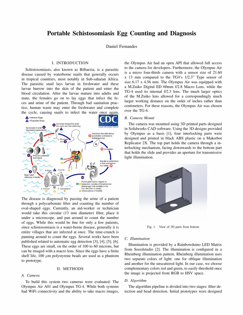
B. Camera Mount
The camera was mounted using 3D printed parts designedin Solidworks CAD software. Using the 3D designs providedby Olympus as a basis [1], four interlocking parts weredesigned and printed in black ABS plastic on a MakerbotReplicator 2X. The top part holds the camera through a in-terlocking mechanism, facing downwards to the bottom partthat holds the slide and provides an aperture for transmissivelight illumination.
Fig. 1. View of 3D parts from bottom
C. Illumination
Illumination is provided by a Rainbowduino LED Matrixfrom Seeedstudio [2]. The illumination is configured in aRheinberg illumination pattern. Rheinberg illumination usestwo separate colors of light: one for oblique illuminationand another for the unscattered light. In our case, we choosecomplementary colors red and green, to easily threshold oncethe image is projected from RGB to HSV space.
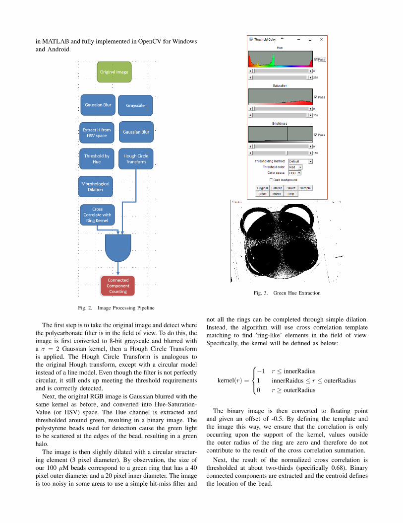
D. Algorithm
The algorithm pipeline is divided into two stages: filter de-tection and bead detection. Initial prototypes were designed
in MATLAB and fully implemented in OpenCV for Windowsand Android.
Fig. 2. Image Processing Pipeline
The first step is to take the original image and detect wherethe polycarbonate filter is in the field of view. To do this, theimage is first converted to 8-bit grayscale and blurred witha σ = 2 Gaussian kernel, then a Hough Circle Transformis applied. The Hough Circle Transform is analogous tothe original Hough transform, except with a circular modelinstead of a line model. Even though the filter is not perfectlycircular, it still ends up meeting the threshold requirementsand is correctly detected.
Next, the original RGB image is Gaussian blurred with thesame kernel as before, and converted into Hue-Saturation-Value (or HSV) space. The Hue channel is extracted andthresholded around green, resulting in a binary image. Thepolystyrene beads used for detection cause the green lightto be scattered at the edges of the bead, resulting in a greenhalo.
The image is then slightly dilated with a circular structur-ing element (3 pixel diameter). By observation, the size ofour 100 µM beads correspond to a green ring that has a 40pixel outer diameter and a 20 pixel inner diameter. The imageis too noisy in some areas to use a simple hit-miss filter and
Fig. 3. Green Hue Extraction
not all the rings can be completed through simple dilation.Instead, the algorithm will use cross correlation templatematching to find ’ring-like’ elements in the field of view.Specifically, the kernel will be defined as below:
kernel(r) =
−1 r ≤ innerRadius1 innerRaidus ≤ r ≤ outerRadius0 r ≥ outerRadius
The binary image is then converted to floating pointand given an offset of -0.5. By defining the template andthe image this way, we ensure that the correlation is onlyoccurring upon the support of the kernel, values outsidethe outer radius of the ring are zero and therefore do notcontribute to the result of the cross correlation summation.
Next, the result of the normalized cross correlation isthresholded at about two-thirds (specifically 0.68). Binaryconnected components are extracted and the centroid definesthe location of the bead.
Fig. 4. Template Matching Result
Fig. 5. Zoomed in result on template match
III. RESULTS
The algorithm was implemented and tested on Androidwith OpenCV. For the toy example given below, there were33 connected components detected, with 20 true positives, 13false positives, 4 false negatives, and (4608x3456)-33 truenegatives (corresponding to all the remaining pixels). Thisyields an sensitivity of 83.33% and specificity of 100.00%.
This algorithm works fairly well for the simple methodsthat it uses. There are clearly many ways forward to improvethe algorithm. First, the algorithm could be improved byautomatically generating the kernel based on the size of
Fig. 7. Before and After
the filter. Second, we could use more modern segmentationmethods that use Convolutional Neural Nets. Finally, theimaging setup could be improved so that the illuminationfrom the LEDs is more uniform, or possibly HDR imagingcould be employed to reduce noise in the darker regions.
[2] http://wiki.seeed.cc/Rainbowduino_v3.0/[3] Coulibaly, J. T., Ouattara, M., DAmbrosio, M. V., Fletcher, D.
A., Keiser, J., Utzinger, J., Bogoch, I. I. (2016). Accuracyof Mobile Phone and Handheld Light Microscopy for the Di-agnosis of Schistosomiasis and Intestinal Protozoa Infections inCte dIvoire. PLOS Neglected Tropical Diseases, 10(6), e0004768.https://doi.org/10.1371/journal.pntd.0004768
[4] Linder, E., Grote, A., Varjo, S., Linder, N., Lebbad, M.,Lundin, M., Lundin, J. (2013). On-Chip Imaging of Schis-tosoma haematobium Eggs in Urine for Diagnosis by Com-puter Vision. PLoS Neglected Tropical Diseases, 7(12), e2547.https://doi.org/10.1371/journal.pntd.0002547
[5] Li, Z., Gong, H., Zhang, W., Chen, L., Tao, J., Song, L., & Wu,Z. (2015). A robust and automatic method for human parasite egg