Biliopatia Hipertensiva Portal: Uma Causa Infrequente de Icterícia Obstrutiva
ResumoIntroducão: A causa mais comum de icterícia obstrutiva é a coledocolitíase. No entanto, nocontexto clínico adequado, devem ser consideradas etiologias alternativas ou concomitantes,nomeadamente a biliopatia hipertensiva portal (BHP).
Jaundice is the most frequent presentation of liver andbiliary disease and the diagnosis of obstructive jaundicecan usually be established with simple laboratory tests andultrasonography.1 Choledocholithiasis is by far the mostcommon cause of biliary obstruction but other importantconditions to consider are biliary or extrabiliary malig-nancy, pancreatitis, non-neoplastic biliary strictures andparasites.2 However, as evidenced by our case report, imag-ing investigations may bring to light an uncommon causeof biliary obstruction such as portal hypertensive biliopathy(PHB) that requires not only the relief of the obstructivejaundice but also the diagnosis of the cause of portal hyper-tension and its management.3
2. Clinical case
A 36-year-old woman presented to the emergency depart-ment due to jaundice that had first become apparent threedays earlier. She also reported pruritus for the last week,pale stools and dark discoloration of the urine. While deny-ing any abdominal pain, the patient did complain of nauseaand vomiting.
At the time of presentation the patient did not recall anysignificant past medical history including blood transfusionsand was taking no prescription or over-the-counter drugs.She also denied unprotected sexual intercourse.
Physical examination was notable for marked jaundiceand scratch induced excoriations. The gallbladder was notpalpable but a nontender hepatomegaly extending 2 cmbelow the ribcage was noted. The remaining of the physicalexamination was unremarkable.
The initial laboratory test results consisted of a nor-mal complete blood count and a normal prothrombin timeof 10.2 s (control 12.0 s). Total bilirubin was elevated at9.9 mg/dL (0.3---1.2 mg/dL) with a conjugated bilirubin of8.7 mg/dL. Alkaline phosphatase was also raised at 237 IU/L(35---105 IU/L) as was aspartate aminotransferase with avalue of 170 IU/L (<32 IU/L).
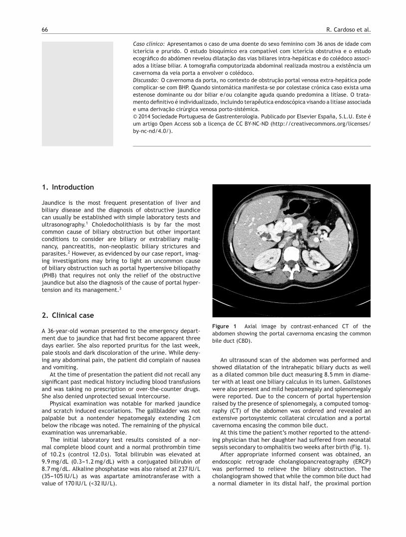
Figure 1 Axial image by contrast-enhanced CT of theabdomen showing the portal cavernoma encasing the commonbile duct (CBD).
An ultrasound scan of the abdomen was performed andshowed dilatation of the intrahepatic biliary ducts as wellas a dilated common bile duct measuring 8.5 mm in diame-ter with at least one biliary calculus in its lumen. Gallstoneswere also present and mild hepatomegaly and splenomegalywere reported. Due to the concern of portal hypertensionraised by the presence of splenomegaly, a computed tomog-raphy (CT) of the abdomen was ordered and revealed anextensive portosystemic collateral circulation and a portalcavernoma encasing the common bile duct.
At this time the patient’s mother reported to the attend-ing physician that her daughter had suffered from neonatalsepsis secondary to omphalitis two weeks after birth (Fig. 1).
After appropriate informed consent was obtained, anendoscopic retrograde cholangiopancreatography (ERCP)was performed to relieve the biliary obstruction. Thecholangiogram showed that while the common bile duct hada normal diameter in its distal half, the proximal portion
Portal hypertensive biliopathy 67
Figure 2 Cholangiogram during ERCP showing a sacculardilatation in the middle of the CBD with subtraction imagescompatible with calculi dilatation in its interior. The proximalportion of the CBD was also dilated while the diameter of thedistal half remained normal.
was dilated at 10 mm and subtraction images compatiblewith calculi were present. In the transition between thesetwo segments of the common bile duct a saccular dilationwas formed and also contained calculi. After sphincterotomycalculi removal was ensued requiring mechanical lithotripsyand a plastic stent was left in the common bile duct since itwas not possible to remove all calculi.
In order to investigate conditions contributing for thecavernoma’s development, a thrombophilia screen wasordered and its results were negative. Further anatomicdefinition was attained with the help of a magnetic res-onance cholangiography that revealed, in addition to thepersistence of calculi in the biliary ducts, an anatomicalvariant with the biliary drainage of the posterior segmentsof the right hepatic lobe being done to the left hepatic duct(Fig. 2). An esophagogastroduodenoscopy was performedand was unremarkable, showing no signs of portal hyper-tension namely esophageal varices.
The patient had an excellent outcome after biliarystenting with total remission of symptoms, jaundice and nor-malization of biochemical markers of cholestasis. The casewas presented to a hepatobiliary surgeon and the patient iscurrently waiting for portosystemic shunt surgery.
3. Discussion
In this clinical report we present the case of a patient withbiliary obstruction caused by PHB which in turn resulted fromextrahepatic portal venous obstruction (EHPVO).
Portal cavernoma, also designated as portal vein cav-ernomatous transformation, consists in the formation of
varicose collaterals around an occluded segment of theportal vein and is the hallmark feature of EHPVO.4 Thiscompensatory but pathological phenomenon representsan important cause of portal hypertension, especially indeveloping countries where it accounts for 40% of portalhypertension cases.5 In most instances it presents in chil-dren as major upper gastrointestinal bleeding.6 However, ina minority of individuals from which our patient is an exam-ple, EHPVO may only manifest itself in adulthood as a causeof chronic liver disease, PHB or an incidental finding.7 Theetiology of the portal cavernoma itself is only identified inabout half the cases, being omphalitis and intra-abdominalsepsis the leading causes in the pediatric population whilein adults a wide array of conditions such as thrombophilia,intra-abdominal infection or malignancy, myeloproliferativedisorders and cirrhosis have been implicated.7,8 In this casewe believe that the driving force for the portal cavernomadevelopment was the omphalitis episode that occurred inthe patient’s infancy. Concurring conditions such as throm-bophilia, abdominal sepsis or malignancy were excluded dueto clinical observation, evolution and appropriate laboratorytesting and imaging.
PHB, also commonly named pseudosclerosing cholangi-tis, portal ductopathy or simply portal biliopathy, is usedto describe the abnormalities observed in the full extentof the biliary tract including intrahepatic and extrahepaticbile ducts, gallbladder and cystic duct in patients with portalhypertension.3 This condition is mainly a feature of EHPVOwhere radiological evidence of PHB is present in 81---100%of patients but it can be found in a small proportion ofindividuals with other causes of portal hypertension.9
The pathogenesis of PHB is based on three mechanismsthat may act individually or simultaneously: compression ofthe pliable common bile duct by the collaterals that form theportal cavernoma and the connective tissue that developswith time and encases this duct; ischemia induced by portalthrombosis and subsequent sclerosis of the vessels that drainthe bile ducts resulting in the formation of biliary strictures;inflammation and deposition of connective tissue caused bybacterial cholangitis.9
Choledocholithiasis is a common feature of PHB and canbe present in as much as 84% of patients even though thishigh value may represent a selection bias.10 The increasedoccurrence of biliary stones in PHB is likely justified bychronic cholestasis and changes in the constituents of bilewhile reduced portal flow and liver dysfunction may alsoplay a role.11
The proportion of patients with EHPVO that experiencesymptoms related to PHB ranges between 5% and 38%.12,13
When symptoms do occur, the clinical presentation is usuallyeither one of chronic cholestasis when strictures dominateor of biliary pain or acute cholangitis when choledocholithi-asis is the main culprit.3 In this case, while there was nocomplaint of significant pain or features of acute cholan-gitis, we believe that the presentation was dominated byacute obstruction of a dominant stricture of the commonbile duct by a biliary stone. This view is not only supportedby the radiological diagnosis of choledocholithiasis but alsoby the fast resolution of jaundice and cholestasis followingbiliary stenting. Individuals who do present with symptomsof PHB tend to do so at an older age when compared withchildren presenting with upper gastrointestinal bleeding or
68 R. Cardoso et al.
splenomegaly as features of EHPVO, and at a mean age of36 years old, the same age as our patient.14
In this case the suspicion of biliary obstruction elicited byappropriate clinical signs and symptoms, biochemical abnor-malities and abdominal ultrasonography findings justifiedobtaining a CT scan than in its turn diagnosed portal caver-noma as the cause of jaundice and associated biliary calculiwhich further drove testing and therapy.
ERCP has been the traditional method of investigatingPHB and was pivotal in the recognition of this condition. Sev-eral characteristic findings have been described and includesmooth strictures of variable lengths, degrees and number,saccular dilations, indentations, dilated intrahepatic bileducts, as well as filling defects mainly in the common bileduct that may represent calculi or varices.14,15 While itsdiagnostic use has been hindered by the availability of non-invasive and equally accurate alternatives, ERCP still has amajor role when therapy is anticipated.
With magnetic resonance it is now possible to couplecholangiography and portography thus allowing the evalu-ation of all components of PHB with a single noninvasivetest.4 Since several studies corroborate that magnetic res-onance cholangiography has a similar sensitivity to ERCP inthis setting, it has become the first line imaging test in thiscondition.16
Endoscopic ultrasonography does not yet have a definedrole in PHB and its use is limited to the cases where otherimaging tests are unable to give a clear answer, namely in thedifferential diagnosis of common bile duct stones, calculi ortumors.17
Therapy is currently not recommend for asymptomaticindividuals with normal liver function tests. However,patients presenting with symptoms require an integratedmanagement addressing not only biliary obstruction but alsoportal hypertension. Endoscopic treatment is the therapyof choice when there is choledocholithiasis, acute cholangi-tis or if shunt surgery is not possible.3,18---21 ERCP is usuallyeffective in stone extraction and treatment of dominantstrictures.3,14,18 However, the relief of biliary obstructionwith balloon dilation and stenting is only a temporarymeasure and mandates the construction of a portosys-temic venous shunt or regular sessions of endotherapy sincestents tend to become clogged over time.14 Increased riskof bleeding is also an issue due to presence of venouscollaterals.22
Patients that have a shuntable vein should be offeredportosystemic shunt surgery since this procedure effec-tively addresses portal hypertension and may be the onlytreatment needed for dominant biliary strictures.23---25 Tran-sjugular intrahepatic portosystemic shunt may be a validalternative, albeit with less supporting evidence, and maybe considered in selected cases when surgery is not feasi-ble or is declined.3 In some cases additional biliary bypasssurgery or cholecystectomy may be required.
Since our patient exhibited biliary stones in associationwith PHB she was initially managed endoscopically withgreat success and is now awaiting shunt surgery without anysymptoms. The need for an effective management of por-tal hypertension in this patients, even when jaundice hasresolved, is emphasized by the diagnosis of secondary biliarycirrhosis and liver dysfunction in patients with longstandingand untreated PHB.16,18
In summary, PHB is an uncommon entity that seldomcauses symptoms. However, when symptomatic, the mana-gement must be tailored individually and should includeshunt surgery. Magnetic resonance is the imaging modalityof choice and ERCP is very useful when biliary stones, acutecholangitis or contraindications for surgery are present.
Ethical disclosures
Protection of human and animal subjects. The authorsdeclare that no experiments were performed on humans oranimals for this study.
Confidentiality of data. The authors declare that they havefollowed the protocols of their work center on the publica-tion of patient data.
Right to privacy and informed consent. The authors haveobtained the written informed consent of the patients orsubjects mentioned in the article. The corresponding authoris in possession of this document.
Conflict of interest
The authors declare that there are no conflicts of interest.
References
1. Beckingham IJ, Ryder SD. Investigation of liver and biliary dis-ease. BMJ. 2001;322:33---6.
3. Dhiman RK, Behera A, Chawla YK, Dilawari JB, Suri S. Portalhypertensive biliopathy. Gut. 2007;56:1001---8.
4. Shin SM, Kim S, Lee JW, Kim CW, Lee TH, Lee SH, et al.Biliary abnormalities associated with portal biliopathy: eval-uation on MR cholangiography. Am J Roentgenol. 2007;188:W341---7.
6. Chawla YK, Dilawari JB, Ramesh GN, Kaur U, Mitra SK, Walia BN.Sclerotherapy in extrahepatic portal venous obstruction. Gut.1990;31:213---6.
7. Rangari M, Gupta R, Jain M, Malhotra V, Sarin SK. Hepatic dys-function in patients with extrahepatic portal vein obstruction.Liver Int. 2003;23:434---9.
8. Valla DC, Condat B, Lebrec D. Spectrum of portal vein throm-bosis in the West. J Gastroenterol Hepatol. 2002;17 Suppl3:S224---7.
9. Chattopadhyay S, Nundy S. Portal biliopathy. World J Gastroen-terol. 2012;18:6177---82.
10. Oo YH, Olliff S, Haydon G, Thorburn D. Symptomatic portalbiliopathy: a single centre experience from the UK. Eur J Gas-troenterol Hepatol. 2009;21:206---13.
11. Harmanci O, Bayraktar Y. How can portal vein cavernous trans-formation cause chronic incomplete biliary obstruction? WorldJ Gastroenterol. 2012;18:3375---8.
13. Khuroo MS, Yattoo GN, Zargar SA, Javid G, Dar MY, Khan BA,et al. Biliary abnormalities associated with extrahepatic portalvenous obstruction. Hepatology. 1993;17:807---13.
Portal hypertensive biliopathy 69
14. Sezgin O, Oguz D, Attintas E, Saritas U, Sahin B. Endo-scopic management of biliary obstruction caused by cavernoustransformation of the portal vein. Gastrointest Endosc.2003;68:602---8.
15. Nagi B, Kochhar R, Bhasin D, Singh K. Cholangiopathy in extra-hepatic portal venous obstruction. Radiological appearances.Acta Radiol. 2000;41:612---5.
17. Umphress JL, Pecha RE, Urayama S. Biliary stricture caused byportal biliopathy: diagnosis by EUS with Doppler US. Gastroin-test Endosc. 2004;60:1021---4.
18. Chandra R, Kapoor D, Tharakan A, Chaudhary A, Sarin SK. Portalbiliopathy. J Gastroenterol Hepatol. 2001;16:1086---92.
19. Khare R, Sikora SS, Srikanth G, Choudhuri G, Saraswat VA, KumarA, et al. Extrahepatic portal venous obstruction and obstructivejaundice: approach to management. J Gastroenterol Hepatol.2005;20:56---61.
20. Thervet L, Faulques B, Pissas A, Bremondy A, Monges B, SalducciJ, et al. Endoscopic management of obstructive jaundice dueto portal cavernoma. Endoscopy. 1993;25:423---5.
21. Lopez RR, Cosenza CA, Lois J, Hoffman AL, Sher LS, NoguchiH, et al. Long-term results of metallic stents for benign biliarystrictures. Arch Surg. 2001;136:664---9.
22. Mutignani M, Shah SK, Bruni A, Perri G. Costamagna endoscopictreatment of extrahepatic bile duct strictures in patients withportal biliopathy carries a high risk of hemobilia: report of 3cases. Dig Liver Dis. 2002;34:587---91.
23. Chaudhary A, Dhar P, Sarin SK, Sachdev A, Agarwal AK, Vij JC,et al. Bile duct obstruction due to portal biliopathy in extra-hepatic portal hypertension: surgical management. Br J Surg.1998;85:326---9.
24. Agarwal AK, Sharma D, Singh S, Agarwal S, Girish SP. Portal bil-iopathy: a study of 39 surgically treated patients. HPB (Oxford).2011;13:33---9.
25. Vibert E, Azoulay D, Aloia T, Pascal G, Veilhan LA, Adam R, et al.Therapeutic strategies in symptomatic portal biliopathy. AnnSurg. 2007;246:97---104.