Post-infectious irritable bowel syndrome: Mechanistic insights into chronic disturbances following enteric infection
Jennifer K Beatty, Amol Bhargava, Andre G Buret
Jennifer K Beatty, Amol Bhargava, Andre G Buret, Depart-ment of Biological Sciences, University of Calgary, Calgary, AB T2N 1N4, CanadaAuthor contributions: Beatty JK, Bhargava A and Buret AG wrote this paper.Supported by Natural Sciences and Engineering Research Council of Canada (individual operating and CREATE)Correspondence to: Andre G Buret, PhD, Professor, Depart-ment of Biological Sciences, University of Calgary, 2500 Univer-sity Drive, NW, Calgary, AB T2N 1N4, Canada. [email protected]: +1-403-2202817 Fax: +1-403-2899311Received: November 21, 2013 Revised: January 9, 2014Accepted: February 17, 2014Published online: April 14, 2014
AbstractIrritable bowel syndrome (IBS) is a commonly encoun-tered chronic functional gastrointestinal (GI) disorder. Approximately 10% of IBS patients can trace the onset of their symptoms to a previous a bout of infectious dysentery. The appearance of new IBS symptoms fol-lowing an infectious event is defined as post-infectious-IBS. Indeed, with the World Health Organization estimating between 2 and 4 billion cases annually, in-fectious diarrheal disease represents an incredible inter-national healthcare burden. Additionally, compounding evidence suggests many commonly encountered en-teropathogens as unique triggers behind IBS symptom generation and underlying pathophysiological features. A growing body of work provides evidence supporting a role for pathogen-mediated modifications in the resi-dent intestinal microbiota, epithelial barrier integrity, effector cell functions, and innate and adaptive immune features, all proposed physiological manifestations that can underlie GI abnormalities in IBS. Enteric pathogens must employ a vast array of machinery to evade host protective immune mechanisms, and illicit successful in-fections. Consequently, the impact of infectious events on host physiology can be multidimensional in terms
of anatomical location, functional scope, and duration. This review offers a unique discussion of the mecha-nisms employed by many commonly encountered en-teric pathogens that cause acute disease, but may also lead to the establishment of chronic GI dysfunction compatible with IBS.
Core tip: This review discusses the long-term conse-quences of acute enteric infections that may serve to trigger post-infectious irritable bowel syndrome, a rou-tinely diagnosed disorder. This unique discussion elu-cidates novel initiation mechanisms, underlying patho-physiological features of post-infectious irritable bowel syndrome, employed by commonly encountered enteric pathogens.
Beatty JK, Bhargava A, Buret AG. Post-infectious irritable bowel syndrome: Mechanistic insights into chronic distur-bances following enteric infection. World J Gastroenterol 2014; 20(14): 3976-3985 Available from: URL: http://www.wjgnet.com/1007-9327/full/v20/i14/3976.htm DOI: http://dx.doi.org/10.3748/wjg.v20.i14.3976
INTRODUCTIONIrritable bowel syndrome (IBS) is among the most com-monly encountered chronic functional gastrointestinal (GI) disorders afflicting individuals in westernized na-tions. Based on the Rome Ⅲ criteria abdominal pain accompanied by sustained changes in bowel habit consti-tute IBS, whose diagnosis is achieved in the absence of
biochemical markers of disease[1]. Clinical presentation of constipation, diarrhea, or a combination, constitutes the different subtypes of IBS: IBS with constipation (IBS-C), diarrheal IBS subtype (IBS-D), mixed IBS (IBS-M), re-spectively[2]. Often perceived as a female-dominant dis-order, IBS is thought to afflict between 5%-10% of the population[3], especially in westernized nations. Elucidat-ing the mechanisms underlying the typical multifaceted clinical presentation of IBS is a topic of considerable research efforts in the medical community[4]. A growing body of evidence implicates numerous triggering events in contributing to IBS pathophysiology, including an initiating bout of infectious enteritis, low grade inflam-mation, altered functionalities in GI cell types, increases in epithelial permeability, and alterations in the GI mi-crobiota, although the precise mechanisms of underlying each remain obscure[2,5-8]. Approximately 10% of IBS patients believe that their symptoms began following a bout of infectious dysentery[6], leading to the coinage of the term; Post infectious (Pi)-IBS. While many enteric pathogens cause self-limiting, acute diarrheal disease, subsequent chronic physiological consequences may per-sist in some individuals[9]. Many commonly encountered enteric pathogens can produce physiological changes that may provide important initiation mechanisms underlying chronic GI conditions, such as Pi-IBS. This article criti-cally reviews the evidence supporting a role for key physi-ological changes initiated during enteric infection, that may in turn be responsible for IBS symptom.
Pi-IBSBased on the Rome criteria for diagnosis, any onset of new IBS symptoms subsequently following an infectious event is defined as Pi-IBS[6]. Pi-IBS cases often exhibit characteristics of the IBS-D, and can occur in 4%-31% of patients following acute gastroenteritis[6,10-12]. A large body of work provides evidence supporting a role for pathogen-mediated modifications in the resident intes-tinal microbiota, epithelial barrier integrity, enterochro-maffin cell function, and innate immune features[5,13,14] in Pi-IBS manifestation. Any number of these pathogenic consequences have been reported following enteric infec-tion incited by an array of pathogens such as Shigella spp., pathogenic Escherichia coli, Salmonella, Campylobacter jejuni, and Giardia duodenalis[14-18]. Enteric pathogens must employ a vast array of machinery to evade the host protective immune mechanisms, and illicit successful infections. Re-cent work identifying genetic mutations, namely in genes responsible for epithelial and innate immune function-alities, in patients experiencing both the post-infectious, and traditional forms of IBS, point to defects in innate immunity and epithelial homeostasis as an important risk factor for IBS susceptibility[19,20]. The impact of infectious events on host physiology can be multidimensional in terms of anatomical location, functional scope, and dura-tion. Indeed, anatomical, immunological, and neurologi-cal dysfunctions, or combinations of such, have all been shown as risk factors determining Pi-IBS manifestation.
This review will provide an in-depth discussion surround-ing the potential roles in which a variety of commonly encountered enteric pathogens may play in initiating im-portant pathophysiological features of Pi-IBS.
CLINICAL PRESENTATIONS OF IBS FOLLOWING ENTERIC INFECTION: ALTERED INTESTINAL MOTILITY AND HYPERSENSITIVITYAbnormal bowel habits and abdominal hypersensitivity, or reduced threshold of pain, are the hallmark clinical signs of IBS. The classification of IBS as a functional disorder stems from a lack of determinant histopatho-logical, or structural biomarkers in afflicted patients. The Rome criteria requires the incidence of abdominal pain, accompanied by alterations in bowel habit for complete IBS diagnosis[21].
Altered intestinal motilityAbnormal GI motility is commonly associated with al-tered bowel habits producing diarrheal, constipation, and mixed IBS subtypes[22]. The potential for dysfunctional intestinal motility in contributing to altered bowel hab-its in IBS is supported by studies looking at intestinal transit rates between healthy and IBS individuals, with IBS-D subtypes exhibiting enhanced rates of SI transit, and the opposite trend observed for IBS-C patients[22,23]. Moreover, a recent report demonstrated that the nor-mal colorectal reflex (normal increase in rectal tone in response to phasic colonic distention) was largely abol-ished in IBS patients, regardless of bowel habit, provid-ing some evidence for altered colonic motility in these individuals[24]. Interestingly, muscle hypercontractility and abnormal motility patterns are observed subsequent to Trichinella spiralis infection in a commonly used murine model of PI-IBS[13,25-27], suggesting that persistent dys-functional intestinal motility can be incited following an acute infection.
Abdominal hypersensitivityLower thresholds for pain tolerance in IBS patients have been documented along the entire length of the GI tract[22], an effect that is thought to occur in upwards of 60% of afflicted individuals[7]. Hypersensitivity often oc-curs locally in response to colonic distention[7]. Further-more, overall visceral hypersensitivity, even upon brief stimuli such as the ingestion of food, is well documented in IBS patients, and may contribute to additional bloating, nausea, and urgency symptoms[8,28].
Stressful events can drastically affect the processing of visceral stimuli[29,30] and result in dysfunctional cen-tral neural processes culminating into heightened pain perception. Injury to visceral afferents, for example, is a common cause underlying visceral hypersensitivity[7]. Studies using a rat model of TNBS-induced transient colonic inflammation have highlighted that persistent tis-
3977 April 14, 2014|Volume 20|Issue 14|WJG|www.wjgnet.com
Beatty JK et al . Chronic consequences following enteric infections
sue injury may directly produce heightened visceral pain perception[31]. Importantly, chemically induced colonic inflammation models have stark parallels to many of the physiological events accompanying enteric infections. Initial processes of inflammation, for instance, may act to first sensitize effector, neuronal, and immune cells within the GI tract.
Interestingly, many of the physiological consequences that can result from infectious events within the GI tract have also been proposed as determinants capable of contributing to abnormal motility and hypersensitivity symptoms seen in IBS patients. The major mechanisms currently thought to underlie IBS pathogenesis, and the evidence surrounding possible contributions made to each by distinct enteric pathogens, will be discussed in the following sections (Figure 1).
PATHOPHYSIOLOGICAL FEATURES OF IBS FOLLOWING ENTERIC INFECTIONImmune system alterationsAccumulating evidence suggests subtle alterations in the immune system in both the gut, and peripheral circulation of PI-IBS patients[32]. Pathogen-mediated disruptions of the mucosal barrier have the ability to allow for persistent immune activation within the intestine, largely due to in-creased exposure to luminal antigens. Likewise, the host inflammatory response towards perceived pathogens, while meant to be protective, may result in detrimental, perpetuated activation of effector cells and inflammatory mediators. The incidence of PI-IBS symptoms in many patients following enteric infection has fuelled interest in looking at persistent immune infiltrate, and/or altered
3978 April 14, 2014|Volume 20|Issue 14|WJG|www.wjgnet.com
Commensal Microbiota
(A)(B)
(C)(D)
(E)
?
? ?
Enterocytes
MacrophageSCV
Pyroptosis
NF-κB
M-cell
IL-1β, IL-8 release
IFN-γ release
Dendritic cell
Pyroptosis
NLRC4-dependent inflammasome activation
IL-1β, IL-18 release
TNF-α, IFN-γ, IL-1β release
NF-κB,ERK-1/2 MLCK
EPEC EHEC
Enteric nerve fibres
Portal vein
Mast cells
5-HT
Enterochromaffin cell5-HT in vesicles
IL-18 release
Basolateral
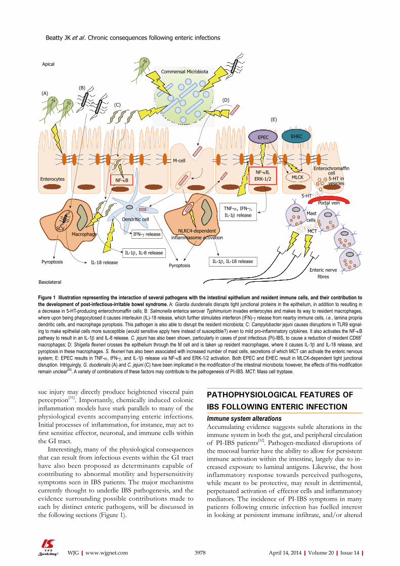
Figure 1 Illustration representing the interaction of several pathogens with the intestinal epithelium and resident immune cells, and their contribution to the development of post-infectious-irritable bowel syndrome. A: Giardia duodenalis disrupts tight junctional proteins in the epithelium, in addition to resulting in a decrease in 5-HT-producing enterochromaffin cells; B: Salmonella enterica serovar Typhimurium invades enterocytes and makes its way to resident macrophages, where upon being phagocytosed it causes interleukin (IL)-18 release, which further stimulates interferon (IFN)-γ release from nearby immune cells, i.e., lamina propria dendritic cells, and macrophage pyroptosis. This pathogen is also able to disrupt the resident microbiota; C: Campylobacter jejuni causes disruptions in TLR9 signal-ing to make epithelial cells more susceptible (would sensitive apply here instead of susceptible?) even to mild pro-inflammatory cytokines. It also activates the NF-κB pathway to result in an IL-1β and IL-8 release. C. jejuni has also been shown, particularly in cases of post infectious (Pi)-IBS, to cause a reduction of resident CD68+ macrophages; D: Shigella flexneri crosses the epithelium through the M cell and is taken up resident macrophages, where it causes IL-1β and IL-18 release, and pyroptosis in these macrophages. S. flexneri has also been associated with increased number of mast cells, secretions of which MCT can activate the enteric nervous system; E: EPEC results in TNF-α, IFN-γ, and IL-1β release via NF-κB and ERK-1/2 activation. Both EPEC and EHEC result in MLCK-dependent tight junctional disruption. Intriguingly, G. duodenalis (A) and C. jejuni (C) have been implicated in the modification of the intestinal microbiota; however, the effects of this modification remain unclear[90]. A variety of combinations of these factors may contribute to the pathogenesis of PI-IBS. MCT: Mass cell tryptase.
Beatty JK et al . Chronic consequences following enteric infections
MCT
Apical
3979 April 14, 2014|Volume 20|Issue 14|WJG|www.wjgnet.com
and dendritic cells (DCs) for phagocytosis upon which activation of the nucleotide-binding oligomerization domain (NOD)-like receptor protein (NLRC4) inflam-masome occurs[44,45] (Figure 1). Consequently, the result-ing activation of pro-inflammatory cytokines, interleukin (IL)-18 and IL-1β, are thought to be major determinants of the high inflammatory conditions characteristic of early Shigella infection[45]. Inflammasome activation can also produce heightened rates of macrophage cell death via pyroptosis, which acts as an “inflammatory” form of programmed cell death (Figure 1). Thus, Shigella infection promotes a high status of inflammation, while simultane-ously resulting in the detrimental loss of lamina propria (LP) macrophages. LP macrophages have an important regulatory, and anti-inflammatory role in maintaining in-testinal homeostasis[45]. Furthermore, as a consequence of resident LP macrophage depletion, additional circulating monocytes may be recruited to the site of infection, and often differentiate into macrophages possessing a more pro-inflammatory capacity[45]. Considering ample reports documenting low-grade inflammation IBS patients[46,47], pathogen-mediated inflammatory conditions, in addition to the promotion of pro-inflammatory cell phenotypes, may be especially relevant triggers underlying Pi-IBS de-velopment.
In contrast to Shigella, Salmonella is seemingly less cyto-toxic to macrophages[48], yet Pi-IBS symptoms have been reported following anywhere between 6%-32% of con-firmed infections[2,19]. Following phagocytosis, Salmonella forms the characteristic Salmonella Containing Vacuole (SCV) in macrophages, in which it replicates while effec-tively evading host immune machinery, and pyroptosis[48] (Figure 1). While capable of avoiding certain immune pa-rameters, Salmonella still evokes a strong IL-18 response[48] which has important implications in exerting paracrine effects on surrounding immune cells to induce IFN-γ ex-pression, and also result in increased levels of activated T cells in the infected intestine, accumulation of which has been documented in many examinations of IBS[9,32,33,42,49].
Cytokine profiles: Substantial regulation exists within the GI tract in order to maintain a functional balance between pro- and anti-inflammatory mediators under ho-meostatic conditions. Engagement of the Toll-like recep-tors (TLRs), NOD-like receptors (NLRs), and other host pathogen-recognition-receptors (PRRs) occurs through li-gation by various pathogen-associated-molecular-patterns (PAMPs). Shigella, for instance, is known to stimulate excess production of IL-1β from immune cells during infection via the NLRC4 inflammasome[44,45] (Figure 1). Also, excessive IL-8 secretion is a hallmark of Campylo-bacter pathogenesis[50], and is initiated upon host recogni-tion of the pathogen-associated lipooligosaccharide[51]. Interestingly, a recent report demonstrated a disruption in TLR9 expression on epithelial cells to be implicated in the enhanced susceptibility to mild pro-inflammatory stimuli post-campylobacteriosis in mice[52]. C. jejuni is also know to promote the translocation of non-invasive commensal
immune functionalities, as plausible driving forces in the generation of IBS symptoms[33].
Mast cells/macrophages/dendritic cells: Certain enteric pathogens have been shown to promote mast cell accumulation. A recent study found that a large propor-tion of patients experiencing Shigellosis, caused by inva-sive Shigella spp., go on to develop PI-IBS, and that this effect is accompanied by augmented mast cell numbers[34]. Under normal conditions, mucosal mast cells are highly involved in wound-healing, and defense against patho-gens[5]. However, multiple reports document heightened numbers of mast cells within the small[35,36], and large in-testines[37-39] of IBS patients. One study, which observed increased mast cells specifically within the duodenum of IBS patients suggested that infiltration of these cells may provide some explanation behind the observation that symptoms differ depending upon the affected site along the GI tract[36]. Also, mast cells can secrete serotonin, therefore increased populations of these cells may pro-vide a link between cellular infiltrate and altered serotonin signaling leading to changes along the brain-gut axis, and dysmotility, characteristic of either IBS-D or IBS-C[36]. Furthermore, augmented numbers of mast cells, and par-ticularly those closely associated with nerve fibers, have been reported in both IBS and Pi-IBS[38] (Figure 1), an ef-fect which may be correlated with enhanced bloating and pain perception symptoms[2,40-42].
The T. spiralis mouse model of Pi-IBS has provided important insight into many pathophysiological changes following acute enteric infection. A recent study, for in-stance, documented numerical and phenotypic alterations in lamina propria dendritic cells (LPDC), following acute T. spiralis infection[43]. In what the authors defined as the “Pi-IBS stage” of infection, i.e., no recovery of nematode in the stool, LPDCs exhibited enhanced expression of co-stimulatory molecules, and greater ability to migrate to and drive CD4+ T cell proliferation[43]. Furthermore, the altered LPDC phenotype was proposed to underlie enhanced levels of pro-inflammatory interferon (IFN)-γ, IL-23 and tumor necrosis factor (TNF)-α production in the Pi-IBS stage[43]. The important role that these cells play in directing T-cell responses may have implications in promoting a low-grade inflammatory milieu, and requires further investigation in relation to IBS pathogenesis.
Monocytes and macrophages are at the forefront of initiating an inflammatory response to pathogens, in ad-dition to providing essential directives to the adaptive immune system[5]. In Pi-IBS cases confirmed following C. jejuni infection the numbers of resident CD68+ mac-rophages are diminished, perhaps owing to the cytotoxic nature of the pathogen inside host cells[9]. Likewise, Shigella spp.[15,16] and Salmonella infections have been im-plicated in causing Pi-IBS, and both are obligate intracel-lular pathogens, which preferentially exploit phagocytic machinery of the macrophage. Specifically, Shigella is transported into the lamina propria through M cells in the epithelium, and presented to resident macrophages
Beatty JK et al . Chronic consequences following enteric infections
3980 April 14, 2014|Volume 20|Issue 14|WJG|www.wjgnet.com
bacteria via paracellular and transcellular pathways[53,54]. Campylobacter has also been shown to activate copious amounts of nuclear factor (NF)-κB and IL-1β from im-mune cells, in vitro[51]. Likewise, recognition of EPEC flagellin and endotoxin results in NF-κB and extracellular signal regulated kinase (ERK)-1/2 –driven IL-8 release, and enhanced TNF-α, IFN-γ and IL-1β in the infected mucosa[55,56] (Figure 1). Interestingly, at least some of the pro-inflammatory cytokines, including TNF-α, IL-1β, and IFN-γ may themselves disrupt the epithelial bar-rier through alterations of the tight junctions (TJs), and promote increased permeability[57-59]. Thus, residual pro-inflammatory infiltrate following enteric infection com-bined with the sub-epithelial penetration of commensal bacteria, can create extensive damage to surrounding intestinal tissues, and likely promote chronic pathophysi-ological consequences. Consequently, many reports have drawn links between altered cytokine profiles and IBS generation[60], and findings include increased levels of pro-inflammatory IL-6, IL-8, and TNF-α in plasma and circulating blood mononuclear secretions from IBS pa-tients[47,61]. Lower detection of typical anti-inflammatory cytokines, IL-10 and transforming growth factor (TGF)-β, at the level of mRNA has also been reported[62]. Also, evi-dence from the T. spiralis Pi-IBS murine infection model has shown greater levels of IFN-γ, IL-23 and TNF-α produced by DCs in the Pi-IBS stage[43]. Additionally, sus-tained levels of pro-inflammatory mediators have been documented in a 21-d Citrobacter rodentium model of mu-rine E. coli pathogenesis[63]. Regardless of these promis-ing observations, the implications of pathogen-mediated alterations in normal cytokine profiles in providing suf-ficient trigger for IBS symptom establishment requires further investigation.
Mucosal barrier alterations The intestinal epithelium provides an interface between the luminal space and the dynamic environment of the underlying subepithelial compartment. This physical barrier is intricately involved in regulating the controlled passage of vital nutrients, molecules, and water, via a semipermeable function maintained by TJs. TJs actively maintain the polarized characteristic of the epithelial bar-rier, and are composed of over 40 proteins consisting of occludin, junctional adhesion molecule (JAM), and claudins[64]. Patients with a history of infectious events experiencing Pi-IBS show drastic increases in perme-ability[65,66]. A prospective study, however, following a large waterborne outbreak of bacterial gastroenteritis, incited by mixed infection of EHEC O157:H7 and C. jejuni, documented increased permeability to be associ-ated with IBS, regardless of whether symptoms were post-infectiously initiated[65]. Enterohemorrhagic E. coli (EHEC) is known to have deleterious impacts on the epithelial barrier through number of mechanisms, in-cluding TJ disruptions, and abnormal rates of intestinal epithelial cell (IEC) apoptosis[67,68]. These effects can be mediated directly via physical interaction through EHEC
formation of characteristic attaching and effacing lesions (A/E lesions), and/or diffusely through toxin release[64,69]. EHEC, and its close relative: Enteropathogenic E. coli (EPEC), are known to hijack various pathways regulating the semi-permeable profile of TJs, and both have been shown to activate myosin light chain kinase (MLCK) to produce abnormally leaky barrier functionalities[70-72] (Figure 1). Additionally, Giardia duodenalis, a protozoan pathogen recently implicated in promoting Pi-IBS devel-opment[18,73], is well-known to disturb homeostatic bar-rier function through alterations in key TJ elements[74]. Specifically, Giardia has been shown to disrupt zonula oc-cludins protein (ZO)-1, numerous transmembrane clau-din proteins, and alter F-actin and α-actinin in order to disrupt paracellular flow[75,76] (Figure 1), which may have important implications in providing a mechanistic link between initial giardiasis, and subsequent development of IBS symptoms. Indeed, recent analysis of colonic bi-opsies from IBS patients indicated decreased expression of ZO-1, which was associated with increased permeabil-ity[77]. Moreover, an earlier report examining fecal extracts indicated higher levels of serine proteases in samples from IBS-D patients. When these extracts were applied to healthy colonic mucosa, they could elicit a proteinase activated receptor (PAR)-2 dependent increase in para-cellular permeability in mice via increased myosin light chain (MLC) phosphorylation and delayed redistribution of ZO-1[78]. Numerous pathogens, including both EPEC and EHEC, produce potentially cytotoxic serine prote-ases[79], suggesting another possible link between enteric infection and IBS pathogenesis. Proteases are known to be involved in the infectious processes of pathogens such as EHEC and EPEC where they can prove detri-mental to the epithelial barrier via modifications of the extracellular matrix[80], and or by activating protease-activated receptors, which have been shown to stimulate sensory neurons to produce hypersensitivity reactions[81]. Consequently, the possibility of residual pathogen media-tors, such as inherent proteases, contributing to persistent changes in GI function requires further examination.
Enterochromaffin cells: Enterochromaffin cells (ECs) lining the GI mucosa are primary sources of Serotonin (5-HT) within the body. Alterations in the biosynthesis of 5-HT, in its release from ECs and degradation, and/or in its re-uptake, may have severe ramifications and per-turb normal GI function[82]. Multiple studies have shown significantly higher 5-HT levels in the plasma of Pi-IBS patients compared with that of healthy controls, even in comparison to patients of the sporadic IBS-C subtype[83]. Recent studies have observed such significant alterations in EC counts and 5-HT levels, that the authors declared Pi-IBS as a distinct IBS subtype[10,11]. Augmented num-bers of 5-HT-contatining ECs have been observed in co-lonic biopsies from patients following C. jejuni infection[9]. Up to 25% of C. jejuni infections are known to result in IBS[9], and the resulting implications on EC hyperplasia and excessive 5-HT bioavailability suggest a possible
Beatty JK et al . Chronic consequences following enteric infections
3981 April 14, 2014|Volume 20|Issue 14|WJG|www.wjgnet.com
mechanism whereby enteric infection may provide suf-ficient trigger for IBS symptom generation. Additionally, numerous reports have suggested a defect in the sero-tonin reuptake transporter (SERT) expression, and func-tion in IBS patients[82,84,85], which may dictate inadequacies in homeostatic serotonin turnover. Interestingly, in the T. spiralis model of Pi-IBS, mice develop chronic abnormal motility patterns subsequent to infection, an effect that is accompanied by EC hyperplasia and 5-HT release[6,13], and blocked upon administration of a 5-HT antagonist[86]. In contrast, patients with persisting abdominal symptoms after acute Giardia infection have lower duodenal 5-HT-containing ECs, and lower plasma 5-HT postprandially, compared to controls[87], further underscoring the com-plexity of IBS pathophysiology.
Intestinal microbiota disruptions: The intestinal mi-crobiota have extensive protective capacities[88] that are maintained by a diverse species profile. The characteristic high fat, high protein diets employed by the majority of people living in westernized countries facilitates the establishment of distinct microbiota species profile, as compared to that of those living in rural areas of devel-oping countries, with a polysaccharide-rich diet[89]. Par-ticular bacterial groups, mainly Bacteroidetes are known to harbor significant genetic capabilities to hydrolyse xylo-ses, making it an important constituent of the microbiota of people subsisting on carbohydrate-dominant food sources. The relative sensitivity of these distinct micro-biota to enteropathogens, and how in turn disruptions in their respective flora may differentially regulate post-infectious disorders, is unknown.
Interestingly, changes in the relative Firmicutes to Bacteroidetes ratio[90,91], loss of Bifidobacteria spp. and Faecalibacterium[91], and overall diminished diversity[92], are all apparent in the microbiota profile of IBS patients. Additionally, numerous studies have demonstrated small intestinal bacterial overgrowth in IBS patients, where excessive colonization of the small intestine occurs with colonic flora[33,93]. There is the possibility that en-teropathogens may disrupt the indigenous microbiota, either directly through pathogen-microbiota interactions, indirectly via the host mucosal immune response to the pathogen, or by a combination of the two[94]. For exam-ple, S. enterica serovar Typhimurium induced the loss of 95% of total bacterial numbers throughout the murine intestinal tract, 7 d following infection[94]. Findings from ongoing research also indicate that G. duodenalis and C. jejuni are able to directly alter species distribution of hu-man commensal microbiota[95]. Pathogenic effects, how-ever, may only provide a suitable trigger, and ultimately require the accompaniment of a host inflammatory re-sponse in order to markedly alter the microbiota ecosys-tem. The necessity of these compounding factors is ex-emplified in contrasting C. rodentium and C. jejuni murine infection models, where the former induces overt host inflammation, while the later can successfully colonize without producing inflammatory reactions[96]. It appears
then, that both enteropathogen assault, combined with pathogen-mediated intestinal inflammation, can elicit dramatic changes in the total abundance of the intestinal microbiota, and shift in anaerobic:aerobic species[96].
CONSIDERATIONMany studies that classify patients as experiencing Pi-IBS do so based upon questionnaires, highlighting the fact that they rely exclusively on a patient’s recall of past medi-cal events, including infections and/or prescription drug use. Some antibiotics, for example, have established cau-sality in disturbing the overall fecal microbial composition through drastic reduction of Firmicutes and Bacteroidetes, and a corresponding promotion of Proteobacteria spp.[97].
Also, the classification of IBS as biopsychosocial disorder challenges the mantra of body and mind being distinct entities, and suggests an equal consideration of both when examining disease manifestation. The risk of developing IBS symptoms following enteric infection may also differ in individuals depending on psychologi-cal parameters such as stress level, emotional status, and upbringing. High stress and anxiety levels, for instance, are associated with IBS development following Campylo-bacter infection[98]. Anxiety, as well as depression, is also correlated with altered pain perception in IBS patients[30]. Additionally, anxiety and depressive states in IBS patients were recently shown to lead to changes in serum levels of gastrointestinal hormones. Indeed, the authors sug-gest increased secretion of somatostatin and vasoactive intestinal peptide seen in IBS patients exhibiting anxiety-depression emotional state ratings, may contribute to altered gastrointestinal motility and function[99]. An important mediator in the endocrine arm of the stress response, corticotropin-releasing factor, may also con-tribute to Pi-IBS development through direct local action on specific cellular targets, namely mast cells, and conse-quently lead to the modification the intestinal inflamma-tory process[100].
Additionally, as Pi-IBS is defined based upon the de-velopment of exclusively new IBS symptom presentation, researchers must be certain that no preceding presenta-tion of IBS occurred. Indeed, clear cause-to effect rela-tionship studies need to establish mechanistic causalities in Pi-IBS.
CONCLUSIONUnfortunately, the link between physiological conse-quences of enteric infection and altered gut function (sensitivity and motility) seen in IBS remains largely circumstantial. As many as 30%-40% of patients ex-periencing enteritis can go on to develop chronic GI abnormalities compatible with IBS; however, this means that a greater percentage of patients make a full recovery. Susceptibility, in turn, to developing IBS is determined by a number of factors, with enteric pathogens constituting only one possible route of initiation. Regardless of the
Beatty JK et al . Chronic consequences following enteric infections
3982 April 14, 2014|Volume 20|Issue 14|WJG|www.wjgnet.com
heterogeneous initiation mechanisms culminating into disease, the pathophysiological implications of enteric infection provide important clues towards elucidating the mechanics underlying IBS manifestation. Animal models are becoming increasingly appreciated as divergent means in which IBS triggering mechanisms may be elucidated. Indeed, the maternal separation stress model in rodents is well documented in mimicking early life stress that can result in lifelong dysfunctions in the brain-gut axis, and is implicated in predisposing to IBS development[101]. Furthermore, animal models of post-infectious, or post-inflammatory conditions, such as those using T. spiralis or TNBS, are proving useful in examining the mechanisms underlying motility and pain perception changes subse-quent to diverse stimuli, without the challenges associated with patient recall, or the need for complex psychological status analyses.
This is especially relevant in terms of developing treatment technologies to combat IBS, most of which currently target overt symptomology. Many of the physi-ological consequences of GI infections represent paral-lels with fundamental triggering mechanisms currently though to contribute to IBS. Understanding the simi-larities between remnants of enteric infections, and the detrimental outcomes, can lead to the development of prevention strategies and therapeutic techniques to target IBS generation; before it can even start.
REFERENCES1 Bixquert Jiménez M. Treatment of irritable bowel syn-
drome with probiotics. An etiopathogenic approach at last? Rev Esp Enferm Dig 2009; 101: 553-564 [PMID: 19785495]
2 Spiller R, Campbell E. Post-infectious irritable bowel syndrome. Curr Opin Gastroenterol 2006; 22: 13-17 [PMID: 16319671]
3 Chang FY, Lu CL. Irritable bowel syndrome and migraine: bystanders or partners? J Neurogastroenterol Motil 2013; 19: 301-311 [PMID: 23875096 DOI: 10.5056/jnm.2013.19.3.301]
4 Ford AC, Talley NJ. IBS in 2010: advances in pathophysi-ology, diagnosis and treatment. Nat Rev Gastroenterol Hepatol 2011; 8: 76-78 [PMID: 21293507 DOI: 10.1038/nrgas-tro.2010.216]
5 Ohman L, Simrén M. Pathogenesis of IBS: role of inflam-mation, immunity and neuroimmune interactions. Nat Rev Gastroenterol Hepatol 2010; 7: 163-173 [PMID: 20101257 DOI: 10.1038/nrgastro.2010.4]
6 Spiller R, Garsed K. Postinfectious irritable bowel syndro-me. Gastroenterology 2009; 136: 1979-1988 [PMID: 19457422 DOI: 10.1053/j.gastro.2009.02.074]
7 Zhou Q, Verne GN. New insights into visceral hypersen-sitivity--clinical implications in IBS. Nat Rev Gastroenterol Hepatol 2011; 8: 349-355 [PMID: 21643039 DOI: 10.1038/nrgastro.2011.83]
8 Zhou Q, Fillingim RB, Riley JL, Malarkey WB, Verne GN. Central and peripheral hypersensitivity in the irritable bow-el syndrome. Pain 2010; 148: 454-461 [PMID: 20074857 DOI: 10.1016/j.pain.2009.12.005]
9 Spiller RC, Jenkins D, Thornley JP, Hebden JM, Wright T, Skinner M, Neal KR. Increased rectal mucosal enteroendo-crine cells, T lymphocytes, and increased gut permeability following acute Campylobacter enteritis and in post-dysen-teric irritable bowel syndrome. Gut 2000; 47: 804-811 [PMID: 11076879]
10 Dunlop SP, Jenkins D, Neal KR, Spiller RC. Relative im-portance of enterochromaffin cell hyperplasia, anxiety, and depression in postinfectious IBS. Gastroenterology 2003; 125: 1651-1659 [PMID: 14724817]
11 Dunlop SP, Jenkins D, Spiller RC. Distinctive clinical, psy-chological, and histological features of postinfective irritable bowel syndrome. Am J Gastroenterol 2003; 98: 1578-1583 [PMID: 12873581 DOI: 10.1111/j.1572-0241.2003.07542.x]
12 Zanini B, Ricci C, Bandera F, Caselani F, Magni A, Laronga AM, Lanzini A. Incidence of post-infectious irritable bowel syndrome and functional intestinal disorders following a water-borne viral gastroenteritis outbreak. Am J Gastro-enterol 2012; 107: 891-899 [PMID: 22525306 DOI: 10.1038/ajg.2012.102]
13 Bercík P, Wang L, Verdú EF, Mao YK, Blennerhassett P, Khan WI, Kean I, Tougas G, Collins SM. Visceral hyperal-gesia and intestinal dysmotility in a mouse model of postin-fective gut dysfunction. Gastroenterology 2004; 127: 179-187 [PMID: 15236184]
14 Parry SD, Stansfield R, Jelley D, Gregory W, Phillips E, Barton JR, Welfare MR. Does bacterial gastroenteritis pre-dispose people to functional gastrointestinal disorders? A prospective, community-based, case-control study. Am J Gastroenterol 2003; 98: 1970-1975 [PMID: 14499773 DOI: 10.1111/j.1572-0241.2003.07664.x]
15 Ji S, Park H, Lee D, Song YK, Choi JP, Lee SI. Post-infectious irritable bowel syndrome in patients with Shigella infection. J Gastroenterol Hepatol 2005; 20: 381-386 [PMID: 15740480 DOI: 10.1111/j.1440-1746.2005.03574.x]
16 Kim HS, Kim MS, Ji SW, Park H. [The development of irri-table bowel syndrome after Shigella infection: 3 year follow-up study]. Korean J Gastroenterol 2006; 47: 300-305 [PMID: 16632982]
17 Wang H, Chang L. The Walkerton outbreak revisited at year 8: predictors, prevalence, and prognosis of postinfec-tious irritable bowel syndrome. Gastroenterology 2011; 140: 726-78; discussion 726-78; [PMID: 21182958 DOI: 10.1053/j.gastro.2010.12.008]
18 Hanevik K, Dizdar V, Langeland N, Hausken T. Develop-ment of functional gastrointestinal disorders after Giardia lamblia infection. BMC Gastroenterol 2009; 9: 27 [PMID: 19383162 DOI: 10.1186/1471-230X-9-27]
19 Thabane M, Marshall JK. Post-infectious irritable bowel syndrome. World J Gastroenterol 2009; 15: 3591-3596 [PMID: 19653335]
20 Villani AC, Lemire M, Thabane M, Belisle A, Geneau G, Garg AX, Clark WF, Moayyedi P, Collins SM, Franchimont D, Marshall JK. Genetic risk factors for post-infectious ir-ritable bowel syndrome following a waterborne outbreak of gastroenteritis. Gastroenterology 2010; 138: 1502-1513 [PMID: 20044998 DOI: 10.1053/j.gastro.2009.12.049]
21 Simrén M, Barbara G, Flint HJ, Spiegel BM, Spiller RC, Vanner S, Verdu EF, Whorwell PJ, Zoetendal EG. Intestinal microbiota in functional bowel disorders: a Rome founda-tion report. Gut 2013; 62: 159-176 [PMID: 22730468 DOI: 10.1136/gutjnl-2012-302167]
22 Posserud I, Ersryd A, Simrén M. Functional findings in irritable bowel syndrome. World J Gastroenterol 2006; 12: 2830-2838 [PMID: 16718806]
23 Small PK, Loudon MA, Hau CM, Noor N, Campbell FC. Large-scale ambulatory study of postprandial jejunal motil-ity in irritable bowel syndrome. Scand J Gastroenterol 1997; 32: 39-47 [PMID: 9018765]
24 Ng C, Danta M, Kellow J, Badcock CA, Hansen R, Malcolm A. Attenuation of the colorectal tonic reflex in female pa-tients with irritable bowel syndrome. Am J Physiol Gastro-intest Liver Physiol 2005; 289: G489-G494 [PMID: 15905412 DOI: 10.1152/ajpgi.00527.2004]
25 Akiho H, Deng Y, Blennerhassett P, Kanbayashi H, Collins SM. Mechanisms underlying the maintenance of muscle hy-
Beatty JK et al . Chronic consequences following enteric infections
3983 April 14, 2014|Volume 20|Issue 14|WJG|www.wjgnet.com
percontractility in a model of postinfective gut dysfunction. Gastroenterology 2005; 129: 131-141 [PMID: 16012943]
26 Akiho H, Blennerhassett P, Deng Y, Collins SM. Role of IL-4, IL-13, and STAT6 in inflammation-induced hypercon-tractility of murine smooth muscle cells. Am J Physiol Gastro-intest Liver Physiol 2002; 282: G226-G232 [PMID: 11804843]
28 Verne GN, Robinson ME, Price DD. Hypersensitivity to vis-ceral and cutaneous pain in the irritable bowel syndrome. Pain 2001; 93: 7-14 [PMID: 11406333]
29 Elsenbruch S. Abdominal pain in Irritable Bowel Syn-drome: a review of putative psychological, neural and neuro-immune mechanisms. Brain Behav Immun 2011; 25: 386-394 [PMID: 21094682 DOI: 10.1016/j.bbi.2010.11.010]
30 Elsenbruch S, Rosenberger C, Enck P, Forsting M, Sched-lowski M, Gizewski ER. Affective disturbances modulate the neural processing of visceral pain stimuli in irritable bowel syndrome: an fMRI study. Gut 2010; 59: 489-495 [PMID: 19651629 DOI: 10.1136/gut.2008.175000]
31 Zhou Q, Price DD, Caudle RM, Verne GN. Visceral and somatic hypersensitivity in a subset of rats following TNBS-induced colitis. Pain 2008; 134: 9-15 [PMID: 17481818 DOI: 10.1016/j.pain.2007.03.029]
32 Chadwick VS, Chen W, Shu D, Paulus B, Bethwaite P, Tie A, Wilson I. Activation of the mucosal immune system in irri-table bowel syndrome. Gastroenterology 2002; 122: 1778-1783 [PMID: 12055584]
33 Törnblom H , Lindberg G, Nyberg B, Veress B. Full-thickness biopsy of the jejunum reveals inflammation and enteric neuropathy in irritable bowel syndrome. Gastroenter-ology 2002; 123: 1972-1979 [PMID: 12454854 DOI: 10.1053/gast.2002.37059]
34 Wang LH, Fang XC, Pan GZ. Bacillary dysentery as a caus-ative factor of irritable bowel syndrome and its pathogen-esis. Gut 2004; 53: 1096-1101 [PMID: 15247174 DOI: 10.1136/gut.2003.021154]
35 Guilarte M, Santos J, de Torres I, Alonso C, Vicario M, Ramos L, Martínez C, Casellas F, Saperas E, Malagelada JR. Diarrhoea-predominant IBS patients show mast cell activa-tion and hyperplasia in the jejunum. Gut 2007; 56: 203-209 [PMID: 17005763 DOI: 10.1136/gut.2006.100594]
36 Walker MM, Talley NJ, Prabhakar M, Pennaneac’h CJ, Aro P, Ronkainen J, Storskrubb T, Harmsen WS, Zinsmeister AR, Agreus L. Duodenal mastocytosis, eosinophilia and intraepithelial lymphocytosis as possible disease markers in the irritable bowel syndrome and functional dyspepsia. Ali-ment Pharmacol Ther 2009; 29: 765-773 [PMID: 19183150 DOI: 10.1111/j.1365-2036.2009.03937.x]
37 Barbara G, Stanghellini V, De Giorgio R, Cremon C, Cottrell GS, Santini D, Pasquinelli G, Morselli-Labate AM, Grady EF, Bunnett NW, Collins SM, Corinaldesi R. Activated mast cells in proximity to colonic nerves correlate with abdomi-nal pain in irritable bowel syndrome. Gastroenterology 2004; 126: 693-702 [PMID: 14988823]
38 Barbara G, Wang B, Stanghellini V, de Giorgio R, Cremon C, Di Nardo G, Trevisani M, Campi B, Geppetti P, Tonini M, Bunnett NW, Grundy D, Corinaldesi R. Mast cell-dependent excitation of visceral-nociceptive sensory neurons in irrita-ble bowel syndrome. Gastroenterology 2007; 132: 26-37 [PMID: 17241857 DOI: 10.1053/j.gastro.2006.11.039]
39 O'Sullivan M, Clayton N, Breslin NP, Harman I, Bountra C, McLaren A, O’Morain CA. Increased mast cells in the ir-ritable bowel syndrome. Neurogastroenterol Motil 2000; 12: 449-457 [PMID: 11012945]
40 Rijnierse A, Nijkamp FP, Kraneveld AD. Mast cells and nerves tickle in the tummy: implications for inflammatory bowel disease and irritable bowel syndrome. Pharmacol Ther
41 Park JH, Rhee PL, Kim HS, Lee JH, Kim YH, Kim JJ, Rhee JC. Mucosal mast cell counts correlate with visceral hyper-sensitivity in patients with diarrhea predominant irritable bowel syndrome. J Gastroenterol Hepatol 2006; 21: 71-78 [PMID: 16706815 DOI: 10.1111/j.1440-1746.2005.04143.x]
42 Cremon C, Gargano L, Morselli-Labate AM, Santini D, Co-gliandro RF, De Giorgio R, Stanghellini V, Corinaldesi R, Barbara G. Mucosal immune activation in irritable bowel syndrome: gender-dependence and association with diges-tive symptoms. Am J Gastroenterol 2009; 104: 392-400 [PMID: 19174797 DOI: 10.1038/ajg.2008.94]
43 Long Y, Wang W, Wang H, Hao L, Qian W, Hou X. Char-acteristics of intestinal lamina propria dendritic cells in a mouse model of postinfectious irritable bowel syndrome. J Gastroenterol Hepatol 2012; 27: 935-944 [PMID: 22141367 DOI: 10.1111/j.1440-1746.2011.07046.x]
44 Suzuki T, Franchi L, Toma C, Ashida H, Ogawa M, Yo-shikawa Y, Mimuro H, Inohara N, Sasakawa C, Nuñez G. Differential regulation of caspase-1 activation, pyroptosis, and autophagy via Ipaf and ASC in Shigella-infected mac-rophages. PLoS Pathog 2007; 3: e111 [PMID: 17696608 DOI: 10.1371/journal.ppat.0030111]
45 Sasakawa C. A new paradigm of bacteria-gut interplay brought through the study of Shigella. Proc Jpn Acad Ser B Phys Biol Sci 2010; 86: 229-243 [PMID: 20228623]
46 Kindt S, Van Oudenhove L, Broekaert D, Kasran A, Ceup-pens JL, Bossuyt X, Fischler B, Tack J. Immune dysfunction in patients with functional gastrointestinal disorders. Neu-rogastroenterol Motil 2009; 21: 389-398 [PMID: 19126184 DOI: 10.1111/j.1365-2982.2008.01220.x]
47 Liebregts T, Adam B, Bredack C, Röth A, Heinzel S, Lester S, Downie-Doyle S, Smith E, Drew P, Talley NJ, Holtmann G. Immune activation in patients with irritable bowel syn-drome. Gastroenterology 2007; 132: 913-920 [PMID: 17383420 DOI: 10.1053/j.gastro.2007.01.046]
48 Miao EA, Rajan JV. Salmonella and Caspase-1: A complex Interplay of Detection and Evasion. Front Microbiol 2011; 2: 85 [PMID: 21833326 DOI: 10.3389/fmicb.2011.00085]
49 Ohman L, Isaksson S, Lindmark AC, Posserud I, Stotzer PO, Strid H, Sjövall H, Simrén M. T-cell activation in patients with irritable bowel syndrome. Am J Gastroenterol 2009; 104: 1205-1212 [PMID: 19367268 DOI: 10.1038/ajg.2009.116]
50 Hickey TE, McVeigh AL, Scott DA, Michielutti RE, Bixby A, Carroll SA, Bourgeois AL, Guerry P. Campylobacter jejuni cytolethal distending toxin mediates release of interleu-kin-8 from intestinal epithelial cells. Infect Immun 2000; 68: 6535-6541 [PMID: 11083762]
51 Young KT, Davis LM, Dirita VJ. Campylobacter jejuni: mo-lecular biology and pathogenesis. Nat Rev Microbiol 2007; 5: 665-679 [PMID: 17703225 DOI: 10.1038/nrmicro1718]
52 O'Hara JR, Feener TD, Fischer CD, Buret AG. Campylo-bacter jejuni disrupts protective Toll-like receptor 9 signal-ing in colonic epithelial cells and increases the severity of dextran sulfate sodium-induced colitis in mice. Infect Immun 2012; 80: 1563-1571 [PMID: 22311925 DOI: 10.1128/IAI.06066-11]
53 Kalischuk LD, Leggett F, Inglis GD. Campylobacter jejuni induces transcytosis of commensal bacteria across the intes-tinal epithelium through M-like cells. Gut Pathog 2010; 2: 14 [PMID: 21040540 DOI: 10.1186/1757-4749-2-14]
54 Kalischuk LD, Inglis GD, Buret AG. Campylobacter jejuni induces transcellular translocation of commensal bacteria via lipid rafts. Gut Pathog 2009; 1: 2 [PMID: 19338680 DOI: 10.1186/1757-4749-1-2]
55 Sharma R, Tesfay S, Tomson FL, Kanteti RP, Viswanathan VK, Hecht G. Balance of bacterial pro- and anti-inflamma-tory mediators dictates net effect of enteropathogenic Esch-erichia coli on intestinal epithelial cells. Am J Physiol Gastro-
Beatty JK et al . Chronic consequences following enteric infections
3984 April 14, 2014|Volume 20|Issue 14|WJG|www.wjgnet.com
56 Savkovic SD, Koutsouris A, Hecht G. Activation of NF-kap-paB in intestinal epithelial cells by enteropathogenic Escherich-ia coli. Am J Physiol 1997; 273: C1160-C1167 [PMID: 9357759]
57 Bruewer M, Luegering A, Kucharzik T, Parkos CA, Madara JL, Hopkins AM, Nusrat A. Proinflammatory cytokines disrupt epithelial barrier function by apoptosis-indepen-dent mechanisms. J Immunol 2003; 171: 6164-6172 [PMID: 14634132]
58 Bruewer M, Utech M, Ivanov AI, Hopkins AM, Parkos CA, Nusrat A. Interferon-gamma induces internalization of epi-thelial tight junction proteins via a macropinocytosis-like process. FASEB J 2005; 19: 923-933 [PMID: 15923402 DOI: 10.1096/fj.04-3260com]
59 Al-Sadi RM, Ma TY. IL-1beta causes an increase in intes-tinal epithelial tight junction permeability. J Immunol 2007; 178: 4641-4649 [PMID: 17372023]
60 Bashashati M, Rezaei N, Andrews CN, Chen CQ, Daryani NE, Sharkey KA, Storr MA. Cytokines and irritable bowel syndrome: where do we stand? Cytokine 2012; 57: 201-209 [PMID: 22178716 DOI: 10.1016/j.cyto.2011.11.019]
61 Scully P, McKernan DP, Keohane J, Groeger D, Shanahan F, Dinan TG, Quigley EM. Plasma cytokine profiles in fe-males with irritable bowel syndrome and extra-intestinal co-morbidity. Am J Gastroenterol 2010; 105: 2235-2243 [PMID: 20407431 DOI: 10.1038/ajg.2010.159]
62 Macsharry J, O’Mahony L, Fanning A, Bairead E, Sherlock G, Tiesman J, Fulmer A, Kiely B, Dinan TG, Shanahan F, Quigley EM. Mucosal cytokine imbalance in irritable bowel syndrome. Scand J Gastroenterol 2008; 43: 1467-1476 [PMID: 18752146 DOI: 10.1080/00365520802276127]
63 Guttman JA, Samji FN, Li Y, Vogl AW, Finlay BB. Evidence that tight junctions are disrupted due to intimate bacterial contact and not inflammation during attaching and effacing pathogen infection in vivo. Infect Immun 2006; 74: 6075-6084 [PMID: 16954399 DOI: 10.1128/IAI.00721-06]
64 Lapointe TK, O’Connor PM, Buret AG. The role of epithe-lial malfunction in the pathogenesis of enteropathogenic E. coli-induced diarrhea. Lab Invest 2009; 89: 964-970 [PMID: 19620958 DOI: 10.1038/labinvest.2009.69]
65 Marshall JK, Thabane M, Garg AX, Clark W, Meddings J, Collins SM. Intestinal permeability in patients with irri-table bowel syndrome after a waterborne outbreak of acute gastroenteritis in Walkerton, Ontario. Aliment Pharmacol Ther 2004; 20: 1317-1322 [PMID: 15606393 DOI: 10.1111/j.1365-2036.2004.02284.x]
66 Dunlop SP, Hebden J, Campbell E, Naesdal J, Olbe L, Perkins AC, Spiller RC. Abnormal intestinal permeabil-ity in subgroups of diarrhea-predominant irritable bowel syndromes. Am J Gastroenterol 2006; 101: 1288-1294 [PMID: 16771951 DOI: 10.1111/j.1572-0241.2006.00672.x]
67 Roxas JL, Koutsouris A, Bellmeyer A, Tesfay S, Royan S, Falzari K, Harris A, Cheng H, Rhee KJ, Hecht G. Entero-hemorrhagic E. coli alters murine intestinal epithelial tight junction protein expression and barrier function in a Shiga toxin independent manner. Lab Invest 2010; 90: 1152-1168 [PMID: 20479715 DOI: 10.1038/labinvest.2010.91]
68 Flynn AN, Wang A, McKay DM, Buret AG. Apoptosis-in-ducing factor contributes to epithelial cell apoptosis induced by enteropathogenic Escherichia coli. Can J Physiol Pharmacol 2011; 89: 143-148 [PMID: 21326346 DOI: 10.1139/y11-002]
69 O'Brien AO, Lively TA, Chen ME, Rothman SW, Formal SB. Escherichia coli O157: H7 strains associated with haemor-rhagic colitis in the United States produce a Shigella dysen-teriae 1 (SHIGA) like cytotoxin. Lancet 1983; 1: 702 [PMID: 6132054]
70 Manjarrez-Hernandez HA, Baldwin TJ, Williams PH, Haigh R, Knutton S, Aitken A. Phosphorylation of myosin light chain at distinct sites and its association with the cyto-
73 Wensaas KA, Langeland N, Hanevik K, Mørch K, Eide GE, Rortveit G. Irritable bowel syndrome and chronic fatigue 3 years after acute giardiasis: historic cohort study. Gut 2012; 61: 214-219 [PMID: 21911849 DOI: 10.1136/gutjnl-2011-300220]
74 Cotton JA, Beatty JK, Buret AG. Host parasite interactions and pathophysiology in Giardia infections. Int J Parasitol 2011; 41: 925-933 [PMID: 21683702 DOI: 10.1016/j.ijpara.2011.05.002]
75 Teoh DA, Kamieniecki D, Pang G, Buret AG. Giardia lam-blia rearranges F-actin and alpha-actinin in human colonic and duodenal monolayers and reduces transepithelial elec-trical resistance. J Parasitol 2000; 86: 800-806 [PMID: 10958459 DOI: 10.1645/0022-3395(2000)086[0800:GLRFAA]2.0.CO;2]
76 Buret AG, Mitchell K, Muench DG, Scott KG. Giardia lam-blia disrupts tight junctional ZO-1 and increases perme-ability in non-transformed human small intestinal epithelial monolayers: effects of epidermal growth factor. Parasitology 2002; 125: 11-19 [PMID: 12166516]
77 Piche T, Barbara G, Aubert P, Bruley des Varannes S, Dai-nese R, Nano JL, Cremon C, Stanghellini V, De Giorgio R, Galmiche JP, Neunlist M. Impaired intestinal barrier integ-rity in the colon of patients with irritable bowel syndrome: involvement of soluble mediators. Gut 2009; 58: 196-201 [PMID: 18824556 DOI: 10.1136/gut.2007.140806]
78 Gecse K, Róka R, Ferrier L, Leveque M, Eutamene H, Cart-ier C, Ait-Belgnaoui A, Rosztóczy A, Izbéki F, Fioramonti J, Wittmann T, Bueno L. Increased faecal serine protease activity in diarrhoeic IBS patients: a colonic lumenal factor impairing colonic permeability and sensitivity. Gut 2008; 57: 591-599 [PMID: 18194983 DOI: 10.1136/gut.2007.140210]
79 Navarro-García F, Canizalez-Roman A, Sui BQ, Nataro JP, Azamar Y. The serine protease motif of EspC from entero-pathogenic Escherichia coli produces epithelial damage by a mechanism different from that of Pet toxin from enteroag-gregative E. coli. Infect Immun 2004; 72: 3609-3621 [PMID: 15155671 DOI: 10.1128/IAI.72.6.3609-3621.2004]
80 Steck N, Hoffmann M, Sava IG, Kim SC, Hahne H, Tonko-nogy SL, Mair K, Krueger D, Pruteanu M, Shanahan F, Vogelmann R, Schemann M, Kuster B, Sartor RB, Haller D. Enterococcus faecalis metalloprotease compromises epi-thelial barrier and contributes to intestinal inflammation. Gastroenterology 2011; 141: 959-971 [PMID: 21699778 DOI: 10.1053/j.gastro.2011.05.035]
81 Cenac N, Andrews CN, Holzhausen M, Chapman K, Cot-trell G, Andrade-Gordon P, Steinhoff M, Barbara G, Beck P, Bunnett NW, Sharkey KA, Ferraz JG, Shaffer E, Vergnolle N. Role for protease activity in visceral pain in irritable bowel syndrome. J Clin Invest 2007; 117: 636-647 [PMID: 17304351 DOI: 10.1172/JCI29255]
82 Coates MD, Mahoney CR, Linden DR, Sampson JE, Chen J, Blaszyk H, Crowell MD, Sharkey KA, Gershon MD, Mawe GM, Moses PL. Molecular defects in mucosal serotonin con-tent and decreased serotonin reuptake transporter in ulcer-ative colitis and irritable bowel syndrome. Gastroenterology 2004; 126: 1657-1664 [PMID: 15188158]
83 Dunlop SP, Coleman NS, Blackshaw E, Perkins AC, Singh G, Marsden CA, Spiller RC. Abnormalities of 5-hydroxy-tryptamine metabolism in irritable bowel syndrome. Clin Gastroenterol Hepatol 2005; 3: 349-357 [PMID: 15822040]
Beatty JK et al . Chronic consequences following enteric infections
3985 April 14, 2014|Volume 20|Issue 14|WJG|www.wjgnet.com
LA. Altered 5-hydroxytryptamine signaling in patients with constipation- and diarrhea-predominant irritable bowel syndrome. Gastroenterology 2006; 130: 34-43 [PMID: 16401466 DOI: 10.1053/j.gastro.2005.09.031]
85 Sikander A, Rana SV, Prasad KK. Role of serotonin in gas-trointestinal motility and irritable bowel syndrome. Clin Chim Acta 2009; 403: 47-55 [PMID: 19361459 DOI: 10.1016/j.cca.2009.01.028]
86 Wheatcroft J, Wakelin D, Smith A, Mahoney CR, Mawe G, Spiller R. Enterochromaffin cell hyperplasia and decreased serotonin transporter in a mouse model of postinfectious bowel dysfunction. Neurogastroenterol Motil 2005; 17: 863-870 [PMID: 16336502 DOI: 10.1111/j.1365-2982.2005.00719.x]
87 Dizdar V, Spiller R, Singh G, Hanevik K, Gilja OH, El-Salhy M, Hausken T. Relative importance of abnormalities of CCK and 5-HT (serotonin) in Giardia-induced post-infectious irritable bowel syndrome and functional dyspepsia. Ali-ment Pharmacol Ther 2010; 31: 883-891 [PMID: 20132151 DOI: 10.1111/j.1365-2036.2010.04251.x]
88 Hooper LV, Macpherson AJ. Immune adaptations that maintain homeostasis with the intestinal microbiota. Nat Rev Immunol 2010; 10: 159-169 [PMID: 20182457 DOI: 10.1038/nri2710]
89 De Filippo C, Cavalieri D, Di Paola M, Ramazzotti M, Poul-let JB, Massart S, Collini S, Pieraccini G, Lionetti P. Impact of diet in shaping gut microbiota revealed by a comparative study in children from Europe and rural Africa. Proc Natl Acad Sci USA 2010; 107: 14691-14696 [PMID: 20679230 DOI: 10.1073/pnas.1005963107]
90 Salonen A, de Vos WM, Palva A. Gastrointestinal microbi-ota in irritable bowel syndrome: present state and perspec-tives. Microbiology 2010; 156: 3205-3215 [PMID: 20705664 DOI: 10.1099/mic.0.043257-0]
91 Rajilić-Stojanović M, Biagi E, Heilig HG, Kajander K, Kekkonen RA, Tims S, de Vos WM. Global and deep mo-lecular analysis of microbiota signatures in fecal samples from patients with irritable bowel syndrome. Gastroenterol-ogy 2011; 141: 1792-1801 [PMID: 21820992 DOI: 10.1053/j.gastro.2011.07.043]
92 Carroll IM, Ringel-Kulka T, Keku TO, Chang YH, Packey CD, Sartor RB, Ringel Y. Molecular analysis of the luminal- and mucosal-associated intestinal microbiota in diarrhea-predominant irritable bowel syndrome. Am J Physiol Gastro-
93 Pimentel M, Chatterjee S, Chang C, Low K, Song Y, Liu C, Morales W, Ali L, Lezcano S, Conklin J, Finegold S. A new rat model links two contemporary theories in irritable bow-el syndrome. Dig Dis Sci 2008; 53: 982-989 [PMID: 17934822 DOI: 10.1007/s10620-007-9977-z]
94 Barman M, Unold D, Shifley K, Amir E, Hung K, Bos N, Sal-zman N. Enteric salmonellosis disrupts the microbial ecol-ogy of the murine gastrointestinal tract. Infect Immun 2008; 76: 907-915 [PMID: 18160481 DOI: 10.1128/IAI.01432-07]
95 Andre G, Buret SVA, Feener T, McKnight G, Wallace J, Rioux K, Beatty JK. Campylobacter Jejuni- or Giardia duo-denalis-Mediated Disruptions of Human Intestinal Microbi-ota Biofilms: Novel Mechanisms Producing Post-Infectious Intestinal Inflammatory Disorders? Gastroenterology 2013; 144: s-309
96 Lupp C, Robertson ML, Wickham ME, Sekirov I, Champion OL, Gaynor EC, Finlay BB. Host-mediated inflammation disrupts the intestinal microbiota and promotes the over-growth of Enterobacteriaceae. Cell Host Microbe 2007; 2: 204 [PMID: 18030708]
97 Cotter PD, Stanton C, Ross RP, Hill C. The impact of antibi-otics on the gut microbiota as revealed by high throughput DNA sequencing. Discov Med 2012; 13: 193-199 [PMID: 22463795]
98 Spence MJ, Moss-Morris R. The cognitive behavioural model of irritable bowel syndrome: a prospective investiga-tion of patients with gastroenteritis. Gut 2007; 56: 1066-1071 [PMID: 17324974 DOI: 10.1136/gut.2006.108811]
99 Han B. Correlation between gastrointestinal hormones and anxiety-depressive states in irritable bowel syndrome. Exp Ther Med 2013; 6: 715-720 [PMID: 24137253 DOI: 10.3892/etm.2013.1211]
100 Kiank C, Taché Y, Larauche M. Stress-related modulation of inflammation in experimental models of bowel disease and post-infectious irritable bowel syndrome: role of corticotro-pin-releasing factor receptors. Brain Behav Immun 2010; 24: 41-48 [PMID: 19698778 DOI: 10.1016/j.bbi.2009.08.006]
101 O'Mahony SM, Hyland NP, Dinan TG, Cryan JF. Maternal separation as a model of brain-gut axis dysfunction. Psycho-pharmacology (Berl) 2011; 214: 71-88 [PMID: 20886335 DOI: 10.1007/s00213-010-2010-9]
P- Reviewers: Jadallah KA, Kohen R, Sinagra E S- Editor: Qi Y L- Editor: A E- Editor: Ma S
Beatty JK et al . Chronic consequences following enteric infections