39
Post op Complications Following a Craniotomy
Post op Complications Following a Craniotomy

The Neurosurgery and Education Outreach Network (NEON)
• The Neurosurgery Education and Outreach Network (NEON) is comprised of Neurosurgical Nurse Educators (NNEs), Clinical Outreach Specialists/Advanced Practice Nurses, and hospital Administrators dedicated to the neurosurgical nursing program implementation and on-going educational and clinical support of nursing staff in the neurosurgical centers and the non-neurosurgical referral centers.
• As a neurosurgical educational support program, NEON reports directly to and works in conjunction with Critical Care Services Ontario (CCSO) and the Provincial Neurosurgery Advisory Committee who support system wide improvements for Ontario’s neurosurgical services.

Disclosure Statement
• The Neurosurgery Education and Outreach Network (NEON) and Critical Care Services Ontario (CCSO) have no financial interest or affiliation concerning material discussed in this presentation.
• This presentation provides direction for how to provide nursing care to adult and paediatric patients experiencing complications post neurosurgery to ensure consistency within and across organizations. It was developed by a sub-group of clinical neurosurgical nurses and neurosurgical educators for Registered Nurses (RN) across Ontario. This presentation is not meant to be exhaustive and its contents are recommended but not mandated for use. RNs should use their clinical judgment and utilize other assessment parameters if determined necessary.
Definitions
• Craniotomy: defines a procedure where the cranial cavity is accessed through removal of bone to perform a variety of brain surgeries. Once the surgery is completed, the bone flap is returned to its previous position
• Craniectomy: differs from a craniotomy in that the bone is not replaced to its previous position: instead it is stored for future insertion or may be discarded depending on pathology (i.e.. Infection). This results in cranial defect
• If the bone flap is discarded, it is replaced with a custom made implant
• Cranioplasty:replacement of the bone flap
Learning Objectives
The learner will be able to:
1. Explain the complications that can arise from a craniotomy
2. Recognize signs and symptoms of post operative complications following a craniotomy
3. Understand and perform appropriate nursing interventions and treatments

Complications
The major risks following a craniotomy that will be discussed in this webinar include the following:
1. Infection
i. Surgical Site Infection
ii. Meningitis
2. Cerebrospinal Fluid (CSF) Leaks
3. Hydrocephalus
4. Swelling/Expansion
5. Elevated Intracranial Pressure (ICP)
6. Herniation

Infections
There are many sources for potential acquired bacterial infections including:
• exposure during a procedure
• poor hand hygiene
• improper sterile technique
• and a whole host of patient factors

Surgical Site Infection Symptoms
• fever• swollen or painful incision site• drainage from the incision site• headache• Change in neurological status

Nursing Interventions
• Assess patients level of consciousness (LOC) using the GCS• Monitor for fever• Assess wound and surrounding area• Maintain strict aseptic technique when changing the dressing.• Antibiotics
Meningitis
Occurs when bacteria invade the meninges and subarachnoid space, the immune system eventually reacts to the invaders and the immune cells gather to defend the body against them. The result is inflammation of the meninges (meningitis).

Symptoms• Fever
• Headache
• Neck stiffness
• Cognitive impairment
• Photophobia
• Skin lesions
• Seizure

Brudzinski and Kernig Sign
Complications of Meningitis
• Blood Clots
• Cerebral Edema (Swelling in the brain)
• Increased pressure within the skull (increased ICP)
• Excess fluid in the brain
• Inflammation of cranial nerves
• Sepsis

Treatment of Meningitis
• Rapid treatment
• Antibiotic consideration
• Administration of corticosteroid
• IV fluid to prevent dehydration
Nursing Interventions
• Administer IV fluids and medications as ordered by the physician
• Antibiotics should be started immediately
• Record intake and output carefully to observe the patient closely for signs of dehydration
• Perform a complete neurological assessment including GCS, Pupils, strength and vital signs. Maintain dim lightening to reduce photophobic discomfort
CSF Leaks
• CSF leaks are caused by an opening in the Dura to the subarachnoid space and can occur at any time in the post operative period.
• Patients with CSF leaks are at risk for wound breakdown and infection, particularly meningitis
• There is the potential for a contained CSF leak (no obvious drainage)

CSF Leaks
• The wound site will be monitored for signs of infection, dehiscence and possibly CSF leak
• The most common surgeries that a CSF leak will occur will be from :
i. Pituitary adenoma
ii. Acoustic neuroma
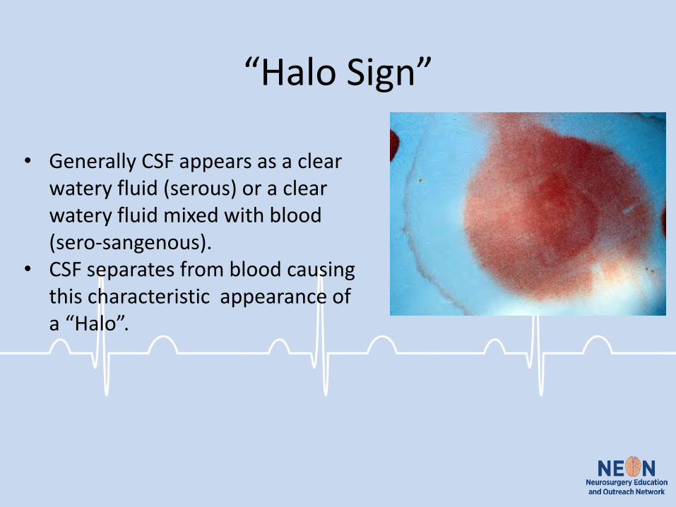
“Halo Sign”
• Generally CSF appears as a clear watery fluid (serous) or a clear watery fluid mixed with blood (sero-sangenous).
• CSF separates from blood causing this characteristic appearance of a “Halo”.

Signs and Symptoms
• Patients may complain of a salty taste in the back of the throat
• Often seen as CSF leakage from the operative site.
• Headache
• Nausea
• Change in hearing
• Visual Disturbances
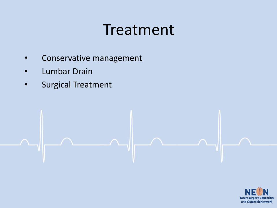
Treatment
• Conservative management
• Lumbar Drain
• Surgical Treatment
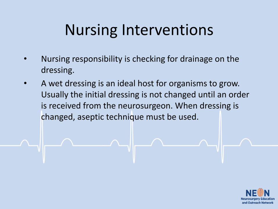
Nursing Interventions
• Nursing responsibility is checking for drainage on the dressing.
• A wet dressing is an ideal host for organisms to grow. Usually the initial dressing is not changed until an order is received from the neurosurgeon. When dressing is changed, aseptic technique must be used.

Hydrocephalus
Definition: An active distension of the ventricular system resulting from inadequate passage of CSF from its point of production within the cerebral ventricles to its point of absorption into the systemic circulation.
1) Communicating (absorption)
2) Non-Communicating (flow)
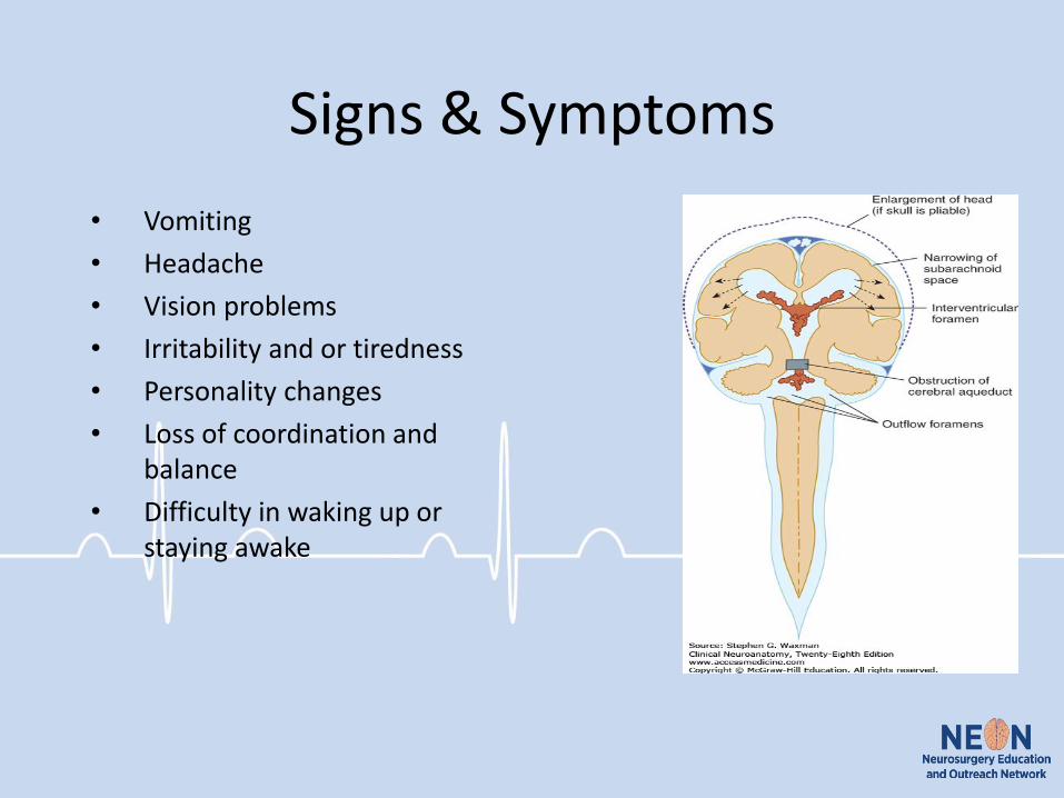
Signs & Symptoms
• Vomiting
• Headache
• Vision problems
• Irritability and or tiredness
• Personality changes
• Loss of coordination and balance
• Difficulty in waking up or staying awake

Treatment
• Surgery Surgical treatment is either:
• insertion of a shunt or
• establishment or a bypass to alleviate obstruction of CSF flow. This may be either an endoscopic third ventriculostomy (ETV) or temporizing measure such as extraventricular drains (EVD) or ventricular reservoirs.
• These are likely to be done at neurosurgical center's.

Nursing Interventions
• Frequent neurological assessments
• Ongoing monitoring of patients ability to perform ADL’s
• Safety considerations
• Anticipation of surgery
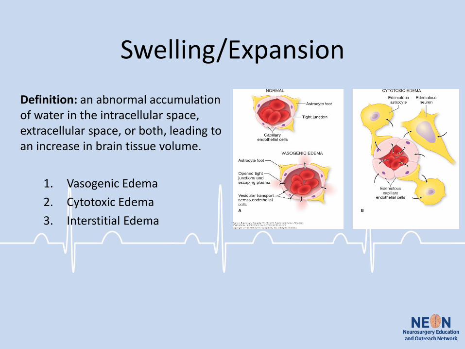
Swelling/Expansion
Definition: an abnormal accumulation of water in the intracellular space, extracellular space, or both, leading to an increase in brain tissue volume.
1. Vasogenic Edema
2. Cytotoxic Edema
3. Interstitial Edema

Treatments
Peaks 2-4 days post injury
S&S ↑ICP
1. Vasogenic Edema
• Corticosteroids
• Mannitol
• Hypertonic Saline
2. Cytotoxic Edema
• Mannitol
3. Interstitial Edema
• ↓ CSF

Intracranial pressure
• Intracranial pressure is the pressure exerted by the brain tissue, blood and CSF against the inside of the skull
• Is measured as the pressure exerted by the CSF inside of the skull

Common Signs and Symptoms of Elevated ICP
• Headache
• Vomiting
• Restlessness and irritability
• Confusion
• Double vision
• Decreased LOC
• Coma
• Temperature changes

Signs and Symptoms of Increased ICP
Early Signs
• Yawning, restlessness
• Confusion
• Ipsilateral pupil change
• Paresis, plegia
Late Signs• Diminishing LOC
• Posturing
• Dilated, non-reactive pupils

Treatment for ICP
Goal: to reduce swelling
• It is reversible
• Mannitol and 3%NS
• Monitor serum electrolytes serum osmolality
Nursing Interventions
Remember…
• Conduct regular neurological assessments on patients post craniotomy
• Inform most responsible physician when patient deteriorates

Herniation
• As ICP (intracranial Pressure) continues to rise, it can lead to herniation
• When the brain shifts across structures—occurs during time of high intracranial pressure
• Is a medical emergency due to potential for coma or death
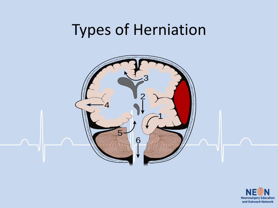
Types of Herniation

Recognizing Herniation
Cushing’s Triad is a loss of autoregulation, resulting in increased ICP (intrinsic monitoring occurs in neurosurgical centers) or by close monitoring of vital signs and neurological status

Cushing’s Triad (Symptoms)

Treatments
• Osmotic Diuretics (Mannitol)
• Hypertonic Saline (3%)
• Maintain adequate blood pressure
• Oxygen
• Nutrition

Your Role
Conduct neuro-vital sign checks more often to detect, document and identify the trend in status
Enact nursing interventions to decrease ICP Communicate Be persistent Work with MD to treat underlying causes Support family Document

References
• Athanasakis, E., & Despina Ermidou BSc, R. N. (2011). Post-Operative Complications of Ventriculoperitoneal Shunt in Hydrocephalic Pediatric Patients-Nursing Care. International Journal of Caring Sciences, 4(2), 66.
• Del Bigio, M. R. (1993). Neuropathological changes caused by hydrocephalus. Acta neuropathologica, 85(6), 573-585.
• Hashimoto, M., Ishikawa, M., Mori, E., & Kuwana, N. (2010). Diagnosis of idiopathic normal pressure hydrocephalus is supported by MRI-based scheme: a prospective cohort study. Cerebrospinal fluid research, 7(1), 18.
• Hickey, J. (2014). The clinical practice of neurological and neurosurgical nursing. Philadelphia, PA: Lippincott Williams & Wilkins.
• McCance, K. L., & Huether, S. E. (2015). Pathophysiology: The biologic basis for disease in adults and children. Elsevier Health Sciences.
• Neurophysiology & Anesthesia. In: Butterworth IV JF, Mackey DC, Wasnick JD. Butterworth IV J.F., Mackey D.C., Wasnick J.D. Eds. John F. Butterworth IV, et al.eds. Morgan & Mikhail's Clinical Anesthesiology, 6e New York, NY: McGraw-Hill; . http://accessmedicine.mhmedical.com/content.aspx?bookid=2444§ionid=193560975. Accessed January 18, 2019.
• Orešković, D., & Klarica, M. (2011). Development of hydrocephalus and classical hypothesis of cerebrospinal fluid hydrodynamics: facts and illusions. Progress in neurobiology, 94(3), 238-258.
• Pudenz, R. H., & Foltz, E. L. (1991). Hydrocephalus: overdrainage by ventricular shunts. A review and recommendations. Surgical neurology, 35(3), 200-212.
• Schwamb, R., Dalpiaz, A., Miao, Y., Gonka, J., & Khan, S. A. (2014). Clinical manifestations of hydrocephalus: A review. Neurology and Clinical Neuroscience, 2(6), 173-177.
• Tymianski, D. & Sarro, A. (2012). Navigating neuroscience nursing: A Canadian perspective. Pembroke, ON: Pappin Communications.
• Ventricles and Coverings of the Brain. In: Waxman SG. Waxman S.G. Ed. Stephen G. Waxman.eds. Clinical Neuroanatomy, 28e New York, NY: McGraw-Hill; . http://accessmedicine.mhmedical.com/content.aspx?bookid=1969§ionid=147037023. Accessed January 18, 2019.
• Yavuz, C., Demırtas, S., Calıskan, A., Kamasak, K., Karahan, O., & Guclu, O. (2013). Reasons, procedures, and outcomes in ventriculoatrialshunts: A single-center experience. Surgical neurology international, 4.


