97
Posterior fossa Gerhard van der Westhuizen Medical officer (3 Military Hosp) Department of Radiology
| Date post: | 18-Dec-2015 |
| Category: |
Documents |
| Upload: | derick-jennings |
| View: | 218 times |
| Download: | 1 times |

Posterior fossa
Gerhard van der WesthuizenMedical officer (3 Military Hosp)Department of Radiology

Posterior fossa - Outline
• Calvarium▫Posterior skull base
• Brainstem anteriorly▫Midbrain, pons and medulla
• Cerebellum posteriorly▫2 Hemispheres and midline vermis
• Divided into:▫Mesencephalon (midbrain)▫Rhomboencephalon (pons, medulla and cerebellum)
• Cerebral aquaduct and fourth ventricle• CSF cisterns containing vertebrobasilar arteries and
veins
Posterior skull base• Formed by posterior temporal and occipital
bones• Anterior - Dorsum sellae medially, petrous ridges
laterally• Posterior - Groove for transverse sinus on
occipital bone• Transmits CN 7-12, medulla oblangata and
jugular veins• Multiple foramina and fissures

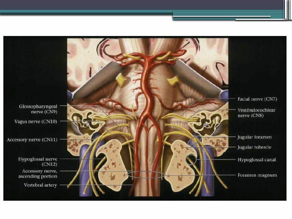
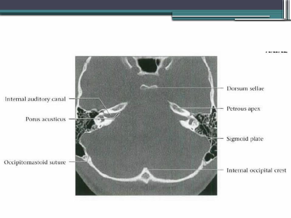
Posterior skull base -Foramina• Internal acoustic meatus
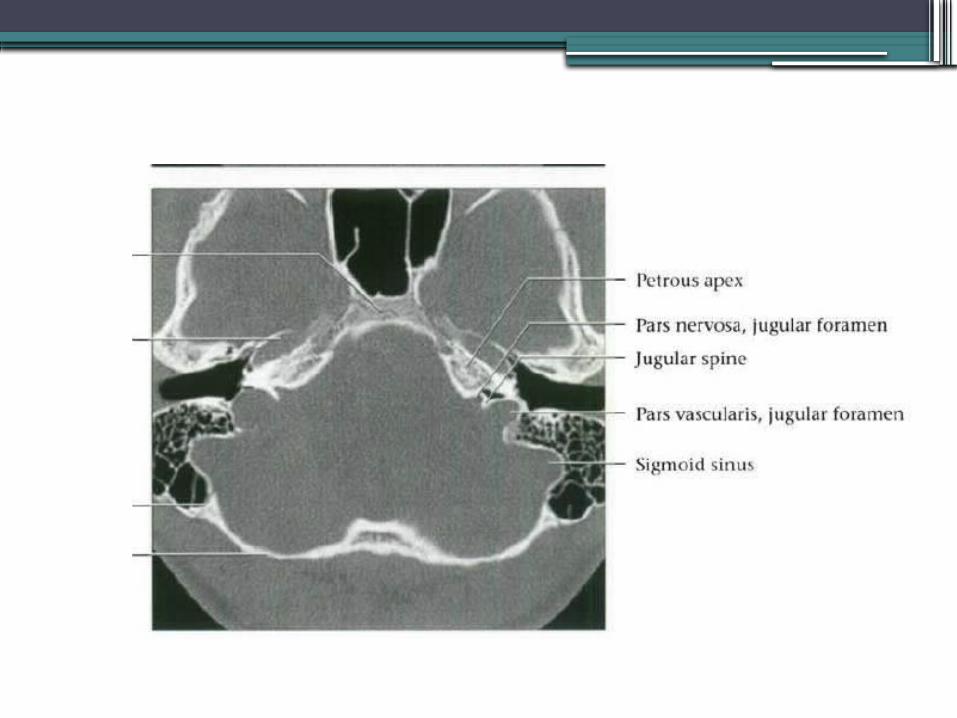
▫ Porus acusticus – CN VII & VIII, labyrinthine artery• Jugular foramen
▫ Pars nervosa - anteromedial CN IX, Jacobson’s nerve and inferior petrosal sinus
▫ Pars vascularis - posterolateral Jugular bulb, CN X & XI, Arnold’s nerve, posterior meningeal
artery, meningeal branch of ascending pharyngeal artery• Hypoglossal canal
▫ CN XII• Stylomastoid foramen
▫ CN VII• Foramen magnum
▫ Medulla oblangata, CN XI and vertebral arteries


Brainstem and cerebellum

Brainstem
•Midbrain▫Connects pons and cerebellum with
forebrain•Pons
▫Relays information from brain to cerebellum
•Medulla▫Relays information from spinal cord to
brain


Midbrain (Mesenchephalon)• “Butterfly-shaped”, passes through tentorium cerebelli
• 3 Main parts:▫ Cerebral peduncles
White matter tracts - Corticospinal, corticobulbar & corticopontine tracts
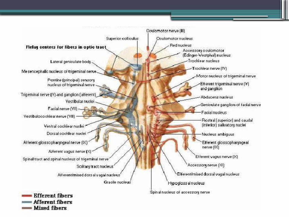
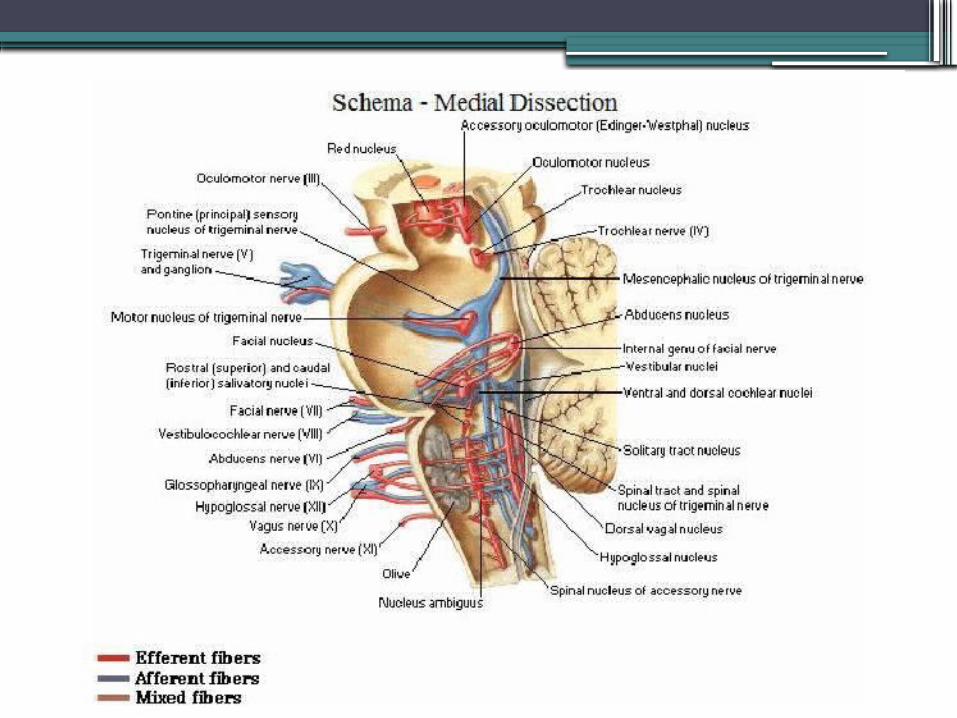
▫ Tegmentum CN nuclei: III – Level of superior colliculus;
IV – Level of inferior colliculus Accessory oculomotor (Edinger-Westphal)
Gray matter nuclei Substantia nigra - Motor planning, eye movement, reward seeking, learning and
addiction Red nucleus – Relay and control centre of cortiomotor impulses. Periaquaductal gray matter – Pain and defensive behaviour
White matter tracts Spinothalamic Medial and lateral lemniscus Somatosensory Medial longitudinal fasciculus – Vestibulo-ocular and optokinetic reflexes

Midbrain▫ Tectum
Superior colliculus ( visual pathway) Inferior colliculus (auditory pathway)
Cerebral aquaduct passes between tectum and tegmentum
CSF cisterns associated with midbrain Ambient – Lateral, CN IV Quadrigeminal – Posterior, CN IV Interpeduncular – Anterior, CN III.
Connections: Superior – Cerebral hemispheres, basal ganglia and thalami Posterior – Cerebellum via superior cerebellar peduncle (brachium
conjuntivum) Inferior – Pons
Blood supply via vertebrobasilar circulation Perforating branches of basilar, SCA, PCA.


Axial T2
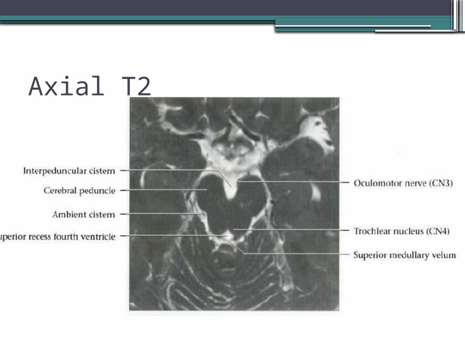
Axial T2
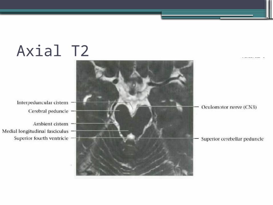
Axial T2

Axial T2

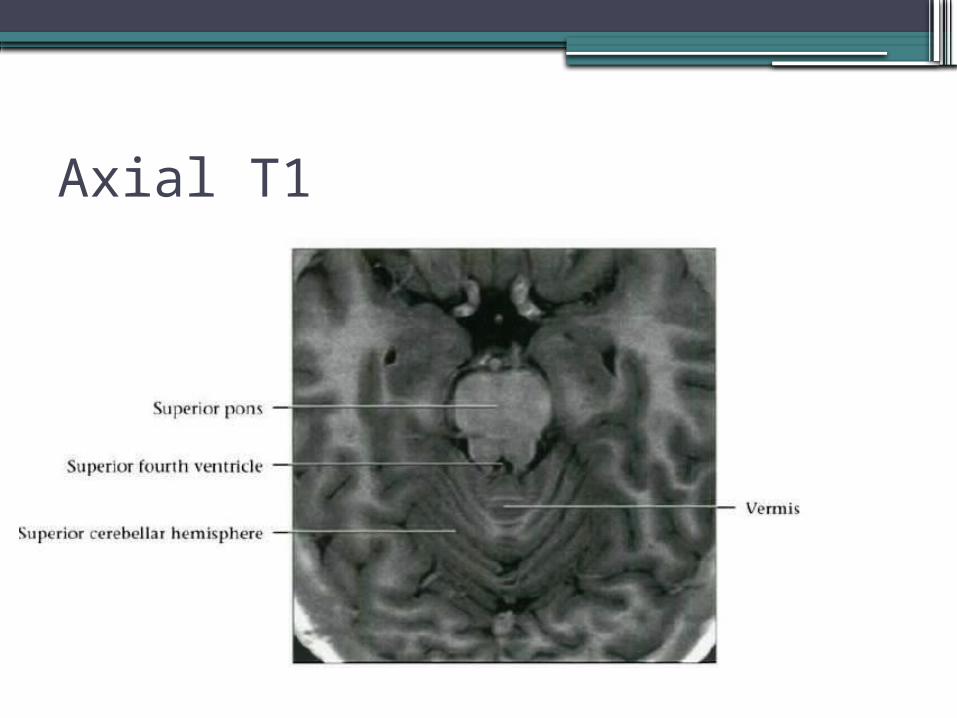
Axial T1

Pons•Relays info from brain to cerebellum.•Middle cerebellar peduncle – Brachium pontis•Bulbous midportion of brainstem•Two main parts:
▫Ventral pons – White matter tracts continuous with cerebral peduncles and medullary pyramids.
▫Dorsal tegmentum– CN nuclei, gray matter nuclei and white matter tracts. Continuation of midbrain tegmentum superiorly and medullary tegmentum inferiorly.

Pons
•Tranverse fibres make up bulk•Dorsal surface forms rostral half of 4th
ventricle.•Adjacent CSF cisterns:
▫Prepontine – CN V & VI ▫CP angle – CN VII & VIII
•Blood supply▫Medial branches SCA, perforating branches
of basilar artery, thalamoperforator arteries.

Pons• CN nuclei:
▫ V – Throughout brainstem and upper cord. Bulk of motor and sensory in pons. Enters and exits at level of midlateral pons
▫ VI – In pontine tegmentum, near midline, anterior to fourth ventricle. Exits anterior at ponto-medullary junction
▫ VII – Ventrolateral aspect of pons Motor, superior salivatory, solitary tract Exits laterally at ponto-medullary junction
VIII – Vestibular along floor of 4th ventricle Cochlear on lateral surface of inferior cerebellar peduncle
Exits at ponto-medullary junction, posterior to VII


Axial T1

Axial T1

Axial T2

Axial T2

Axial T2
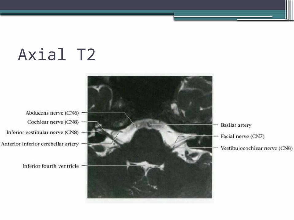
Axial T2

Axial T2
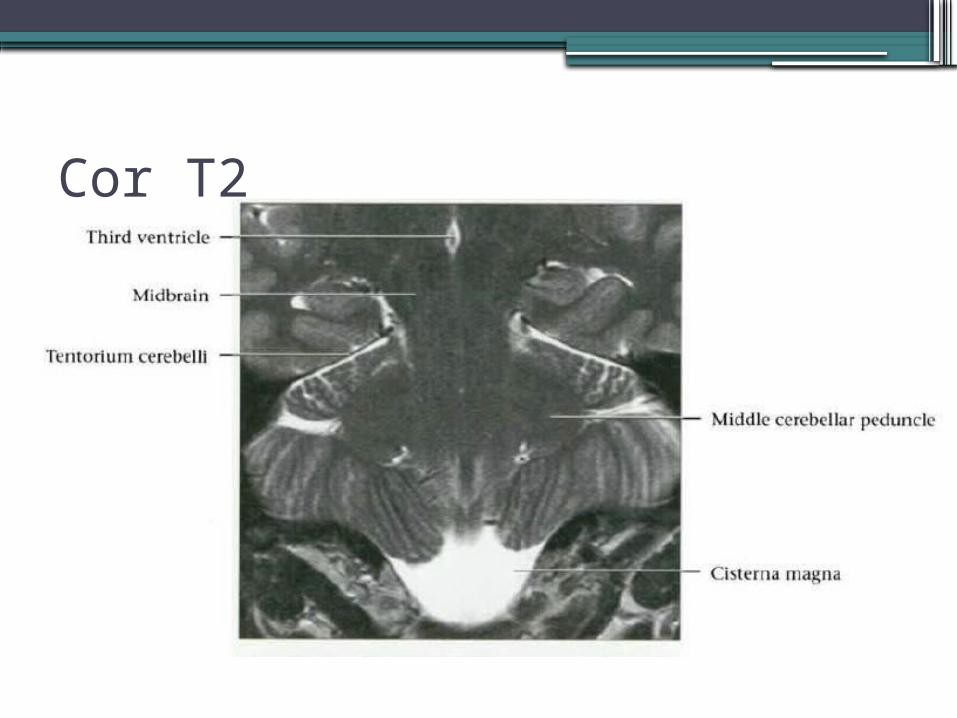
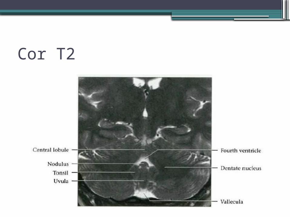
Cor T2
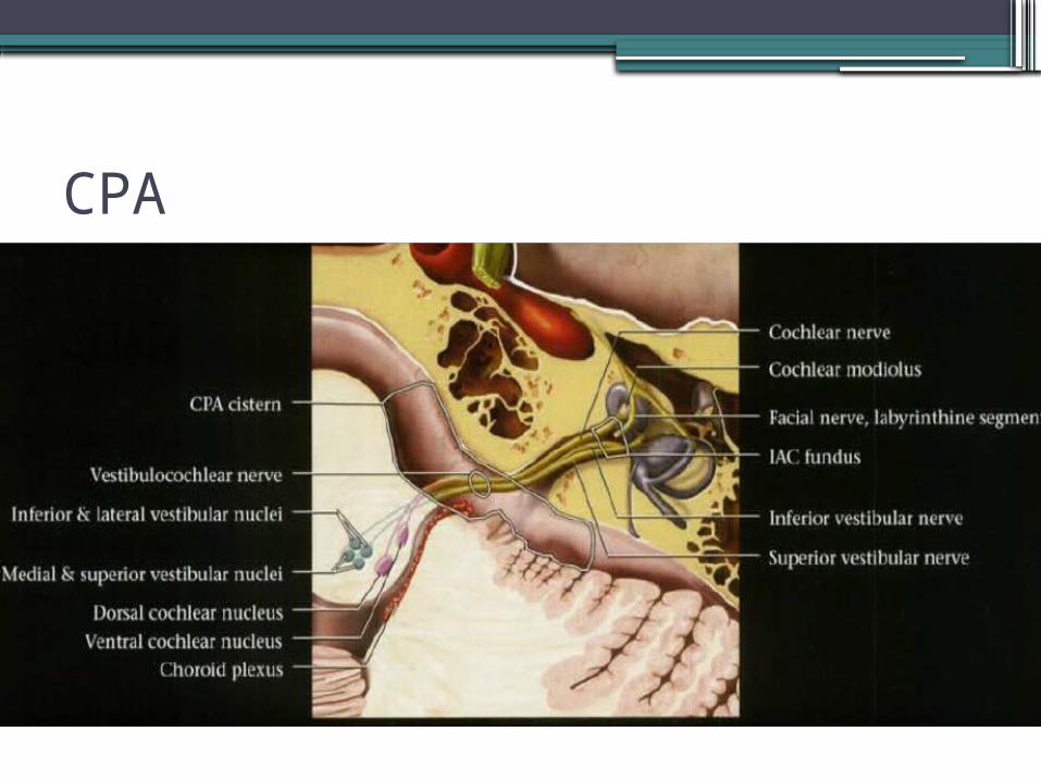
CPA

IAM

IAM

Medulla• Caudal part of brainstem composed of gray
matter formations, CN nuclei IX – XII and white matter tracts.
• Between pons and spinal cord.• 4th ventricle and cerebellum posteriorly• Connected to cerebellum via inferior
cerebellar peduncle (restiform body).• 2 Main parts:
▫Ventral – olive and pyramidal tract▫Dorsal tegmentum – CN nuclei and white
matter tracts

Medulla•Ventral medulla:
•Pyramid▫Paired; anterior surface; midline ventral median
fissure▫Ipsilateral corticospinal tracts prior to
decussation•Olive
▫Lateral to pyramids, venterolateral sulcus (pre-olivary) and posterolateral sulcus (post-olivary)
▫Inferior olivary complex of nuclei
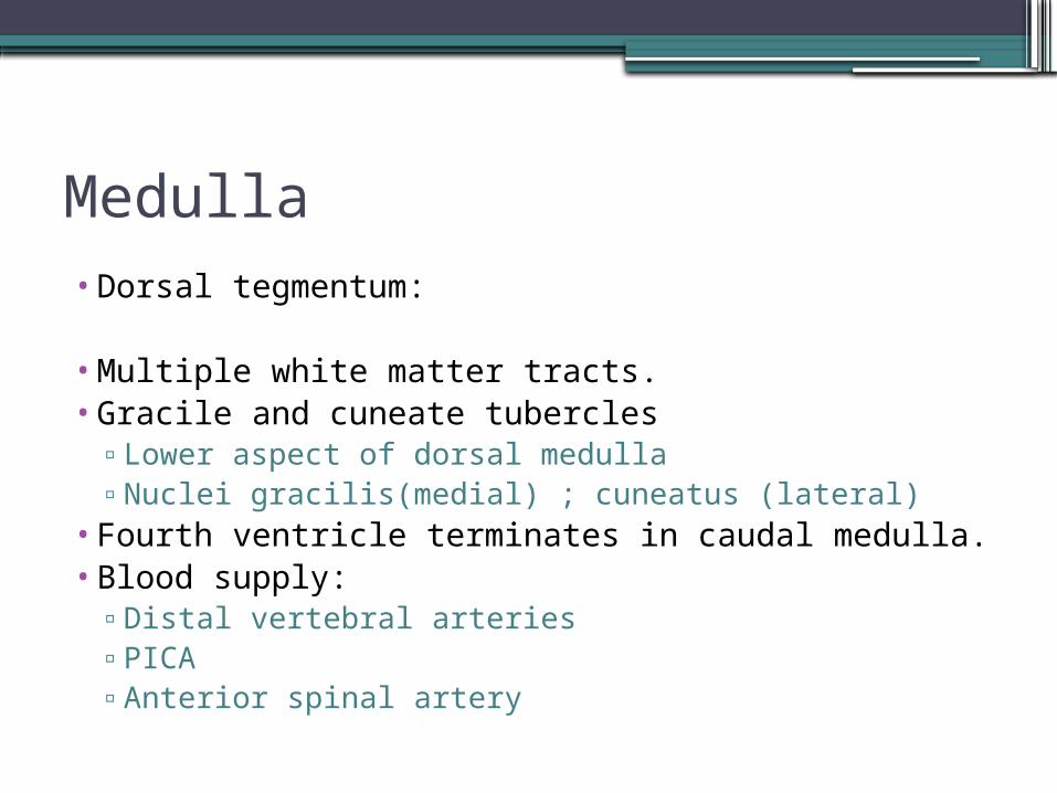
Medulla• Dorsal tegmentum:
• Multiple white matter tracts.• Gracile and cuneate tubercles
▫Lower aspect of dorsal medulla▫Nuclei gracilis(medial) ; cuneatus (lateral)
• Fourth ventricle terminates in caudal medulla.• Blood supply:
▫Distal vertebral arteries▫PICA ▫Anterior spinal artery
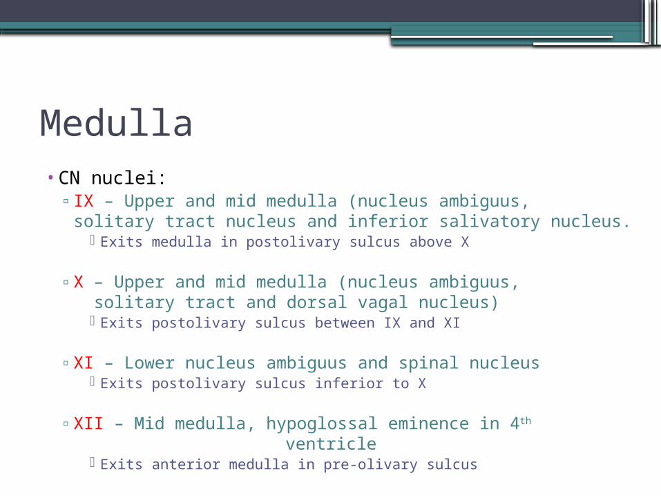
Medulla• CN nuclei:
▫ IX – Upper and mid medulla (nucleus ambiguus, solitary tract nucleus and inferior salivatory nucleus.
Exits medulla in postolivary sulcus above X
▫ X – Upper and mid medulla (nucleus ambiguus, solitary tract and dorsal vagal nucleus)
Exits postolivary sulcus between IX and XI
▫ XI – Lower nucleus ambiguus and spinal nucleus Exits postolivary sulcus inferior to X
▫ XII – Mid medulla, hypoglossal eminence in 4th ventricle
Exits anterior medulla in pre-olivary sulcus


Axial T2

Axial T2

Axial T2

Axial T2

Cerebellum•Function: Integrates coordination and fine-
tuning of movement and regulation of muscle tone.
•2 Hemispheres and midline vermis•Three surfaces – superior,inferior and anterior•Divided into 3 lobes and 9 lobules by
transverse fissures.•3 Cerebellar peduncles•Cortical gray matter, central white matter and
4 paired deep gray nuclei.
Cerebellum
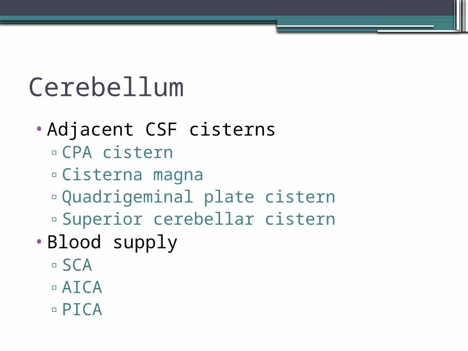
•Adjacent CSF cisterns▫CPA cistern▫Cisterna magna▫Quadrigeminal plate cistern▫Superior cerebellar cistern
•Blood supply▫SCA▫AICA▫PICA

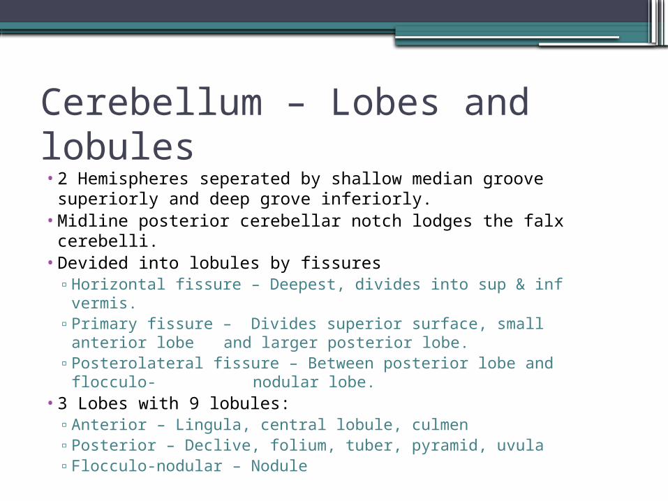
Cerebellum – Lobes and lobules• 2 Hemispheres seperated by shallow median groove
superiorly and deep grove inferiorly.• Midline posterior cerebellar notch lodges the falx
cerebelli.• Devided into lobules by fissures
▫ Horizontal fissure – Deepest, divides into sup & inf vermis.▫ Primary fissure – Divides superior surface, small anterior
lobe and larger posterior lobe.▫ Posterolateral fissure – Between posterior lobe and flocculo-
nodular lobe.• 3 Lobes with 9 lobules:
▫ Anterior – Lingula, central lobule, culmen▫ Posterior – Declive, folium, tuber, pyramid, uvula▫ Flocculo-nodular – Nodule

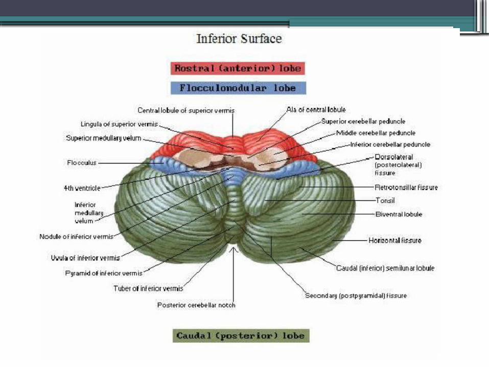

Cerebellum – Lobes and lobules
Vermis lobules Associated hemispheric lobules
Superior vermis:
Lingula Wing of lingula
Central lobule Wing of central lobule Anterior
Culmen Quadrangular lobule lobe
Primary fissure
Declive Simple lobule
Folium Superior semilunar lobule
Horizontal fissure
Inferior vermis: Posterior
Tuber Inferior semilunar lobule lobe
Pyramid Biventral lobule
Uvula Tonsils
Posterolateral (dorsolateral) fissure
Nodule Flocculus Flocculo-nodular lobe

Lobules

“Like cats catch dogs for the party up north”
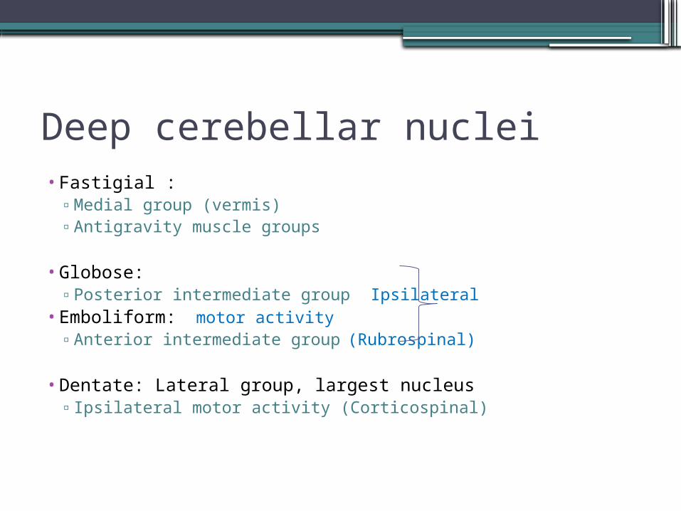
Deep cerebellar nuclei• Fastigial :
▫ Medial group (vermis)▫ Antigravity muscle groups
• Globose: ▫ Posterior intermediate group Ipsilateral
• Emboliform: motor activity▫ Anterior intermediate group (Rubrospinal)
• Dentate: Lateral group, largest nucleus▫ Ipsilateral motor activity (Corticospinal)

Deep cerebellar nuclei

Sag T2

Sag T2

Sag T2

Cor T2
Cor T2

Cor T2
Axial T1

Axial T1

Axial T1

Axial T1

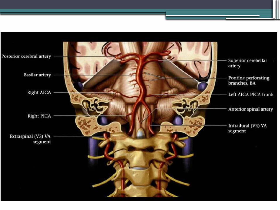
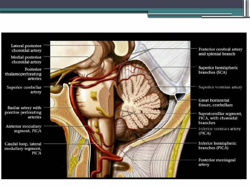
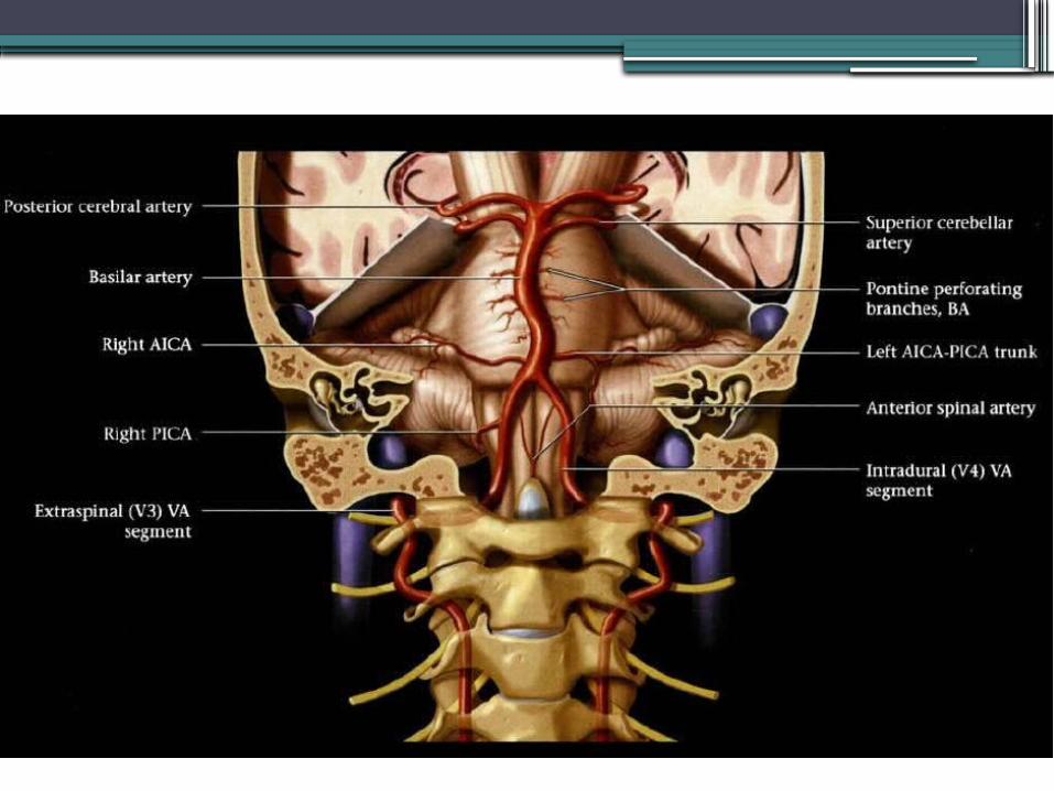
Arterial supply – Vertebrobasilar system•V4 segments of bilateral vertebral
arteries enters through foramen magnum.•Courses superomedially posterior to
clivus•Unites – forms basilar artery.•Terminates into 2 posterior cerebral
arteries in interpeduncular/suprasellar cistern above dorsum sellae

Arterial supply – Vertebrobasilar system• Branches:
▫ Vertebral artery segment V4 Meningeal branch Anterior and posterior spinal arteries Perforating branches to medulla PICA (largest branch)
Lateral, hemispheric branches, inferior vermian artery▫ Basilar
Pontine and midbrain perforating branches Labyrinthine artery AICA
Lateral and medial branches. SCA
Perforating, marginal and hemispheric branches, superior vermian artery▫ PCA’s
Terminal branches of BA. Perforating – Posterior thalamoperforating, thalamogeniculate Choroidal – Medial posterior, lateral posterior Cortical branches – Anterior & posterior temporal Two terminal trunks - Medial: Medial occipital, parieto-occipital, calcarine, posterior
splenial - Lateral: Lateral occipital, temporal


Variants- Persistent trigeminal artery
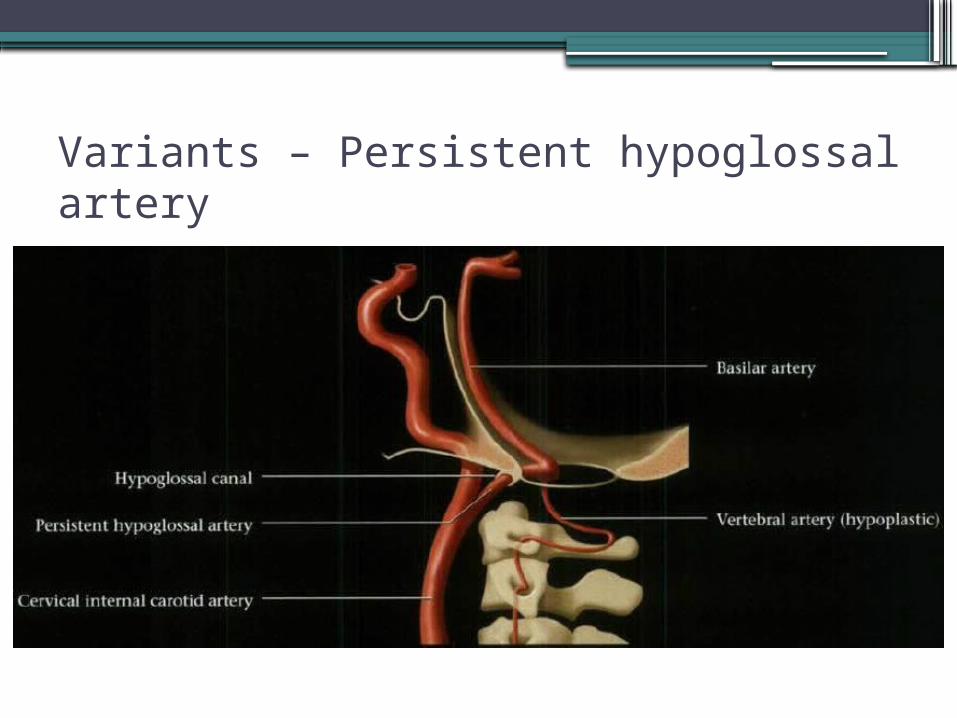
Variants – Persistent hypoglossal artery

Variants – Proatlantal intersegmental artery

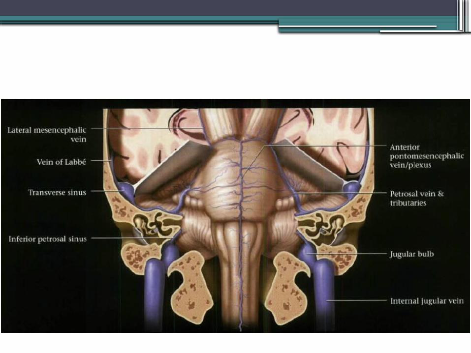
Venous drainage• Three major drainage systems:
▫ Superior (galenic) group Drains into vein of Galen, 3 major veins Precentral cerebellar – single, between lingula and central
lobule Superior vermian – originates near declive, course superiorly
over culmen Anterior pontomesencephalic – anterior to pons and midbrain;
in relation to basilar artery▫ Anterior (petrosal) group
Petrosal vein – in CPA, tributaries from cerebellum, pons and medulla
▫ Posterior (tentorial) group Inferior vermian veins – Paired, paramedian. Curves
posterosuperiorly under pyramids and uvula


References • Netter, F.H. (2011). Atlas of Human Anatomy,
5th ed. Philadelphia: Saunders Elsevier• Ryan, S., McNicholas, M., Eustace, S. (2011).
Anatomy for diagnostic imaging, 3rd ed. London: Saunders Elsevier
• Butler, P., Mitchell, A.W.M., Ellis, H. (1999). Applied Radiological Anatomy. Cambridge: Cambridge University Press
• Harnsberger,H.R., Osborn, A.G., (2006). Imaging anatomy – Brain, head and neck, spine, 1st ed. Utah: Amirsys


