International Journal of Science and Research (IJSR) ISSN (Online): 2319-7064
Index Copernicus Value (2013): 6.14 | Impact Factor (2013): 4.438
Volume 4 Issue 5, May 2015
www.ijsr.net Licensed Under Creative Commons Attribution CC BY
Postpartem Psychosis with Sheehan’s Syndrome:
A Rare Case Report and a Brief Review of
Literature
Dr. Mahendra Wawhal1, Dr. Vajed Mogal
2, Dr. Mahendra Sonawane
3, Dr. Pratap Gole
4,
Dr. Aniket Kurhade5, Dr. Anirudh Londhe
6
1Senior Consultant and Associate Professor ,MD (General medicine) in the Department of Medicine, Mahatma Gandhi Mission's Medical
College and Hospital, CIDCO, N-6,Aurangabad, Maharashtra, India - 431003
2Chief Resident in the Department of Medicine, Mahatma Gandhi Mission's Medical College and Hospital, Aurangabad
3Consultant Internist, MD (General Medicine) Tuljai hospital, Latur, Maharashtra, India
4Consultant Internist and Gastroenterologist, Meenakshi Multispeciality Hospital, Satara, Maharashtra, India
5, 6 Senior Resident in the Department of Medicine, Mahatma Gandhi Mission's Medical College and Hospital, Aurangabad,
Abstract: We report a case of sheehan’s syndrome due to postpartum psychosis. A 28 year old female G2 P2 L2 with a significant
history of intrauterine fetal death (IUFD), confusion, altered sensorium, shortness of breath, bilateral leg sweeling and severe anemia.
Early diagnosis and adequate medical treatment are crucial to reduce morbidity and mortality of the disease. We report unusual case
with worse symptoms after IUFD in sheehan’s syndrome.
Keywords: Sheehan’s, hypopituitarism, post partem ischemic necrosis.
1. Introduction
Sheehan’s syndrome is postpartum hypopituitarism caused
by necrosis of the pituitary gland due to sudden
hypovolemia. It is usually the result of severe hypotension or
shock caused by massive hemorrhage during or after
delivery. 40 years ago, it was estimated that the prevalence
of sheehan’s syndrome was about 100-200 per 1,000,000
women1. In 2009, retrospective nationwide investigation in
Iceland reported that the prevalence of sheehan’s syndrome
was estimated to be 5.1 per 100,000 women2.
The criteria for diagnosis of sheehan’s syndrome includes
typical obstetric history of severe postpartum vaginal
bleeding, severe hypotension or shock for which blood
transfusion or fluid replacement is necessary, failure of
postpartum lactation, failure to resume regular menses after
delivery, partial or panhypopituitarism and empty sella on
CT scan or MRI3. We report a unusual case with symptoms
of sheehan’s syndrome and postpartum psychosis.
2. Case Report
A 28 year old female G2 P2 L2 with 8 month ANC
complaints of fever with chills, dry cough, breathlessness on
exertion, vomiting, abdominal pain, malaise and loss of
apetite since 15 days. She was admitted to other hospital and
diagnosed as viral hemorhagic fever with intrauterine fetal
death where she was intubated and ventilated due to acute
respiratory distress syndrome. She was treated with I.V.
antibiotics, transfusion of blood products ( packed cell
volume, platelet ). She undergone tracheostomy after 7 days
due to prolong intubation.
On 10th
day, patient was referred to us with tracheostomy
tube in situ presented with confusion, shortness of breath
,facial puffiness, bilateral leg oedema, anemia. Ultimately
she was send to emergency department and admitted in
intensive care unit with hemorrhagic shock. A detailed
history from relatives revealed that patient had excessive
bleeding in the course of her first delivery at the age of 22
years. Patient’s relatives noticed progressive increasing
weakness, skin pallor and gradual loss of weight for past 1
month before she came to hospital.
Menstrual and Obstetric history showed Menarche at 16
year, regular cycle of 30+2 days with flow for 5-6 days. G2
P2 L2 with history of normal deliveries in past. The family
history of similar symptoms and previous history of diabetes
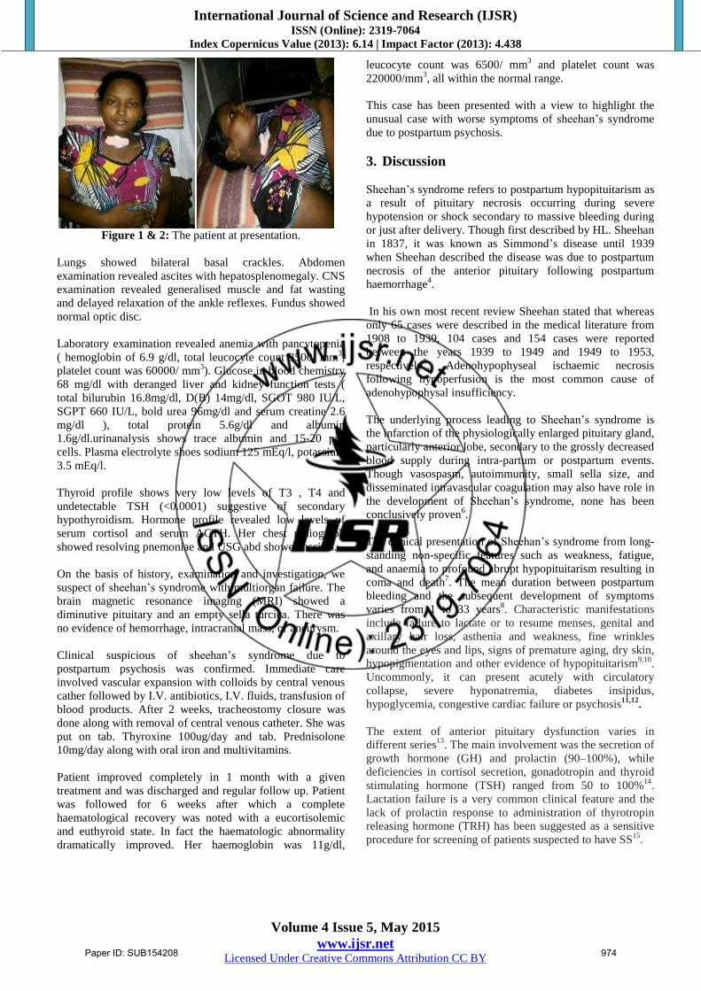
or hypertension were not found. Physical examination
showed a thin, pale, middle-aged women who appeared
somewhat lethargic. Her face was expressionless. The skin
was pale light-brown and of a smooth, delicate texture.
Patchy pigmentation was seen. There was yellowish of skin,
sclera and urine. Her breast tissue was normal but the
areolae were depigmented. She had no pubic or axillary
hairs.
The clinical examination on arrival confirmed the state of
shock with a pulse rate of 80/min, blood pressure of 80/60
mm of Hg, respiratory rate 32/min, cold periphery and pale
conjunctivae. Patient was conscious with eye opening,
confused, not responding to verbal commands and was ill
looking.
Paper ID: SUB154208 973
International Journal of Science and Research (IJSR) ISSN (Online): 2319-7064
Index Copernicus Value (2013): 6.14 | Impact Factor (2013): 4.438
Volume 4 Issue 5, May 2015
www.ijsr.net Licensed Under Creative Commons Attribution CC BY
Figure 1 & 2: The patient at presentation.
Lungs showed bilateral basal crackles. Abdomen
examination revealed ascites with hepatosplenomegaly. CNS
examination revealed generalised muscle and fat wasting
and delayed relaxation of the ankle reflexes. Fundus showed
normal optic disc.
Laboratory examination revealed anemia with pancytopenia
( hemoglobin of 6.9 g/dl, total leucocyte count 3500/ mm3,
platelet count was 60000/ mm3). Glucose in blood chemistry
68 mg/dl with deranged liver and kidney function tests (
total bilurubin 16.8mg/dl, D(B) 14mg/dl, SGOT 980 IU/L,
SGPT 660 IU/L, bold urea 96mg/dl and serum creatine 2.6
mg/dl ), total protein 5.6g/dl and albumin
1.6g/dl.urinanalysis shows trace albumin and 15-20 pus
cells. Plasma electrolyte shoes sodium 125 mEq/l, potassium
3.5 mEq/l.
Thyroid profile shows very low levels of T3 , T4 and
undetectable TSH (<0.0001) suggestive of secondary
hypothyroidism. Hormone profile revealed low levels of
serum cortisol and serum ACTH. Her chest radiograph
showed resolving pnemoniae and USG abd showed ascites.
On the basis of history, examination and investigation, we
suspect of sheehan’s syndrome with multiorgan failure. The
brain magnetic resonance imaging (MRI) showed a
diminutive pituitary and an empty sella turcica. There was
no evidence of hemorrhage, intracranial mass, or aneurysm.
Clinical suspicious of sheehan’s syndrome due to
postpartum psychosis was confirmed. Immediate care
involved vascular expansion with colloids by central venous
cather followed by I.V. antibiotics, I.V. fluids, transfusion of
blood products. After 2 weeks, tracheostomy closure was
done along with removal of central venous catheter. She was
put on tab. Thyroxine 100ug/day and tab. Prednisolone
10mg/day along with oral iron and multivitamins.
Patient improved completely in 1 month with a given
treatment and was discharged and regular follow up. Patient
was followed for 6 weeks after which a complete
haematological recovery was noted with a eucortisolemic
and euthyroid state. In fact the haematologic abnormality
dramatically improved. Her haemoglobin was 11g/dl,
leucocyte count was 6500/ mm3 and platelet count was
220000/mm3, all within the normal range.
This case has been presented with a view to highlight the
unusual case with worse symptoms of sheehan’s syndrome
due to postpartum psychosis.
3. Discussion
Sheehan’s syndrome refers to postpartum hypopituitarism as
a result of pituitary necrosis occurring during severe
hypotension or shock secondary to massive bleeding during
or just after delivery. Though first described by HL. Sheehan
in 1837, it was known as Simmond’s disease until 1939
when Sheehan described the disease was due to postpartum
necrosis of the anterior pituitary following postpartum
haemorrhage4.
In his own most recent review Sheehan stated that whereas
only 65 cases were described in the medical literature from
1908 to 1939, 104 cases and 154 cases were reported
between the years 1939 to 1949 and 1949 to 1953,
respectively5. Adenohypophyseal ischaemic necrosis
following hypoperfusion is the most common cause of
adenohypophysal insufficiency.
The underlying process leading to Sheehan’s syndrome is
the infarction of the physiologically enlarged pituitary gland,
particularly anterior lobe, secondary to the grossly decreased
blood supply during intra-partum or postpartum events.
Though vasospasm, autoimmunity, small sella size, and
disseminated intravascular coagulation may also have role in
the development of Sheehan’s syndrome, none has been
conclusively proven6.
The clinical presentation of Sheehan’s syndrome from long-
standing non-specific features such as weakness, fatigue,
and anaemia to profound abrupt hypopituitarism resulting in
coma and death7. The mean duration between postpartum
bleeding and the subsequent development of symptoms
varies from 1 to 33 years8. Characteristic manifestations
include failure to lactate or to resume menses, genital and
axillary hair loss, asthenia and weakness, fine wrinkles
around the eyes and lips, signs of premature aging, dry skin,
hypopigmentation and other evidence of hypopituitarism9,10
.
Uncommonly, it can present acutely with circulatory
collapse, severe hyponatremia, diabetes insipidus,
hypoglycemia, congestive cardiac failure or psychosis11,12
.
The extent of anterior pituitary dysfunction varies in
different series13
. The main involvement was the secretion of
growth hormone (GH) and prolactin (90–100%), while
deficiencies in cortisol secretion, gonadotropin and thyroid
stimulating hormone (TSH) ranged from 50 to 100%14
.
Lactation failure is a very common clinical feature and the
lack of prolactin response to administration of thyrotropin
releasing hormone (TRH) has been suggested as a sensitive
procedure for screening of patients suspected to have SS15
.
Paper ID: SUB154208 974
International Journal of Science and Research (IJSR) ISSN (Online): 2319-7064
Index Copernicus Value (2013): 6.14 | Impact Factor (2013): 4.438
Volume 4 Issue 5, May 2015
www.ijsr.net Licensed Under Creative Commons Attribution CC BY
Figure 3: Pathophysiology of Sheean’s syndrome
Hyponatremia is the most common electrolyte disturbance
occurring in 33 69% of all cases16
. hypothyroidism and
glucocorticoid deficiency by decreasing free water clearance
independent of vasopressin cause hyponatremia17
. Syndrome
of inappropriate antidiuretic hormone secretion (SIADH)
and volume depletion are the other factors leading to
hyponatremia18
. Clinical diabetes insipidus is apparently an
uncommon complication of postpartum pituitary necrosis
occurring in about 5% of all cases19
. Hematological
abnormalities are common and include normocytic
normochromic anemia, pancytopenia, and acquired factor
VIII and von Willebrand factor (aFVIII–VWF) deficiency20
.
In a study of 40 patients with SS, anemia, thrombocytopenia
and pancytopenia were described in 87%, 60% and 15%,
respectively21
.
Anemia is well recognized as a feature of hypopituitarism.
Gokalp et al. have recently reported hematological
abnormalities in 65 patients with Sheehan’s syndrome, 80%
of whom presented with anemia, compared with 25% of
controls22
. Many hormonal deficiencies, such as
hypothyroidism, adrenal insufficiency and gonadal hormonal
deficiency, can explain normochromic anemia in
hypopituitarism23
. Pancytopenia as a result of an anterior
hormone deficiency has not been clearly investigated. It is a
consequence of the loss of effect of pituitary hormones on
metabolic reactions to hematopoiesis, which is
related to hypopituitarism24
.
The presence of anti-pituitary antibodies (APAs) has been
demonstrated in some patients with SS, suggesting that an
autoimmune pituitary process could be involved in this
syndrome25
. It is proposed that sequestered antigens due to
tissue necrosis could trigger autoimmunity and may cause
delayed hypopituitarism in these patients26
.
The criteria for the diagnosis of SS are as follows:
(1) typical obstetric history of intrapartum or postpartum
bleeding, (2) hypotension or shock, (3) agalactia, (4) failure
to resume regular menses after delivery, (5) hypopituitarism,
and (6) empty sella on CT or MRI27
.
The diagnosis of Sheehan’s syndrome is based on the
features of hormone deficiency, a suggestive obstetric
history, and decreased basal hormone levels (freeT3, free
T4, TSH, cortisol, ACTH). The diagnosis can be made
reliably in the presence of lactational failure, prolonged
amenorrhea and hypoglycaemic crises28
. The finding of a
normal pulse rate in the presence of significant postural drop
was noteworthy as she had coexisting hypothyroidism and
hypocortisolism. The delay in diagnosis was probably due to
her vague symptoms and inadequate obstetric history29
.
The main radiological finding of SS is the image of an
empty sella (around 70% of patients) or partially empty sella
(30%). These findings on MR imaging characterize SS and
provide early confirmation of the clinical diagnosis30
.
Kaplun et al31
. reportedthat sequential MRI demonstrates
evidence of ischemic infarct in the pituitary gland with
enlargement, followed by gradual shrinkage to pituitary
atrophy.
Figure 4: MRI showing an empty sella turcica
Komatsu et al32
. reported that serum antipituitary antibodies
were positive in 70% of patients with empty sella and
Paper ID: SUB154208 975
International Journal of Science and Research (IJSR) ISSN (Online): 2319-7064
Index Copernicus Value (2013): 6.14 | Impact Factor (2013): 4.438
Volume 4 Issue 5, May 2015
www.ijsr.net Licensed Under Creative Commons Attribution CC BY
suggested that antipituitary antibodies might be related to the
development of pituitary atrophy and the consequent empty
sella.
The goal of therapy is to replace deficient hormones.
Treatment is important not only to correct endocrine
abnormalities, but also to reduce mortality due to
hypopituitarism33
. The treatment of Sheehan’s syndrome is
replacement of the deficient hormones. ACTH and TSH
deficiencies should be replaced with glucocorticoids and
thyroxin respectively. Gonadotropin deficiency and
hypogonadism should be treated with a hormone
replacement therapy. Patients who wish to become pregnant
may be directed to the service of fertility for ovulation
induction followed by successful pregnancy15
. Patients with
diabetes insipidus, treatment of choice is 1-desamino-8-d-
arginine vasopressin or desmopressin (DDAVP).
Replacement of GH should be considered in patients with
GH deficiency. See et al. reported that spontaneous
pregnancy could bring partial recovery of pituitary function
in the patient with Sheehan’s syndrome34
.
4. Conclusion
Diagnosis of postpartum psychosis may be missed in a case
of multiorgan failure or in a case of sheehan’s with
dyselectronemia. There is a great need for increased
awareness of postpartum hypopituitarism as a relatively
common sequela of pregnancy complicated by hemorrhage.
Adequate measures should be taken to combat severe
bleeding during delivery. History of postpartum hemorrhage,
failure to lactate and cessation of menses are important clues
to the diagnosis. A simple replacement therapy with thyroid
and cortisol hormones results in complete recovery. Early
diagnosis and appropriate treatment are necessary to reduce
the morbidity and mortality of patients. The patient can look
forward to a normal life expectancy.
References
[1] F. Keles¸timur, “Sheehan’s syndrome,”
Pituitary,vol.6,no.4,pp. 181–188, 2003
[2] H. L. Kristjansdottir, S. P. Bodvarsdottir, and H. A.
Sigur- jonsdottir, “Sheehan’s syndrome in modern
times: a nationwide retrospective study in Iceland,”
European Journal of Endocrinology, vol. 164, no. 3,
pp. 349–354, 2011.
[3] Kelestimur F. Sheehan's syndrome. Pituitary 2003; 6
:181-88
[4] Sheehan HL. Simmonds disease due to postpartum
necrosis of the anterior pituitary following postpartum
haemorrhage. Q JMed 1939; 32: 277-309
[5] SHEEHAN, H. L. The Incidence of Postpartum
Hypopituitarism. Am. J. Obst. & Gyn. 68:202-223,
1954.
[6] Kelestimur F. Sheehan’s syndrome. Pitutary 2003;
6:181-188
[7] Gei-Guardia O, Soto-Herrera E, Gei-Brealey A, Chen-
Ku CH. Sheehan’s Syndrome in Costa Rica: Clinical
experience on 60 cases. Endocr Pract 2010;1:1-27
[8] O. Gei-Guardia, E. Soto-Herrera, A. Gei-Brealey, and
C. K. Chih Hao, “Sheehan syndrome in Costa Rica:
clinical experience with 60 cases,” Endocrine Practice,
vol. 17, no. 3, pp. 337–344, 2011.
[9] Collins ML, O’Brien P. Diabetes insipidus following
obstetric shock. Obstet Gynecol 1979;53:16-7.
[10] Wang SY, Hsu SR, Su SL, Tu ST. Sheehan’s
syndrome presenting with early postpartum congestive
heart failure. J Chin Med Assoc 2005;68:386-91.
[11] Weston G, Chaves N, Bowditch J. Sheehan’s
syndrome presenting post-partum with diabetes
insipidus. Aust N Z J Obstet Gynaecol 2005;45:249-
50.
[12] Sas AM, Meynaar IA, Laven JS, Bakker SL, Feelders
RA. Irreversible coma following hypoglycemia in
Sheehan syndrome with adrenocortical insufficiency.
Ned Tijdschr Geneeskd 2003;147: 1650-3.
[13] Sert M, Tetik T, Kirim S, Kocak M. Clinical report of
28 Patients with Sheehan’s syndrome. Endocr J
2003;50:297-301.
[14] Dökmetas HS, Kilicli F, Korkmaz S, Yonem O.
Characteristic features of 20 patients with Sheehan’s
syndrome. Gynecol Endocrinol 2006;22:279-83.
[15] Soares DV, Conceição FL, Vaisman M. Clinical,
laboratory and therapeutics aspects of Sheehan’s
syndrome. Arq Bras Endocrinol Metabol 2008;52:872-
8.
[16] Shoji M, Kimura T, Ota K. Cortical laminar necrosis
and central pontine myelinolysis in a patient with
Sheehan syndrome and severe hyponatremia. Intern
Med 1996;35:427-31.
[17] Singhania P, Singh S, Banerjee R, Singhania B,
Banerjee I, Maitra S. Hyponatremia – A rare and
emergency presentation of Sheehan’s Syndrome. Pak J
Med Sci 2010;26:713-5.
[18] Anfuso S, Patrelli TS, Soncini E, Chiodera P, Fadda
GM, Nardelli GB. A case report of Sheehan’s
syndrome with acute onset, hyponatremia and severe
anemia. Acta Biomed 2009;80:73-6.
[19] Miwa S, Kaseki S, Kazeto S, Sunouchi S, Iwasaki Y.
A case of Sheehan’s syndrome complicated by
diabetes insipidus. Nippon Sanka Fujinka Gakkai
Zasshi 1989;41:625-8.
[20] Oliveira MC, Kramer CK, Marroni CP, Leães CG,
Viana L, Roithman S, et al. Acquired factor VIII and
von Willebrand factor (aFVIII/VWF) deficiency and
hypothyroidism in a case with hypopituitarism. Clin
Appl Thromb Hemost 2010;16:107-9.
[21] Laway BA, Mir SA, Bashir MI, Bhat JR, Samoon J,
Zargar AH. Prevalence of hematological abnormalities
in patients with Sheehan’s syndrome: Response to
replacement of glucocorticoids and thyroxine. Pituitary
2011;14:39-43.
[22] Gokalp D, Tuzcu A, Bahceci M, Arikan S, Bahceci S,
Pasa S: Sheehan’s syndrome as a rare cause of anemia
secondary to hypopituitarism. Ann Hematol 2009,
88(5):405-410.
[23] Kim DY, Kim JH, Park YJ, Jung KH, Chung HS, Shin
S, Yun SS, Park S, Kim BK: Case of complete
recovery of pancytopenia after treatment of
hypopituitarism. Ann Hematol 2004, 83(5):309-312.
[24] Laway BA, Bhat JR, Mir SA, Khan RS, Lone MI,
Zargar AH: Sheehan’s syndrome with pancytopenia–
complete recovery after hormone replacement (case
Paper ID: SUB154208 976
International Journal of Science and Research (IJSR) ISSN (Online): 2319-7064
Index Copernicus Value (2013): 6.14 | Impact Factor (2013): 4.438
Volume 4 Issue 5, May 2015
www.ijsr.net Licensed Under Creative Commons Attribution CC BY
series with review). Ann Hematol 2010, 89(3):305-
308.
[25] De Bellis A, Kelestimur F, Sinisi AA, Ruocco G,
Tirelli G, Battaglia M, et al. Anti-hypothalamus and
anti-pituitary antibodies may contribute to perpetuate
the hypopituitarism in patients with Sheehan’s
syndrome. Eur J Endocrinol 2008;158:147-52.
[26] Goswami R, Kochupillai N, Crock PA, Jaleel A, Gupta
N. Pituitary autoimmunity in Patients with Sheehan’s
syndrome. J Clin Endocrinol Metab 2002;87:4137-41.
[27] F. Keles¸timur, “Sheehan’s syndrome,”
Pituitary,vol.6,no.4, pp. 181–188, 2003.
[28] Assan R.Urgences métaboliques et endocriniennes In :
Kleinknecht D,éd. Principes de réanimation médicales,
3e éd.Paris : Flammarion médecine sciences, 1984
:292326.
[29] Kelestimur F. Sheehan’s syndrome. Pitutary 2003;
6:181-188
[30] Kaplun J, Fratila C, Ferenczi A, Yang WC, Lantos G,
Fleckman AM, et al. Sequential pituitary MR imaging
in Sheehan syndrome: Report of 2 cases. AJNR Am J
Neuroradiol 2008;29:941-3.
[31] J.Kaplun,C.Fratila,A.Ferenczietal.,“Sequentialpituitary
MR imaging in Sheehan syndrome: report of 2 cases,”
American Journal of Neuroradiology, vol. 29, no. 5,
pp. 941– 943, 2008.
[32] M. Komatsu, T. Kondo, K. Yamauchi et al.,
“Antipituitary antibodies in patients with the primary
empty sella syndrome,” Journal of Clinical
Endocrinology and Metabolism,vol.67, no. 4, pp. 633–
638, 1988.
[33] Kelestimur F. Sheehan’s syndrome. Pituitary
2003;6:181-8.
[34] T. T. See, S. P. Lee, and H. F. Chen, “Spontaneous
pregnancy and partial recovery of pituitary function in
a patient with Sheehan’s syndrome,” Journal of the
Chinese Medical Association, vol. 68, no. 4, pp. 187–
190, 2005.
Paper ID: SUB154208 977