21
Postpartum Haemorrhage 4-Stage Approach: Practical Guide

Postpartum Haemorrhage 4-Stage Approach: Practical Guide

© Healthcare Improvement Scotland 2018 First published February 2018 This document was developed in collaboration with our colleagues in OBS Cymru. This document is licensed under the Creative Commons Attribution-Noncommercial-NoDerivatives 4.0 International Licence. This allows for the copy and redistribution of this document as long as Healthcare Improvement Scotland is fully acknowledged and given credit. The material must not be remixed, transformed or built upon in any way. To view a copy of this licence, visit https://creativecommons.org/licenses/by-nc-nd/4.0/

3
Contents Introduction and background ............................................................................................................................ 4
How to use the guide ......................................................................................................................................... 4
Where do I start? ............................................................................................................................................... 5
Stage 0......................................................................................................................................................... 5
Stage 1....................................................................................................................................................... 11
Stage 2....................................................................................................................................................... 13
Stage 3....................................................................................................................................................... 15
Post-event checklist ......................................................................................................................................... 17
Appendix 1: Approach to quantitative measurement of blood loss ............................................................... 18
List of abbreviations ......................................................................................................................................... 19
References ....................................................................................................................................................... 20

4
Introduction and background
Welcome to this practical guide to using the 4-stage approach to postpartum haemorrhage (PPH). The risk
of obstetric haemorrhage is present in every pregnancy. Early identification of abnormal blood loss creates
the potential to intervene and prevent major blood loss.
Early intervention requires:
1. recognition of risk factors leading to heightened surveillance
2. appropriate preparation
3. a standardised approach to accurately determine cumulative blood loss, and
4. recognition of clinical findings suggestive of, or indicating, hypovolaemia.
To have the best chance of preventing heavy bleeding progressing to a massive haemorrhage, which
carries the risk of more devastating sequelae, all four areas need to be integrated into the care of the
woman who is giving birth.
What is the PPH 4-stage approach?
The MBRRACE-UK Confidential Enquiry into Maternal Deaths and Morbidity in 20161 placed emphasis on
the importance of basic clinical skills and prompt recognition of severity of haemorrhage, with
communication and teamwork being an essential component in the management of PPH. Data in Scotland
gathered between 2013 and 2017 identified variation not just in the management of PPH but also in risk
assessment, early recognition of deterioration and escalation, and management by the multidisciplinary
team (MDT).
With this in mind, the 4-stage tool was developed by the Maternity and Children Quality Improvement
Collaborative (MCQIC), a key component of the Scottish Patient Safety Programme (SPSP), in collaboration
with colleagues from OBS Cymru, the Obstetric Bleeding Strategy for Wales
(http://www.1000livesplus.wales.nhs.uk/obs-cymru). The tool encompasses the recommendations from
the MBRRACE-UK report, as it is designed to facilitate an MDT approach to recognising, responding to and
managing PPH.
How to use the guide
This practical guide uses visual examples of a paper version of the tool in practice.
Appendix 1 is included as a resource you may wish to use in clinical practice, and a list of abbreviations at
the end of the guide helps to explain some of the terminology.
For more information about MCQIC and their work, please visit: http://ihub.scot/spsp/maternity-children-
quality-improvement-collaborative-mcqic/

5
Where do I start?
Prevention of PPH starts with recognition of risk and preparation (Stage 0). Although more than half of
women who haemorrhage due to uterine atony have no known risk factors, identification of associated risk
factors during the antenatal and intrapartum periods can improve readiness to respond for those with
known risks. For women with abnormal placentation, an MDT approach to care is vital.
All pregnant women should have an admission assessment of PPH risk that is based on previous
pregnancies and antenatal history. An admission assessment should be completed for all women admitted
in spontaneous labour, admission for induction and augmentation of labour and elective lower segment
caesarean section (LSCS). If any of the 11 risks are present, tick or mark an X next to the corresponding risk.
Perinatal factors
The risk of PPH can change as labour progresses. Continuous assessment during labour and delivery will
assess for the presence of any of the six risks listed below. Vigilance in the immediate postnatal period is
essential if any perinatal risk factors are identified. Place a tick or an X in the box to highlight the risk(s).
During shift handover or communication to the MDT, Stage 0 should be communicated using the Situation,
Background, Assessment, Recommendation (SBAR) format.
X
X
Stage 0

6
Treat
Plan the active third stage of labour in line with the unit’s protocol. If two or more increased risk factors
are present, consider the use of additional uterotonics. Refer to local policy for further guidance. Early
intravenous (IV) access should be considered and documented, including the time of insertion.
Act
Document measured blood loss (MBL) ≤500ml. All other loss will be documented according to stage.
Appendix 1 provides guidance on a quantitative approach to MBL. Check the blood bank. Can blood be
issued on electronic release or is group and save/x-match required?
Completion of Stage 0
Time of birth and when the third stage is complete should be completed for all deliveries irrespective of
blood loss. Total MBL is documented here only if it is ≤500ml.
Before moving on to Stages 1-3, it is important to be familiar with the tool’s action logs and additional
clinical notes and comments section, and the Rule of 30.
Action logs
The action logs on pages 6 and 7 of the tool are designed to document the administration of all
uterotonics, IV fluids and blood products, and for documenting blood results and MBL. The purpose of
each action log is for teams to look at care given in one documented area, in a couple of pages only, which
will facilitate quick decisions on the next steps of care.
09:00
09:15
7
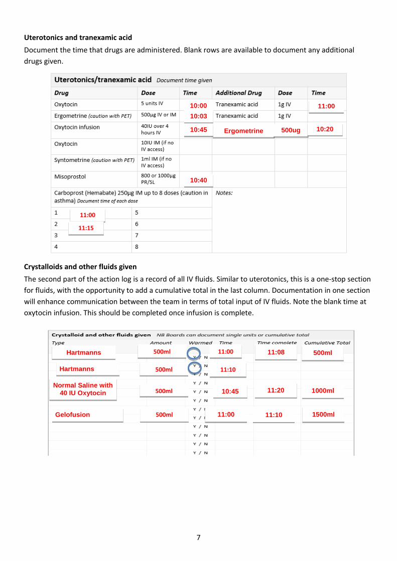
Uterotonics and tranexamic acid
Document the time that drugs are administered. Blank rows are available to document any additional
drugs given.
Crystalloids and other fluids given
The second part of the action log is a record of all IV fluids. Similar to uterotonics, this is a one-stop section
for fluids, with the opportunity to add a cumulative total in the last column. Documentation in one section
will enhance communication between the team in terms of total input of IV fluids. Note the blank time at
oxytocin infusion. This should be completed once infusion is complete.
Hartmanns solution
500ml 11:00 11:08 500ml
Hartmanns 500ml
11:20
11:10
1000ml Normal Saline with
40 IU Oxytocin 500ml
11:00 Gelofusion 500ml
10:45
11:10 1500ml
Ergometrine
10:00
10:03
10:45
10:40
11:00
500ug 10:20
11:00
11:15

8
Blood products
For similar reasons cited on the previous page, blood and blood products given should be documented in
allocated columns.
Measured blood loss
Accurate measurement of blood loss is essential for:
recognising potentially life-threatening haemorrhage, and
managing blood product replacement2.
Although multiple methods for estimating blood loss are available, most are inaccurate, for example visual
estimation. This practice has consistently been shown to significantly underestimate large-volume blood
loss by 33–50% when compared with direct measurement. Similarly, small measures of blood loss are
frequently overestimated, resulting in overtreatment. It is recommended practice for MBL to be
documented every 15 minutes in Stages 1–3 until the bleeding is arrested. Delay in recognition of large
blood loss is a common finding in cases of maternal morbidity and mortality from haemorrhage. A policy of
waiting to quantify blood loss only after the excessive loss is appreciated does not address this problem.
RPC
RPC
FFP
332
278
301
11:45
12:00
12:12
11:52
12:10
12:25
10:15
10:30
10:45
372
300
245
11:00
11:15
400
372
672
917
1371
11:30
11:45
12:00
250
356
400
300
1621
1977
2377
2677
12:13 100 2777

9
Cell savage
Where applicable, clinicians can document the use of cell savage.
Blood test results
Document the results in the box. If required, any other blood test results can be documented in the right-
hand box, but this is optional.
Additional clinical notes and comments
Available for clinicians to record any care not covered by the tool.
09:00
10:30
13.1 – lab
9.7 - haemacue
NA
1mmol/L
NA
NA
NA
400mg/dl
CRP - 197

10
Rule of 30
It is important to know booking or 36-week maternal weight to determine the percentage of circulating
blood volume lost. Figure 1 represents some examples of weight and when 15%, 30% and 40% volume loss
occurs. A 750ml blood loss may be well tolerated by a woman weighing 70kg with minimal signs and
symptoms, but for a woman weighing 50kg this is 15% of her circulating blood volume lost. Figure 2
represents signs, symptoms and Maternity Early Warning Score (MEWS) for each percentage of circulating
blood volume lost.
Figure 1: Circulating blood volume and % blood loss
% blood loss guide
% blood loss 15% 30%: Follow
‘Rule of 30’
40%
Signs and
symptoms
Possible anxiety,
dizziness, palpitations
Unwell, anxious, weakness,
faint, clammy, restless,
sweating
Confused, drowsy,
unconscious, clammy skin,
air hunger, pallor, cold,
peripheral cyanosis
MEWS Blood pressure Normal 30mmHg drop in systolic Hypotensive
Respiratory rate Normal >20 or 30% increase >30
Pulse Normal >100 or 30% increase >20
Urine output Normal <30ml/h <15ml/h
Figure 2: Signs, symptoms and MEWS: % blood loss guide

11
Move through treatment simultaneously: mobilise help, act and treat accordingly and initiate unit
protocols for PPH. Stage 1 represents blood loss of 500–999ml without clinical shock or ≤15% blood loss.
This stage is not applicable for LSCS.
Mobilise help
The midwife in charge will be the first person to contact for help. The midwife in the room and midwife in
charge will clearly communicate to each other the next steps and who is assigned as scribe. If further
assistance is requested, their name, status and grade will be documented.
Possible cause
This visual aide is designed as prompt or reminder to think of potential cause of the bleeding. This
essentially follows the 4T approach: Tone (atony), Trauma (vaginal tears), Tissue (retained placenta), and
Thrombin (coagulopathy). No documentation is required in this box. You may wish to circle causes that
apply.
Joe Bloggs
Mary Bloggs, Midwife
10:00
ST6
10:03
10:10
Mary Doe
Stage 1

12
Act
MEWS will be used for all women, with observations recorded every 15 minutes until the bleeding stops.
Consider IV access if blood loss continues, especially if progression to Stage 2 is anticipated. The same
principles apply for ranitidine. The actions are in order of the clinical actions that are required at this stage,
rather than chronological preference of treatment. These can be performed simultaneously with the
midwife in charge allocating tasks to the team. Document the time when each of the care indicators
started or NA (not applicable) if not required.
Treat
Treat the identified cause of haemorrhage. Document the time performed or leave blank or document NA
if not required. At any time during treatment, bimanual compression should be discussed with the team
and considered as a possible treatment option if bleeding persists.
Completion of Stage 1
The tool can stop at Stage 1 once the team are reassured that bleeding has stopped. The PPH post-event
checklist should be completed for Stages 1–3 and is explained later in this document. If MBL is >1000ml OR
there is clinical concern, progress to Stage 2.
.
10:15
NA
10:15
09:15
10:10
10:03
10:20
10:10
400ml

13
This stage applies to all blood loss ≥1000–1499ml OR clinical concern OR <30% blood loss.
Restart here after Stage 0 for all LSCS. More information about the Rule of 30 and guide to percentage
blood loss can be found on page 10 of the guide.
Stage 2 is focused on sequentially advancing through medications and procedures, mobilising help, blood
bank support and keeping ahead with volume and blood product. Here, the woman is beginning to
decompensate and senior obstetric and anaesthetic involvement is vital. This is where theatre should be
considered. In cases of atony with continued bleeding after second-line uterotonics, an intrauterine
balloon needs to be considered, and other causes need to be excluded, rather than going straight to third
and fourth-line uterotonics.
Mobilise help
Additional help will be required during this stage because of the moderate amount of blood loss. If
members of the team are present from Stage 1, tick or place an X in the time box. There is no need to
duplicate names. It is expected that an obstetrician(s) and anaesthetist will be involved in care. Document
the name of the scribe.
Act
Document the time started for all or NA if not relevant. Similar to Stage 1, the central banner has clinical
indications of PPH. When the cause is known, circle or place an X next to all that are relevant.
10:50 11:00
11:03
11:00
10:50
10:50
NA
X
Dr Peter Bones
Dr Betty Bones 10:50
10:50
Mary Bones Midwife 10:50
Dr John Doe
Bridget Bones
ST6 10:50
10:50 MCA
11:00
11:00
Stage 2

14
Blood tests
Tick, circle or place an X, whatever is easiest electronically, next to the tests that are ordered, with a time
entered in the last column. Write how many units of x-matched bloods have been requested.
.
Treat
To emphasise, treatment during this stage should be timely, with act and treat carried out simultaneously.
Document the time of each treatment in the right-hand box. Leave blank or document NA if not required.
Document in the action log any uterotonics given and additional clinical notes as required.
Completion of Stage 2
Once the bleeding has stabilised and final total MBL is between 1000–1500ml, the tool is complete and can
be signed as below. The post-event checklist will be completed by a member of the obstetric or
anaesthetic team. It is recommended practice for a management plan to be documented in the clinical
notes and clearly communicated to the MDT using structured communication tools such as SBAR. Increase
postnatal surveillance for any woman with a Stage 2 haemorrhage.
NA
10:08
10:10
NA NA
NA NA

15
Stage 3 focuses on all blood loss >1500ml OR ≥30% blood loss OR ongoing clinical concern. This stage is
critical as the woman may be in extremis. Teamwork, communication and collaboration between all
members of the MDT are critical at this stage. Effective communication of major obstetric haemorrhage
(MOH) is critical between the theatre team and laboratory staff. The key messages are as follows:
Do not delay other interventions while waiting for a response to medication(s).
Do not wait for laboratory values to initiate transfusions:
1. transfuse based on clinical signs and patient response.
2. transfuse aggressively with a high ratio of fresh frozen plasma (FFP) to packed red blood cells
(PRBC)2.
Communicate and document:
1. Verbally acknowledge the actions you will take and the orders received.
2. Provide ongoing updates about the patient’s status with other departments.
3. Record care on the action log.
Mobilise help
Tick or place an X if staff from Stages 1 and 2 are already present. If any staff have left, this can be
documented in the additional clinical notes. Document the name of the scribe. The column on the left-
hand box is prepopulated, as these members of the MDT are required to attend.
Plan further treatment
Consultant presence is essential in severe PPH cases. Document the time contacted. In the case of
continuing or worsening haemorrhage, it is critical to consider timely transfer to theatre and activation of
the local MOH protocol.
Betty Bloggs LW Co Coordinator
Mary Marple
X
X
X
Jane Doe, Midwife
11:10
10:30 FY2
11:10
X
11:10
11:13
Stage 3

16
Act
Act and treat are simultaneous and are not in chronological order of preference. Enter the time that each
element started. Leave blank or document NA if not required for each relevant element of clinical care.
During severe haemorrhage, the primary goals are to provide adequate and early blood product
replacement and to either prevent or correct disseminated intravascular coagulation (DIC). Delays in
recognising and treating haemorrhage frequently lead to inadequate blood product administration. After
the first several units of PRBCs and in the face of continuing or worsening haemorrhage, aggressive
transfusion therapy becomes critical.
Treat
Adhere to local MOH protocol for severe haemorrhage. A crucial step is reviewing the care given, such as
ongoing resuscitation, uterotonics and IV fluids administered, while continually updating the action log and
communicating this to the team. Document the time this occurred in the appropriate box. The focus during
this stage is surgical interventions. Document the time each started in each box or NA if not required.
11:15
10:40
11:30
NA
11:00
NA
11:08 11.20
11:15
11:10
NA
NA 11:16
11:10
17
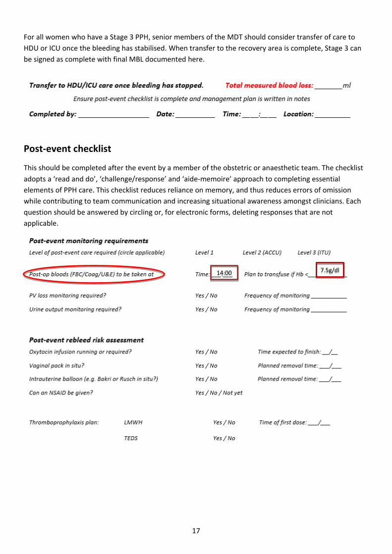
For all women who have a Stage 3 PPH, senior members of the MDT should consider transfer of care to
HDU or ICU once the bleeding has stabilised. When transfer to the recovery area is complete, Stage 3 can
be signed as complete with final MBL documented here.
Post-event checklist
This should be completed after the event by a member of the obstetric or anaesthetic team. The checklist
adopts a ‘read and do’, ‘challenge/response’ and ‘aide-memoire’ approach to completing essential
elements of PPH care. This checklist reduces reliance on memory, and thus reduces errors of omission
while contributing to team communication and increasing situational awareness amongst clinicians. Each
question should be answered by circling or, for electronic forms, deleting responses that are not
applicable.
14:00 7.5g/dl

18
Appendix 1: Approach to quantitative measurement of blood loss
This is not a prescriptive guide to blood loss but an example that could be incorporated into practice.
Identification of dry weights is essential to establish an accurate measurement of blood loss. Figures A1
and A2 can be used as pocket cards or posters. Figure A1 can be adapted for local use. Formally measure
blood loss as per local methods, for example using scales or graduated measurement containers.
Remember to add any blood loss from placental dishes. Visual estimates will be required for blood loss on
the floor.
Dry weights (adapt for local use)
Item Dry Weight
Inco Pads
Small swabs (10cm × 10cm)
Medium swabs (30cm ×
30cm)
Large swabs (45cm × 45cm)
Sanitary pads
Pillows
Bed sheets
Drapes
Figure A1: Establishing dry weight Figure A2: Procedure for weighing blood loss
Figure A3: Rule of 30
Procedure
Weigh all bloody items in
grams
Subtract dry weights in
grams
Remaining weight in grams
= ml blood loss
o 1 gram = 1ml
For example 400g = 400ml blood loss
‘Rule of 30’
30% of blood volume is probably lost if: • Fall of systolic BP by 30 • Heart rate rises by 30 • Respiratory rate rises >30 • Hb or Haematocrit drops by 30% • Urine output <30ml/h
Moderate to severe shock is likely

19
List of abbreviations
4T Tissue, Tone, Thrombin and Trauma
ACCU adult critical care unit
Coag coagulation
DIC disseminated intravascular coagulation
FBC full blood count
FFP fresh frozen plasma
FY foundation year
Hb haemoglobin
HDU high-dependency unit
ICU intensive care unit
ITU intensive therapy unit
IV intravenous
LMWH low-molecular-weight heparin
LSCS lower (uterine) segment caesarean section
MBL measured blood loss
MBRRACE-UK Mothers and Babies: Reducing Risk through Audits and Confidential Enquiries across the UK.
MDT multidisciplinary team
MEWS maternity early warning score
MOH major obstetric haemorrhage
NA not applicable
NSAID non-steroidal anti-inflammatory drug
PLT platelets
PRBC packed red blood cells
PV per vagina
RPC red packed cells
SBAR Situation, Assessment, Background, Recommendation
ST specialty trainee
TEDS thromboembolic disease stockings
U&E urea and electrolytes

20
References
1. MBRRACE-UK. Saving Lives, Improving Mothers’ Care: Surveillance of maternal deaths in the UK
2012–14 and lessons learned to inform maternity care from the UK and Ireland Confidential
Enquiries into Maternal Deaths and Morbidity 2009–14. 2016 [cited 1 June 2016]. Available from:
https://www.npeu.ox.ac.uk/downloads/files/mbrrace-uk/reports/MBRRACE-
UK%20Maternal%20Report%202016%20-%20website.pdf
2. Californian Maternal Quality Care Collaborative (CMQCC). Improving Health Care Response to
Obstetric Haemorrhage Version 2.0: A California Quality Improvement Toolkit. 2015 [cited 1 June
2016]. Available from: https://www.cmqcc.org/

This document was developed in collaboration with our colleagues in OBS Cymru.
http://ihub.scot/spsp/maternity-children-quality-improvement-collaborative-mcqic/
http://www.1000livesplus.wales.nhs.uk/obs-cymru


