J Am Soc Nephrol 9: 1242-1248, 1998 Potassium Citrate/Citric Acid Intake Improves Renal Function in Rats with Polycystic Kidney Disease GEORGE A. TANNER Department of Physiology and Biophysics, Indiana University School of Medicine, Indianapolis, Indiana. Abstract. Polycystic kidney disease (PKD) has been shown to be exacerbated by acidosis or a low potassium intake, and there is evidence that administration of alkali might have a beneficial effect. This study determined whether ingestion of potassium citrate and citric acid would ameliorate PKD. Healthy normal and heterozygous littermate Han:SPRD rats with autosomal dominant PKD were provided with either tap water or 55 mM K3citrate/67 mM citric acid solution (KCitr) to drink starting at the age of 1 mo. Renal clearance measurements and histologic assessments were performed when the rats were 3 mo old. KCitr intake did not affect body weight or urine flow, but completely prevented the decline in GFR found in untreated rats with PKD. In rats that drank tap water, left kidney GFR averaged (in p.1/mm per 100 g body wt) 503 ± 78 (ii = 9) in normal animals and 242 ± 56 (ii = 6) in rats with PKD. In rats that drank KCitr, GFR averaged 562 ± 123 (n = 7) in normal animals and 534 ± 103 (ii = 7) in rats with PKD. Kidneys of rats with PKD were approximately double normal size. KCitr treatment did not affect kidney size, but led to fewer interstitial abnormalities and smaller cysts in cystic kidneys. KCitr inges- tion led to a significantly lower (P < 0.001) plasma [K] in rats with PKD (3.3 ± 0.2 versus 4. 1± 0.2 mEq/L in rats on tap water). Chronic KCitr intake in the young heterozygous Han: SPRD rat with PKD yields a modest improvement of kidney histology and a dramatic improvement in GFR. The mecha- nism of action of KCitr and the long-term effects of this treatment on renal structure and function in PKD deserve further study. Autosomal dominant polycystic kidney disease (PKD) is a common genetic disease that often leads to renal failure. The expression of this disease is quite variable, probably because of differences in the primary gene defect, modifier genes, and environmental factors. Practical treatments to halt or slow the progression of PKD in patients are extremely limited ( 1). The discovery of the Han:SPRD rat with autosomal domi- nant PKD has provided a unique opportunity for testing various treatments (2-5). PKD in heterozygous rats of this strain closely mimics the disease in humans. Major advantages of this model are that treatments can be tested in the young animal, before advanced renal failure develops, and that the usual time course (months) of the disease is much shorter than in people (decades). Using these rats, Torres et al. (4) found that acido- sis, induced by NH4CI ingestion, resulted in a diminished GFR and greater kidney size in heterozygous rats with PKD. These investigators also found that treatment with alkalinizing salts, e.g.. KHCO3 and NaHCO3, diminished enlargement of cystic kidneys, but they did not find a statistically significant im- provement in endogenous creatinine clearance. A potential Received November 1 1. 1997. Accepted January 13, 1998. Portions of this study were presented at the XlVth meeting of the International Society of Nephrology. May 1997. Sydney. Australia and at the National Institute of Diabetes and Digestive and Kidney Diseases Workshop on Poly- cystic Kidney Disease, September 1997. Crystal City. Virginia. Correspondence to Dr. George A. Tanner. Department of Physiology and Biophysics. Indiana University School of Medicine. 635 Barnhill Drive, Indi- anapolis. IN 46202. l046-6673/()907- I 242$03.00/0 Journal of the American Society of Nephrology Copyright (1) 1998 by the American Society of Nephrology problem with the alkalizers was that large doses of bicarbonate led to precipitation of calcium stones in the kidneys. Also, rats ingesting KHCO1 solutions (200 or 300 mM) showed impaired growth. Recent studies by Cowley et al. (5) demonstrate the detrimental effects of NH4C1 ingestion or a low potassium intake on renal cystic disease in the Han:SPRD rat. In the present study, the effects of ingestion of a potassium citrate/citric acid solution (KCitr) on renal structure and func- tion in Han:SPRD rats were investigated. KCitr was chosen for several reasons. Citrate, like bicarbonate or carbonate, is an alkalizer. Citrate forms a soluble complex with calcium in the urine and so prevents intrarenal calculi formation (6). The potassium salt was selected because it would be expected to have less of an effect on BP than the sodium salt. Also, administration of potassium might serve to correct potassium deficiency, a condition known to favor the development of renal cysts (5,7). This study hypothesized that administration of KCitr would ameliorate renal cystic disease. Chronic treat- ment of cystic Han:SPRD rats with KCitr led to complete normalization of GFR and less severe histologic changes in the kidneys. Materials and Methods Experiments were performed on 13 male heterozygous Han:SPRD rats with PKD and 16 normal littermates. The breeding stock was obtained from the Polycystic Kidney Program at the University of Kansas. courtesy of Dr. Benjamin D. Cowley Jr. All animals were allowed free access to a diet containing 24% protein and 6% fat (Teklad 6% mouse/rat diet 7002, Harlan, Madison, WI). Beginning at I mo of age and until 3 mo of age. animals were provided with a solution of 55 mM tripotassium citrate/67 mM citric acid (KCitr) or tap water to drink. The KCitr had an osmolality of 240 ± 3
Transcript
J Am Soc Nephrol 9: 1242-1248, 1998
Potassium Citrate/Citric Acid Intake Improves Renal Function
in Rats with Polycystic Kidney Disease
GEORGE A. TANNERDepartment of Physiology and Biophysics, Indiana University School of Medicine, Indianapolis, Indiana.
Abstract. Polycystic kidney disease (PKD) has been shown to
be exacerbated by acidosis or a low potassium intake, and there
is evidence that administration of alkali might have a beneficial
effect. This study determined whether ingestion of potassium
citrate and citric acid would ameliorate PKD. Healthy normal
and heterozygous littermate Han:SPRD rats with autosomal
dominant PKD were provided with either tap water or 55 mM
K3citrate/67 mM citric acid solution (KCitr) to drink starting at
the age of 1 mo. Renal clearance measurements and histologic
assessments were performed when the rats were 3 mo old.
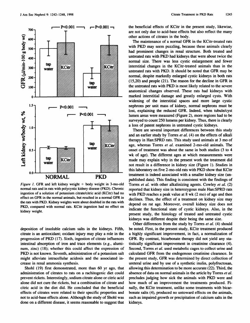
KCitr intake did not affect body weight or urine flow, but
completely prevented the decline in GFR found in untreated
rats with PKD. In rats that drank tap water, left kidney GFR
averaged (in p.1/mm per 100 g body wt) 503 ± 78 (ii = 9) in
normal animals and 242 ± 56 (ii = 6) in rats with PKD. In rats
that drank KCitr, GFR averaged 562 ± 123 (n = 7) in normal
animals and 534 ± 103 (ii = 7) in rats with PKD. Kidneys of
rats with PKD were approximately double normal size. KCitr
treatment did not affect kidney size, but led to fewer interstitial
abnormalities and smaller cysts in cystic kidneys. KCitr inges-
tion led to a significantly lower (P < 0.001) plasma [K�] in
rats with PKD (3.3 ± 0.2 versus 4. 1 ± 0.2 mEq/L in rats on tap
water). Chronic KCitr intake in the young heterozygous Han:
SPRD rat with PKD yields a modest improvement of kidney
histology and a dramatic improvement in GFR. The mecha-
nism of action of KCitr and the long-term effects of this
treatment on renal structure and function in PKD deserve
further study.
Autosomal dominant polycystic kidney disease (PKD) is a
common genetic disease that often leads to renal failure. The
expression of this disease is quite variable, probably because of
differences in the primary gene defect, modifier genes, and
environmental factors. Practical treatments to halt or slow the
progression of PKD in patients are extremely limited ( 1).
The discovery of the Han:SPRD rat with autosomal domi-
nant PKD has provided a unique opportunity for testing various
treatments (2-5). PKD in heterozygous rats of this strain
closely mimics the disease in humans. Major advantages of this
model are that treatments can be tested in the young animal,
before advanced renal failure develops, and that the usual time
course (months) of the disease is much shorter than in people
(decades). Using these rats, Torres et al. (4) found that acido-
sis, induced by NH4CI ingestion, resulted in a diminished GFR
and greater kidney size in heterozygous rats with PKD. These
investigators also found that treatment with alkalinizing salts,
e.g.. KHCO3 and NaHCO3, diminished enlargement of cystic
kidneys, but they did not find a statistically significant im-
provement in endogenous creatinine clearance. A potential
Received November 1 1. 1997. Accepted January 13, 1998.Portions of this study were presented at the XlVth meeting of the International
Society of Nephrology. May 1997. Sydney. Australia and at the National
Institute of Diabetes and Digestive and Kidney Diseases Workshop on Poly-
cystic Kidney Disease, September 1997. Crystal City. Virginia.Correspondence to Dr. George A. Tanner. Department of Physiology and
Biophysics. Indiana University School of Medicine. 635 Barnhill Drive, Indi-
anapolis. IN 46202.
l046-6673/()907- I 242$03.00/0Journal of the American Society of Nephrology
Copyright (1) 1998 by the American Society of Nephrology
problem with the alkalizers was that large doses of bicarbonate
led to precipitation of calcium stones in the kidneys. Also, rats
ingesting KHCO1 solutions (200 or 300 mM) showed impaired
growth. Recent studies by Cowley et al. (5) demonstrate the
detrimental effects of NH4C1 ingestion or a low potassium
intake on renal cystic disease in the Han:SPRD rat.
In the present study, the effects of ingestion of a potassium
citrate/citric acid solution (KCitr) on renal structure and func-
tion in Han:SPRD rats were investigated. KCitr was chosen for
several reasons. Citrate, like bicarbonate or carbonate, is an
alkalizer. Citrate forms a soluble complex with calcium in the
urine and so prevents intrarenal calculi formation (6). The
potassium salt was selected because it would be expected to
have less of an effect on BP than the sodium salt. Also,
administration of potassium might serve to correct potassium
deficiency, a condition known to favor the development of
renal cysts (5,7). This study hypothesized that administration
of KCitr would ameliorate renal cystic disease. Chronic treat-
ment of cystic Han:SPRD rats with KCitr led to complete
normalization of GFR and less severe histologic changes in the
kidneys.
Materials and MethodsExperiments were performed on 13 male heterozygous Han:SPRD
rats with PKD and 16 normal littermates. The breeding stock was
obtained from the Polycystic Kidney Program at the University of
Kansas. courtesy of Dr. Benjamin D. Cowley Jr. All animals were
allowed free access to a diet containing 24% protein and 6% fat
(Teklad 6% mouse/rat diet 7002, Harlan, Madison, WI). Beginning atI mo of age and until 3 mo of age. animals were provided with asolution of 55 mM tripotassium citrate/67 mM citric acid (KCitr) ortap water to drink. The KCitr had an osmolality of 240 ± 3
J Am Soc Nephrol 9: 1242-1248. 1998 Citrate Treatment in PKD Rats 1243
mosmol/kg H,O (ii = 10) and a pH of 4.2 ± 0.04 (‘z = 9). In 12
3-mo-old rats, two animals per cage, daily consumption of KCitr
averaged 16.7 ± 2.5 mb/bOO g body wt (ii = 6), a normal fluid intake.
The experimental design included four groups, because both normal
rats and rats with PKD drank tap water or the KCitr. Whether the
animals were normal or had PKD was determined by gross inspection
of the kidneys after completion of renal clearance measurements. All
experiments were conducted in accordance with National Institutes of
Health Guide for the Care and Use of Laboratory Animals.Before experiments. the rats were deprived overnight of food but
had free access to tap water or KCitr. They were anesthetized with the
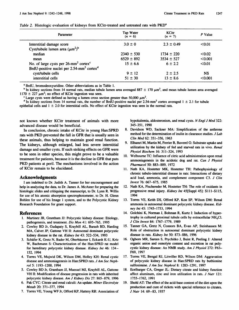
Lumen Area (I03�m2) Lumen Area (1OMm2) Lumen Area ( I0’�un2)
Figure 2. Photographs ( X77) of representative hematoxylin and eosin-stained sections of outer kidney cortex from normal rats (A) and from
KCitr-treated (B) and untreated (C) rats with PKD. Interstitial spaces were wider, filled with more inflammatory cells, and fibrotic, and the cysts
were larger in the untreated than in the KCitr-treated rats with PKD. Below are histograms of tubule/cyst lumen areas from these experiments( 14 normal rats. seven rats with PKD treated with KCitr, and six untreated rats with PKD). which demonstrate a shift to larger lumen sizes in
the cystic kidneys.
Normal RatsRats with PKD
KCitrate/citric acid
L
Rats with PKD
Tap Water
L
250
200>�
� 150
�. 100
a.
50 L
1246 Journal of the American Society of Nephrology J Am Soc Nephrol 9: 1242-1248, 1998
Although treatment with KCitr proved to be remarkably
effective in preventing a decline in GFR in young Han:SPRD
rats with PKD, it remains to be seen whether this treatment
would be effective over a more extended period of time, and
whether it would be beneficial in patients with PKD. Oral
potassium citrate and citric acid therapy is not new; this com-
bination is widely used for treating patients with renal calculi
or renal tubular acidosis (RTA). Igarashi and coworkers
(23,24) found that renal cysts are a common complication in
patients with primary distal RTA. They reported that 5 yr of
alkali therapy with sodium and potassium citrate was effective
in stabilizing the number and size of renal cysts in one patient
(23). In a later report (24), however, they state that alkali
therapy did not prevent an increase in number of renal cysts in
three patients with distal RTA. The effect of KCitr ingestion on
the progression of autosomal dominant PKD in patients de-
serves study.
The dose of KCitr administered in the present rat experi-
ments produced no detrimental effect on body growth or ap-
pearance and no obvious gastrointestinal or BP disturbances.
Assuming a daily fluid intake of 50 ml/d for a 300-g rat (body
surface area 0.039 m2), the rats consumed 23 g of potassium
citrate and 16.5 g of citric acid per m2 body surface area per
day. In adult patients, the usual daily oral dose of potassium
citrate and citric acid, for use as an antiurolithic or systemic or
urinary alkalizer, is approximately 7 to 14 g of potassium
citrate and 2 to 4 g of citric acid per m2 body surface area (25),
assuming an average body surface area of 1 .73 m2. Whether
such lower doses would be beneficial in slowing renal cystic
disease is an open question. One caveat is that administration
of potassium citrate to patients with advanced PKD may be
dangerous because of the risk of hyperkalemia. It is important
to note that in the present study KCitr treatment was begun at
a young age, at a time when GFR seems to be normal in
heterozygous cystic rats, as judged from measurements of
serum creatinine and urea nitrogen concentrations (2,3). It is
J Am Soc Nephrol 9: 1242-1248, 1998 Citrate Treatment in PKD Rats 1247
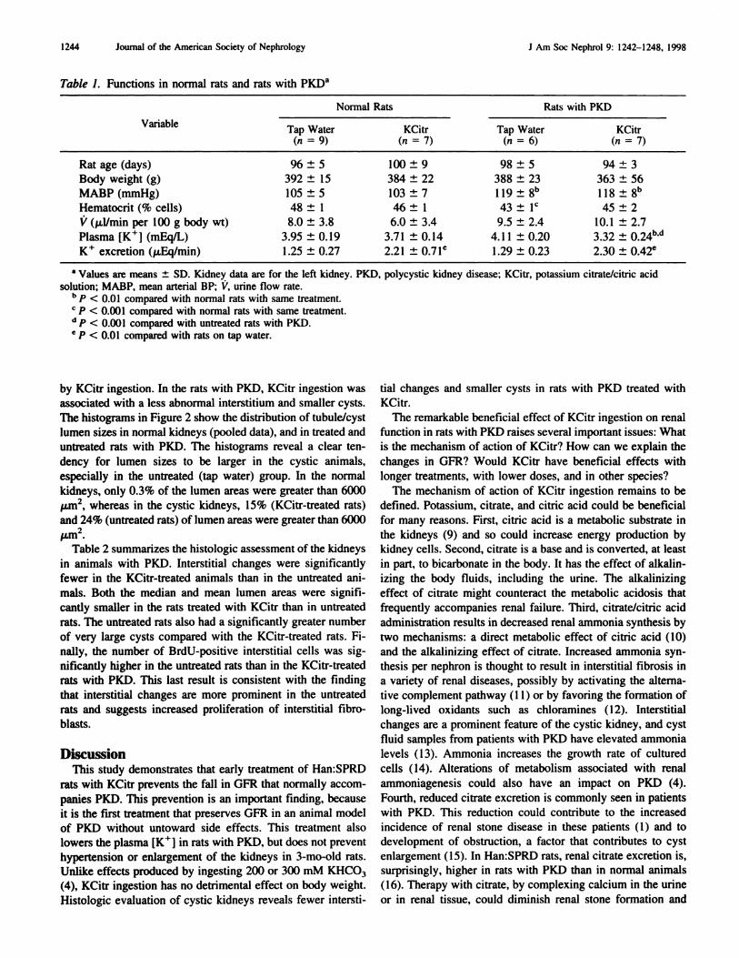
Table 2. Histologic evaluation of kidneys from KCitr-treated and untreated rats with PKD�
No. of large cysts per 26-mm2 cortex’ 15 ± 6.6 6 ± 2.2 <0.01
BrdU-positive nuclei per 2.54-mm2 cortex’�
cyst/tubule cells 9 ± 12 2 ± 2.5 NS
interstitial cells 51 ± 30 13 ± 8.6 <0.001
a BrdU. bromodeoxyuridine. Other abbreviations as in Table I.1� In kidney sections from 14 normal rats, median tubule lumen area averaged 887 ± 170 �m2, and mean tubule lumen area averaged
I I 70 ± 227 �m2; no effect of KCitr ingestion was seen.
C Large cysts were defined as having a lumen cross section greater than 50,000 �m2.
d In kidney sections from 14 normal rats, the number of BrdU-positive nuclei per 2.54-mm2 cortex averaged I ± 2.1 for tubule
epithelial cells and 1 ± 2.0 for interstitial cells. No effect of KCitr ingestion was seen in the normal rats.
not known whether KCitr treatment of animals with more
advanced disease would be beneficial.
In conclusion, chronic intake of KCitr in young Han:SPRD
rats with PKD prevented the fall in GFR that is usually seen in
these animals, thus helping to maintain good renal function.
The kidneys, although enlarged, had less severe interstitial
damage and smaller cysts. If such striking effects on GFR were
to be seen in other species, this might prove to be a valuable
treatment for patients, because it is the decline in GFR that puts
PKD patients at peril. The mechanisms involved in the action
of KCitr remain to be elucidated.
AcknowledgmentsI am indebted to Dr. Judith A. Tanner for her encouragement and
help in analyzing the data. to Dr. James A. McAteer for preparing thehistologic slides and critiquing the manuscript, to Dr. Lynn R. Willis
for use of his atomic absorption spectrophotometer, to Dr. H. Glenn
Bohlen for use of his Image I system, and to the Polycystic Kidney
Research Foundation for grant support.
References1 . Martinez JR. Grantham II: Polycystic kidney disease: Etiology,
pathogenesis, and treatment. Dis Mon 41 : 695-765, 1995
2. Cowley BD Jr. Gudapaty S. Kraybill AL, Barash BD, Harding