Rev. sci. tech. Off. int. Epiz., 16 (1), 65-78 Potential animal health hazards of pork and pork products S. Farez & R.S. Morley Animal and Plant Health Risk Assessment Network, Canadian Food Inspection Agency, 3851 Fallowfield Road, Nepean, Ontario K2H 8P9, Canada Summary The animal health hazards associated with the importation of pork and pork products include four viral agents: foot and mouth disease, classical swine fever (hog cholera), African swine fever, and swine vesicular disease viruses. The safety of importing pork from a zone infected with one or more of these diseases can be adequately determined only through risk assessment. This also applies for the safety of importing pork products which have undergone some form of processing (fully cooked pork products are not counted here). For each disease, the agent (pH and temperature lability), target organs, agent survival in pork and pork products, and agent quantification are discussed. Agent quantification is an input of the risk assessment which measures the viral titres in waste pork and pork products in relation to the oral infective dose estimated for each disease. Two other viral diseases, transmissible gastroenteritis of pigs and porcine reproductive and respiratory syndrome, are presented to illustrate why these two diseases are not hazards when associated with pork and pork products. Keywords African swine fever - Classical swine fever (hog cholera) - Foot and mouth disease - Porcine reproductive and respiratory syndrome - Risk assessment - Swine vesicular disease - Transmissible gastroenteritis of pigs. Introduction The introduction of animal diseases through the importation of pork and pork products is a concern for all swine-rearing countries. While the trade in pork is intended for human consumption, the intentional or inadvertent feeding of any uncooked waste to pigs could introduce animal disease. The Office International des Epizooties (OIE) International Animal Health Code (63, 69) describes the sanitary measures for the safe importation of pork and pork products from zones infected with certain swine diseases. The importation of pork (other than fully cooked products) from these zones is generally considered a high risk. It is therefore advisable to conduct a risk assessment for both chilled or frozen pork and for pork products which have been subjected to a process such as heat treatment, freezing, maturation and ageing, deboning, acidification, additives, curing, desiccation, etc. Preceding risk assessment is the hazard identification. Hazard identification is the process which identifies potential risk agents and the conditions under which they may produce adverse reactions. Each hazard necessitates a risk assessment. The risk assessment must determine, characterise, and quantify the following factors: a) the potential of the source to release a risk hazard or risk agent b) the intensity, frequency, and duration of exposure, and the nature of the animal and human populations which might be exposed c) the relationship between exposure and the resulting biological and economic consequences. The final outputs of this process are estimates of the magnitudes of possible adverse health and economic conse- quences, including a characterisation of the probabilities, uncertainties, or degree of confidence associated with these estimates (16). In this paper, the authors discuss the potential hazards associated with trade in pork and pork products and present some of the inputs and the relevant information for a risk assessment on the safety of importing from zones where these hazards exist. The paper is restricted to a discussion of the
Transcript
Rev. sci. tech. Off. int. Epiz., 16 (1), 65-78
Potential animal health hazards of pork and pork products
S. Farez & R.S. Morley
Animal and Plant Health Risk Assessment Network, Canadian Food Inspection Agency, 3851 Fallowfield Road, Nepean, Ontario K2H 8P9, Canada
Summary The animal health hazards associated with the importation of pork and pork products include four viral agents: foot and mouth disease, classical swine fever (hog cholera), African swine fever, and swine vesicular disease viruses. The safety of importing pork from a zone infected with one or more of these diseases can be adequately determined only through risk assessment. This also applies for the safety of importing pork products which have undergone some form of processing (fully cooked pork products are not counted here). For each disease, the agent (pH and temperature lability), target organs, agent survival in pork and pork products, and agent quantification are discussed. Agent quantification is an input of the risk assessment which measures the viral titres in waste pork and pork products in relation to the oral infective dose estimated for each disease. Two other viral diseases, transmissible gastroenteritis of pigs and porcine reproductive and respiratory syndrome, are presented to illustrate why these two diseases are not hazards when associated with pork and pork products.
Introduction The introduct ion of animal diseases th rough the impor ta t ion of pork and p o r k p roduc t s is a concern for all swine-rearing countries. Whi le the trade in p o r k is in tended for h u m a n consumption, the intentional or inadvertent feeding of any uncooked waste to pigs could in t roduce animal disease.
The Office International des Epizooties (OIE) International Animal Health Code (63 , 69) describes the sanitary measures for the safe importa t ion of p o r k and p o r k produc ts from zones infected wi th certain swine diseases. The impor ta t ion of p o r k (other than fully cooked products) from these zones is generally considered a h igh risk. It is therefore advisable to conduct a risk assessment for b o t h chilled or frozen p o r k and for pork p roduc ts wh ich have been subjected to a process such as heat treatment, freezing, matura t ion and ageing, deboning, acidification, additives, curing, desiccation, etc.
Preceding risk assessment is the hazard identification. Hazard identification is the process wh ich identifies potential risk agents and the condit ions u n d e r which they m a y p roduce adverse reactions. Each hazard necessitates a risk assessment.
The risk assessment m u s t determine, characterise, a n d quantify the following factors:
a) the potential of the source to release a risk hazard or risk agent
b) the intensity, frequency, and dura t ion of exposure, and the na ture of the animal and h u m a n populat ions which might b e exposed
c) the relationship be tween exposure and the resulting biological and economic consequences.
The final ou tputs of this process are estimates of the magni tudes of possible adverse heal th a n d economic consequences, including a characterisation of the probabilities, uncertainties, or degree of confidence associated wi th these estimates (16).
In this paper , the authors discuss the potential hazards associated wi th t rade i n p o r k a n d p o r k produc ts and present some of the inputs and the relevant information for a risk assessment on the safety of impor t ing from zones where these hazards exist. The paper is restricted to a discussion of the
66 Rev. sci tech. Off. int. Epiz., 16 (1)
commodity-related inputs . These are as follows: agent (pH and temperature lability), target organs, agent survival in p o r k and p o r k products , and agent quantification. Agent quantification is an input of the risk assessment which quantifies the viral titres in waste pork and pork products in relation to the oral infective dose for each disease.
The risk assessment inputs of incidence or prevalence of disease, durat ion of infection/viraemia, swine populat ion demographics, detection systems (in the herd, ante- and pos t -mor tem inspections), surveillance and monitor ing programmes, disease control and eradication programmes and exposure are more clearly elaborated on an individual country basis and are no t discussed in this paper.
Hazard identification
The potential hazards associated with trade in pork and pork products are the viral agents of foot and m o u t h disease (FMD), classical swine fever (CSF) (hog cholera), African swine fever (ASF), and swine vesicular disease (SVD). The viral agents of transmissible gastroenteritis of pigs (TGE) and porcine reproductive and respiratory syndrome (PRRS), al though perceived as potential hazards by some countries, are discussed to illustrate the differences be tween these two agents and actual hazards of p o r k and p o r k products . Excluded from the discussion is the parasitic agent of trichinellosis. Although the agent of this disease represents a potential animal health hazard of pork and pork products , the disease is principally a food safety concern that wou ld manifest itself in h u m a n s m u c h earlier and wi th a m u c h greater impact than in pigs.
In the past and recently, po rk and p o r k products have been incriminated as sources of disease introduct ion for all four hazards. The disease introduct ion often occurs as outbreaks in herds which are fed raw food waste in their swill, despite regulations that may be in place requiring the cooking of such waste before feeding. However, disease introduct ion of PRRS and TGE has never been attributed to the importat ion of p o r k and pork products from infected zones.
Many primary outbreaks of FMD have been attr ibuted to imported meat and meat products , al though pork and p o r k products were no t necessarily implicated (6, 92).
It is almost certain that the long-distance spread of ASF from Angola to Portugal in 1957 took place as a result of uncooked waste food being fed to pigs near Lisbon airport. Subsequent outbreaks in Cuba in 1971 and 1978, Malta, Sardinia, Brazil and the Dominican Republic in 1978 and Belgium in 1985 have also been attributed to feeding pigs on swill from ports or airports (97).
Outbreaks of CSF have often been traced to the feeding of garbage from ships or aircraft. Notable examples include two occasions, i n 1930 a n d 1953 , w h e n the disease was in t roduced into N e w Zealand (47). The feeding of swill, presumably containing uncooked waste p o r k and pork products , to pigs was incriminated in CSF outbreaks in the United Kingdom in 1986 (98), in Switzerland in 1993 , in Bulgaria (65), Germany (66) and Poland (67) in 1994 and in Austria in 1994 (64) and 1995 (68).
Of 518 SVD outbreaks which occurred in Great Britain be tween 1972 and 1 9 8 1 , 80 were attributed to feeding of contaminated waste food (38).
There is n o evidence of oral transmission of PRRS th rough uncooked waste of p o r k or pork products either in an enzootically infected zone or in the spread of the disease to a previously disease-free zone (5 , 24 , 6 1 , 78). Experimental oral transmission is possible following administration of doses of 1 0 7 T C I D 5 0 virus (55).
Similarly, the transmission of TGE through the feeding of waste p o r k or p o r k products has never been reported. Only experimental oral infection of one-week-old piglets wi th homogenates of muscle of the h ind leg, l ymph nodes (internal iliac, sub-maxillary and cervical) and bone mar row from the femur of 16 s ix-month-old pigs which h a d been in contact wi th experimentally infected piglets has been demonstrated. Twelve three-week-old pigs fed about 1.5 kg of the same tissues each over 5 days failed to become infected, yet all became serologically positive for neutralizing ant ibody to TGE virus (27). Cook et al. (13) achieved transmission of TGE in six-day-old piglets by oral dosing daily for 4 days wi th a homogena te of brachiocephalic muscle and parot id l y m p h node . These two studies involved dosing wi th a rather artificial mixture considering the homogenisat ion of the tissues and the high propor t ion of l ymph n o d e tissue.
Inputs for risk assessments on the importation of pork and pork products
Agent pH and temperature lability The p H and temperature lability of the viral agents represent the two most impor tant biological propert ies for an assessment on the safety of import ing meat. The anaerobic glycolysis which ensues in muscle after slaughter results in the conversion of glycogen to lactic acid. In one s tudy on mea t quality in different swine breeds, the p H of the semimembranosus and longissimus dorsi muscles at 4 5 minutes post m o r t e m ranged from a low of 6.17 to a h igh of 6.71 (88). The final p H attained is generally about 5.4-5.5
Rev. sci tech. Off. int. Epiz., 16 (1) 67
in typical mammal ian muscles and is referred to as the ultimate pH' . Between individual pigs there is a considerable variation i n the t ime taken for the p H of latissimus dorsi muscle to fall from a p H of 6.5 to an ult imate p H of 5.5 at 37°C, a range of about 150 to jus t over 4 0 0 minu tes (45). In the commercial p roduc t ion of pork , the t ime and temperatures at wh ich the carcasses are he ld se ldom allow the pork to fall be low a p H of 5.7 (10). FMD virus is the only hazard which is labile to p H levels found in pork: however, viral inactivation as seen in the matura t ion of beef may no t b e as dramatic. Beef ma tu red for 20 -24 hour s safely achieves a p H of 5.7 (3) and any virus present in the muscle tissues would b e readily inactivated. The significance of this is that the p H of p o r k and p o r k produc ts represents an impor tan t input in the risk assessment on p o r k from FMD infected zones. Other organs and tissues such as the parenchymatous organs, l y m p h nodes and bone m a r r o w wou ld be unlikely to achieve the low p H values as evidenced in muscle .
Temperature lability is the mos t impor tant consideration for pork p roduc ts wh ich undergo some thermal processing. Although there is a difference be tween the temperature lability of the viral agents in cul ture media and that in pork, the laboratory data give some indication of the ease of inactivation. Generally the studies involving thermal processing of p o r k in which particular core temperatures achieve an apparent inactivation would be better to assess. For the four viral hazards, a temperature of 69°C which is reached by the very inner core of a por t ion of p o r k appears to readily inactivate the virus. A minimal time per iod of less than 15 minutes at this temperature , however, m a y be necessary to inactivate CSF virus in pork .
Target organs Before discussing the target organs and predilection sites of the viral agents, it is necessary to m a k e a clear distinction between the organs and tissues which constitute p o r k and pork products and n o n - p o r k organs. Pork and p o r k produc ts are comprised principally of skeletal muscle, b o n e and fat. Bone marrow, blood wi thin the capillaries of skeletal muscles and lymph nodes (prepectoral, presternal, precrural, superficial inguinal, popliteal, iliac, lumbar and renal) (32) amount to a very small fraction of the swine carcass. W i t h respect to p o r k port ions, l y m p h nodes and b o n e m a r r o w may not be present as a result of t r imming, deboning and depending on the particular cut. Many of the l y m p h nodes indicated above are removed th rough carcass t r imming due to their superficial, fat-embedded location on the carcass. Obviously, the blood, the respiratory, gastro-intestinal and reproductive tracts, the head, the respective l y m p h nodes of these parts and the tonsils are no t pork tissues.
The cell and tissues for wh ich the agent has an affinity and in which the agent replicates are therefore an impor tant consideration in the importa t ion of pork . Virus titres in pork derived from infected pigs may b e very low in comparison to the predilection sites of the agent (e.g. SVD virus replicates in
the epi thel ium of the coronary band , tongue, snout and lips and in the myocard ium, tonsils and brain s tem (90). Dur ing the incubat ion period, the virus m a y only b e found at the site of entry and the adjacent l y m p h nodes , such as the tonsils a n d mandibu la r and parot id l y m p h nodes for CSF, ASF and SVD or the popliteal l y m p h nodes of the h ind legs for SVD following agent entry th rough b r o k e n skin o n the foot. Despite this tissue tropism, titres are detected in almost all tissues dur ing the state of viraemia. Viral titres in skeletal muscle of viraemic pigs wi th SVD or FMD m a y represent more a function of the viral litre w t h i n b lood capillaries. Viral titres in skeletal muscle of pigs dur ing the viraemic phase of ASF are a reflection of h igh b lood titres, the target cells ( lymphocytes, endothelial cells, reticular cells, monocytes and macrophages) (76) and loss i n vascular integrity. Highly vascularised organs such as the spleen, kidney, and liver and b o n e m a r r o w w o u l d b e expected to exhibit viral titres dur ing the state of viraemia. Generally, only the viraemic per iod and its dura t ion are of significance in the impor ta t ion of pork . The durat ion of viraemia m a y b e quite short , as observed wi th SVD, or prolonged, as in some of the clinical forms of CSF and ASF. The mortality as seen in the peracute , acute and subacute forms of CSF and ASF naturally limits the viraemic period.
A carrier state is no ted wi th ASF b u t no t wi th CSF, SVD or FMD in pigs. The carrier state for diseases such as TGE and PRRS is characterised by predilection and maintenance of the virus in tissues other than pork , that is the tonsils, lungs a n d intestinal tract for TGE and the lung and tonsils for PRRS. The carrier state of ASF in surviving pigs is confined to l y m p h nodes , tonsils and to a lesser extent kidney, spleen and b o n e m a r r o w (53).
Agent survival in pork and pork products Survival in the p roduc t depends on the agent properties, especially lability to time, temperature and p H and on the inherent propert ies of the p roduc t (e.g. p H , water activity, moisture:protein ratio, temperature of processing and storage, salinity, additives, etc.). The interaction of these p roduc t propert ies and the enzymatic proteolysis which occurs in m a n y products are significant factors in the inactivation of viral agents. Survival of agents in various p o r k produc ts has been investigated and repor ted in the scientific literature. Most of these studies m u s t b e statistically interpreted, based o n the n u m b e r of samples tested for virus isolation or the data points on the reduct ion of viral litre of an agent over time. Multiple studies and other information m a y be combined and statistically evaluated to estimate the inactivation curve associated with some form of processing. Agent survival beyond the usual processing time and the t ime required for impor ta t ion a n d distribution in the impor t ing country are part of the considerations of this input . Changes in the technology of meat processing and curing, and the efficiency of m o d e r n abattoir practices and international t ransportat ion and distribution, m u s t be considered here.
68 Rev. sci tech. Off. int. Epiz., 16 (1)
Agent quantification
Viral titres Data o n titres in tissues, in particular muscle, quantify the amoun t of agent present pe r gram. Quantification of viral titres is generally obtained from experimental infection of swine with a specific strain of virus. The viral titres in muscle a n d other tissues are measured, often at the peak of viraemia. Titres at other times in the disease course are no t available, as experimental studies are frequendy looking for the 'worst-case scenario' w h e n titres are expected to be at their highest level. Employing these data to represent the titres for any day of the durat ion of infection or viraemia is an extrapolation. Nonetheless, the data may represent the best available biological information.
Oral infective dose Uncooked waste of pork or pork products being ingested by swine by one or more scenarios represents the only m o d e of transmission of interest here. The target species is the pig, the portals of entry are the tonsils, oral abrasions, respiratory tract and the gastro-intestinal tract. After contact exposure, ASF virus primarily gains entry th rough the tonsils a n d i n some instances across the nasal, bronchial and (possibly) gastric mucosae (34). Similarly with exposure to pigs infected wi th SVD, the tonsils appear to be the main site of entry, however, other entry sites may include the skin of the head and lower limbs, u p p e r respiratory a n d digestive tract epithelium, and intestinal mucosae (56). Virus in pork or p o r k products has to contact the tonsils, the u p p e r respiratory tract epithelium or gain entry through oral abrasions. Failing these routes of entry, the gastro-intestinal mucosae may serve as sites of entry providing the virus survives the low p H of the gastric juices. The amoun t of mastication of the food and the rapidity of swallowing a bolus of p o r k would determine the site of viral entry. It is for this reason that the oral pig infective dose 5 0 % (P1D 5 0 ) for the viral diseases wou ld be expected to b e considerably higher for feeding of infected p o r k than for instilling virus i n culture media o n the tongue of a pig, feeding the virus suspended in a l iquid m e d i u m such as milk or force-feeding homogenised infected tissues. The P I D 5 0 has not been estimated for FMD, CSF, and SVD. Instead, an infective dose at which infection was achieved experimentally in one or more pigs is available in the literature. In the case of SVD, skin abrasions, especially a round the coronary bands of the feet may serve as a portal of entry following contact wi th infected waste pork (56).
Foot and mouth disease Agent pH and temperature lability FMD virus is acid, alkali a n d hea t labile, b u t can survive for long periods at neutral p H (7 to 9) and u n d e r low temperature
condit ions (82). In tissue culture suspensions, the virus survived less t h a n 15 seconds at p H 2 .2 a n d p H 4 , two minutes at p H 6 and for several weeks at p H 7 (25, 85) . At a p H of 7.5, the virus in simple media survived 30 seconds at 61°C, two minutes at 55°C yet as long as 18 weeks at 4°C (1).
Target cells and tissues The virus is distributed th roughout the body of the infected animal and can be found in different concentrations for varying periods in the tissues. In pigs, the greatest quantities of virus are in the blood, epithelium, a n d liver (23 , 86). The specific lesions in their early stages are microscopic and are l imited to the epithel ium at sites of predilection such as the mucosa of the mouth , including the tongue, lips, gums, pharynx , a n d palate (9) .
The incubat ion period of FMD is generally within the range of 2-14 days (82). The virus is excreted one to ten days before clinical signs appear, and continues for four to ten days. Virus was detected i n b lood 32 h o u r s and i n muscle 2 0 h o u r s before the appearance of aphthae or the beginning of a rise in temperature (21) . I n pigs, the virus does n o t persist for m o r e than a m o n t h (57). One characteristic wh ich distinguishes pigs from cattle and sheep is that they appear to ha rbour the virus only dur ing clinical stages of the disease and therefore do no t act as carriers (15).
Survival in pork and pork products The apparent durat ion of survival of FMD virus in the listed p o r k and p o r k products is as follows:
- 30 days in different chilled organs such as lungs, s tomach, tongue, intestine (83)
- 2 4 hour s in chilled spleen, liver and kidney (83)
- 210 days in frozen lungs, intestine, s tomach, tongue, kidney, spleen a n d liver (83)
- 170 days in Parma h a m s (52)
- 182 days in the whi te Serrano h a m (58)
- 168 days in Iberian h a m (58)
- 112 days in Iberian shoulder h a m s (58)
- 42 days in Iberian loins (58)
- 190 days in salted bacon, and 183 days in h a m fat (22)
- 56 days in sausages (22)
- 250 days in processed intestinal casings (49)
- 7 days i n salami (71)
- 10 days in tongue and 1 day in muscle (14).
Apparent thermal inactivation of FMD virus is obtained wi th an internal temperature of 69°C (50).
Agent quantification Viral titres Sellers (86) found h igh titres of 1 0 7 ' 2 p laque forming uni t s (PFU)/ml in blood, 1066 PFU/g in bone m a r r o w and
Rev. sci tech. Off. int. Epiz., 16(1) 69
1 0 5 ' 6 PFU/g in liver of infected pigs. Similarly h igh virus titres of greater than 1 0 5 PFU/ml were detected by extraction from both fat and muscle tissues of infected pigs (71). Viral titres were measured in each of 62 pigs two days after intravenous inoculation wi th 1 m l of a 1:10 dilution of a stock virus (serotype C) having a titre of 1 0 8 ' 9 T C 1 D 5 0 pe r ml. The m e a n and standard deviation of the viral titres in muscle , fat, b o n e marrow, b lood and l y m p h n o d e are presented in Table 1 (58). The mean viral titres in muscle and fat as presented in Table I are significandy lower than the titres detected by Panina et al. (71).
Table I Foot and mouth disease viral titres in tissues of 62 pigs two days after experimental infection (58)
Titre (plaque forming units [PFU] per ml or g) Mean Standard deviation
Blood 1 0 3 . 5 10 1- 5
Lymph node 103-4 1(F Bone marrow 10'-9 101-5
Fat 1 0 0 . 5 1 D 0 . 8
Muscle 1 0 D.O3 1(F
Oral infective dose A viral titre of 1 0 5 0 T C I D 5 0 of FMD O-strain initiated infection in two of 30 pigs fed minced offal (liver, k idney and lymph nodes) (39).
African swine fever Agent pH and temperature lability The sensitivity of the ASF virus to different temperatures has been studied widely. Infected b lood heated for 30 minu tes at 60°C loses infectivity. At 56°C, the resistance of the virus will depend on the presence of serum. Some strains could remain virulent after 3.5 hour s at 56°C (12). Kovalenko et al. (44) found that the virus could survive 6 years at 5°C wi th n o light. ASF virus is very resistant to acid p H , even m o r e than CSF viras. Plowright et al. (74) have found that the ASF virus could survive 22 hour s at p H 3 . 1 . The virus is rapidly inactivated at p H 11.5 and the inactivation rate decreases rapidly at lower p H values (12).
Target cells and tissues The ASF virus usually enters the pig th rough the m o u t h or upper respiratory system, and infection is established in the nasopharyngeal region. The virus spreads rapidly to the mandibular l y m p h nodes and throughout the body th rough lymph and b lood (46). Following the incubat ion period, the duration of viraemia m a y last 1-3 days, 4-8 days, and
6-10 days before death in the hyperacute , acute and subacute forms of the disease, respectively (70). W i t h one ASF viral strain, viraemia was demonst ra ted two days before the onset of fever (34). Pigs surviving the subacute form may exhibit the chronic form of the disease which m a y last for several mon ths . The carrier state in these chronically affected pigs and in pigs manifesting a subclinical or inapparent form of the disease m a y b e confined to l y m p h nodes , tonsils and to a lesser extent kidney, spleen and b o n e mar row (53 , 70, 97) . The virus replicates principally in the cells of the lymphoret icular system, b o t h the fixed cells in the l y m p h nodes , liver and spleen (58, 62) , and in monocytes and macrophages (97). Endothelial cells lining b lood vessels are severely damaged, wi th resulting oedema, haemorrhage a n d necrosis. Lesions are mos t p rominen t i n the spleen, l y m p h nodes , lung and liver, b u t m a y b e found in any lymphoid tissue or infiltrate of lymphoid cells (46).
Survival in pork and pork products The apparent dura t ion of survival of ASF virus in the listed p o r k and pork produc ts is as follows: - 104 days in frozen meat or chilled meat (44) - 140 days in Iberian h a m s (58) - 140 days in Iberian shoulder h a m s (58) - 112 days in Iberian loins (58) - 140 days in whi te Serrano h a m s (58) - 399 days in Parma h a m s (52) - 30 days in pepperon i sausage (49) - 30 days in salami sausage (49).
Apparent thermal inactivation of ASF virus is obtained wi th an internal temperature of 69°C (50).
Agent quantification Viral t i t res Viral titres of meat samples from four pigs infected wi th ASF ranged from 1 0 3 " 2 5 to 1 0 3 ' 7 5 haemadsorb ing uni ts 5 0 % ( H A d 5 0 ) per g in b o t h whole and g round meat , two days after slaughter; 1 0 2 " ° - 1 0 2 ' 5 in salami, three days after slaughter; 1 0 3 " ° - 1 0 3 ' 2 5 in pepperoni , three days after slaughter; 1 0 2 ' 5 - 1 0 3 ' 7 5 in br ined h a m , two days after slaughter; 1 0 2 " 7 5 - 1 0 3 in pepperon i sausage, eight days after slaughter; and 1 0 _ 1 in salami sausage, n ine days after slaughter (49). Viral titres were measured in each of 65 pigs five days after intramuscular inoculat ion of 1 m l of und i lu ted stock virus having a titre of 1 0 6 , 5 H A d 5 0 per ml (58). The m e a n and s tandard deviation of the viral titres in muscle , fat, b o n e mar row, b lood and lymph n o d e are presented in Table II.
Oral infective dose In one experimental study, the P I D 5 0 was 1 0 5 ' 4 H A d 5 0 (34). McVicar (53) found the P I D 3 0 of a moderately virulent strain of ASF virus to b e 1 0 4 ' 3 H A d 5 0 by oral experimental infection.
Experiments were conducted in which l y m p h n o d e suspensions from naturally infected warthogs were administered to domest ic swine. The results of these
70 Rev. sci tech. Off. int. Epiz., 16(1)
Table II African swine fever viral titres in tissues of 65 pigs five days after experimental infection (58)
Tissue Titre (haemadsorbing units 50% [HAdJ per ml or g)
Mean Standard deviation
Blood 107-9 1 0 1 2
Lymph node 1(F 1 0 " Bone marrow 1 0 9 . 5 10° 8
Fat 1(P 1 0 o a
Muscle 1 0 B . B 1 0 o .5
experiments made it evident that well-homogenised tissue suspension, containing 1 0 3 7 to 1 0 6 ' 1 H A d 5 0 of ASF virus failed to infect pigs w h e n administered either in l iquid or moistened solid food (75).
Classical swine fever (hog cholera)
Agent pH and temperature lability While the virus is very resistant at temperatures be low 0°C, research has shown the sensitivity of the virus above this temperature. It can survive three days at 50°C, seven to 15 days at 37°C and years at - 70°C . The virus is, however, susceptible to rapid changes in temperature such as thawing and refreezing. The effect of heat treatment o n CSF virus is influenced by the physical m e d i u m in which the virus is heated. Thus cell culture fluid infectivity is lost after 10 minutes at 60°C, whereas in defibrinated b lood the virus is no t inactivated after 30 minutes at 68°C (94). The virus is stable over a wide p H range, bu t is rapidly inactivated below p H 4 and above p H 11 (89).
Target cells and tissues The virus initially infects epithelial cells of the tonsillar crypts and subsequently spreads to the sur rounding lymphoreticular tissue. From the tonsil, CSF virus is drained to the regional l ymph nodes , where replication occurs. The virus reaches the peripheral b lood and then attains h igh titres in the spleen, bone marrow, visceral l ymph nodes , and lymphoid structures lining the small intestine. As a result of the replication in lymphoid tissue and in circulating leukocytes and mononuclear cells, the level of viraemia is high. The virus probably does not invade parenchymatous organs unt i l late in the viraemic phase (94). Viraemia can b e detected in pigs from between two to four days after infection. Viraemia follows the incubation period as the spread of virulent virus throughout the pig is usually completed in five to six days (77). In the peracute form, death may appear wi th in five days post-infection. In the acute and subacute forms of the disease,
the viraemia persists at a high level until death which may occur be tween 10 a n d 2 0 days post-infection for the acute form and be tween 20 and 29 days post infection for the subacute form. In the chronic form, the durat ion of the disease could be 30 or more days (18). In the chronic form, the viraemia m a y subside dur ing the course of the infection whereas viraemia persists at a h igh level for life in the late-onset form of CSF (94, 96). The late-onset disease, a sequel of congenital CSF virus infection, is characterised by a period of a few mo n t h s dur ing which pigs remain free of disease. Most pigs survive for more than 6 m o n t h s bu t all eventually die. The viraemia persists for life (95). CSF virus strains of low virulence can induce mild disease wi th subsequent recovery wi thout a carrier state (89). Virulent virus infects b o t h epithelial a n d reticular cells, macrophages and tonsillar cells, whereas the growth of virus of reduced virulence is mainly restricted to cells of the epithelial tonsillar crypts (93).
Survival in pork and pork products The apparent dura t ion of survival of CSF virus in the listed p o r k and p o r k products is as follows:
- 4 .5 years in frozen mea t (96) - 1 m o n t h in the meat of salt-cured p o r k (96) - 2 mo n t h s i n the bone mar row of salt-cured p o r k (96) - 90 days in salami (84) - 75 days in Italian salami (72) - 90 days in h a m (muscle a n d fat) (84) - 70 days in neck, lard and b o n e m a r r o w (84) - 226 days in frozen liver at - 4°C and - 6°C (12) - 252 days in Iberian h a m s (58) - 4 0 days in Iberian shoulder h a m s (58) - 126 days in Iberian loins (58) - 140 days in Whi te Serrano h a m s (58) - 189 days in Parma h a m s (52) - 147 days i n intestinal casings processed in water at 42 .2°C for 30 minutes (50).
Apparent thermal inactivation of CSF virus is obtained us ing the following procedures:
- pasteurisation at core temperatures over 67°C of cured and canned h a m s (89)
- exposure of cubes (2 c m 3 ) of h a m to a 'flash' temperature of 71°C for 1 m i n (87)
- heat ing to 69°C for 15 m i n (50).
Agent quantification Viral t i tres
W o o d et al. (99) s tudied the titres of virus in tissues of pigs experimentally inoculated wi th CSF and slaughtered be tween seven and 25 days after infection. Pigs were infected b y intranasal inoculation wi th 1 0 6 5 TCID 5 0 / p ig . CSF virus titres in muscle ranged from 1 0 3 4 to 1 0 4 ' 9 T C I D 5 0 / g a n d titres in l y m p h nodes ranged from 1 0 5 0 to 1 0 7 ' 5 T C I D 5 0 / g . Titres
fíev. sci tech. Off. int. Epiz., 16(1] 71
(TCID 5 0 /g) of CSF viras in quadriceps muscle were obtained on different days after infection as follows: 1 0 3 , 4 on day 7, 1 0 4 ' 8 on day 1 1 , 1 0 4 ' 4 o n day 2 3 and 1 0 4 ' 9 on day 2 5 . A comparison s tudy of the pathogenicity of two strains of CSF virus revealed titres wi th a virulent strain of 1 0 2 ' 5 to 1 0 7 2 5 T Q D 5 0 per m l starting on day three of the infection, whereas wi th a low virulent strain the viraemia was no t detected in pigs until day six. The range of titres in the pigs was 1015 to 1 0 4 T C I D 5 0 pe r ml . (42). W i t h the late-onset form of the disease, the viraemia persists as long as the animal lives, wi th plasma titres ranging be tween 1 0 5 0 and 1 0 6 ' 9
plaque forming uni ts (PFU)/ml. Virus is continuously present in the buffy coat cells (95).
Viral titres were measured in each of 64 pigs five days after intravenous inoculation wi th 1 m l of a 1:100 dilution of a stock virus having a titre of 1 0 5 ' 3 T C I D 5 0 pe r m l (58). The mean and s tandard deviation of the viral titres in muscle , fat, bone marrow, b lood and l y m p h n o d e are presented in Table III.
Table III Classical swine fever (hog cholera) viral titres in tissues of 64 pigs four or five days after experimental infection (58)
Tissue Titre (plaque forming units [PFU] per ml or g)
Mean Standard deviation
Blood 1 0 3 . s 101-5
Lymph nods 1 0 3 9 1 0 1 2
Bone marrow 1 0 " 10 1- 2
Fat 1Qo.a 101'° Muscle 1 0 i . o 1 0 M
Oral infective dose A CSF virus titration in weaner pigs us ing the highly virulent strain Alfort' showed that the minimal infective dose resulting in fatal disease was less than 10 T Q D 5 0 (18). In comparison to the three other viral hazards , this represents a very low oral infective dose. It may indicate that an infective dose for pork and pork products is likewise comparatively very low.
Swine vesicular disease
Agent pH and temperature lability SVD virus is relatively stable over a p H range of 2-12, depending o n temperature and time (38): 164 days at a p H of 5.10 and 7.54 at 5°C, bu t a reduct ion of over 6 log in titre occurred at p H values of 2 .88 and 10.14 by 164 days and at p H values of 1.92 and 11.96 at 3 8 days (40). SVD virus is also stable in infected tissue kept at ambient or higher temperatures for four m o n t h s or m o r e (90). Carcass materials
at - 20°C were sampled after approximately 11 m o n t h s a n d showed n o significant d rop in infectivity resulting from storage (20).
Target cells and tissues Swine vesicular disease virus has an affinity for, and replicates in, the epi thel ium of the coronary band , tongue, snout and lips, as well as the myocard ium, the tonsils and the bra in s tem (90). The s t ra tum sp inosum is the pr imary site of viral replication in the epi thel ium (41). In cell cultures of epithelial and salivary gland tissues, SVD virus reached significant titres whereas little or n o growth was detected in cell cultures of muscle , l y m p h n o d e or parts of the digestive tract (56).
Eight healthy pigs were inoculated i n the coronary b a n d of the h ind legs wi th cell culture SVD virus and were slaughtered at the peak temperature . Samples of different tissues were collected dur ing necropsy. The virus could n o t b e isolated from the muscles, b u t was detected in one fat sample. Tests were performed on fresh samples and at 12, 30 , 4 0 , 70 and 100 days after storage at 4°C (28).
The peak of viraemia occurs 2-4 days post-exposure to SVD virus and persists for six days. Virus persists for at least 10 days in the tissues of the snout , tongue, coronary band , tonsil, cardiac muscle, and central nervous system. Following experimental infection, swine shed virus in their faeces for u p to 2 3 days (41).
Survival in pork and pork products The apparent dura t ion of survival of SVD virus in the listed pork and p o r k products is as follows:
- 300 days in Parma h a m s (51)
- 200 days i n dry salami sausage, dry pepperon i sausage and intestinal casings (48)
- 4 0 0 days in dried pepperon i and salami sausage (33)
- 780 days in processed intestinal casings (33)
- 40 days in salami and pepperon i sausages (38)
- 509 days in unprocessed intestinal casings (38)
- 2 8 days in Iberian loins (59)
- 112 days in Iberian shoulder h a m s (59)
- 560 days in Iberian h a m s (59)
- 539 days in white Serrano h a m s (59).
Apparent thermal inactivation of SVD virus is obtained by heat ing to at least 69°C (48 , 50) .
Agent quantification Viral t i t res
Dawe (20) reported that 11-month-old frozen carcass material h a d 1 0 6 T C I D 5 0 pe r g of SVD virus in the skin, 1 0 4 T C I D 5 0 in intercostal muscle and 1 0 3 T C I D 5 0 pe r g in
7 2 Rev. sci tech. Off. int. Epiz., 16(1)
b o t h rib bone and kidney cortex. These virus titres were no t significantly be low titres from similar samples taken at the beginning of the storage. In h a m s held at 0-4°C for 72 hours after slaughter, m e a n titres, expressed as PFU per g of muscle, bone mar row and fat were 1 0 4 ' 5 , 1 0 2 ' 3 and 1 0 1 ' 1 , respectively.
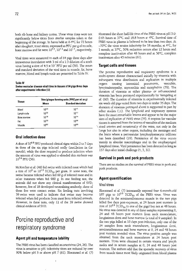
Viral titres were measured in each of 64 pigs three days after intravenous inoculation wi th 3 m l of a 1:3 dilution of a stock virus having a titre of 4.5 X 1 0 7 PFU per m l (59). The m e a n and s tandard deviation of the viral titres in muscle, fat, b o n e marrow, b lood and lymph n o d e are presented in Table IV.
Table IV Swine vesicular disease viral titres in tissues of 64 pigs three days after experimental infection (59)
Tissue Titre (plaque forming units [PFU] per ml or g)
Mean Standard deviation
Blood 1 Q 2 . e 1 0 u
Lymph node 1(F 10 1.2 Bone marrow 1 0 1 D 10 1- 5
Fat 10° 3 10° 8
Muscle 1 Q 0 . 2 1 0 O . B
Oral infective dose A dose of 1 0 6 ' 8 PFU produced clinical signs within 2 to 7 days in three of the six pigs infected orally (instillation in the mou th ) , while the dose required to p roduce infection w h e n the amoun t of virus was applied to abraded skin surfaces was 1036 PFU (56).
McKercher et al. (48) fed swine wi th infected meat wh ich h a d a titre of 1 0 3 to 1 0 4 ' 5 T C I D 5 0 pe r gram. In some tests, the swine became infected w h e n fed 60 g of infected meat and in other instances w h e n fed 900 g. In one feeding test, the animals did no t show any clinical manifestations of SVD; however, five of 18 developed neutralising antibody; three of these five were contact swine. Six feeding tests involving 39 swine were used to indicate that swine may become infected w h e n fed products from meat from infected animals. However, in these tests, only 12 of the 39 swine showed clinical evidence of SVD.
Porcine reproductive and respiratory syndrome
Agent pH and temperature lability The PRRS virus has been classified an arterivirus ( 2 4 , 2 6 ) . The virus is sensitive to p H : infectivity titres are reduced by over 9 0 % below p H 5 or above p H 7 (61). Bloemraad et al. (7)
illustrated the short half-life time of the PRRS virus at p H 5.0: 0.65 hour s at 37°C and 18.8 hour s at 4°C. Survival time of PRRS virus in plasma is believed to b e less than two days. At - 7 0 ° C the virus retains infectivity for 18 mon ths ; at 4°C, for 1 mon th ; at 37°C, 5 0 % reduct ion occurs after 12 hour s and complete inactivation after 4 8 hour s and at 56°C, complete inactivation after 4 5 minutes (61).
Target cells and tissues The porcine reproductive and respiratory syndrome is a multi-system disease characterised initially by viraemia wi th subsequent virus distribution and replication in mult iple organs causing interstitial pneumonia , vasculitis, lymphadenopathy , myocarditis and encephalitis (79). The durat ion of viraemia as either plasma- or cell-associated viraemia has been portrayed experimentally by Mengeling et al. (60). The durat ion of viraemia in experimentally infected six-week-old pigs varied from two days to u n d e r 35 days. The durat ion of viraemia portrayed above is suppor ted in part by other studies (11). The lymphoid and respiratory systems have the mos t remarkable lesions and appear to b e the major sites of replication of PRRS virus (35). A t ropism for vascular tissues is assumed from the lesions of vasculitis of the m e d i u m
\ sized arteries and occasionally of the veins, no t only in the lungs b u t also in other organs, including the meninges and the brain where a perivascular lymphoplasmacytic infiltrate has been described (19). Persistence of the virus occurs mainly in alveolar macrophages and in the oropharyngeal lymphoid tissue. Viral persistence has been detected as long as 157 days after initial challenge (4, 5, 31) .
Survival in pork and pork products There are n o studies on the survival of PRRS virus in p o r k and pork products .
Agent quantification Viral t i tres
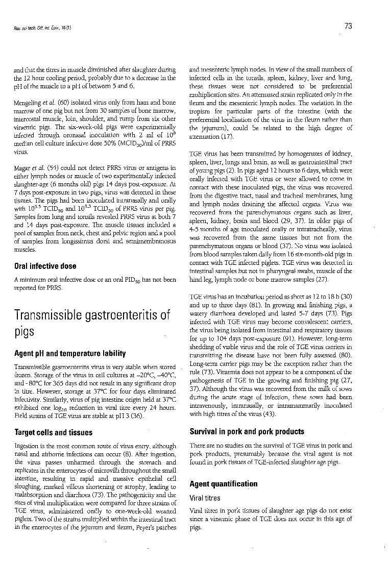
Bloemraad et al. (7) intranasally exposed four 6-month-old SPF pigs to 1 0 5 0 T C I D 5 0 of the PRRS virus. Virus was detected in the semimembranosus muscle in the two pigs killed five days post-exposure, at 2 4 hour s post m o r t e m (a titre of 1029 T C I D 5 0 in one of the pigs) bu t no t at 4 8 hours . N o virus was detected in any of three samples representing 0, 24 and 4 8 hours post m o r t e m from neck muscula ture , longissimus dorsi and bone mar row (a total of 9 samples). In the two pigs killed at 10 days post-infection, only one of the 24 samples from neck musculature , longisssimus dorsi, semimembranosus and bone mar row at 0, 24 and 4 8 hour s post m o r t e m revealed virus. The virus positive sample was obtained from the neck muscula ture at 24 hours post mor tem. Titres were obtained in certain viscera and l y m p h nodes and in se rum samples at 0, 24 and 4 8 hours post mor tem. The authors add that the sporadic recovery of virus from muscle tissue most likely originated from blood plasma
Rev. sci tech. Off. int. Epiz., 16(1) 7 3
and that the titres in muscle diminished after slaughter dur ing the 12 h o u r cooling period, probably due to a decrease in the p H of the muscle to a p H of be tween 5 and 6.
Mengeling et al. (60) isolated virus only from h a m and b o n e marrow of one pig b u t no t from 30 samples of bone marrow, intercostal muscle , loin, shoulder , a n d r u m p from six other viraemic pigs. The six-week-old pigs were experimentally infected th rough oronasal inoculation wi th 2 m l of 1 0 6
median cell cul ture infective dose 5 0 % ( M C I D 5 0 ) / m l of PRRS virus.
Magar et al. (54) could no t detect PRRS virus or antigens in either l ymph nodes or muscle of two experimentally infected slaughter-age (6 m o n t h s old) pigs 14 days post-exposure. At 7 days post-exposure in two pigs, virus was detected in these tissues. The pigs h a d been inoculated intranasally and orally with 1 0 5 ' 5 T C I D 5 0 and 1 0 5 3 T C I D 5 0 of PRRS virus per pig. Samples from lung and tonsils revealed PRRS virus at b o t h 7 and 14 days post-exposure. The muscle tissues inc luded a pool of samples from neck, chest and pelvic region and a pool of samples from longissimus dorsi and semimembranosus muscles.
Oral infective dose A m i n i m u m oral infective dose or an oral P I D 5 0 has no t been reported for PRRS.
Transmissible gastroenteritis of pigs
Agent pH and temperature lability Transmissible gastroenteritis virus is very stable w h e n stored frozen. Storage of the virus in cell cultures at - 2 0 ° C , - 4 0 ° C , and - 8 0 ° C for 3 6 5 days did no t result in any significant d rop in titre. However, storage at 37°C for four days eliminated infectivity. Similarly, virus of pig intestine origin he ld at 37°C exhibited one l o g 1 0 reduct ion in viral titre every 2 4 hours . Field strains of TGE virus are stable at p H 3 (36).
Target cells and tissues Ingestion is the mos t c o m m o n route of virus entry, a l though nasal and airborne infections can occur (8). After ingestion, the virus passes u n h a r m e d th rough the s tomach and replicates in the enterocytes of microvilli th roughout the small intestine, resulting in rapid and massive epithelial cell sloughing, marked villous shortening or atrophy, leading to malabsorption and diarrhoea (73). The pathogenicity and the sites of viral multiplication were compared for three strains of TGE virus, administered orally to one-week-old weaned piglets. Two of the strains multiplied wi th in the intestinal tract in the enterocytes of the j e j u n u m and i leum, Peyer's patches
a n d mesenteric l y m p h nodes . In view of the small n u m b e r s of infected cells in the tonsils, spleen, kidney, liver and lung, these tissues were no t considered to b e preferential multiplication sites. An at tenuated strain replicated only in the i leum and the mesenteric l y m p h nodes . The variation i n the t ropism for particular parts of the intestine (with the preferential localisation of the virus in the i leum rather than the j e junum) , could b e related to the h igh degree of at tenuation (17).
TGE virus has been t ransmit ted b y homogenates of kidney, spleen, liver, lungs and brain, as well as gastrointestinal tract of young pigs (2). In pigs aged 12 h o u r s to 6 days, wh ich were orally infected wi th TGE virus or were allowed to come in contact wi th these inoculated pigs, the virus was recovered from the digestive tract, nasal and tracheal membranes , lung a n d l y m p h nodes draining the affected organs. Virus was recovered from the parenchymatous organs such as liver, spleen, kidney, brain a n d b lood (29, 37) . In older pigs of 4-5 m o n t h s of age inoculated orally or intratracheally, virus was recovered from the same tissues b u t no t from the parenchymatous organs or b lood (37). N o virus was isolated from b lood samples taken daily from 16 s ix-month-old pigs in contact wi th TGE infected piglets. TGE virus was detected i n intestinal samples b u t no t in pharyngeal swabs, muscle of the h i n d leg, l y m p h n o d e or b o n e m a r r o w samples (27).
TGE virus has an incubat ion per iod as short as 12 to 18 h (30) and u p to three days (81). In growing and finishing pigs, a watery diarrhoea developed and lasted 5-7 days (73). Pigs infected wi th TGE virus m a y become convalescent carriers, the virus being isolated from intestinal and respiratory tissues for u p to 104 days post-exposure (91). However, long-term shedding of viable virus and the role of TGE virus carriers in transmitt ing the disease have no t b e e n fully assessed (80). Long-term carrier pigs m a y b e the exception rather than the rule (73). Viraemia does no t appear to b e a componen t of the pathogenesis of TGE in the growing and finishing pig (27, 37) . Al though the virus was recovered from the mi lk of sows dur ing the acute stage of infection, these sows h a d been intravenously, intranasally, or in t ramammari ly inoculated wi th h igh titres of the virus (43).
Survival in pork and pork products There are n o studies on the survival of TGE virus in p o r k and p o r k products , p resumably because the viral agent is no t found in p o r k tissues of TGE-infected slaughter age pigs.
Agent quantification Viral t i t res
Viral titres in p o r k tissues of slaughter age pigs do no t exist since a viraemic phase of TGE does no t occur in this age of pigs.
74 Rev. sci tech. Off. int. Epiz., 16 (1)
Oral infective dose
A m i n i m u m oral infective dose or an oral P I D 5 0 has no t been
reported for TGE, other than a significant difference in the
virus titre required to infect slaughter age pigs versus neonatal
pigs-
Conclusions The potential animal health hazards associated with the
importat ion of p o r k and p o r k products , excepting fully
cooked products , are FMD, ASF, CSF and SVD. PRRS and
TGE viruses are no t hazards of these commodit ies . The
importat ion of any p o r k and p o r k products from zones
infected wi th one of these hazards necessitates a risk
assessment to ensure the animal heal th security of the
impor t ing country. Commodi ty related inputs which are
required for the risk assessment include: the agent p H and
temperature lability, target organs, agent survival in p o r k a n d
p o r k products , and the quantification of the agent as to viral
titres in relation to a m i n i m u m infective dose.
Risques zoosanitaires potentiels liés à la viande de porc et aux produits de charcuterie
S. Farez & R.S. Morley
Résumé Les risques zoosanitaires liés à l'importation de viande de porc et de produits de charcuterie concernent quatre virus : ceux de la fièvre aphteuse, de la peste porcine classique, de la peste porcine africaine et de la maladie vésiculeuse du porc. Seule l'évaluation des risques permet de déterminer correctement l'innocuité des importations de viande de porc à partir d'une zone infectée par une ou plusieurs de ces maladies. Cela vaut également pour l'importation de produits dérivés du porc ayant subi un certain degré de transformation (abstraction faite de la charcuterie cuite). Les auteurs analysent, pour chaque maladie, la sensibilité de l'agent responsable (aux variations de pH et de température), les organes cibles, la persistance de l'agent dans la viande de porc et la charcuterie ainsi qu'une évolution quantifiée de la présence de cet agent. Cette évaluation est une donnée de l'appréciation du risque qui consiste à mesurer le titre de virus présent dans les déchets de viande de porc et de charcuterie par rapport à la dose infectieuse orale estimée pour chaque maladie. Les auteurs présentent également deux autres maladies virales du porc, la gastro-entérite transmissible et le syndrome dysgénésique et respiratoire, et expliquent pourquoi elles ne présentent pas de risque pour la viande de porc et la charcuterie.
Mots-clés Evaluation des risques - Fièvre aphteuse - Gastro-entérite transmissible du porc -Maladie vésiculeuse du porc - Peste porcine africaine - Peste porcine classique -Syndrome dysgénésique et respiratoire du porc.
Rev. sci tech. Off. int. Epiz., KW 7 5
Potenciales riesgos zoosanitarios asociados a la carne de cerdo y embutidos
S. Farez & R.S. Morley
Resumen Cuatro son los agentes víricos que representan los principales riesgos zoosanitarios asociados a la importación de carne de cerdo y embutidos, a saber, los virus de la fiebre aftosa, la peste porcina clásica, la peste porcina africana y la enfermedad vesicular porcina. La evaluación de riesgos constituye el único modo correcto de determinar el nivel de seguridad que ofrece la importación de productos porcinos procedentes de una zona afectada por una o varias de esas enfermedades. Esto también se aplica a la importación de productos porcinos previamente sometidos a alguna forma de tratamiento (excluidos los embutidos cocinados). Para cada una de las enfermedades antedichas, los autores examinan los órganos diana, la labilidad del agente en función del pH y la temperatura, su persistencia en el cerdo y embutidos y por último su cuantificación. La cuantificación del patógeno es un dato de la evaluación de riesgos que calcula la relación entre el título vírico presente en los restos de cerdos y productos porcinos y la dosis infecciosa por vía oral que se estima para cada enfermedad. Se exponen también los casos de otras dos enfermedades porcinas de origen vírico, la gastroenteritis transmisible y el síndrome disgenésico y respiratorio porcino, con objeto de ilustrar por qué estas dos enfermedades no entrañan riesgo alguno ligado a la carne de cerdo y embutidos.
Palabras clave Enfermedad vesicular porcina - Evaluación de riesgos - Fiebre aftosa - Gastroenteritis transmisible del cerdo - Peste porcina africana - Peste porcina clásica - Síndrome disgenésico y respiratorio porcino.
36. Harada K., Kaji T., Kumagai T. & Sarahara J. (1968). -
Studies on transmissible gastroenteritis in pigs. IV.
Physicochemical and biological properties of TGE vims. Natl
Inst. Anim. Hlth Q., 8 , 140-147.
37. Harada K., Furmuchi S., Kumagai T. & Sarahara J. (1969). -
Pathogenicity, immunogenicity and distribution of
transmissible gastroenteritis vims in pigs. Natl Inst Anim. Hlth
Q.,9, 185-192.
Rev. scitech. Off. int. Epiz., 16 (1) 77
38. Hedger R.S. & Mann J.A. (1989). - Swine vesicular disease. In Virus infections of porcines (M.B. Pensaert, ed.). Elsevier Science Publishers, Amsterdam, 241-250.
39. Henderson W.M. & Brooksby J.B. (1948). - The survival of foot and mouth disease vims in meat and offal. J. Hyg., 46 (4), 394-402.
40. Hemiman K.A.J., Medhurst P.M., Wilson J.N. & Sellers R.F. (1973). - The action of heat, chemicals and disinfectants on swine vesicular disease vims. Vet. Ree, 9 3 , 620-625.
41. House J.A. & House C.A. (1992). - Vesicular diseases. In Diseases of swine, 7th Ed. (A.D. Leman, B.E. Straw, W.L. Mengeling, S. D'Allaire & D.J. Taylor, eds). Iowa State University Press, Ames, Iowa, 387-401.
42. Kamolsiriprichaipom S., Hooper P.T., Morrissy C.J. & Westbury H.A. (1992). - A comparison of the pathogenicity of two strains of hog cholera vims. I. Clinical and pathological studies. Aust. vet.J., 69 (10), 240-244.
46. McDaniel H.A. (1986). - African swine fever. In Diseases of swine, 6th Ed. (A.D. Leman, B. Straw, R.D. Glock, W.L. Mengeling, R.H.C. Penny & E. Scholl, eds). Iowa State University Press, Ames, Iowa, 300-309.
47. MacDiarmid S.C. (1991). - The importation into New Zealand of meat and meat products. A review of the risks to animal health. National Agricultural Security Service Publication No. 91-2, Ministry of Agriculture and Fisheries, 180 pp.
48. McKercher P.D., Graves J.H., Callis J.J. & Carmichael F. (1974). - Swine vesicular disease: vims survival in pork products. Proc. Ann. Meet. U.S. Anim. Hlth. Assoc., 78, 213a-213g.
50. McKercher P.D., Morgan D.O., McVicar J.W. & Shuol N.J. (1980). - Thermal processing to inactivate viruses in meat products. Proc. Ann. Meet. U.S. Anim. Hlth Assoc., 84, 320-328.
51. McKercher P.D., Blackwell J.H., Murphy R., Callis J.J., Panina G.F., Civardi A., Bugnetti M., De Simone F. & Scatozza F. (1985). - Survival of swine vesicular disease vims in 'Prosciutto di Parma' (Parma ham). Can. Inst. Food Sci. Technol.J., 18(2) , 163-167.
52. McKercher P.D., Yedloutschnig R.J., Callis J.J., Murphy R., Panina G.F., Civardi A., Bugnetti M., Fonn E.H., Laddomada A., Scarano C. & Scatozza F. (1987). - Survival of viruses in
53. McVicar J.W. (1984). - Quantitative aspects of the transmission of African swine fever. Am. J. vet. Res., 45 , 1535-1541.
54. Magar R , Robinson Y., Dubuc C. & Larochelle R. (1995). -Evaluation of the persistence of porcine reproductive respiratory syndrome vims in pig carcasses. Vet. Rec., 1 3 7 , 559-561.
55. Magar R., Robinson Y., Dubuc C. & Larochelle R. (1995). -Isolation and experimental oral transmission in pigs of a porcine reproductive and respiratory syndrome vims isolate. In Corona- and related viruses: current concepts in molecular biology and pathogenesis (P.J. Talbot & G.A. Levy, eds). Plenum Press, New York, 139-144.
56. Mann J.A. & Hutchings G.H. (1980). - Swine vesicular disease: pathways of infection. J. Hyg., 84, 355-362.
57. Mann J.A. & Sellers R.F. (1989). - Foot and mouth disease vims. In Vims infections of porcines (M.B. Pensaert, ed.). Elsevier Science Publishers, Amsterdam, 251-258.
58. Mebus C.A., House C , Ruiz Gonzalvo F.R., Pineda J.M., Tapiador J., Pire J.J., Bergada J., Yedloutschnig R.J. & Sanchez-Vizcaino J.M. (1993). - Survival of foot-and-mouth disease, African swine fever, and hog cholera viruses in Spanish Serrano cured hams and Iberian cured hams, shoulders and loins. Food Microbiol, 1 0 , 133-143.
59. Mebus C.A., House C , Ruiz Gonzalvo F.R., Pineda J.M., Tapiador J., Pire J.J., Bergada J., Yedloutschnig R.J. & Sanchez-Vizcaino J.M. (1993). - Survival of swine vesicular disease vims in Spanish Serrano cured hams and Iberian cured hams, shoulders and loins. Food Microbiol, 1 0 , 263-268.
60. Mengeling W.L., Lager K.M. & Vorwald A.C. (1995). -Diagnosis of porcine reproductive and respiratory syndrome. J. vet. Diagn. Invest., 7, 3-16.
61. Meredith M. (1993). - Porcine reproductive and respiratory syndrome. Pig Information Centre, University of Cambridge, United Kingdom, 51 pp.
62. Moulton J. & Coggins L. (1968). - Comparison of lesions in acute and chronic swine fever. Cornell vet.J., 58, 364-388.
63. Office International des Epizooties (OIE) (1992). -International Animal Health Code. OIE, Paris, 550 pp.
64. Office International des Epizooties (OIE). (1994). - Hog cholera in Austria. Dis. Info., 7 (38), 161.
65. Office International des Epizooties (OIE) (1994). - Hog cholera in Bulgaria. Dis. Info., 7 (2), 7-8.
66. Office International des Epizooties (OIE) (1994). - Hog cholera in Germany. Dis. Info., 7 (9), 34.
67. Office International des Epizooties (OIE) (1994). - Hog cholera in Poland. Dis. Info., 7 (15), 59.
68. Office International des Epizooties (OIE) (1995). - Hog cholera in Austria. Dis. Info., 8 (32), 91 & 8 (34), 97.
78 Rev. sci tech. Off. int Epiz., 16(1)
69. Office International des Epizooties (OIE) (1996). -International Animal Health Code. Updates 1993-1996. OIE, Paris.
70. Ordas-Alvarez A. & Marcotegui M.A. (1987). - African swine fever - clinical aspects. In African swine fever (Y. Becker, ed.). Martinus Nijhoff Publishing, Boston, 11-21.
71 . Panina G.F., Civardi A., Massino I., Scatozza F., Baldini P. & Palmia F. (1989). - Survival of foot and mouth disease vims in sausage meat products (Italian salami). Int. J. Food Microbiol., 8 ,141-148.
72. Panina G.F., Civardi A., Cordioli P., Massirio I., Scatozza F., Baldini P. & Palmia F. (1991). - Persistence of swine fever vims in various types of Italian salami. Selez. vet, 32, 225-226.
73. Pensaert M.B. & Callebaut P. (1994). - Transmissible gastroenteritis. In Infectious diseases of livestock, Vol. II (J.A.W. Coetzer, G.R. Thomson & R.C. Tustin, eds). Oxford University Press, Cape Town, 866-869.
74. Plowright W. & Parker J. (1967). - The stability of African swine fever vims with particular reference to heat and pH inactivation. Arch. Ges. Virusforsch., 2 1 , 383-402.
75. Plowright W., Parker J. & Pierce M.A. (1969). - The epizoodology of African swine fever in Africa. Vet Rec., 85 , 668-674.
76. Plowright W., Thomson G.R. & Neser J.A. (1994). - African swine fever. In Infectious diseases of livestock, Vol. II (J.A.W. Coetzer, G.R. Thomson & R.C. Tustin, eds). Oxford University Press, Cape Town, 568-599.
77. Ressang A.A. (1973). - Studies on the pathogenesis of hog cholera. I. Demonstration of hog cholera vims subsequent to oral exposure. Zbl. VetMed., 20, 256.
78. Robertson I. (1992). - Transmission of blue eared pig disease. Vet Rec., 130 (21), 478-479.
79. Rossow K., Bautista E.M., Goyal S.M., Molitor T.W., MurtaughM.P., Morrison R.B., Benfield D.A. & Collins J.E. (1994). - Experimental porcine reproductive and respiratory syndrome vims infection in one-, four-, and 10-week-old pigs. J. vet. Diagn. Invest, 6 (1), 3-12.
80. Saif L.J. & Bohl E.H. (1986). - Transmissible gastroenteritis. In Diseases of swine, 6th Ed. (A.D. Leman, B. Straw, R.D. Glock, W.L. Mengeling, R.H.C. Penny & E. Scholl, eds). Iowa State University Press, Ames, Iowa, 255.
81 . Saif L.J. & Wesley R.D. (1992). - Transmissible gastroenteritis. In Diseases of swine, 7th Ed. (A.D. Leman, B. Straw, W.L. Mengeling, S. D'Allaire & D.J. Taylor, eds). Iowa State University Press, Ames, Iowa, 362-386.
82. Sanson R.L. (1994). - The epidemiology of foot and mouth disease: implications for New Zealand. N.Z.. vet. J., 41-53.
83. Savi P., Baldelli B. & Morozzi A. (1962). - Présence et persistance du vims aphteux dans les viandes de porcins et de bovins et dans leurs produits dérivés. Bull. Off. int. Epizoot., 57 (5-6), 853-890.
84. Savi P., Baldelli B. & Morozzi A. (1972). - Présence et persistance du vims aphteux dans les viandes de porcins et de
bovins et dans leurs produits dérivés. Bull. Off. int. Epizoot, 77 (7-8), 1125-1129.
85. Sellers R.F. (1968). - The inactivation of foot-and-mouth disease vims by chemicals and disinfectants. Vet. Rec., 83 , 595-608.
86. Sellers R.F. (1971). - Quantitative aspects of the spread of foot and mouth disease. Vet Bull., 4 1 , 431-439.
87. Stewart W.C., Downing D.R., Carbrey E.A., Kresse J.I. & Snyder B.A. (1978). - Thermal inactivation of hog cholera vims in ham. Am. J. vet. Res., 40 (5), 739-741.
88. Sybesma W. & Eikelenboom G. (1977). - Methods of predicting pale, soft, exudative pork and their application in breeding programmes - a review. Meat Sci., 2, 79-90.
89. Terpstra C. (1991). - Special review series. Hog cholera: an update of present knowledge. Br. vet.J., 147, 397-406.
90. Thomson G.R. (1994). - Swine vesicular disease. In Infectious diseases of livestock, Vol. II (J.A.W. Coetzer, G.R. Thomson & R.C. Tustin, eds). Oxford University Press, Cape Town, 817-819.
91 . Underdahl N.R., Mebus C.A. & Torres-Medina (1975). -Recovery of transmissible gastroenteritis vims from chronically infected experimental pigs. J. vet. Res., 36, 1473-1476.
92. United States Department of Agriculture (USDA) (1994). -Foot and mouth disease: sources of outbreaks and hazard categorization of modes of vims transmission. Centers for Epidemiology and Animal Health, Fort Collins, Colorado, 37 pp.
93. Van Oirschot J.T. (1988). - Description of the vims infection. In Classical swine fever and related viral infections (B. Liess, ed.). Martinus Nijhoff Publishing, Boston, Dordrecht, 1-25.
94. Van Oirschot J.T. (1992). - Hog cholera. In Diseases of swine, 7th Ed. (A.D. Leman, B.E. Straw, W.L. Mengeling, S. D'Allaire & D.J. Taylor, eds). Iowa State University Press, Ames, Iowa, 274-285.
95. Van Oirschot J.T. & Terpstra C. (1977). - A congenital persistent swine fever infection. I. Clinical and virological observations. Vet. Microbiol., 2, 121-132.
96. Van Oirschot J.T. & Terpstra C. (1989). - Hog cholera vims. In Vims infections of porcines, Vol. II (M.B. Pensaert, ed.). Elsevier Science Publishers, Amsterdam, 113-130.
97. Wilkinson P.J. (1989). - African swine fever vims. In Vims infections of porcines, Vol. II (M.B. Pensaert, ed.). Elsevier Science Publishers, Amsterdam, 17-37.
98. Williams D. & Matthews D. (1988). - Outbreaks of classical swine fever in Great Britain in 1986. Vet. Ree, 122 (20), 479-483.
99. Wood L , Brockman S., Harkness J.W. & Edwards S. (1988). - Classical swine fever: virulence and tissue distribution of a 1986 English isolate in pigs. Vet. Rec., 122, 391-394.