Background: The outbreak of a new virus known as severe acute respiratory syndrome coronavirus 2 (SARS-CoV-2)has now become the main health concern all over the world. Since effective antiviral treatments have not beendeveloped until now, SARS-CoV-2 is severely affecting countries and territories around the world.
Methods: At the present review, articles in PubMed were searched with the following terms: mesenchymal stemcells, exosomes, coronavirus, and SARS-CoV-2, either alone or in a combination form. The most relevant selectedfunctions were mesenchymal stem cell-derived exosomes and SARS-CoV-2 virus infection.
Results: SARS-CoV-2 could damage pulmonary cells and induce secretion of different types of inflammatorycytokines. In the following, these cytokines trigger inflammation that damages the lungs and results in lethal acuterespiratory distress syndrome (ARDS). The main characteristic of ARDS is the onset of inflammation in pulmonary,hyaline formation, pulmonary fibrosis, and edema. Mesenchymal stem cell-derived exosomes (MSC-Exo) arebelieved to have anti-inflammatory effects and immune-modulating capacity as well as the ability to induce tissueregeneration, suggesting a significant therapeutic opportunity that could be used to SARS-CoV-2 pneumoniatreatment. Besides, exosomes may serve as a biomarker, drug delivery system, and vaccine for the management ofthe patient with SARS-CoV-2.
Conclusion: MSC-Exo may serve as a promising tool in the treatment of SARS-CoV-2 pneumonia. However, furtherwork needs to be carried out to confirm the efficacy of exosomes in the treatment of SARS-CoV-2 pneumonia.
BackgroundThe exploration of severe acute respiratory syndromecoronavirus 2 (SARS-CoV-2) in the city of Wuhan inChina is done in December 2019. SARS-CoV-2 cantransmit human to human and spread rapidly to otherparts of China and then to other countries. The numberof individuals with SARS-CoV-2 has rapidly increased ina few weeks; as of 8 July 2020, more than 11 million
cases have been reported across 213 countries and terri-tories, resulting in more than 500,000 deaths, and morethan 4 million people have recovered [1]. The WorldHealth Organization (WHO) recognized it on January 12and named it “new novel coronavirus 2019 (2019-nCoV)”; therefore, coronavirus 2019 (2019-nCoV) andCOVID-19 virus are stated as follows: 2019-nCoV is acommon name, and SARS-CoV-2 is a classificationname for this new emerging virus. Because of theincreasing afflicted people, concerns about reserverestrictions, and emerging understanding on how to besttreat SARS-CoV-2, healthcare systems have established
* Correspondence: [email protected] Tumor Research Center, Cellular and Molecular Medicine ResearchInstitute, Urmia University of Medical Sciences, Shafa St, Ershad Blvd., P.O.Box: 1138, Urmia 57147, Iran
Akbari and Rezaie Stem Cell Research & Therapy (2020) 11:356 https://doi.org/10.1186/s13287-020-01866-6
minimal therapeutic cares for both hospital admissionand mechanical ventilation [2]. Currently, there is a sig-nificant lack of antiviral agents that could be used specif-ically for the treatment of SARS-CoV-2 infection.Although symptomatic and supportive cares are recom-mended for severely infected individuals, those with ad-vancing age and co-morbidities such as diabetes andheart diseases remain to be at high risk for adverse out-comes. Therefore, it is urgent to find a safe and effectivetherapeutic approach to patients with severe SARS-CoV-2 virus characterized by a severe acute respiratory im-pairment [3]. It was demonstrated that mesenchymalstem cells (MSCs) and their derivatives such as exo-somes (MSC-Exo) considerably improved lung inflam-mation and pathological damage resulting from differenttypes of lung injuries [4, 5]. MSC-Exo have been shownto harbor different proteins and RNAs that have thera-peutic effects such as regenerative, anti-inflammatory,pro-angiogenic, immunomodulatory, and anti-fibroticproperties on damaged tissues [6–8]. Based on thesepieces of evidence, in this review, we aimed to discussthe possible therapeutic application of exosomes inSARS-CoV-2 virus infection.
CoronavirusesCoronaviruses are enclosed, sphere-shaped, or pleo-morphic viruses, containing single-strand positive-senseRNA genome which is the longest among the RNAviruses [9]. They belong to a large family of virus whichis the leading cause of common cold and severe infec-tion such as Middle East respiratory syndrome (MERS)and severe acute respiratory syndrome (SARS) [10–12].Before the recent pandemic, six types of coronavirusspecies are documented to induce human respiratorydiseases [13]. There are four different coronavirusestypes, namely NL63, human coronaviruses 229E, HKU1,and OC43, which classically infect only the upperrespiratory system and cause fairly light symptoms [14],whereas three other coronaviruses like SARS-CoV,MERS-CoV, and more recently identified SARS-CoV-2can infect the lower respiratory system and cause pneu-monia, which can be lethal. SARS-CoV-2 is a member ofthe betacoronavirus genus. It genetically resembles,among human coronaviruses, SARS-CoV-2 with 79%similarity [15], while among wholly recognized corona-virus sequences, it has about 98% similarity to batcoronavirus RaTG13 [16] and also share high similarityto coronavirus sequences of pangolin [17].
Pathogenesis of SARS-CoV-2The affliction of coronaviruses is mediated by a trimericspike glycoprotein existing on the virion membrane.Similar to the envelope of HIV or hemagglutinin ofinfluenza species, the spike proteins of coronavirus are
one of class I fusion proteins [18]. SARS-CoV-2 entersinto cells when the S protein binds to the ACE2 receptorlocated on the host cell membrane [19] (Fig. 1). Afterdocking, the S protein conformation is changed, whichfacilitates virus entry into the endosomal pathway. Then,viral components such as RNA were uncoated inside thecell and translated into the viral components. Once thestructural proteins of SARS-CoV-2 are formed, nucleo-capsids initially are assembled in the cytoplasm and theninward bud into the lumen of the endoplasmic reticulum(ER)–Golgi transitional compartments. Then, SARS-CoV-2 structural proteins are released out of theinfected cell via exocytosis [19] (Fig. 1). SARS-CoV-2spreads mostly via respiratory droplets and via, likely,but unconfirmed, the orofecal route. During infection,the average latency is around 4–5 days earlier before thesymptom begins [20–22]; however, 97.5% of the symp-toms emerge within 11.5 days [22]. The results reportedfrom hospitals indicate patients with SARS-CoV-2 basic-ally show fever, dry cough, muscle and/or joint pain,difficulty in breathing, diarrhea, nausea, headache, andhemoptysis [23–26]. Between days 5 and 6 after theonset of symptoms, SARS-CoV-2 viral load rises to itspeak—expressively earlier than that of the SARS-CoVone—so around 10 days later, the viral load peak reachesmaximum [27–29]. Within 8–9 days after symptom on-set, severe COVID-19 patients develop acute respiratorydistress syndrome (ARDS) with aggressive inflammatoryresponses, hyaline formation, and pulmonary fibrosis[24, 30]. So, disease severity is mostly caused by the im-mune responses of the host immune system. In addition,the relation between growing severity and age is mostlyalike to that of SARS-CoV and MERS-CoV [24, 26].ARDS caused by SARS-CoV-2 is defined by the strugglein breathing, insufficient blood oxygen level, and alsofailure in the respiratory system that is responsible fordeath in 70% of lethal SARS-CoV-2 patients [31]. Someindividuals may capitulate to secondary fungal and bac-terial infections [26]. Furthermore, inflammatory cyto-kine storm caused by the immune system causes death(28%) followed by viral infection and/or secondary infec-tions [31]. The majority of patients show mild symp-toms; however, some patients develop ARDS likelytriggered by a cytokine storm, increased inflammation,septic shock, failure in organs, and blood clots [32, 33].Interaction between SARS-CoV-2 infection and immunecells leads to dysregulated immune responses and in-creased risk of multi-organ dysfunction. In SARS-CoV-2-infected individuals, different pro-inflammatory cyto-kines and other mediators like IL-2, IL-10, IL-7, IL-6,IL-1β, IFNγ, INF-ɑ, monocyte chemoattractant protein-1 (MCP-1), and induced protein 10 (IP10) significantlyincreased, causing failure in multi-organs [34–36](Fig. 2). Increased level of cytokines and chemokines
Akbari and Rezaie Stem Cell Research & Therapy (2020) 11:356 Page 2 of 10
attracts several immune cells, especially T lymphocytesand monocytes, but not neutrophils, from the circulatorysystem into the infected tissue [37, 38]. The entry of im-mune cells into the lung tissue and the influx of lympho-cytes into the airways may correlate with the highneutrophil-to-lymphocyte ratio, increased T helper-to-Tregulatory cell ratio, and lymphopenia, which are presentin approximately 80% of individuals [20, 39]. Further-more, SARS-CoV-2 virus, which is a cytopathic virus,can induce injury and death in cells and tissues as a con-sequence of the viral life cycle [40]. Viral infection andreplication in epithelial cells of the respiratory systemmay induce pyroptosis associated with vascular leak, asconfirmed in people infected with SARS-CoV [41]. Pyr-optosis, a caspase-1-based cell death, is an inflammatorytype of programmed cell death that could be promptedby cytopathic viruses [42] and activates inflammatory re-sponse [43]. For example, IL-1β as a pyroptosis secretionis increased during SARS-CoV-2 infection [24]. Collect-ively, in addition to the lesion on pulmonary cells, the
cytokine storm triggers inflammatory responses; thus, anincreasing effort is focused on the regeneration ofdamaged cells as well as on the blocking or modulatingof inflammatory responses.
Extracellular vesiclesExtracellular vesicles (EVs) are heterogeneously bi-phospholipid encapsulated vesicles released from almostall eukaryotic cells, which mediate intercellular commu-nication via transferring different biomaterial compo-nents like proteins, different kinds of nucleic acids,lipids, and carbohydrates between cells [44, 45]. Guide-lines released by the International Society for Extracellu-lar Vesicles (ISEV, https://www.isev.org) categorize EVsinto three subtypes such as exosomes, microvesicles orshedding vesicles, and apoptotic bodies based on theirorigin and size. In recent years, scientists have a greatattention to exosomes due to their pivotal roles inbiological systems. Exosomes, 30–120 nm of EVs in size,are originating from endosome compartments (late
Fig. 1 The SARS-CoV-2 entry and life cycle in human lung cells. The S proteins of SARS-CoV-2 bind to the angiotensin-converting enzyme 2(ACE2) located on the cellular membrane and enters the target cells through an endosomal pathway (1). Then, viral components such as RNA areuncoated in the cytoplasm (2). In keeping, the genome encodes RNA-dependent RNA polymerase (RdRp) (3), which, in turn, participates toproducing full-length (+) RNA and full length (−) RNA copies (4). During replication, genome RNA and RNAs which belong to viral structuralcomponents are produced. Then, RNAs are translated into viral components and processed by the endoplasmic reticulum (ER)–Golgi apparatus(GA) (5). Once SARS-CoV-2 structural proteins are formed, nucleocapsids initially are assembled in the cytoplasm, and then they are budding intothe lumen of the ER and GA (6). Finally, mature viruses are secreted out of the infected cell via exocytosis (7). EE, early endosome
Akbari and Rezaie Stem Cell Research & Therapy (2020) 11:356 Page 3 of 10
endosomes) located inside the cytoplasm via a complexregularity system [46]. Once secreted, exosomes distrib-ute into the extracellular matrix and bio-fluids and reachthe neighboring and distantly located target cells [45].Exosomes bear different molecules both on their surfaceand in the lumen. The density of sucrose (1.12–1.18 g/mL) is equivalent to that of exosomes, and they appearas cup-shaped vesicles under an electron microscopy[47]. As exosomes contain active biomolecules fromparent cells, they can regulate function, fate, and shapesof target cells, participating in different pathological andphysiological conditions [45, 48].
Role of MSC-derived exosomes in SARS-CoV-2treatmentMSCs, multipotent and self-renewal cells, are known asadult stem cells that can be obtained from different tis-sues such as the adipose tissue, bone marrow, umbilicalcord tissue, and amniotic fluid. These cells are capableof differentiation into a variety of cells such as cartilage,bone cells, neural cells, muscle cells, and skin cells [49].MSCs are widely considered to be the most useful toolin regenerative medicine because these cells can improvedamaged tissues and organs via differentiation into dif-ferent cells and also producing many types of chemo-kines, cytokines, growth factors, and EVs [50, 51]. MSCs
may improve damaged lung tissue through homing to-ward specific injuries of the lung to maintain homeosta-sis as well as promote regeneration and secretion ofsoluble factors and exosomes to moderate inflammationand induce tissue regeneration [52, 53]. More recently,Leng et al. found that the transplantation of MSCs con-siderably improved the pulmonary function of patientswith 2019-nCoV pneumonia over a period of 2 days[54]. They administered about one million cells per kilo-gram of weight once and declared that MSCs modulatedimmune responses, participating in the improvement ofthe outcome of SARS-CoV-2 pneumonia. Apart fromtransdifferentiation into tissue cells, the other mecha-nisms may involve in repairing tissue [55]; alternatively,paracrine effects (such as exosomes) of stem cells arethe base of the beneficial effect of cell therapies [56].According to the literatures, MSC-Exo contain differ-
ent active biomolecules and can repair and improvemyocardial infarcts, kidney injury, CNS diseases, livercirrhosis, diabetic wound, and lung-associated diseases[57, 58]. The detailed mechanisms involved in exosome-mediated favorable effects and their site of action remainyet unclear. However, reported results from several pre-clinical non-SARS-CoV-2 models propose that MSC-Exo could also have efficiency against SARS-CoV-2 virusinfection. For instance, through the systemic
Fig. 2 Pathogenesis of SARS-CoV-2 virus. During SARS-CoV-2 infection, immune cells produce several pro-inflammatory cytokines. Inflammatorycytokines trigger lethal acute respiratory distress syndrome (ARDS) and failure in multi-organs
Akbari and Rezaie Stem Cell Research & Therapy (2020) 11:356 Page 4 of 10
administration, MSC-Exo can modulate immune re-sponses like increased cytokine storms in related acutelung injury and sepsis models [59–64]. Khatri and co-workers found that the administration of MSC-Exoattenuated influenza virus-induced acute lung injurythrough suppressing influenza virus replication in a pigmodel [65]. Similarly, a study conducted by Li andcolleagues showed that MSC treatment significantlyprevents avian H9N2-induced acute lung injury in micevia decreasing the level of chemokines and pro-inflammatory cytokines as well as reducing the infiltra-tion of inflammatory cells onto the lungs [66]. Inaddition, in Escherichia coli-induced pneumonia mousemodels, MSC-Exo had the ability to increase phagocyt-osis of bacteria [61, 67]. It seems that the hallmark ofthe treatment of SARS-CoV-2 severe pneumonia is tosuppress the pro-inflammatory immune responseinduced by the virus, thus reducing the damage of bothalveolar epithelial cells and capillary endothelial cells,followed by the restoring function of lung cells and alsothe recovery of the lung tissue which may be mediatedby MSC-Exo (Fig. 3). Overall, as shown in Fig. 3, thepossible therapeutic roles of MSC-Exo in SARS-CoV-2virus infection may comprise inhibiting pro-inflammatory cytokines, inducing M2 macrophagesthrough delivering PEG2, increasing secretion of anti-inflammatory cytokines like IL-10, and regeneratingdamaged tissue by producing KGF, VEGF, and HGF [6,62, 68–71].
By June 2020, the public clinical trial database (https://clinicaltrials.gov) prepared three studies (NCT04276987,NCT0438938, and NCT04384445), and the ChineseClinical Trial Registry site (ChiCTR, http://www.chictr.org.cn) records two clinical trials (ChiCTR2000030484and ChiCTR2000030261) aiming to study the inhalationof aerosol and intravenous administration of MSC-Exoin the treatment of severe individuals with SARS-CoV-2pneumonia. However, for SARS-CoV-2 pneumonia,further studies, which take MSC-Exo into account, willneed to be undertaken, because various immune cells re-spond differently against MSC-Exo and these exosomesmay modulate immune responses. For instance, Di Tra-pani et al. found that MSC-Exo strongly inhibited thefunction of B cells compared with the function of NKand T cells [72]. Therefore, the immunological activityof MSC-Exo toward different immune cells is dependenton the type of target cell, the cellular maturity, cellularstatus, and type of diseases among other factors [73, 74].However, different studies declared that MSC-Exo caninhibit immune cell production and support an immu-notolerant microenvironment.
Other potential therapeutic applications ofexosomes in 2019-nCoV treatmentThis section provides some possible application ofexosomes in SARS-CoV-2 virus infection treatment;however, there are some limitations regarding theirapplication, which are discussed in the later section. In
Fig. 3 Possible mechanisms, by which mesenchymal stem cell-derived exosomes may improve adverse effects of SARS-CoV-2 virus infection.These exosomes contain different molecules that can suppress inflammatory responses and repair damage tissue. MSC, mesenchymal stem cell;N, neutrophil; M1, macrophage type I; M2, macrophage type II; T, T lymphocyte
Akbari and Rezaie Stem Cell Research & Therapy (2020) 11:356 Page 5 of 10
addition to exosome therapy, exosomes can serve as adrug delivery system for the treatment of SARS-CoV-2virus pneumonia (Fig. 3). Exosomes exhibit superiorityto other nanocarriers as they are bi-phospholipid vesi-cles, are of cell origin, safe, and have low immunogen-icity and they can pass through the physiological barriers[75]. In this regard, different strategies can be used toconstruct optional exosomes such as (I) direct engineer-ing method and (II) indirect engineering method [76].By direct engineering methods, therapeutic agents
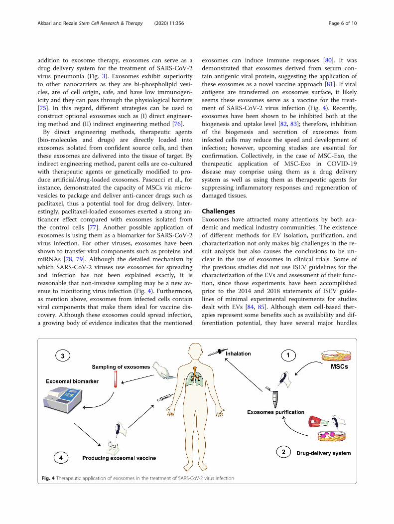
(bio-molecules and drugs) are directly loaded intoexosomes isolated from confident source cells, and thenthese exosomes are delivered into the tissue of target. Byindirect engineering method, parent cells are co-culturedwith therapeutic agents or genetically modified to pro-duce artificial/drug-loaded exosomes. Pascucci et al., forinstance, demonstrated the capacity of MSCs via micro-vesicles to package and deliver anti-cancer drugs such aspaclitaxel, thus a potential tool for drug delivery. Inter-estingly, paclitaxel-loaded exosomes exerted a strong an-ticancer effect compared with exosomes isolated fromthe control cells [77]. Another possible application ofexosomes is using them as a biomarker for SARS-CoV-2virus infection. For other viruses, exosomes have beenshown to transfer viral components such as proteins andmiRNAs [78, 79]. Although the detailed mechanism bywhich SARS-CoV-2 viruses use exosomes for spreadingand infection has not been explained exactly, it isreasonable that non-invasive sampling may be a new av-enue to monitoring virus infection (Fig. 4). Furthermore,as mention above, exosomes from infected cells containviral components that make them ideal for vaccine dis-covery. Although these exosomes could spread infection,a growing body of evidence indicates that the mentioned
exosomes can induce immune responses [80]. It wasdemonstrated that exosomes derived from serum con-tain antigenic viral protein, suggesting the application ofthese exosomes as a novel vaccine approach [81]. If viralantigens are transferred on exosomes surface, it likelyseems these exosomes serve as a vaccine for the treat-ment of SARS-CoV-2 virus infection (Fig. 4). Recently,exosomes have been shown to be inhibited both at thebiogenesis and uptake level [82, 83]; therefore, inhibitionof the biogenesis and secretion of exosomes frominfected cells may reduce the speed and development ofinfection; however, upcoming studies are essential forconfirmation. Collectively, in the case of MSC-Exo, thetherapeutic application of MSC-Exo in COVID-19disease may comprise using them as a drug deliverysystem as well as using them as therapeutic agents forsuppressing inflammatory responses and regeneration ofdamaged tissues.
ChallengesExosomes have attracted many attentions by both aca-demic and medical industry communities. The existenceof different methods for EV isolation, purification, andcharacterization not only makes big challenges in the re-sult analysis but also causes the conclusions to be un-clear in the use of exosomes in clinical trials. Some ofthe previous studies did not use ISEV guidelines for thecharacterization of the EVs and assessment of their func-tion, since those experiments have been accomplishedprior to the 2014 and 2018 statements of ISEV guide-lines of minimal experimental requirements for studiesdealt with EVs [84, 85]. Although stem cell-based ther-apies represent some benefits such as availability and dif-ferentiation potential, they have several major hurdles
Fig. 4 Therapeutic application of exosomes in the treatment of SARS-CoV-2 virus infection
Akbari and Rezaie Stem Cell Research & Therapy (2020) 11:356 Page 6 of 10
for therapy [86], which may include (i) determining andtargeting a reliable source of cells with phenotypic stabil-ity, (ii) the immunological incompatibility that mayresult in rejection, (iii) difficulty of handling and thehigher cost of expansion, and (iv) the potential of cancerrisks or formation of ectopic tissue. Exosome therapy ex-hibits several advantages against cell therapy such as thefollowing: exosomes have a small size, which allowsthem to pass through filter organs including the liver,spleen, and lungs. Furthermore, as exosomes are bi-phospholipid vesicles, their membrane-binding propertyimparts excellent biocompatibility and biostability to theloaded cargos and optional drugs, and also, exosomescan pass through biological barriers like the brain-bloodbarrier. As a hopeful therapeutic tool for novel cell-freetherapy, exosomes may serve as an alternative to stemcells in the treatment of various diseases for mainten-ance of the microenvironment for organ homeostasisand tissue regeneration upon injury [87, 88].There are great developments in clinical exosome-
based studies (https://clinicaltrials.gov); however, untilnow, no Food and Drug Administration (FDA)-approvedexosome products are available (https://www.fda.gov/).In the case of using MSC-Exo, from a clinical applica-
tion point of view, a number of concerns should be con-sidered before administering MSC-Exo to SARS-CoV-2-infected people. These comprise the source of MSC-Exo.MSCs, the heterogeneous cells, can be obtained fromvarious tissues, even though MSCs isolated from thesame sources may vary in entity and functionality [89,90]. For example, several donor-derived bone marrowMSCs release exosomes with distinct cytokine profilecontent [91]. Besides, young MSC-Exo as compared withaged MSC-Exo improved LPS-induced acute lung injuryin a mouse model [92]. In addition to immunomodula-tory properties, MSC-Exo actually also regulate otherbiological processes, some with therapeutic functions[93], and others that may induce unexpected non-targeting effects. In order to inhibit the exosome biogen-esis and secretion, future attempts should focus on silen-cing or eliminating exosomes that selectively encouragediseases which are not useful exosomes. Some researchgroups tried to use exosome inhibitors as versatile toolsfor exosome biology investigation; however, others haveevaluated the inhibitory potential of such drugs in vari-ous disease models [82, 94]. Most of the experimentswere carried out preclinical; therefore, clinical trialsmust be done to confirm their validity. However, thenon-targeting effects of exosome inhibitors could beconsidered as the main issue on exosome biogenesis ofhealthy cells.Another fascinating approach that exosomes can be
used as a therapeutic agent is the drug delivery potentialof them [95–97]. Exosomes from a safe source such as
MSCs may be loaded with optional compounds or gen-etically engineered for targeting infected tissues/cells,suggesting the exosome-based nanocarriers for the treat-ment of infectious diseases. The advent of safe nanocar-riers with high efficiency is the core goal ofnanomedicine. However, most of the studies aredesigned in vitro and animal models; therefore, from aclinical trial application point of view, there are stillmore questions in the specificity, safety, and proficiencyof this method. Moreover, despite various available de-livery strategies for therapeutic anti-viral vaccines, thera-peutic anti-viral vaccines in humans have beenchallenging to implement [98]. In another word, thesevaccines have not been a strong success so far, and it iscertainly valuable to consider more effective adjuvantmethods for refining vaccine efficiency. Furthermore, an-other important thing is the source of immunostimula-tory exosomes for human antiviral vaccines which needsmore considerations [81]. In a nutshell, researches andexplorations in the field of exosomes are in their earlystages; therefore, to implement many of the aforemen-tioned ideas, the biology of EVs (especially exosomes)from infected cells must be explored in-depth.
ConclusionAlthough the exploration of novel specific vaccine forthe treatment of SARS-CoV-2 virus pneumonia iscritical, it seems to take a long time. Therefore, furtherresearch into preventing and controlling the virus isessential as soon as possible. Exosomes from MSCs maybe a useful tool for the treatment of SARS-CoV-2 viruspneumonia due to their pivotal roles in the suppressionof inflammatory responses and the regeneration ofdamaged tissues. Furthermore, exosomes may serve as abiomarker, nanocarrier, and vaccine for the treatment ofSARS-CoV-2 virus. However, for a better outcome,upcoming efforts are vital to carefully weigh the risk inthe use of MSC-Exo for SARS-CoV-2 virus pneumoniaagainst available pre-clinical findings in applicablein vivo models.
AbbreviationsARDS: Acute respiratory distress syndrome; EVs: Extracellular vesicle;FDA: Food and Drug Administration; IP10: Induced protein 10; MCP-1: Monocyte chemoattractant protein-1; MERS: Middle East respiratorysyndrome; MSC-Exo: Mesenchymal stem cell-derived exosomes; SARS-CoV-2: Severe acute respiratory syndrome coronavirus 2; WHO: World HealthOrganization
AcknowledgementsNot applicable.
Authors’ contributionsJ.R. and A.A. collected the data and edited the manuscript and with equalconceptualization. All authors read and approved the final manuscript.
FundingNot applicable.
Akbari and Rezaie Stem Cell Research & Therapy (2020) 11:356 Page 7 of 10
Availability of data and materialsThe datasets used and/or analyzed during the current study are availablefrom the corresponding author on reasonable request.
Ethics approval and consent to participateNot applicable.
Consent for publicationNot applicable.
Competing interestsThe authors have no competing interests to declare.
Received: 17 June 2020 Revised: 9 July 2020Accepted: 30 July 2020
References1. WHO Coronavirus Disease (COVID-19) Dashboard. https://covid19.who.int/.
Accessed 8 July 2020.2. Gattinoni L, Coppola S, Cressoni M, Busana M, Rossi S, Chiumello D. Covid-
19 does not lead to a “typical” acute respiratory distress syndrome. Am JRespir Crit Care Med. 2020;201:1299–300.
3. HuiDS IAE, Madani T, Ntoumi F, Koch R, Dar O. The continuing 2019-nCoVepidemic threat of novel coronaviruses to global health: the latest 2019novel coronavirus outbreak in Wuhan, China. Int J Infect Dis. 2020;91:264–6.
4. Abraham A, Krasnodembskaya A. Mesenchymal stem cell-derivedextracellular vesicles for the treatment of acute respiratory distresssyndrome. Stem Cells Transl Med. 2020;9:28–38.
5. Shah TG, Predescu D, Predescu S. Mesenchymal stem cells-derivedextracellular vesicles in acute respiratory distress syndrome: a review ofcurrent literature and potential future treatment options. Clin Transl Med.2019;8:25.
6. Jabbari N, Nawaz M, Rezaie J. Ionizing radiation increases the activity ofexosomal secretory pathway in MCF-7 human breast cancer cells: a possibleway to communicate resistance against radiotherapy. Int J Mol Sci. 2019;20:3649.
7. Martin-Rufino JD, Espinosa-Lara N, Osugui L, Sanchez-Guijo F. Targeting theimmune system with mesenchymal stromal cell-derived extracellularvesicles: what is the cargo’s mechanism of action? Front Bioeng Biotechnol.2019;7:308.
8. Joo HS, Suh JH, Lee HJ, Bang ES, Lee JM. Current knowledge and futureperspectives on mesenchymal stem cell-derived exosomes as a newtherapeutic agent. Int J Mol Sci. 2020;21:727.
9. Belouzard S, Millet JK, Licitra BN, Whittaker GR. Mechanisms of coronaviruscell entry mediated by the viral spike protein. Viruses. 2012;4:1011–33.
10. Kuiken T, Fouchier RA, Schutten M, Rimmelzwaan GF, Van Amerongen G,van Riel D, Laman JD, de Jong T, van Doornum G, Lim W. Newly discoveredcoronavirus as the primary cause of severe acute respiratory syndrome.Lancet. 2003;362:263–70.
11. Drosten C, Günther S, Preiser W, Van Der Werf S, Brodt H-R, Becker S,Rabenau H, Panning M, Kolesnikova L, Fouchier RA. Identification of a novelcoronavirus in patients with severe acute respiratory syndrome. N Engl JMed. 2003;348:1967–76.
12. de Groot RJ, Baker SC, Baric RS, Brown CS, Drosten C, Enjuanes L, FouchierRA, Galiano M, Gorbalenya AE, Memish ZA. Commentary: Middle Eastrespiratory syndrome coronavirus (MERS-CoV): announcement of theCoronavirus Study Group. J Virol. 2013;87:7790–2.
13. Su S, Wong G, Shi W, Liu J, Lai AC, Zhou J, Liu W, Bi Y, Gao GF.Epidemiology, genetic recombination, and pathogenesis of coronaviruses.Trends Microbiol. 2016;24:490–502.
14. Fehr AR, Perlman S: Coronaviruses: an overview of their replication andpathogenesis. New York: In Coronaviruses Springer; 2015. p. 1–23.
15. of the International CSG. The species severe acute respiratory syndrome-related coronavirus: classifying 2019-nCoV and naming it SARS-CoV-2. NatMicrobiol. 2020;5:536.
16. Zhou P, Yang X-L, Wang X-G, Hu B, Zhang L, Zhang W, Si H-R, Zhu Y, Li B,Huang C-L. A pneumonia outbreak associated with a new coronavirus ofprobable bat origin. Nature. 2020;579:270–3.
17. Andersen KG, Rambaut A, Lipkin WI, Holmes EC, Garry RF. The proximalorigin of SARS-CoV-2. Nat Med. 2020;26:450–2.
18. Whittaker GR, Millet JK: Biochemical characterization of Middle Eastrespiratory syndrome coronavirus spike protein proteolytic processing. NewYork: In MERS Coronavirus Springer; 2020. p. 21–37.
19. La A, Alzoughool F, Atoum M. The human coronavirus disease COVID-19: itsorigin, characteristics, and insights into potential drugs and its mechanisms.Pathogens. 2020;9:331.
20. W-J G, Z-Y N, Hu Y, Liang W-H, Ou C-Q, He J-X, Liu L, Shan H, Lei C-L, HuiDS. Clinical characteristics of coronavirus disease 2019 in China. N Engl JMed. 2020;382:1708–20.
21. Pung R, Chiew CJ, Young BE, Chin S, Chen MI, Clapham HE, Cook AR,Maurer-Stroh S, Toh MP, Poh C. Investigation of three clusters of COVID-19in Singapore: implications for surveillance and response measures. Lancet.2020;395:1039–46.
22. Lauer SA, Grantz KH, Bi Q, Jones FK, Zheng Q, Meredith HR, Azman AS,Reich NG, Lessler J. The incubation period of coronavirus disease 2019(COVID-19) from publicly reported confirmed cases: estimation andapplication. Ann Intern Med. 2020;172:577–82.
23. Chan JF-W, Yuan S, Kok K-H, To KK-W, Chu H, Yang J, Xing F, Liu J, Yip CC-Y,Poon RW-S. A familial cluster of pneumonia associated with the 2019 novelcoronavirus indicating person-to-person transmission: a study of a familycluster. Lancet. 2020;395:514–23.
24. Huang C, Wang Y, Li X, Ren L, Zhao J, Hu Y, Zhang L, Fan G, Xu J, Gu X.Clinical features of patients infected with 2019 novel coronavirus in Wuhan,China. Lancet. 2020;395:497–506.
25. Liu Y, Yang Y, Zhang C, Huang F, Wang F, Yuan J, Wang Z, Li J, Li J, Feng C.Clinical and biochemical indexes from 2019-nCoV infected patients linkedto viral loads and lung injury. Sci China Life Sci. 2020;63:364–74.
26. Chen N, Zhou M, Dong X, Qu J, Gong F, Han Y, Qiu Y, Wang J, Liu Y, Wei Y.Epidemiological and clinical characteristics of 99 cases of 2019 novel coronaviruspneumonia in Wuhan, China: a descriptive study. Lancet. 2020;395:507–13.
27. Pan Y, Zhang D, Yang P, Poon LL, Wang Q. Viral load of SARS-CoV-2 inclinical samples. Lancet Infect Dis. 2020;20:411–2.
28. Kim JY, Ko J-H, Kim Y, Kim Y-J, Kim J-M, Chung Y-S, Kim HM, Han M-G, KimSY, Chin BS. Viral load kinetics of SARS-CoV-2 infection in first two patientsin Korea. J Korean Med Sci. 2019;35:e86.
29. Zou L, Ruan F, Huang M, Liang L, Huang H, Hong Z, Yu J, Kang M, Song Y,Xia J. SARS-CoV-2 viral load in upper respiratory specimens of infectedpatients. N Engl J Med. 2020;382:1177–9.
30. Wang D, Hu B, Hu C, Zhu F, Liu X, Zhang J, Wang B, Xiang H, Cheng Z,Xiong Y. Clinical characteristics of 138 hospitalized patients with 2019 novelcoronavirus–infected pneumonia in Wuhan, China. JAMA. 2020;323:1061–9.
31. Zhang B, Zhou X, Qiu Y, Feng F, Feng J, Jia Y, Zhu H, Hu K, Liu J, Liu Z. Clinicalcharacteristics of 82 death cases with COVID-19. MedRxiv. 2020;15:e0235458.
32. Adhikari SP, Meng S, Wu Y-J, Mao Y-P, Ye R-X, Wang Q-Z, Sun C, Sylvia S,Rozelle S, Raat H. Epidemiology, causes, clinical manifestation and diagnosis,prevention and control of coronavirus disease (COVID-19) during the earlyoutbreak period: a scoping review. Infect Dis Poverty. 2020;9:1–12.
33. Murthy S, Gomersall CD, Fowler RA. Care for critically ill patients withCOVID-19. JAMA. 2020;323:1499–500.
34. Chen G, Wu D, Guo W, Cao Y, Huang D, Wang H, Wang T, Zhang X, ChenH, Yu H. Clinical and immunological features of severe and moderatecoronavirus disease 2019. J Clin Invest. 2020;130:2620–9.
35. Diao B, Wang C, Tan Y, Chen X, Liu Y, Ning L, Chen L, Li M, Liu Y, Wang G.Reduction and functional exhaustion of T cells in patients with coronavirusdisease 2019 (COVID-19). Front Immunol. 2020;11:827.
36. Yang Y, Shen C, Li J, Yuan J, Wei J, Huang F, Wang F, Li G, Li Y, Xing L, PengL. Plasma IP-10 and MCP-3 levels are highly associated with disease severityand predict the progression of COVID-19. J Allergy Clin Immunol 2020.https://doi.org/10.1016/j.jaci.2020.04.027.
37. Tian S, Hu W, Niu L, Liu H, Xu H, Xiao S-Y. Pulmonary pathology of earlyphase 2019 novel coronavirus (COVID-19) pneumonia in two patients withlung cancer. J Thorac Oncol. 2020;15:700–4.
38. Xu Z, Shi L, Wang Y, Zhang J, Huang L, Zhang C, Liu S, Zhao P, Liu H, Zhu L.Pathological findings of COVID-19 associated with acute respiratory distresssyndrome. Lancet Respir Med. 2020;8:420–2.
39. Qin C, Zhou L, Hu Z, Zhang S, Yang S, Tao Y, Xie C, Ma K, Shang K, Wang W:Dysregulation of immune response in patients with COVID-19 in Wuhan,China. Clin Infect Dis. 2020;71:762–8.
40. Park WB, Kwon N-J, Choi S-J, Kang CK, Choe PG, Kim JY, Yun J, Lee G-W,Seong M-W, Kim NJ. Virus isolation from the first patient with SARS-CoV-2 inKorea. J Korean Med Sci. 2019;35:e84.
Akbari and Rezaie Stem Cell Research & Therapy (2020) 11:356 Page 8 of 10
41. Zhang H, Zhou P, Wei Y, Yue H, Wang Y, Hu M, Zhang S, Cao T, Yang C, LiM. Histopathologic changes and SARS-CoV-2 immunostaining in the lung ofa patient with COVID-19. Ann Intern Med. 2020;172:629–32.
42. Fink SL, Cookson BT. Apoptosis, pyroptosis, and necrosis: mechanisticdescription of dead and dying eukaryotic cells. Infect Immun. 2005;73:1907–16.
43. Yang M. Cell pyroptosis, a potential pathogenic mechanism of 2019-nCoVinfection. Available at SSRN 3527420; 2020.
44. Rezaie J, Rahbarghazi R, Pezeshki M, Mazhar M, Yekani F, Khaksar M, ShokrollahiE, Amini H, Hashemzadeh S, Sokullu SE, Tokac M. Cardioprotective role ofextracellular vesicles: a highlight on exosome beneficial effects incardiovascular diseases. J Cell Physiol. 2019;234:21732–45.
45. Kowal J, Tkach M, Théry C. Biogenesis and secretion of exosomes. Curr OpinCell Biol. 2014;29:116–25.
46. Jabbari N, Karimipour M, Khaksar M, Akbariazar E, Heidarzadeh M, Mojarad B,Aftab H, Rahbarghazi R, Rezaie J. Tumor-derived extracellular vesicles:insights into bystander effects of exosomes after irradiation. Lasers Med Sci.2019;35:1–15.
47. Kesimer M, Scull M, Brighton B, DeMaria G, Burns K, O’Neal W, Pickles RJ,Sheehan JK. Characterization of exosome-like vesicles released from humantracheobronchial ciliated epithelium: a possible role in innate defense.FASEB J. 2009;23:1858–68.
48. Hannafon BN, Ding W-Q. Intercellular communication by exosome-derivedmicroRNAs in cancer. Int J Mol Sci. 2013;14:14240–69.
49. Han Y, Li X, Zhang Y, Han Y, Chang F, Ding J. Mesenchymal stem cells forregenerative medicine. Cells. 2019;8:886.
50. Zhao T, Sun F, Liu J, Ding T, She J, Mao F, Xu W, Qian H, Yan Y. Emergingrole of mesenchymal stem cell-derived exosomes in regenerative medicine.Curr Stem Cell Res Ther. 2019;14:482–94.
51. Satija NK, Singh VK, Verma YK, Gupta P, Sharma S, Afrin F, Sharma M,Sharma P, Tripathi R, Gurudutta G. Mesenchymal stem cell-basedtherapy: a new paradigm in regenerative medicine. J Cell Mol Med.2009;13:4385–402.
52. Ullah M, Liu DD, Thakor AS. Mesenchymal stromal cell homing: mechanismsand strategies for improvement. iScience. 2019;15:421.
53. Mohammadipoor A, Antebi B, Batchinsky AI, Cancio LC. Therapeuticpotential of products derived from mesenchymal stem/stromal cells inpulmonary disease. Respir Res. 2018;19:218.
54. Leng Z, Zhu R, Hou W, Feng Y, Yang Y, Han Q, Shan G, Meng F, Du D,Wang S. Transplantation of ACE2-mesenchymal stem cells improves theoutcome of patients with COVID-19 pneumonia. Aging Dis. 2020;11:216.
55. Rogers TB, Pati S, Gaa S, Riley D, Khakoo AY, Patel S, Wardlow RD II,Frederick CA, Hall G, He L-P. Mesenchymal stem cells stimulate protectivegenetic reprogramming of injured cardiac ventricular myocytes. J Mol CellCardiol. 2011;50:346–56.
56. Uemura R, Xu M, Ahmad N, Ashraf M. Bone marrow stem cells prevent leftventricular remodeling of ischemic heart through paracrine signaling. CircRes. 2006;98:1414–21.
57. Keshtkar S, Azarpira N, Ghahremani MH. Mesenchymal stem cell-derivedextracellular vesicles: novel frontiers in regenerative medicine. Stem Cell ResTher. 2018;9:63.
58. Baglio SR, Pegtel DM, Baldini N. Mesenchymal stem cell secreted vesicles providenovel opportunities in (stem) cell-free therapy. Front Physiol. 2012;3:359.
59. Khaksar M, Sayyari M, Rezaie J, Pouyafar A, Montazersaheb S, Rahbarghazi R.High glucose condition limited the angiogenic/cardiogenic capacity ofmurine cardiac progenitor cells in in vitro and in vivo milieu. Cell BiochemFunct. 2018;36:346–56.
60. Mansouri N, Willis GR, Fernandez-Gonzalez A, Reis M, Nassiri S, Mitsialis SA,Kourembanas S. Mesenchymal stromal cell exosomes prevent and revertexperimental pulmonary fibrosis through modulation of monocytephenotypes. JCI insight. 2019;4:e128060.
61. Monsel A, Zhu Y-G, Gennai S, Hao Q, Hu S, Rouby J-J, Rosenzwajg M,Matthay MA, Lee JW. Therapeutic effects of human mesenchymal stemcell–derived microvesicles in severe pneumonia in mice. Am J Respir CritCare Med. 2015;192:324–36.
62. Park K-S, Svennerholm K, Shelke GV, Bandeira E, Lässer C, Jang SC,Chandode R, Gribonika I, Lötvall J. Mesenchymal stromal cell-derivednanovesicles ameliorate bacterial outer membrane vesicle-induced sepsisvia IL-10. Stem Cell Res Ther. 2019;10:231.
63. Varkouhi AK, Jerkic M, Ormesher L, Gagnon S, Goyal S, Rabani R, MastersonC, Spring C, Chen PZ, Gu FX. Extracellular vesicles from interferon-γ–primed
human umbilical cord mesenchymal stromal cells reduce Escherichia coli–induced acute lung injury in rats. Anesthesiology. 2019;130:778–90.
64. Willis GR, Fernandez-Gonzalez A, Anastas J, Vitali SH, Liu X, Ericsson M,Kwong A, Mitsialis SA, Kourembanas S. Mesenchymal stromal cell exosomesameliorate experimental bronchopulmonary dysplasia and restore lungfunction through macrophage immunomodulation. Am J Respir Crit CareMed. 2018;197:104–16.
65. Khatri M, Richardson LA, Meulia T. Mesenchymal stem cell-derivedextracellular vesicles attenuate influenza virus-induced acute lung injury in apig model. Stem Cell Res Ther. 2018;9:1–13.
66. Li Y, Xu J, Shi W, Chen C, Shao Y, Zhu L, Lu W, Han X. Mesenchymal stromalcell treatment prevents H9N2 avian influenza virus-induced acute lunginjury in mice. Stem Cell Res Ther. 2016;7:159.
67. Hao Q, Gudapati V, Monsel A, Park JH, Hu S, Kato H, Lee JH, Zhou L, He H,Lee JW. Mesenchymal stem cell–derived extracellular vesicles decrease lunginjury in mice. J Immunol. 2019;203:1961–72.
68. Zhou F, Yu T, Du R, Fan G, Liu Y, Liu Z, Xiang J, Wang Y, Song B, Gu X.Clinical course and risk factors for mortality of adult inpatients with COVID-19 in Wuhan, China: a retrospective cohort study. Lancet. 2020;395:1054–1062.
69. Harrell CR, Sadikot R, Pascual J, Fellabaum C, Jankovic MG, Jovicic N, DjonovV, Arsenijevic N, Volarevic V. Mesenchymal stem cell-based therapy ofinflammatory lung diseases: current understanding and future perspectives.Stem Cells Int. 2019;4236973.
70. Devaney J, Horie S, Masterson C, Elliman S, Barry F, O’Brien T, Curley GF,O’Toole D, Laffey JG. Human mesenchymal stromal cells decrease the severityof acute lung injury induced by E. coli in the rat. Thorax. 2015;70:625–35.
71. Vasandan AB, Jahnavi S, Shashank C, Prasad P, Kumar A, Prasanna SJ.Human mesenchymal stem cells program macrophage plasticity by alteringtheir metabolic status via a PGE 2-dependent mechanism. Sci Rep. 2016;6:38308.
72. Di Trapani M, Bassi G, Midolo M, Gatti A, Kamga PT, Cassaro A, Carusone R,Adamo A, Krampera M. Differential and transferable modulatory effects ofmesenchymal stromal cell-derived extracellular vesicles on T, B and NK cellfunctions. Sci Rep. 2016;6:24120.
73. Doeppner TR, Herz J, Görgens A, Schlechter J, Ludwig A-K, Radtke S, deMiroschedji K, Horn PA, Giebel B, Hermann DM. Extracellular vesiclesimprove post-stroke neuroregeneration and prevent postischemicimmunosuppression. Stem Cells Transl Med. 2015;4:1131–43.
74. Xie M, Xiong W, She Z, Wen Z, Abdirahman AS, Wan W, Wen C.Immunoregulatory effects of stem cell-derived extracellular vesicles onimmune cells. Front Immunol. 2020;11:13.
75. Malhotra H, Sheokand N, Kumar S, Chauhan AS, Kumar M, Jakhar P, BoradiaVM, Raje CI, Raje M. Exosomes: tunable nano vehicles for macromoleculardelivery of transferrin and lactoferrin to specific intracellular compartment. JBiomed Nanotechnol. 2016;12:1101–14.
76. Bunggulawa EJ, Wang W, Yin T, Wang N, Durkan C, Wang Y, Wang G.Recent advancements in the use of exosomes as drug delivery systems. JNanobiotechnology. 2018;16:81.
77. Pascucci L, Coccè V, Bonomi A, Ami D, Ceccarelli P, Ciusani E, Viganò L,Locatelli A, Sisto F, Doglia SM. Paclitaxel is incorporated by mesenchymalstromal cells and released in exosomes that inhibit in vitro tumor growth: anew approach for drug delivery. J Control Release. 2014;192:262–70.
78. Naqvi AR, Shango J, Seal A, Shukla D, Nares S. Herpesviruses andmicroRNAs: new pathogenesis factors in oral infection and disease? FrontImmunol. 2018;9:2099.
79. Patters BJ, Kumar S. The role of exosomal transport of viral agents inpersistent HIV pathogenesis. Retrovirology. 2018;15:1–13.
80. Gunasekaran M, Xu Z, Nayak DK, Sharma M, Hachem R, Walia R, BremnerRM, Smith MA, Mohanakumar T. Donor-derived exosomes with lung self-antigens in human lung allograft rejection. Am J Transplant. 2017;17:474–84.
81. Devhare PB, Ray RB. A novel role of exosomes in the vaccination approach.Ann Transl Med. 2017;5:23.
82. Zhou X, Zhang W, Yao Q, Zhang H, Dong G, Zhang M, Liu Y, Chen J-K,Dong Z. Exosome production and its regulation of EGFR during woundhealing in renal tubular cells. Am J Physiol Renal Physiol. 2017;312:F963–70.
83. Zheng Y, Tu C, Zhang J, Wang J. Inhibition of multiple myeloma-derivedexosomes uptake suppresses the functional response in bone marrowstromal cell. Int J Oncol. 2019;54:1061–70.
84. Lötvall J, Hill AF, Hochberg F, Buzás EI, Di Vizio D, Gardiner C, Gho YS,Kurochkin IV, Mathivanan S, Quesenberry P. Minimal experimental
Akbari and Rezaie Stem Cell Research & Therapy (2020) 11:356 Page 9 of 10
requirements for definition of extracellular vesicles and their functions: aposition statement from the International Society for Extracellular Vesicles:Taylor & Francis. J Extracell Vesicles. 2014;3:26913.
85. Théry C, Witwer KW, Aikawa E, Alcaraz MJ, Anderson JD, AndriantsitohainaR, Antoniou A, Arab T, Archer F, Atkin-Smith GK. Minimal information forstudies of extracellular vesicles 2018 (MISEV2018): a position statement ofthe International Society for Extracellular Vesicles and update of the MISEV2014 guidelines. J Extracell Vesicles. 2018;7:1535750.
86. Yu B, Zhang X, Li X. Exosomes derived from mesenchymal stem cells. Int JMol Sci. 2014;15:4142–57.
87. Teixeira FG, Carvalho MM, Sousa N, Salgado AJ. Mesenchymal stem cellssecretome: a new paradigm for central nervous system regeneration? CellMol Life Sci. 2013;70:3871–82.
88. Zhang ZG, Buller B, Chopp M. Exosomes—beyond stem cells for restorativetherapy in stroke and neurological injury. Nat Rev Neurol. 2019;15:193–203.
89. Phinney DG. Functional heterogeneity of mesenchymal stem cells:implications for cell therapy. J Cell Biochem. 2012;113:2806–12.
90. Radtke S, Görgens A, Liu B, Horn PA, Giebel B. Human mesenchymal andmurine stromal cells support human lympho-myeloid progenitor expansionbut not maintenance of multipotent haematopoietic stem and progenitorcells. Cell Cycle. 2016;15:540–5.
91. Kordelas L, Rebmann V, Ludwig A, Radtke S, Ruesing J, Doeppner T, EppleM, Horn P, Beelen D, Giebel B. MSC-derived exosomes: a novel tool to treattherapy-refractory graft-versus-host disease. Leukemia. 2014;28:970–3.
92. Huang R, Qin C, Wang J, Hu Y, Zheng G, Qiu G, Ge M, Tao H, Shu Q, Xu J.Differential effects of extracellular vesicles from aging and youngmesenchymal stem cells in acute lung injury. Aging (Albany). 2019;11:7996.
93. Arslan F, Lai RC, Smeets MB, Akeroyd L, Choo A, Aguor EN, Timmers L, vanRijen HV, Doevendans PA, Pasterkamp G. Mesenchymal stem cell-derivedexosomes increase ATP levels, decrease oxidative stress and activate PI3K/Akt pathway to enhance myocardial viability and prevent adverseremodeling after myocardial ischemia/reperfusion injury. Stem Cell Res.2013;10:301–12.
94. Catalano M, O’Driscoll L. Inhibiting extracellular vesicles formation andrelease: a review of EV inhibitors. J Extracell Vesicles. 2020;9:1703244.
95. Lv L-H, Wan Y-L, Lin Y, Zhang W, Yang M, Li G-L, Lin H-M, Shang C-Z, ChenY-J, Min J. Anticancer drugs cause release of exosomes with heat shockproteins from human hepatocellular carcinoma cells that elicit effectivenatural killer cell antitumor responses in vitro. J Biol Chem. 2012;287:15874–85.
96. Gnecchi M, He H, Noiseux N, Liang OD, Zhang L, Morello F, Mu H, Melo LG,Pratt RE, Ingwall JS. Evidence supporting paracrine hypothesis for Akt-modified mesenchymal stem cell-mediated cardiac protection andfunctional improvement. FASEB J. 2006;20:661–9.
97. Lamichhane TN, Jeyaram A, Patel DB, Parajuli B, Livingston NK,Arumugasaamy N, Schardt JS, Jay SM. Oncogene knockdown via activeloading of small RNAs into extracellular vesicles by sonication. Cell MolBioeng. 2016;9:315–24.
98. Hung C-F, Ma B, Monie A, Tsen S-W, Wu TC. Therapeutic humanpapillomavirus vaccines: current clinical trials and future directions. ExpertOpin Biol Ther. 2008;8:421–39.
Publisher’s NoteSpringer Nature remains neutral with regard to jurisdictional claims inpublished maps and institutional affiliations.
Akbari and Rezaie Stem Cell Research & Therapy (2020) 11:356 Page 10 of 10