21
X-Ray Diffraction
| Date post: | 22-Jan-2018 |
| Category: |
Education |
| Upload: | department-of-biochemistry-veer-bahadur-singh-purvanchal-univarsity-jaunpur |
| View: | 52 times |
| Download: | 0 times |

X-Ray Diffraction

An electromagnetic wave of high energy and very shortwavelength (between ultraviolet light and gamma rays),which is able to pass through many materials opaque tolight.
Energy : 100 eV to 100keV
Wavelength : 0.01 to 10 nanometer
X Ray
Why are x-rays used?
λ ~ Å

The process by which a beam of light or other
system of waves is spread out as a result of passing
through a narrow aperture or across an edge,
typically accompanied by interference between the
wave forms produced.
Diffraction

X Ray DiffractionA technique used to determine the atomic and molecularstructure of a crystal, in which the crystalline atoms cause abeam of incident X-rays to diffract into many specificdirections.
The atomic planes of a crystal cause an incident beam of X-rays to interfere with one another as they leave the crystal. Thephenomenon is called X-ray diffraction.
A stream of X-rays directed at a crystal diffract and scatter asthey encounter atoms. The scattered rays interfere with eachother and produce spots of different intensities that can berecorded on film.

X-ray crystallography is a tool used for identifying the atomic
and molecular structure of a crystal, in which the
crystalline atoms cause a beam of incident X-rays to diffract into
many specific directions. By measuring the angles and
intensities of these diffracted beams, acrystallographer can
produce a three-dimensional picture of the density
of electrons within the crystal. From this electron density, the
mean positions of the atoms in the crystal can be determined,
as well as their chemical bonds, their disorder and various other information.

X- Ray : X-rays absorption
UV-Far : UV- absorption (Spectrophotometer)
UV- Near : UV absorption (Spectrophotometer)
Visible : Absorption (Colorimeter and Spectrophotometer)
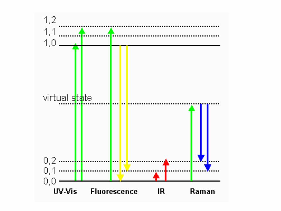
Infra-Red : IR and Raman: changes in the molecular and rotational and vibrational status
Microwave : Changes in rotational status of: Electron Spin (Microwave Spectroscopy and ESR)
Radio : Changes in rotational status of: Nuclear Spin (NMR)

Working Principle
Bragg’s LawBragg's law was used to explain the interference
pattern of X-rays scattered by crystals
Nλ = 2dhkl sinθq q d
hkl
dh
kl
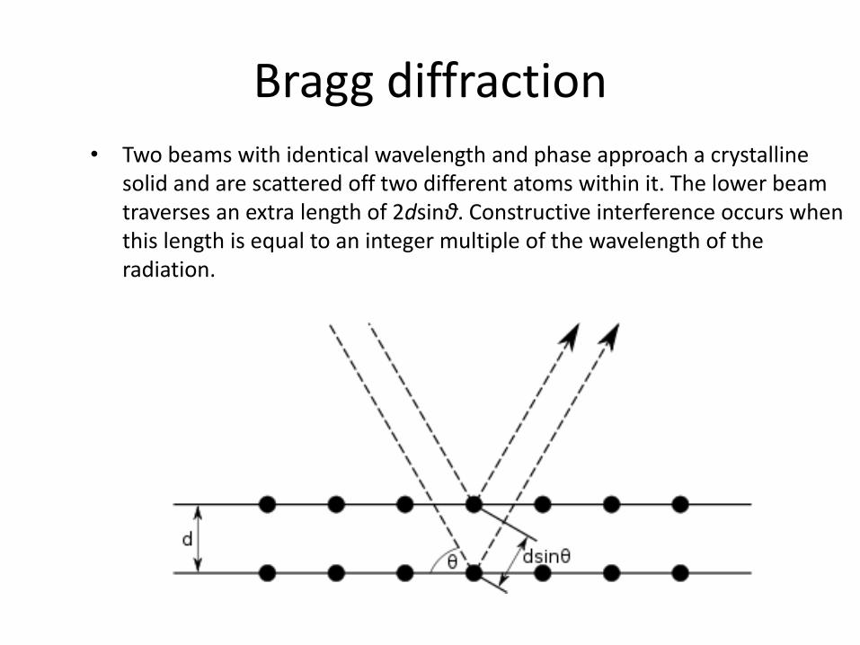
Bragg diffraction• Two beams with identical wavelength and phase approach a crystalline
solid and are scattered off two different atoms within it. The lower beam traverses an extra length of 2dsinθ. Constructive interference occurs when this length is equal to an integer multiple of the wavelength of the radiation.

Experimental Setup
Source: http://pruffle.mit.edu/atomiccontrol/education/xray/xray_diff.php

How Does It WorkSo
urc
e: h
ttp
://e
n.w
ikip
edia
.org
/wik
i/Fi
le:X
_ra
y_d
iffr
acti
on
.pn
g
Crystal (regular array of atoms) is
mounted on a Goniometer
Bombarded with X-Ray while
rotating
Production of diffraction pattern
of regularly spaced spots
The 2-D images taken at different
rotation are converted to 3-D
models of the electron density map
by the method of Fourier
Transform

Some Example Images

UsesXRD is a non-destructive technique.
To determine structural properties such as Lattice
parameters (10-4Å), strain, grain size
Biological macromolecular crystallography
To determine atomic arrangement.
To measure thickness of thin films and multi-layers.

Raman spectroscopy
Raman spectroscopy (/ˈrɑːmən/; named after Sir C. V. Raman) is
a spectroscopic technique used to observe vibrational, rotational, and other low-
frequency modes in a system.[1] Raman spectroscopy is commonly used in
chemistry to provide a fingerprint by which molecules can be identified
It relies on inelastic scattering, or Raman scattering, of monochromatic light,
usually from a laser in the visible, near infrared, or near ultraviolet range. The
laser light interacts with molecular vibrations, phonons or other excitations in the
system, resulting in the energy of the laser photons being shifted up or down.
The shift in energy gives information about the vibrational modes in the
system. Infrared spectroscopy yields similar, but complementary, information.