Linköping Dissertation on Health and Sciences. Thesis No. 1364 Practical and clinical use of opioids Salumeh Bastami Department of Medical and Health Sciences Linköping University, Sweden Linköping 2013
Transcript
Linköping Dissertation on Health and Sciences. Thesis No. 1364
Practical and clinical use of opioids
Salumeh Bastami
Department of Medical and Health Sciences Linköping University, Sweden
Linköping 2013
Contents
Practical and clinical use of opioids Dedicated to: Ali, Aydin, Ayda Sarkohi, my parents and my siblings for your unconditional love and support.
Salumeh Bastami, 2013 Cover/picture/Illustration/Design: Fereidon Bahramian Published article has been reprinted with the permission of the copyright holder. Printed in Sweden by LiU-Tryck, Linköping, Sweden, 2013 ISBN: 978-91-7519-603-9 ISSN: 0345-0082
ABSTRACT Pain is a common symptom of a number of conditions including cancer and one of the most frequent reasons for seeking healthcare. Acute and chronic pain result in considerable discomfort with a detrimental impact on the quality of life. Opioids are the mainstay of pain management for many patients with severe pain. Opioids are, unfortunately, also commonly abused drugs, and are well-represented in forensic toxicology investigations.
Side effects related to the central nervous system are the major reasons for discontinuation of opioid treatment. In this thesis, we tested the hypothesis that local analgesic treatment by opioids, without the usual opioid-related side effects, could be a potential alternative to systemic opioid treatment. We examined the analgesic effect of topically applied morphine in a randomized, double blind, cross over study in patients with painful leg ulcers. Significant reduction of pain was obtained after application of both morphine and placebo gel. Morphine reduced pain more than placebo but the difference was not statistically significant. However, morphine could reduce pain considerably more than placebo in those cases where VAS (Visual analog scale) was higher initially.
Another issue with opioid therapy is the substantial individual variability in response to opioids including morphine and tramadol. We investigated the significance of UGT2B7, CYP2D6, OPRM1 and ABCB1 polymorphisms for pharmacokinetic and pharmacodynamic properties of morphine and tramadol. We showed that genetic variants in CYP2D6 and UGT2B7 have an important role in the metabolism of tramadol and morphine respectively. While the role of SNPs in ABCB1 remained unclear, genetic variants in OPRM1 gene were correlated with the required dose of morphine. Taken together, these findings suggest that genotypes should be taken into consideration when interpreting clinical pharmacology and forensic toxicology results.
Opioids, besides their analgesic properties, have other pharmacological effects including effects on immune system. We evaluated potential differences between commonly used opiates with regard to their effect on the immune system. We found an inhibition of cytokine release, in the order of potency as follows: tramadol > ketobemidone > morphine >fentanyl. All opioids with the exception of fentanyl were capable of inhibiting production of mRNAs for TNF-alpha and IL-8. Further studies are needed to understand the clinical implications of the observed immunosuppressive effects of opioids and to improve opioid treatment strategies in patients with cancer.
Here, we have found that individual genotype matters and affects the individual response. Further research is warranted to tailor individualized treatment. Personalized medicine has increased in importance and will hopefully in the near future become standard procedure to improve and predict the outcome of treatment by opioids.
4
POPULÄRVETENSKAPLIG SAMMANFATTNING Smärta är ett vanligt förekommande symptom och en av de vanligaste orsakerna till sjukvårdsbesök. Akut och kronisk smärta försämrar patienternas sociala samvaro och livskvaliteten. För många patienter med svår smärta, är behandling med opioider grundläggande princip för smärtlindring. Det är tyvärr också vanligt med missburk utav opioider och dessa förekommer ofta inom rättsmedicinska och toxikologiska utredningar.
Biverkningar relaterade till centrala nervsystemet är bland de viktigaste orsakerna till varför man avslutar behandlingen med opioider. Lokal behandling med opioider, med förhoppningsvis mindre biverkningarna kan vara ett potentiellt komplement till systemisk behandling. Vi har i en randomiserad, dubbelblind, cross over studie undersökt effekten av lokal behandling med morfin hos patienter med smärtsamma bensår. Resultat visade signifikant minskning av smärta efter applicering av både morfin och placebo gel. Morfin minskade smärtan mer än placebo, men denna skillnad var inte statistiskt signifikant. I de fall där VAS var initialt högre, minskades smärtan med morfin betydligt mer än placebo.
Den interindividuella variationen i svaret på opioidbehandling är ett kliniskt problem. I denna avhandling har betydelsen av UGT2B7, CYP2D6, OPRM1 och ABCB1 polymorfism för farmakokinetiska och farmakodynamiska egenskaper av morfin och tramadol undersökts. Resultatet visade att CYP2D6 och UGT2B7 har en viktig roll i metabolismen av tramadol respektive morfin. Vidare fann vi att de genetiska variationerna i OPRM1 genen var korrelerade till den dos patienterna behövde för smärtlindring. Däremot återstod betydelsen av de olika polymorfierna i ABCB1 oklar. Sammantaget, tyder resultatet på att hänsyn bör tas till genotyper vid tolkning av resultat vid klinisk farmakologiska, rättmedicinska och toxikologiska utredningar.
Opioider har förutom smärtstillande egenskaper andra farmakologiska effekter så som effekter på immunsystemet. Vi har utvärderat potentiella skillnader mellan vanligen använda förekommande opioder avseende dess effekt på olika komponenter i immunsystemet. Resultatet visade en minskad frisättning av cytokin, i storleksordningen som följer: tramadol> ketobemidon> morfin> fentanyl. Samtliga opioder förutom fentanyl minskade produktionen av mRNA för TNF och IL-8. Ytterligare studier behövs för att både förstå den kliniska betydelsen av den observerade immunosuppressiva effekten av opioider och även förbättra behandlingsstrategierna för cancer patienter.
Opioider är fortfarande en av de mest potenta läkemedelsgrupperna vid smärtlindring i många kliniska sammanhang. Ytterligare forskning behövs för individuellt anpassad behandling. Betydelsen av individualiserade medicinering har ökat och kommer förhoppningsvis inom en snar framtid att bli standardförfarande i syfte att förbättra behandlingsresultatet med opioider.
5
PAPERS IN THE PRESENT THESIS This thesis is based on the following original papers, which are referred to in the text by their Roman numerals:
Paper I Bastami S, Frödin T, Ahlner J, Uppugunduri S. Topical morphine gel in the
treatment of painful leg ulcers, a double-blind, placebo-controlled clinical trial: a pilot study. Int Wound J. 2012 Aug;9(4):419-27
Paper II Bastami S, Gupta A, Zackrisson A.L, Ahlner J, Osman A, S Uppugunduri S.
Influence of UGT2B7, OPRM1 and ABCB1gene polymorphisms on morphine use. Submitted
Paper III Bastami S, Haage P, Kronstrand R, Kugelberg FC Anna-Lena Zackrisson A.L,
Uppugunduri S. Influence of genetic polymorphism on tramadol pharmacokinetics and pharmacodynamics. Submitted
Paper IV Bastami S, Norling C, Trinks C, Holmlund B, Walz TM, Ahlner J,
Uppugunduri S. Inhibitory effect of opiates on LPS mediated release of TNF and IL-8. Acta Oncol. 2013 Jun;52(5):1022-33.
6
7
ABBREVIATIONS ABC ATP-Binding Cassette
AP Alkaline Phosphates
APL Apotekets Produktion och Laboratorier AB
APS Adenosine 5' Phosphosulfate
AUC Area under the curve
BBB Blood Brain Barrier
cAMP Cyclic adenosine monophosphate
CGRP Calcitonin gene-related peptide
CNS Central Nervous System
CYP2D6 Cytochrome 2D6
CYP3A4 Cytochrome P3A4
DNA Deoxyribonucleotide acid
dNTP Deoxyribonucleotide triphosphate
ELISA Enzyme-linked immunosorbent assay
ERK Extracellular signal-regulated kinases
GAPDH Glyceraldehyde 3-phosphate dehydrogenase
GDP Guanosine diphosphate
GPCR G protein-coupled receptors
GTP Guanosine triphosphate
HPLC High Performance Liquid Chromatography
HRP Horseradish peroxidase
huCYC Housekeeping cyclophilin
IL-6 Interleukin 6
IL-8 Interleukin-8
M3G Morphine-3-Glucuronide
M6G Morphine-6-Glucuronide
MAPK Mitogen activated protein kinase
8
MDR Multi drug resistance
MMP Matrix metalloproteinase
MOR μ-opioid receptor
MPQ McGill Pain questionnaire
MR Metabolic ratio ODT/TRA
NMDA N-methyl-D-aspartate
NOP Nociceptin Receptor
NRS Numeric Rating Scale
NSAID Non-Steroid Anti Inflammatory Drugs
ODT O-desmethyl tramadol
OPRM1 Opioid receptor μ-1
ORL1 Opioid receptor like 1
PCA Patient-controlled analgesia
P-gp P-glycoprotein
PI3K Phosphatidyl Inositol 3'-Kinase
PKB Protein kinase B
SLC Solute carrier
SNP Single Nucleotide Polymorphism
TENS Transcutaneous Electrical Nerve Stimulation
TNF Tumor necrosis factor
TRA Tramadol
UGT2B7 UDP- glucuronosyltransferase 2B7
UV Ultraviolet
VAS Visual Analog Scale
WHO World Health Organization
VRS Verbal Analog Scale
9
INTRODUCTION Pain is a common symptom and one of the most frequent reasons for seeking healthcare. About 20-30% of patients seeking general practitioners have clinically significant pain that requires medical intervention [1]. According to the data published by the US National Center for Health Statistics, 25-30% of adults with pain at selected body sites report fair or poor health, 15-22% report being unable to work, 12-17% sleep less than 5 hours per day and 6-13% report psychological distress (http://www.cdc.gov/features/). Studies indicate that every second patient in most hospitals suffers from pain and at least every third patient complains of severe pain [2-4].
Since pain has a tremendous impact on the patient’s physical and psychological well-being [5], inadequate pain therapy can greatly increase healthcare costs. About 10% of drug sales in the USA are attributed to pain and the annual cost of pain includes more than $100 billion for pain-related healthcare and $61.2 billion for lost productivity [6, 7]. The International Association for the Study of Pain has called unrelieved pain “a major global healthcare problem”[8].
Fortunately, adequate pain therapy can improve the patient's performance of daily activities and promote faster recovery and earlier discharge from hospital [9]. Providing a rapid and effective pain relief without compromising the patient’s general condition is a challenge for physicians. In order to be able to tailor analgesic treatment for the individual patient it is important to understand the underlying causes of interindividual variability in drug response. In the research on which this thesis is based, we studied various aspects of pain treatment with an emphasis on opioids
PAIN The International Association for the Study of Pain defines pain as "an unpleasant sensory and emotional experience associated with actual or potential tissue damage, or described in terms of such damage". The response to pain can be highly variable among persons as well as in the same person at different times. A variety of classification schemes have been used for the purpose of understanding, predicting, and treating pain.
CLASSIFICATION OF PAIN Pain has been classified according to pathophysiology (e.g., nociceptive or neuropathic pain), diagnostic purpose (acute or persistent pain), etiology (e.g. postoperative or cancer pain), or the affected area (e.g. headache, low back pain, etc.).
For treatment purposes, it has been more helpful to categorize pain in two categories, nociceptive and neuropathic. Such classifications are useful in the selection of treatment and drug therapy. For diagnostic purposes, pain has been categorized as acute or persistent pain.
10
The term nociception is derived from noci (Latin for harm or injury) and is used to describe pain problems that result largely from stimulation of pain receptors, so called nociceptors. Nociceptive pain can commonly arise from tissue injury or inflammation, for example trauma, burns, infection, arthritis and ischemia. Pain from nociception usually responds well to common analgesic medications. Neuropathic pain can occur after damage to and dysfunction of the peripheral and central nervous system. Diabetic neuralgia, post-herpetic neuralgia and post-traumatic neuralgia are some examples. In contrast to nociceptive pain, neuropathic pain syndromes are often persistent and difficult to treat. They may, however, respond to non-conventional analgesic medications such as tricyclic antidepressants and anticonvulsant drugs [10].
Acute pain is defined by a distinct onset, obvious cause and short duration. It occurs primarily after an injury, and treating the illness or injury will often reduce or eliminate the pain symptoms. Symptoms such as tachycardia, diaphoresis, or elevation in blood pressure are often associated with stimulation of autonomic nerve system. Acute injury commonly results in acute pain. The effective management of acute pain is important to facilitate diagnostic tests. In some cases management of acute pain can help prevent development of chronic pain syndrome. In persistent pain, the psychological and behavioral factors often play a major role. Persistent pain is almost always prolonged and continues beyond the expected normal healing time. It is more complex and difficult to manage and requires multimodal pain treatment strategies. In these patients the intense, repeated, or prolonged stimuli can lower the threshold for activating primary afferent nociceptors, which leads to increased response to painful or non-painful stimulus (hyperalgesia/allodynia).
PAIN ASSESSMENT Assessment of pain severity is a crucial component in providing effective pain management and evaluating the efficacy of treatment. However, this is a challenge as pain is a subjective experience that is influenced by psychological, cultural, and other variables. A variety of pain assessment scales are available to help categorize and quantify the magnitude of pain. The visual analog scale (VAS), numerical rating scale (NRS), verbal rating scale (VRS), faces rating scale and the McGill Pain Questionnaire (MPQ) are the most commonly used pain measurement instruments. The Visual Analog Scale has been used for measurement of pain intensity in this work.
VAS is a simple assessment tool consisting of a 100 mm line with 0 on one end, representing no pain, and 10 on the other, representing the worst pain ever experienced. The patient is asked to place a mark on the line at the point that best represents the pain level. It has been frequently used to assess pain intensity in treatment outcome research. It has a large number of response categories, which makes it potentially more sensitive to changes in pain intensity compared to the other scales and easy to administer. However, some patients have difficulty in understanding and using the scale and therefore tend to require more explanation and assistance than for the other scores [11].
11
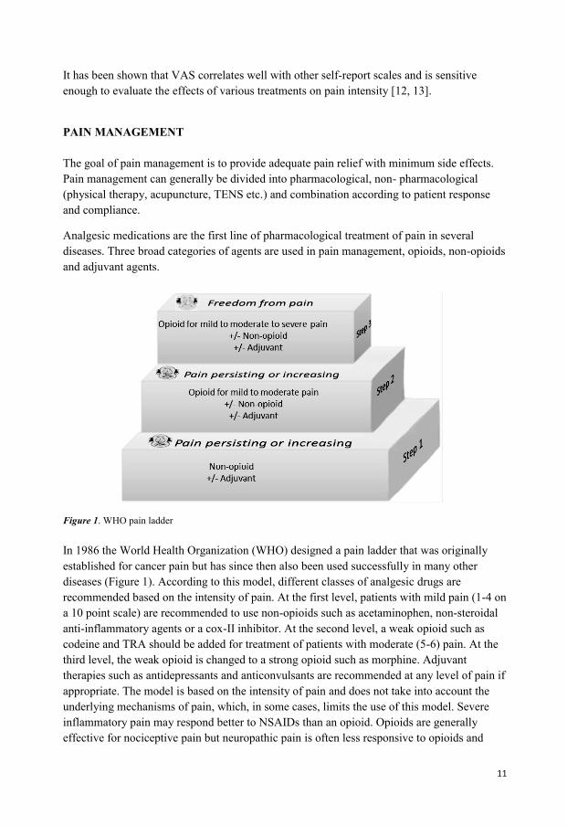
It has been shown that VAS correlates well with other self-report scales and is sensitive enough to evaluate the effects of various treatments on pain intensity [12, 13].
PAIN MANAGEMENT The goal of pain management is to provide adequate pain relief with minimum side effects. Pain management can generally be divided into pharmacological, non- pharmacological (physical therapy, acupuncture, TENS etc.) and combination according to patient response and compliance.
Analgesic medications are the first line of pharmacological treatment of pain in several diseases. Three broad categories of agents are used in pain management, opioids, non-opioids and adjuvant agents.
Figure 1. WHO pain ladder In 1986 the World Health Organization (WHO) designed a pain ladder that was originally established for cancer pain but has since then also been used successfully in many other diseases (Figure 1). According to this model, different classes of analgesic drugs are recommended based on the intensity of pain. At the first level, patients with mild pain (1-4 on a 10 point scale) are recommended to use non-opioids such as acetaminophen, non-steroidal anti-inflammatory agents or a cox-II inhibitor. At the second level, a weak opioid such as codeine and TRA should be added for treatment of patients with moderate (5-6) pain. At the third level, the weak opioid is changed to a strong opioid such as morphine. Adjuvant therapies such as antidepressants and anticonvulsants are recommended at any level of pain if appropriate. The model is based on the intensity of pain and does not take into account the underlying mechanisms of pain, which, in some cases, limits the use of this model. Severe inflammatory pain may respond better to NSAIDs than an opioid. Opioids are generally effective for nociceptive pain but neuropathic pain is often less responsive to opioids and
12
requires the use of adjuvant medications which include tricyclic antidepressants and anticonvulsants such as gabapentin.
PHARMACOLOGICAL AND PHARMACODYNAMICAL EFFECTS OF OPIOIDS
OPIOIDS The term opiate refers to compounds structurally related to products found in opium, a word derived from opos, the Greek word for "juice". Opium is obtained from the unripe seed capsules of the poppy plant, Papaver somniferum. The milky juice is dried and powdered to make powdered opium, which contains a number of alkaloids. Opioids can be categorized into three subgroups; 1) naturally occurring compounds (termed opiates) such as morphine and codeine, 2) chemically modified natural compounds (semisynthetic) such as hydrocodone, buprenorphine and oxycodone, 3) completely artificial compounds (synthetic) such as fentanyl, tramadol and ketobemidone. Some opioids act as agonists to all kind of opioid receptors (morphine) and some act as both agonist and antagonist (buprenorphine).
Opioids are the most effective pain-relieving drugs, however side effects are common. Fortunately most of these are reversible. The most frequently appearing side effects are nausea, vomiting, pruritus, and constipation; these also happen to be the most bothersome. Respiratory depression is uncommon at standard analgesic doses, but can however be life-threatening. Opioids produce analgesia by actions in the CNS. They activate pain-inhibitory neurons and directly inhibit pain-transmission neurons. The pharmacology of the opioids is quite similar. They differ mainly in potency, duration of action and optimal route of administration. Intravenous administration provides the most rapid relief. Opioid effects are dose-related, and there is great variability among patients in the doses that relieve pain and produce side effects. Thus, initiation of therapy requires titration to optimal dose and interval.
Patient-controlled analgesia (PCA) is an innovative approach to achieve adequate pain relief. PCA infusion device delivers a continuous baseline dose. Patients can administer additional preprogrammed doses by pushing a button. PCA is used for treatment of postoperative pain and also for short-term home care of patients with metastatic cancer.
Recent additions to the arsenal for treating opioid-induced side effects are the peripherally acting opioid antagonists alvimopan (Entereg) and methylnaltrexone (Rellistor). Alvimopan is available as an orally administered agent that is restricted to the intestinal lumen by limited absorption. Methylnaltrexone is available in a subcutaneously administered form that has virtually no penetration into the CNS.
13
Morphine Morphine is one of the most effective and widely used drugs for the relief of severe or prolonged pain. It was first isolated in 1804 and took its name from the Greek god of dreams Morpheus. Morphine is the most abundant alkaloid found in opium. The drug is still obtained from opium or extracted from poppy straw because the synthesis of morphine is difficult. Even though there are many compounds with pharmacological properties similar to those of morphine, it remains as the gold standard against which new analgesics are measured.
Morphine is primarily used to treat both acute and persistent severe pain by acting directly on the central nervous system. Morphine has a high potential for addiction, tolerance and psychological dependence but there is a minimal chance of becoming addicted when used appropriately.
Approximately 25% of orally administered dose is absorbed from the GI tract. The absorption through the rectal mucosa is also adequate and a few agents are available as suppositories (not in Sweden). Morphine has been widely used for spinal delivery to produce analgesia through a spinal action. The effect of an oral dose is less than after parenteral administration because of first passage metabolism in the liver. About one-third of morphine in the plasma is protein-bound. Morphine itself does not persist in tissues, and 24 hours after the last dose, tissue concentrations are low. The half-time of morphine in plasma is about two hours.
Figure 2. Chemical structure of morphine and morphine’s metabolites
The major pathway for the metabolism of morphine is conjugation (Figure 2). Morphine is converted to two major metabolites by UGT2B7, morphine-6-glucuronide (M6G) and
14
morphine-3-glucuronide (M3G), which are eliminated by glomerular filtration in kidney. Although the 3- and 6-glucuronides are quite polar, both can still cross the blood-brain barrier to exert significant clinical effects [14, 15]. N-demethylation and N-dealkylation are other minor metabolic pathways.
Experimental and clinical studies indicate that M6G is approximately twice as potent as morphine but with significantly fewer side effect.[15-17]. Morphine-3-glucuronide has little affinity for opioid receptors but may contribute to excitatory effects of morphine.
Tramadol Tramadol (TRA) is a synthetic codeine analog that is a weak μ-receptor agonist. It’s affinity for the mu-opioid receptor is only 1/6000 that of morphine. Other mechanisms that contribute to its analgesic effect are inhibition of neuronal reuptake of noradrenaline and enhancement of serotonin release. TRA is as effective as morphine in the treatment of mild to moderate pain. However, TRA is less effective for the treatment of severe or chronic pain.
TRA is a racemic mixture; (–)-TRA is about 10 times more potent than(+)-TRA in inhibiting noradrenalin uptake and (+)-TRA is about 4 times stronger than (–)-TRA in inhibiting serotonin uptake [18]. Both enantiomers act synergistically to improve analgesia. More than 90% of TRA is absorbed after oral administration. About 20% of TRA is protein bound. Analgesic effect begins an hour after oral administration and peaks after 2-3 hours. Duration of analgesia is almost 6 hours.
Figure 3. Chemical structure of tramadol and tramadol’s main metabolites.
TRA undergoes extensive hepatic metabolism by a number of pathways, including CYP2D6 and CYP3A4 (Figure 3). TRA is metabolized through the N-and O-demethylation. The O-desmethyltramadol (ODT) is the main metabolite and has a higher affinity for opioid
15
receptor. (+)-enantiomer of ODT has 300–400 times greater affinity for opioid receptors than TRA, whereas (–) ODT mainly inhibits noradrenalin reuptake. TRA and its metabolites are excreted almost entirely by the kidneys.
Figure 4. Chemical structure of A) fentanyl and B) ketobemidone.
Fentanyl Fentanyl (Figure 4A) is a strong, relatively short-acting synthetic opioid nearly 100 times more potent than morphine. Like morphine fentanyl interacts predominantly with μ-receptor.
The time to peak analgesic effect after intravenous administration of fentanyl (~5 minutes) is notably less than that for morphine (~15 minutes). Fentanyl is popular in anesthetic practice because of its short time to peak analgesic effect and the rapid termination of effect after small bolus doses (3-4 hours). Its use in chronic pain has become more widespread.
Several methods of administration exist including oral preparations, intravenous injections and the transdermal patch.
Fentanyl is metabolized primarily in the liver by CYP3A4. The major metabolite, norfentanyl, is inactive. Approximately 75% of fentanyl is excreted in the urine, mostly as metabolites and less than 10% as unchanged drug. About 9% of the dose was recovered in the faces, primarily as metabolites.
Ketobemidone Ketobemidone hydrochloride (Figure 4B) is a synthetic opioid. It has been used mainly in Scandinavian countries for treatment of severe pain like cancer pain. The pain relieving properties of ketobemidone are almost in the same range as morphine. It has a somewhat
16
higher lipophilicity than morphine. Besides its μ-receptor agonism, it has even NMDA-antagonist properties.
Analgesic effect is achieved 1-3 hours after orally administered dose and 10-30 minutes after intravenous administration. The effect lasts for 3-5 hours. Ketobemidone undergoes hepatic metabolism and is a substrate for CYP2C9 and CYP3A4 [19]. The major metabolite, norketobemidone, is generally considered to be inactive, which can be beneficial in patients with renal insufficiency or immature renal function [20].
Opioid receptors Opioid receptors are a group of G protein-coupled receptors (GPCR). Each receptor consists of an extracellular N-terminus, seven transmembrane helices, three extra- and intracellular loops, and an intracellular C-terminus characteristic of the GPCRs. The opioid receptor types are approximately 70% identical with differences located at N and C termini. The greatest diversity is found in their extracellular loops.
Three major type of opioid receptor have been identified, mu (μ), delta (δ) and kappa (κ). The G-protein coupled opiate receptor-like protein (ORL1 or NOP) was included to other members of the opioid receptor family, based on its structural homology (48-49% identity) to the other opioid receptors. μ-opioid receptor was identified in binding assays in 1973 and was cloned about 20 years later (Simon 1973, Chen 1993). Other opioid receptors have also been proposed, such as zeta (ζ) opioid receptor, which has been shown to be a cellular growth factor modulator and epsilon (ε) opioid receptor. However, efforts to locate a gene for ε-receptor have been unsuccessful and epsilon-mediated effects were absent in μ/δ/ κ.
The human opiate receptors have been mapped to chromosome 1p355-33 (δ-opioid receptor), chromosome 8q11.23-21 (κ-opioid receptor), and chromosome 6q25-26 (μ-opioid receptor) [21]. Pharmacological studies have suggested more than one class of mu opiate receptor. Although only a single μ- receptor gene has been reported, this gene undergoes extensive splicing. It has been suggested that μ-, δ- and κ- receptors have several subtypes, μ1-3, delta 1-2 and κ- 1-3. It has also been postulated that μ-1 receptors produce analgesia while μ-2 mediates respiratory depression. The function of the μ-3 receptor is unknown. The existence of opioid receptor subtypes has not been confirmed in either cloning studies or experiments with knock out animals.
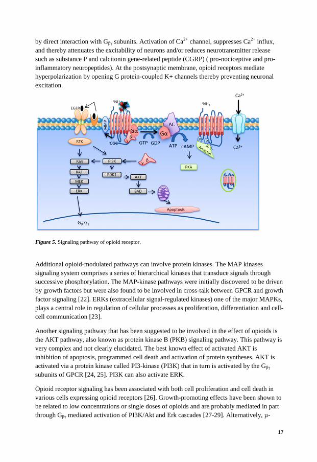
Signal transduction The signaling pathways of opioid receptors are well characterized (Figure 5). When ligand binds to the receptor, conformation changes of receptor occurs that causes replacement of GDP bound to G α-subunit by GTP. The activated Gα dissociates from trimeric G-protein complex and inhibits the adenylyl cyclase and thereby inhibits the formation of cAMP, which subsequently activates the ion channels in the membrane. Ion channels can also be regulated
17
by direct interaction with Gβγ subunits. Activation of Ca2+
channel, suppresses Ca2+
influx,
and thereby attenuates the excitability of neurons and/or reduces neurotransmitter release
such as substance P and calcitonin gene-related peptide (CGRP) ( pro-nociceptive and pro-
inflammatory neuropeptides). At the postsynaptic membrane, opioid receptors mediate
hyperpolarization by opening G protein-coupled K+ channels thereby preventing neuronal
excitation.
Figure 5. Signaling pathway of opioid receptor.
Additional opioid-modulated pathways can involve protein kinases. The MAP kinases
signaling system comprises a series of hierarchical kinases that transduce signals through
successive phosphorylation. The MAP-kinase pathways were initially discovered to be driven
by growth factors but were also found to be involved in cross-talk between GPCR and growth
factor signaling [22]. ERKs (extracellular signal-regulated kinases) one of the major MAPKs,
plays a central role in regulation of cellular processes as proliferation, differentiation and cell-
cell communication [23].
Another signaling pathway that has been suggested to be involved in the effect of opioids is
the AKT pathway, also known as protein kinase B (PKB) signaling pathway. This pathway is
very complex and not clearly elucidated. The best known effect of activated AKT is
inhibition of apoptosis, programmed cell death and activation of protein syntheses. AKT is
activated via a protein kinase called PI3-kinase (PI3K) that in turn is activated by the Gβγ
subunits of GPCR [24, 25]. PI3K can also activate ERK.
Opioid receptor signaling has been associated with both cell proliferation and cell death in
various cells expressing opioid receptors [26]. Growth-promoting effects have been shown to
be related to low concentrations or single doses of opioids and are probably mediated in part
through Gβγ mediated activation of PI3K/Akt and Erk cascades [27-29]. Alternatively, µ-
18
opioid receptor-induced mitogenesis may be mediated through cross-activation of growth
factor receptors [22]. On the other hand, growth inhibitory effects observed in chronic opioid
treatment or at relatively high in vitro concentrations were shown to be closely associated
with opioid receptor desensitization and internalization [30, 31].
PAIN AND INFLAMMATION
Inflammation
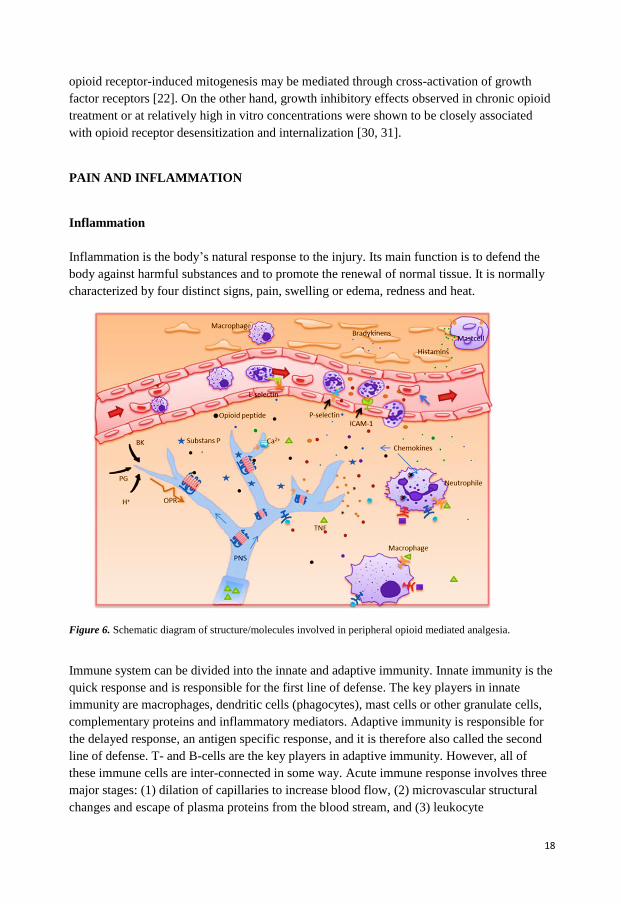
Inflammation is the body’s natural response to the injury. Its main function is to defend the
body against harmful substances and to promote the renewal of normal tissue. It is normally
characterized by four distinct signs, pain, swelling or edema, redness and heat.
Figure 6. Schematic diagram of structure/molecules involved in peripheral opioid mediated analgesia.
Immune system can be divided into the innate and adaptive immunity. Innate immunity is the
quick response and is responsible for the first line of defense. The key players in innate
immunity are macrophages, dendritic cells (phagocytes), mast cells or other granulate cells,
complementary proteins and inflammatory mediators. Adaptive immunity is responsible for
the delayed response, an antigen specific response, and it is therefore also called the second
line of defense. T- and B-cells are the key players in adaptive immunity. However, all of
these immune cells are inter-connected in some way. Acute immune response involves three
major stages: (1) dilation of capillaries to increase blood flow, (2) microvascular structural
changes and escape of plasma proteins from the blood stream, and (3) leukocyte
19
transmigration through endothelium and accumulation at the site of injury. Pathogen infiltration into the body leads to degranulation of mast cells, releasing histamine and bradykinin resulting in a transient increase in vascular permeability and vasodilation in capillaries (Figure 6). Tissue macrophages that are also able to detect the pathogen begin secreting cytokines. Cytokines, responsible for communication with other cells, cause increased vascular permeability that attracts more immune cells such as monocytes and neutrophils to the site of injury. Proinflammatory cytokines such as TNF-alfa stimulate endothelial cells to express various cell adhesion molecules like selectins and integrins. Neutrophils, the most abundant leucocytes, bind to these receptors and begin to move along the endothelium via transient, reversible, adhesive interactions. Tissue macrophages also release other kinds of proinflammatory components such as chemokines like IL-8. IL-8 is instrumental in accumulation of neutrophils at the site of injury [32-34].
Leukocyte extravasation involves three families of adhesion molecules: (1) Selectins, which are glycosylated proteins that bind to other glycosylated proteins and glycolipids and mediate the low-affinity interactions that enable leukocyte rolling, (2) Integrins which are heterodimeric proteins that bind to the immunoglobulin family of adhesion molecules, and together mediate firm adhesion, arrest and extravasation, (3) Chemokines like IL-8 that bind to respective receptors and play an important role in leukocyte extravasation by mediating migration of specific sets of leukocytes like neutrophils and eosinophils.
Immune cells and pain Activation of an immune response modulates the excitability of pain pathways by forming an integrated network between the immune cells, glia, and neurons. This process is very complex and not fully understood. Only a few parts of this process are described below.
The degranulation of mast cells requires direct interaction between mast cells and peripheral nerve terminals, which is mediated by a cell adhesion molecule, N-cadherin. N-cadherin is expressed by both mast cells and primary sensory neurons and is cleaved by metalloproteinase (MMP), which is expressed by neurons [35]. In turn, MMP-9 promotes migration of macrophages to the injured site via breakdown of the blood-brain barrier. The recruitment of macrophages after nerve injury is mediated by several inflammatory cytokines like TNF-α, which is released from Schwann cells immediately after nerve injury [36].
Neutrophils migrate within the first hour of the onset of inflammation through the vascular endothelium and accumulate at the site of injury. Recruitment of neutrophils is enhanced by primary afferent neurons [37, 38]. Stimulation of these neurons generates impulses that spread through neighboring nerve terminals, resulting in the release of the vasoactive neuropeptides such as substance P, CGRP at the peripheral nerve branches. Substance P and CGRP stimulate adhesion of neutrophils to the endothelial cells. These neuropeptides can also facilitate the degranulation of mast cells [39].
20
Other components of immune system that interact with nociceptors are lymphocytes and proteins of complement system. Although there is some evidence that these components contribute to the sensitization of peripheral nociceptors, the data are less conclusive than for other immune cells [40, 41].
Immune cells and endogenous opioids Opioid receptors have been detected in lymphocytes, monocytes, macrophages and granulocytes. Opioid receptors on leucocytes appear to have similar pharmacological and biochemical characteristics as neuronal opioid receptors and are encoded by the same genes [42]. However, the level of receptors expressed by immune cells is much lower than those present in neurons [43].
It has been suggested that binding of opioid ligands triggers the same signaling pathways as in neuronal cells, in other words modulation of cAMP, Ca2+ channels and kinases. Stimulation of opioid receptors on leucocytes modulates proliferation, chemotaxis and cytotoxicity of leucocytes. It has also been observed that cytokine and chemokine expression have been affected by these receptors in vitro. However, the results of these studies are contradictory, depending on experimental conditions such as cultured cell types, doses and timing of opioid exposure [33, 42, 44].
After injury, the immune system also releases factors that suppress inflammation and reduce pain. Opioid peptides are found in many leucocyte subpopulations including lymphocytes, monocytes and granulocytes. It has been suggested that granulocytes are involved in opioid peptide production in early inflammation, while monocytes and lymphocytes seem to be involved in the later inflammatory reaction [45]. Migration of opioid peptide-containing leucocytes are facilitated by chemokines. Neuropeptides and complementary proteins are other mediators in opioid peptide-containing leucocyte recruitment. Opioid peptides released from granulocytes has been shown to have anti-nociceptive effects in vivo [46].
Peripheral opioid analgesia Opioid receptors are expressed throughout the body in various tissues and cell types. Opioid receptors have also been demonstrated on peripheral terminals of sensory nerves in human synovia, dermal and epidermal nerve fibers and dental pulp [47, 48]. Originally it was thought that opioids exert their effects solely through binding to opioid receptors in the central nervous system. During the past two decades several studies have shown that the analgesic effects of opioids can be mediated by opioid receptors located on peripheral sensory neurons [49-51]. A number of studies have shown that opioid receptors are expressed in small, medium, and large-diameter dorsal root ganglia neurons [52-60]. These receptors have been suggested to be synthesized by sensory neurons in the dorsal root ganglia and transported to, both, central and peripheral nerve terminals [61]. It has also been suggested that they have the same structure and functionality as receptors in the central nervous system,
21
exerting their effect by inhibition of ion channels [62]. Painful inflammation in peripheral tissue can induce upregulation of opioid receptors, specifically the μ-opioid receptor [63]. The duration of inflammation has been shown to be an important factor in mediating upregulation of opioid receptors. Short-lasting inflammation (30 minutes) could not change opioid receptor expression on sensory nerve terminals [64]. It was shown that the upregulation of μ-opioid receptor binding sites in dorsal root ganglia neurons is due to an increase in both the number of neurons expressing μ-opioid receptor and the number of μ-opioid receptors per neuron while affinity of opioid agonist to the μ-opioid receptor remained unchanged [65]. It has been suggested that the cytokine-induced binding of transcription factors to opioid receptor gene may be one of the explanations behind the upregulation of opioid receptors [66]. In addition, inflammatory components such as bradykinin and cytokines have been suggested to enhance the peripherally directed axonal transport of μ-opioid receptor and also induced μ-opioid receptor G-protein coupling on dorsal root ganglion [67-69]. Further, inflammation can cause disruption in the perineural barrier which facilitates the access of opioid agonists to their receptors.
The fact that these receptors mediate analgesia has been demonstrated in patients with various types of pain such as persistent rheumatoid arthritis, osteoarthritis, oral mucositis, bone pain, complex regional pain syndrome and after dental , urinary bladder and knee surgery [62, 70-72]. Several studies indicate that about 50-80% of the analgesic effects produced by systemically administered opioids can be mediated by peripheral opioid receptors [64, 73-75]. Intra articular injection of morphine into inflamed knee joints has been one of the most extensively studied approaches and the most successful application of peripheral opioid analgesia. Further, blocking intra articular opioid receptors by local administration of naloxone in patients undergoing knee surgery showed a significant increase in postoperative pain. Administration of intra articular morphine has been recommended as a routine clinical practice by American Society of Anesthesiologists [76].
VARIABILITY IN DRUG RESPONSE Response to a drug depends on the complex interplay between environmental factors and genetic factors. Variation in drug response may be explained by these factors alone or in combination.
GENETIC POLYMORPHISM Genetic polymorphism is a variation in the DNA sequence within the population, leading to the occurrence of at least two alleles. Most of the genetic polymorphisms are single nucleotide polymorphisms (SNP) [77]. SNPs occur every 100–300 base pairs and account for approximately 90% of human genetic variation. A SNP can consist of a base pair substitution, a nucleotide insertion or deletion. Synonymous SNPs will alter the nucleotide without changing the resulting amino acid (also called a “silent mutation”). Non-synonymous SNPs
22
are produced when the nucleotide substitution alters the resulting amino acid. Different types of SNPs exist, depending of where they are located in the genome. These alterations can occur in promotor, exonic, or intergenic regions and, consequently, may affect the function of the corresponding gene product.
The majority of the SNPs do not have any function. The type of SNPs that are important, for example by giving differential response to drugs, are SNPs located in a gene’s coding regions. Less than one percent of SNPs occur in coding regions and alter the genetic product
GENETIC POLYMORPHISM IN DRUG DISPOSITION Genetic variation is one of the several factors contributing to variability in drug disposition. Alteration in drug disposition influences circulating drug concentration, as well as concentration at the sites of action.
Drug disposition is mainly affected by drug metabolizing enzymes, drug transport proteins, plasma binding and receptors for the drug. The most important mechanism by which genetic variation modifies drug response is alternation in drug metabolizing enzymes. Genetic variation in drug metabolizing enzymes can either increase or decrease drug metabolism.
Drug metabolizing enzymes Metabolism is a biochemical reaction that converts lipophilic drug to hydrophilic variant so that the drug is easily excreted from the body [78]. Although all tissues are capable of metabolizing drugs, the liver is the major site of metabolism. The liver, GI tract, kidney and lungs also metabolize a certain fraction of a drug. The chemical reactions involved in drug metabolism are divided into two categories, Phase 1 and Phase 2 reactions.
Phase 1 reactions include oxidation, reduction, hydrolysis, cyclization and decyclization reactions. Various enzymes of cytochrome P450 (CYP), including CYP2C9, CYP2C19, and CYP2D6 are among the most common enzymes involved in phase 1 metabolism.
Cytochrome P450 system refers to a family of enzymes (usually hepatic) which are located on the endoplasmic reticulum. Human CYP enzymes account for more than 75% of the total drug metabolism [79]. CYP3A4 is involved in about 50% of CYP mediated drug metabolism [80, 81]; while CYP2D6, CYP2C9 and CYP2C19 together mediate 40% of drug metabolism. Other families involved in drug metabolism are CYP2A6 CYP2E1 and CYP1A2. Polymorphism in CYP2D6, CYP2C9 and CYP2C19 has been found to be of clinical relevance [82, 83].
CYP2D6 was the first human P450 enzyme recognized and most studied in relation to polymorphisms [84]. Opioids such as codeine and TRA have been shown to be substrates for this enzyme. Other substrates for CYP2D6 include beta blockers, antipsychotics, antidepressants and antiarrythmics [81, 85].
23
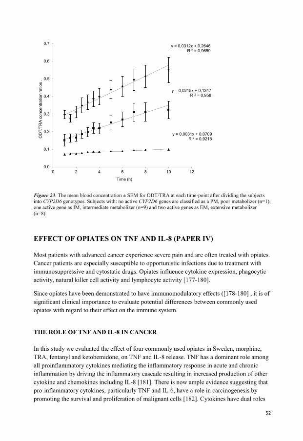
The gene encoding for CYP2D6 is located on chromosome 22q13.1 and spans a 4.2-kb in humans [86]. It consists of nine exons which give rise to a polypeptide containing 497 amino acids. Four different phenotypes have been described, poor metabolizers (PMs), intermediate metabolizers (IMs), extensive metabolizers (EMs) and ultra-rapid metabolizers (UMs). PMs lack a functional enzyme, IMs are heterozygous for a defective allele or carry two alleles with reduced activity, EMs carry two active alleles and UMs carry repeated sequences of the two active alleles. So far 105 different human CYP2D6 allelic variants have been detected [Home Page of the Human Cytochrome P450 (CYP) Allele Nomenclature Committee (http://www.cypalleles.ki.se/cyp2d6.htm)]. Most of them are rare and their effect on enzyme activity is not yet clear.
The CYP2D6 genotypes were assigned based on the alleles identified. Alleles not carrying any of the determined polymorphisms were classified as *1 (wild-type). The outcomes of the genotype analysis were categorized into three groups; individuals carrying no active gene (i.e. carrier of only the *3, *4, *5 or *6 alleles, also known as poor metabolizers, PMs), individuals carrying one active gene (i.e. carrier of *1 in combination with one of the alleles *3, *4, *5 or *6, also known as intermediate metabolizers, IMs) and individuals with two active genes (i.e. carrier of two *1 alleles, also known as extensive metabolizers, EMs). It has been suggested that *4, *5,*3 and *6 are the most frequent alleles in PM and predict about 93-98% of PM phenotypes in Caucasians [87, 88].
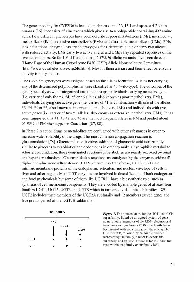
In Phase 2 reaction drugs or metabolites are conjugated with other substances in order to increase water solubility of the drugs. The most common conjugation reaction is glucuronidation [78]. Glucuronidation involves addition of glucuronic acid (structurally similar to glucose) to xenobiotics and endobiotics in order to make a hydrophilic metabolite. After glucuronidation, these conjugated substances/metabolites are readily excreted by renal and hepatic mechanisms. Glucuronidation reactions are catalyzed by the enzymes uridine 5'-diphospho-glucuronosyltransferase (UDP- glucuronosyltransferase, UGT). UGTs are intrinsic membrane proteins of the endoplasmic reticulum and nuclear envelope of cells in liver and other organs. Most UGT enzymes are involved in detoxification of both endogenous and foreign chemicals but some of them like UGT8A1 have a biosynthetic role, such as synthesis of cell membrane components. They are encoded by multiple genes of at least four families UGT1, UGT2, UGT3 and UGT8 which in turn are divided into subfamilies. [89]. UGT2 includes three members of the UGT2A subfamily and 12 members (seven genes and five pseudogenes) of the UGT2B subfamily.
Figure 7. The nomenclature for the UGT- and CYP superfamily. Based on an agreed system of gene nomenclature, members of the UDP- glucuronosyl transferase or cytochrome P450 superfamily have been named with each gene given the root symbol UGT or CYP, followed by an Arabic number representing the family, a letter to denote the subfamily, and an Arabic number for the individual gene within that family or subfamily [89].
24
UGT2B7 is highly expressed in the human liver, with lower levels of expression in other extra hepatic organs and is able to glucuronidate various steroid hormones and fatty acids. It is also able to conjugate major classes of drugs such as analgesics, carboxylic nonsteroidal anti-inflammatory drugs (ketoprofen), anticarcinogens (all-trans retinoic acid) and anticancer drugs [90]. Morphine is the prototypical UGT2B7 substrate which means that UGT2B7 is likely to be the major isoform responsible for morphine glucuronidation in humans [91]. Experimental studies with rats have shown that UGT1 also catalyzes opioid glucuronidation, but does not catalyze the formation of morphine-metabolites [92].
The gene encoding UGT2B7 is localized within a cluster of UGT2B genes on chromosome 4q13. The gene contains six exons and spans nearly 16kb. Polymorphisms have been identified in the coding and regulatory regions [93-96]. One of the most studied SNPs in the coding region in the UGT2B7 gene is T802C in exon 2. This polymorphism leads to a histidine to tyrosine substitution in codon 268 (His268Tyr). The functional impact of this polymorphism is unclear. Studies have shown both lower [97-100], similar [101-103], and higher enzyme activity of the UGT2B7 268Tyr isoform [91, 104-106]. Another polymorphism that has been shown to be associated with increased M6G/ morphine plasma ratio is the polymorphism in promoter region -161C/T. This polymorphism is in complete linkage disequilibrium with C802T suggesting the existence of a haplotype in the UGT2B7 gene. Patients homozygous for both the -161C and 802C alleles had reduced M6G/M ratios compared with patients with C/T and T/T genotypes. Even in this case the functional impact of this polymorphism is not yet clear. No significant association between UGT2B7 and either morphine glucuronides or analgesic response was found in patients with cancer [94, 107]. From the studies conducted so far, there is no compelling evidence for the existence of functional UGT2B7 variants.
Drug receptors Another factor that contributes to alteration in drug response is polymorphism in the gene coding drug receptors. Polymorphism can alter receptor sensitivity to the specific drug and thus have a profound effect on drug efficacy and toxicity. G protein coupled receptors are the most diverse and therapeutically important family of receptors, playing major roles in physiology of various organ and tissues. Opioid receptors (G-protein coupled receptor) are the target for opioid drugs.
The human gene encoding the μ-opioid receptor (OPRM1) has been localized to chromosome 6 (6q24-q25) and spans over 200 kb with at least 9 exons and 19 different splice variants under the control of multiple promoters [108]. The initial receptor subtype, MOR-1, containing 4 exons, is abundantly expressed and has been most intensely studied. More than 100 polymorphisms have been reported but only a few of these have been shown to be functional (http://genome.ucsc.edu). The most frequently studied OPRM1 variant is the A118G (rs1799971) that occurs in exon 1, in which an adenine to guanine substitution (A118G) exchanges an asparagine for an aspartic acid at a putative N-glycosylation site (N40D).[77]. It is common in persons of European (15–30%) and Asian ancestry (40–50%),
25
with lower prevalence in African American and Hispanic populations (1–3%) [109-111]. Two promoter polymorphisms, have been shown to affect transcription of MOR G-554A that decreases the transcription but is extremely rare (minor allele frequency < 0.001) and A-1320G variant that increases transcription (MAF = 0.21). The clinical importance of these polymorphisms is not yet known [112]. Other OPRM1 polymorphisms that have been shown to decrease receptor signaling are G779A (R260H), G794A (R265H), and T802C (S268P) in exon 3.[59, 113, 114]. C17T (A6V) and C440G (S147C) are other polymorphisms in OPRM1 gene that have been associated with pain or opioid dependence which is extremely rare in the general population [115]. To date, none of these polymorphisms have in vitro evidence supporting a functional consequence.
A number of studies have examined OPRM1 as a candidate for a genetic contribution to the risk for drug and alcohol dependence [116-122]. These studies have reported positive associations with the A118G SNP [123, 124] and, no association [125], or a protective effect [126, 127] in individuals possessing the G118 allele.
Drug transport protein In addition to metabolizing enzymes, drug transport proteins also contribute to alteration in drug disposition. These proteins are embedded in the cell membrane and are responsible for transport of endogenous compounds and drug across the cell membrane. There are two superfamilies’ of transport proteins, the solute-carrier (SLC) which are responsible for the transport of ions and organic substrate and ATP-binding cassette (ABC) that are responsible for either importing or removing of substrates through membranes. Variability in drug transporters can contribute to resistance to a variety of medicines, most commonly cytostatic drugs. There are at least 49 ABC transporter genes.
P-glycoprotein (P-gp) is one of the most clinically important trans- membrane proteins. It contains 1280 amino acids [128, 129], two similar halves of 610 amino acids each joined by a linker region of 60 charged residues. Each of the two halves forms six transmembranes. The presence of both halves is essential for the transport activity. P-gp reduces the brain penetration of many drugs by regulating efflux of various substances and has a significant role in drug pharmacokinetics. P-gp is expressed in the epithelia of the liver, kidney, intestine and the brain capillaries that form the blood brain barrier (BBB) [130]. One of the most interesting characteristics of P-gp is that its substrates vary greatly in their structure and functionality, ranging from small molecules such as organic cations, carbohydrates, amino acids and some antibiotics, to macromolecules such as polysaccharides and proteins [131]. Opioids act both as a substrates and inhibitors [132]. P-gp has been shown to be one of the major determinants of the intracellular concentration of morphine and its two main metabolites [133].
P-gp is a product of the ABCB1 (alternate name MDR1) gene. This gene is located on chromosome 7at q21, with 28 exons. Numerous SNPs has been identified during the last decades. The first SNPs, G2677T and G2995A, was reported 1998.[134]. Among the SNPs
26
reported so far, C1236T (rs1128503, exon 12), G2677T/A (rs2032582, exon 21) and C3435T (rs 1045642, exon 26) are well studied. G2677T/A is located on the intra cellular side of P-gp, resulting in an amino acid change from Ala to Ser. C3435T variant leads also to amino acid exchange but not C1236T that has shown to be synonymous with C3435T. The allelic frequency distribution of these SNPs is reported to be dependent on ethnicity. The importance of these genetic variations on pharmacodynamics and pharmacokinetic properties of drugs has been investigated in several diseases such as, epilepsy, myalgia, cancer, inflammatory bowel disease, depression, psychosis, liver transplantations and other disease [135]. The results of these studies have shown divergent effects on the pharmacokinetic properties of tested drugs.
27
AIMS OF THE THESIS Different aspects of pain treatment with opioids were studied in the research on which this thesis is based.
SPECIFIC AIMS
Paper I To evaluate the effect of topically applied morphine on chronic painful leg ulcers in a double-blind, placebo controlled, and cross over clinical trial.
Paper II To investigate the significance of UGT2B7 T-802C, OPRM1 A118G and ABCB1 G1199A, C1236T, G2677T/A and C3435T polymorphisms for interindividual variability in morphine-induced analgesia and for interpretation of morphine concentrations in potential intoxication cases.
Paper III To study the pharmacokinetics of tramadol and the association of genetic variation on drug distribution and tramadol’s side effects.
Paper IV To evaluate the effect of opiates (morphine, tramadol, fentanyl and ketobemidone) on the functioning of the immune system with special reference to TNF and CXCL8 (IL-8) release.
28
29
MATERIALS AND METHODS
SUBJECTS
The study for the first paper was a double blind, placebo controlled, cross- over clinical
study. Patients with painful ulcers were treated by morphine gel, manufactured by APL,
Apotekets Produktion och Laboratorier AB.The effect was evaluated during 24 hours after
application of gel. Each patient received four consecutive treatments either by morphine or
placebo gel (Figure 8). Patients were recruited both from the hospital and primary care units.
Twenty one patients were enrolled of whom 17 completed the study.
Figure 8. Twenty one patients were randomly assigned to receive either morphine or placebo. Four patients
were excluded. Each patient was treated four times, two times with placebo and two times with morphine gel.
Pain was measured by the visual analogue score (VAS) before application of gel, directly after and after 2, 6, 12
and 24 hours. A wash out period of at least three days and at most ten days was allowed between each treatment
occasion. The study period for each patient was two to four weeks.
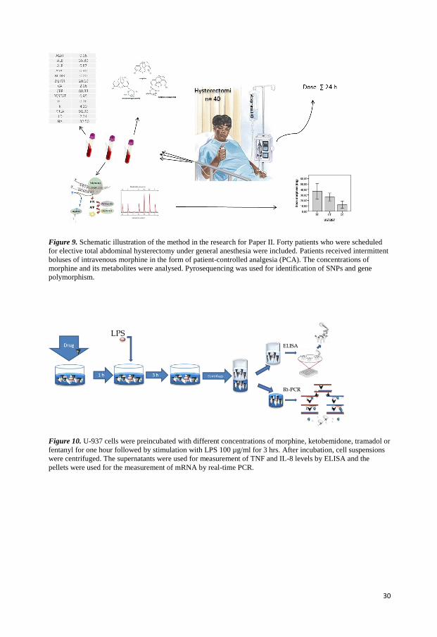
In the study for the second paper 40 patients undergoing total abdominal hysterectomy were
included (Figure 9). Patients received analgesia by PCA (patient controlled analgesia)-
device. Blood samples were taken from each patient 24 hours after the start of PCA. Totally,
three blood sample tubes were collected; one for measuring morphine and its metabolites in
plasma, one for defining creatinin and liver status, and a third tube for DNA extraction prior
to genotype analysis. Morphine is, unfortunately, also a common drug finding in forensic
autopsy cases and interpretation of the postmortem result may be facilitated by additional
information about an individual’s genotypes. We genotyped approximately 200 autopsy cases
that were found positive for morphine to find out if the genotype distribution was similar in
this group compared to a group of postoperative patients given intravenously administered
morphine.
30
Figure 9. Schematic illustration of the method in the research for Paper II. Forty patients who were scheduled
for elective total abdominal hysterectomy under general anesthesia were included. Patients received intermittent
boluses of intravenous morphine in the form of patient-controlled analgesia (PCA). The concentrations of
morphine and its metabolites were analysed. Pyrosequencing was used for identification of SNPs and gene
polymorphism.
Figure 10. U-937 cells were preincubated with different concentrations of morphine, ketobemidone, tramadol or
fentanyl for one hour followed by stimulation with LPS 100 µg/ml for 3 hrs. After incubation, cell suspensions
were centrifuged. The supernatants were used for measurement of TNF and IL-8 levels by ELISA and the
pellets were used for the measurement of mRNA by real-time PCR.
31
In the study for the third paper, 19 healthy volunteers were randomly divided in two groups.
TRA 50 or 100 mg was administered orally and blood samples were collected during 72
hours after administered dose. The concentration of TRA and its main metabolite ODT were
measured in blood. Blood samples were also used for genotyping analysis.
The study for the fourth paper was an in vitro study (Figure 10). The human histiocytic
lymphoma U-937 cell line was used in all experiments. The human monocytoid cell line U-
937-1 displays monocytic characteristics and has served as a robust in-vitro model for the
study of various aspects of monocyte and macrophage differentiation, intracellular signaling
pathways and intracellular kinases etc [136]. U937 cells secrete many cytokines, chemokines
and growth factors, thereby resembling human monocytes and macrophages and has served
as a suitable model to study biochemical and therapeutic aspects of cytokines. A recent study
has, through cytokine antibody array analysis, confirmed that TNF and IL-8 are secreted by
U-937 cells and more specifically that IL-8 secreted by U-937 cells seems to be involved in
fibronectin expression in breast cancer cells by stimulating PI3K/Akt pathway [137]. The
present study was designed to examine the dose response relationships between opiates and
their effects on the immune system, more specifically cytokine release. Although, we did plan
to use human peripheral blood leukocytes initially, we chose instead to use the U-937 cells as
a more robust system where we could perform many repetitive experiments with low inter-
individual variation. Our assumption was that once we had a clear idea of the optimal dose of
individual substances, we could then move on to other cancer cell lines and finally human
leukocyte sub-sets. Since all these experiments cannot be performed in parallel, we chose
therefore to perform comprehensive dose-response studies using only one reliable cell-line.
EXPERIMENTAL METHODS
Since all of the techniques used in this research are both well-known and established, only a
brief summary describing the principle will be presented here. Detailed information regarding
the use of individual techniques is provided in the individual studies.
PYROSEQUENCING TECHNOLOGY
Pyrosequencing is a method of determining the order of nucleotides in DNA based on
sequencing by synthesis [138, 139]. This method requires a single stranded sequencing
template. In the first step, the DNA sequence of interest is amplified by PCR by using one of
the primers biotinylated (Figure 11). The PCR amplicon is immobilised to streptavidine-
coated sepharose beads and denatured with NaOH.
A sequencing primer is hybridized to the single-stranded PCR amplicon. All necessary
components for amplification such as, DNA polymerase, ATP sulfurylase, luciferase,
apyrase, adenosine 5' phosphosulfate (APS), and luciferin are added. Deoxribonucleotide
triphosphate (dNTP) is added to the reaction, one sort at a time, in a predetermined
dispensing order (Figure 12). DNA polymerase catalyzes the incorporation of the dNTPs into
32
Figure 11. Single stranded template preparation by polymerase chain reaction (PCR), using one of the primers
biotinylated (B). The vials containing DNA are heated to a temperature of 90-94 °C to both activate the DNA
polymerase and to separate the two DNA strands (denaturation). In the next step the temperature is lowered to
approximately 50-65 °C for 20-40 seconds to allow binding of the primers to the single stranded DNA
(annealing). In the next step the temperature is raised to an optimal temperature for DNA-polymerase activity,
75-80 °C (normally 72°C). In this step the DNA polymerase synthesizes a new DNA strand complementary to
the DNA template strand by adding dNTPs (elongation)
Figure 12. Schematic diagram of the pyrosequencing reaction.
Published with permission from Qiagen
33
the DNA strand only if it is complementary to the base in the template strand. Pyrophosphate
(PPi) is released in a quantity equimolar to the amount of incorporated nucleotide after each
incorporation event, and is then converted to ATP in the presence of APS. A burst of visible
light is generated when ATP converts luciferin to oxyluciferin and is detected by a charge
coupled device camera and seen as a peak in a pyrogram. The height of each peak is
proportional to the number of nucleotides incorporated. Apyrase, a nucleotide-degrading
enzyme, continuously degrades unincorporated nucleotides and ATP. As the process
continues, another nucleotide is added, the complementary DNA strand is built up and the
nucleotide sequence is determined from the signal peaks in the pyrogram trace.
Figure 13. representative pyrogram for different allelic variants of OPRM1 A108G gene. A. homozygous for
118A; B. heterozygous; C. homozygous for 118G. R= A/G.
34
The method is simple, usually suited for sequencing of shorter fragments, 20-30 base pairs of
the target sequence. An important feature of this method is that the technique is very rapid,
making it feasible for large-scale studies. It is possible to analyze 96 samples simultaneously
within 10 minutes. The technique is less cumbersome compared to for example Sanger
sequencing as there is no need for electrophoresis or labeled primers and sequencing
reactions are continuously monitored in real-time.
This method was used for genotyping analysis in the studies for papers II and III. Genotyping
of CYP2D6 and UGT2B7 T802C (rs 7439366) were performed at the Department of Forensic
Genetics and Forensic Toxicology, Linköping. Genotyping of CYP2D6 included three SNPs;
CYP2D6*3 (rs35742686), CYP2D6*4 (rs3892097) and CYP2D6*6 (rs5030655), and
determination of copy number variation (CNV), which includes identification of whole gene
deletion, CYP2D6*5, and multiple gene copies (CYP2D6xN) [140, 141].
Genotyping of OPRM1 included one SNP A118G, (rs1799971) and ABCB1 four SNPs
G1199A, C1236T, G2677T/A and C3435T were performed at the Department of Clinical
Chemistry; County Council Linköping.
Some details in template preparation varied between these two departments, like denaturing,
annealing and elongation time, annealing temperature and the sample’s final volume.
Detailed steps are described in the individual studies.
The allele identification was performed by the instrument based on the height of each peak in
the programs. Figure 13 shows representative pyrograms for individuals with AA (A), AG
(B) or GG (C) allelic variants to OPRM1 A118G gene.
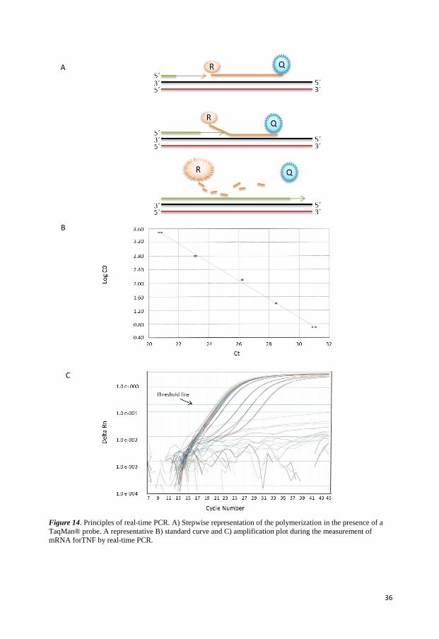
REAL-TIME PCR
Real-time polymerase chain reaction is used to amplify and simultaneously quantify a
targeted molecule [142]. The procedure follows the general principle of PCR with the
difference that the amplified DNA is detected as the reaction progresses in real-time. The
simplest and cheapest way to monitor a PCR in real-time is to add fluorescent dyes to pieces
of DNA complimentary to the gene of interest, which is known as a probe. The most
commonly used type of probe is the Taqman- probe that is labeled with a fluorescent reporter
molecule at one end and a quencher molecule (capable of quenching the fluorescence of the
reporter) at the other end. The quencher rapidly absorbs any light energy emitted by the
fluorescent molecule, as long as it remains in close proximity. During the annealing process
the probe binds to its complimentary sites on DNA between the primers. As the polymerase
pass through, the probe is disassembled and the quencher dye is separated from the receptor
dye. In the absence of a nearby quencher, the fluorescent molecule can now emit detectable
light the intensity of which is directly proportional to the number of PCR products generated
in the exponential phase of the reaction. The real-time PCR instrument generates an
amplification plot that represents the accumulation of product over the duration of the entire
PCR reaction by plotting fluorescence against the cycle number (Figure 14). Quantification is
35
possible by measuring the amount of amplified product at each stage during the PCR cycle.
For distinguishing relevant amplification signal from the background a threshold of the PCR
reaction is usually determined. The threshold can be set at any point in the experimental
phase of PCR reaction. Threshold cycle (Ct the cycle number at which the fluorescent signal
cross the threshold) is used to calculate the initial DNA copy number. Ct value is inversely
related to the amount of starting template, the lower Ct value the more starting template.
Thus if two amplification plots are compared it is easy to deduce which sample contained the
greatest amount of the DNA of interest by the Ct value. For example if a sample has Ct
value=30, and another sample Ct value=34. The sample with Ct value 34 contained 16 (24)
times more of the gene of interest than the red sample. A dilution series of known template
concentration is normally used to establish a standard curve for determining the initial
starting amount of the target template. To achieve accurate and reproducible expression level
between experiments, it is critical to use reliable internal control genes. Frequently used
reference genes for real-time PCR are GAPDH, β-actin and 18S rRNA.
The output from a real-time PCR is in the form of a graph showing each PCR cycles (1 cycle
consist of: denaturation (90°C), annealing (50°C) and elongation (72°C)) against the
fluorescence intensity.
In the study for paper IV we used two-step real-time PCR to quantify the level of mRNA for
TNF and IL-8. RNA from U-937 cells was isolated after stimulation of the cells with
different concentration of studied opioids. A cDNA strand was synthesized from the RNA
using purified reverse transcriptase. The cDNA was then used in real-time PCR. This method
is known as two-step real-time PCR. We chose this method because it provides an accurate
and reproducible quantification of gene expression. This method has been widely used for
quantification of gene and genomes [143]. This method offers faster and higher throughput
assays compared to the other methods. Unlike the other quantitative PCR methods, real-time
PCR does not require post-PCR handling such as gel electrophoresis or plate capture
hybridization.
Two housekeeping genes, cyclophilin (huCYC) and GAPDH, were tested as an internal
control; GAPDH was chosen as there were no considerable differences between these two
genes in our experiments. An absolute real-time quantification method was used in which a
standard curve was generated. The standard curve that produced a linear relationship between
Ct and initial amount of total cDNAwas used for determination of the cDNA concentration
for TNF and IL-8. The mRNA can also be used as a standard but it has been shown that
cDNA have a larger quantification range and greater sensitivity, reproducibility and stability
than RNA standards. However, using cDNA is more time consuming with a higher risk for
laboratory contaminations.
36
Figure 14. Principles of real-time PCR. A) Stepwise representation of the polymerization in the presence of a
TaqMan® probe. A representative B) standard curve and C) amplification plot during the measurement of
mRNA forTNF by real-time PCR.
A
C
B
37
ENZYME-LINKED IMMUNOSORBENT ASSAY (ELISA)
ELISA is a sensitive and specific method for the detection and quantification of substances
such as peptides, proteins, antibodies, hormones, cytokines, drug of abuse and their
metabolites. In an ELlSA, an antigen or antibody is coated on a solid phase, i.e. the well of a
microtiter plate (Figure 15). Samples containing the substance are added to the well and
incubated allowing the antibody to form a complex with the coated material on the surface.
The complex remains after washing and can be detected by an enzyme-antibody conjugate.
The most common enzymes used are alkaline phosphates (AP) or horseradish peroxidase
(HRP). The resulting enzyme activity is measured by substrate color development. Color
formation is related to the concentration of the target substance.
We used ELISA for quantification of TNF and IL-8 in the paper IV. TNF release from U-937
cells was analyzed using an ELISA-kit for human TNF-α from Mabtech AB (Stockholm,
Sweden, # 3510-1H-20). For IL-8 analysis an ELISA kit from R&D Systems (Minneapolis,
USA, Cat. No DY208) was used.
Both assays were based on the use of a combination of two coating antibodies. The substrate
used for streptavidine horseradish peroxidase was Enhanced K-blu®, a ready to use substrate
from Neogen Corporation (Lansing, MI, product # 308175), and stop solution was HCl (1
M). Optical density was read at 450 nM in the ELISA plate reader
Figure 15. Illustration of indirect. ELlSA
38
HIGH PERFORMANCE LIQUID CHROMATOGRAPHY (HPLC)
HPLC is a chromatographic technique used to separate a mixture of compounds with the
purpose of quantifying and identifying the individual components of the mixture. This
technique is based on forcing the analyte in a liquid (mobile phase) through a column packed
with small round particles with a certain surface chemistry (stationary phase). The analyte is
retarded by specific chemical or physical interactions with the stationary phase as it passes
through the column. The amount retarded depends on the nature of the analyte, stationary
phase and mobile phase. In order to identify any compound by HPLC a detector must be first
selected. The choice of a detector depends upon the characteristics and concentrations of the
compounds that need to be separated and analyzed. Mass spectrometry is a powerful detector
for the analysis of organic compounds. It is designed to separate ions in gas phase according
to their mass. The mass spectrometer consists of three major components, an ion source that
generates ions at atmospheric pressure, a mass analyser which filters ions, and a detector that
detects ions. HPLC-tandem mass spectrometry (LC-MS/MS) offer greater detection limit,
higher specificity compared to the other analytical methods such as immunoassays or
conventional UV [144]. However the technique can only be used as long as the analyte can
be suitably ionized. The complexity of the instrumentation, long throughput, risk for ion
suppression and large variation between laboratories are some of the factors limiting its use.
LC-MS was used for analysis of TRA and its metabolite ODT in paper III.
WESTERN BLOT
Western blot, also called immunoblot is an analytical technique used to detect specific
proteins in a cells, tissues, organs or body fluid. The technique is based on the reaction
between antibody and the target protein. The protein is usually applied on a gel matrix and
separated by electrophoresis. Then the gel is placed next to a thin, synthetic membrane that
has a strong affinity for proteins (Figure 16). As a result, the proteins in the gel are
transferred to the membrane that is incubated with an antibody to a specific protein. Then a
horseradish peroxidase-conjugated secondary antibody is added which binds to the primary
antibody. The membrane is incubated with a substrate that is converted to a luminescent
compound after reaction with this enzyme. The signal is captured on a film which is usually
developed in dark room. It is important to be aware that the data produced with a western blot
is typically considered to be semi-quantitative for two reasons; first there are variations in
loading and transfer rates between the samples in separate lanes; second the signal generated
by detection is not linear across the concentration range of samples.
39
Figure 16. Illustration of Western blotting
STATISTICS
All statistical calculations during the research for this thesis have been performed in
consultation with a statistician from the Department of Computer and Information Science at
the Linköping University
The statistical analyses were performed with the SPSS software package version 15 or higher
(IBM SPSS Statistics). P <0.05 was considered as statistically significant.
Analysis of variance (ANOVA) was used in Paper I and III to investigate the difference
within and between groups. Bonferroni’s, Tukey’s and Scheffé’s Post Hoc test was used for
pairwise comparisons.
Chi Square test was used to estimate whether the studied populations in paper II met the
principle of Hardy-Weinberg equilibrium (HWE). The Mann-Whitney test, a non-
paramethric test, was used to compare allelic variation in genes between groups. Regression
analysis was performed to analyze associations between the different variables such as allelic
variation in genes and dose and concentration of morphine. Multiple regression analysis was
used to investigate the association between all polymorphism on all three genes and dose and
concentration of morphine. All the assumptions for the linear regression analysis were
verified (normality and linearity of the residuals as well as collinearity).
40
41
RESULTS AND DISCUSSION In this part the most important results from the studies leading to this thesis are discussed. Most of the results are discussed in detail in the individual studies.
EFFICACY OF TOPICAL APPLIED MORPHINE (PAPER I) Chronic leg ulcers are a major health problem and have a detrimental impact on quality of life due to associated pain. Some clinical reports have suggested that local administration of morphine could be beneficial due to limited local effect and thereby potentially lower frequency of side effects in many clinical conditions such as pressure ulcers. In the study for first paper we investigated the effect of topically applied morphine gel in painful leg ulcers. The study was a randomized, placebo controlled, double blind cross-over study (Fig. 8). Twenty one patients were randomly assigned to receive either morphine or placebo of which 17 completed the study. A low dose of morphine was administered, as we wanted to avoid systemic effects of morphine. The amount of morphine applied was calculated based on the ulcer size (0.5mg/cm2). A gel containing either 1, 2 or 3 mg/ml morphine was used depending on the size and depth of ulcers. The pain was measured by the VAS. Patients were asked to report their pain experience on a documentation sheet during 24 hours after application of gel. The morphine/placebo gel was applied on leg ulcers at four consecutive dressing changes.
The aim of this study was to compare the treatments results on pain intensity. We found the VAS most suitable and reliable for this purpose compared to the other pain assessment scales. Self-reporting of pain by patients is strongly influenced by multiple contextual factors. We measured only one dimension of pain -the intensity of pain- by using VAS. Initially, pain increased after application of both morphine and placebo gel which could be partly due to the dressing change procedures such as cleaning and washing the ulcers. Pain was significantly reduced in both groups of patients two hours after application of gel (Figure 17). The mean pain scores were lower in the morphine group compared to the placebo group but the difference was not statistically significant.
We opted for a cross-over design as we wanted to evaluate morphine versus placebo under similar conditions. More specifically we had hoped that each individual patient would have similar pain scores before entering the morphine or placebo arm of the study. Unfortunately, most patients varied in their baseline pain scores at different treatment occasions. Indeed when patients had an initial high pain score (occasion 1 and 2), morphine gel induced a significant reduction of pain. The mean pain value in the third and fourth treatment occasions was lower initially. It could, therefore, be speculated that a parallel group design with a single-point comparison would have been more suitable. However, a parallel group design would probably require even higher number of patients to obtain sufficient power.
42
Figure 17. Overall efficacy of morphine/placebo gel. Mean pain scores ± SE at different measurement time points for all patients.
Another factor which could have an impact on our results is the degree of inflammation which has been identified as an important regulatory stimulus of opioid receptor expression. [63, 64, 69]. We assumed that most leg ulcers are almost always inflamed and we had therefore not opted to have signs of inflammation as obligatory inclusion criteria. However, to our knowledge there are no established, objective, methods for measurement of the degree of inflammation available today. The method that has been used earlier is a subjective assessment of swelling, heat sensitization, loss of function and redness [145].
Leg ulcers are a very common type of chronic wound [146]. One of the problematic features of leg ulcers is that they can take years to heal and the pain can become persistent/ chronic [147] Recent neurophysiology studies have shown that chronic pain can be associated with substantial functional and structural changes, or plasticity, in the central nervous system [148], a process also known as central sensitization. The effect of opioids alone on persistent pain is relatively limited [149]. It could be argued that even if the administered opioid had been effective in relieving persistent pain, a low dose of morphine administered locally can hardly be expected to affect central sensitization. It is possible that an analgesic effect could have been obtained if topical morphine had been applied before the patient had developed persistent pain.
Only a few case reports and no clinical studies were reported at the time we planned and designed this study (2002). As far as we know, this pilot study is among the first and one of the largest randomized clinical studies conducted in patients with chronic leg ulcers. Although an overall, clinically relevant, reduction of pain was observed upon treatment with
43
morphine, the difference was not statistically significant. A power calculation based on these observations revealed that we would have needed to have a large number of patients (1000) to ensure the statistical significance of treatment with topical morphine. Such a large study was beyond the scope of this research and the question remains open for other groups to pursue.
The relatively small number of patients included in our study and other methodological limitations makes it difficult for us to draw general conclusions regarding the efficacy of topically applied morphine as an effective treatment for some painful ulcers. Nevertheless, we cannot exclude the possibility that topically applied morphine could be useful in some individuals. Some of patients included in this study found this treatment effective and therefore continued even after the study period. It is important to note that this gel is already in use today for treatment of painful ulcers in clinical practice.
Further studies are however warranted to evaluate the value of topically applied morphine in the treatment of patients with chronic painful leg ulcers.
GENETIC POLYMORPHISM AND RESPONSE TO OPIOIDS (PAPER II AND III) Opioids are the drug of choice and are commonly prescribed for the treatment of pain. Each patient may respond differently to specific opioids. The minimal effective analgesic concentration of opioids required for satisfactory analgesia varies considerably among patients [150]. At present, we cannot predict which patients are likely to achieve good analgesia or develop adverse effects. Many factors, including genetic variation, have been proposed to contribute to these differences in opioid responsiveness. It has been suggested that the allelic variants in the genes involved in opioid metabolism, absorption and receptor function may affect the efficacy of morphine in humans.
In Paper II and III we have performed a comprehensive analysis of SNPs in four genes CYP2D6, UGT2B7, OPRM1, and ABCB1 simultaneously and investigated their influence on pharmacokinetic and pharmacodynamic properties of morphine and TRA. The results presented in paper III are part of a larger study. The aim of the study is to measure concentrations of TRA and its metabolite in blood, urine and hair. We also intend to follow enantiomers of both TRA and metabolites in these biological matrixes.
44
DRUG METABOLIZING ENZYMES AND RESPONSE TO OPIOIDS
Polymorphism in UGT2B7 and morphine in hysterectomy patients Polymorphism in UGT2B7 that primarily controls the metabolism of morphine has been associated with interindividual variability in the pharmacokinetics of morphine and its side effects [93-95, 106, 151-153]. In the study for paper II, we investigated the association of polymorphism in UGT2B7 T802C gene to dose and concentration of morphine and its metabolite. As mentioned before, this polymorphism leads to an amino acid change, histidine to tyrosine that may have an effect on the activity of UGT2B7.
In our study, the cumulative 24- hour postoperative dose of morphine varied considerably among patients (range 3-80), which confirms previous findings about the great interindividual variability. There was a clear association between polymorphism in UGT2B7 and dose of morphine. The dose of morphine required for pain relief decreased with increasing number of C alleles. Patients with CC allele required significantly lower dose of morphine compared to the wild type (TT). Consequently the mean concentration of morphine was also lowest in these patients (Figure 18) but these differences were not statistically significant. The lack of statistically significant differences could be due to the limited number of patients included in this study and the great variation in concentration of morphine in each genotyping group. However, even if we had a larger group of patients, there is no guarantee that the distribution between the groups would be as we had expected. It could be speculated that it is better to select patients based on their genotype. On the other hand, such a study would be difficult to implement.
We also observed that patients with CC allele had the highest ratio of concentration M6G/morphine. The higher concentration of M6G, the active metabolite of morphine, can partly explain why these patients required a lower dose of morphine. Our results were in agreement with previous finding that suggested a higher enzymatic activity of UGT2B7 in individuals with CC allele [72, 91].
Paradoxically, in some cases despite administration of the same dose, patients with CC allele had a higher concentration of morphine. It is possible that blood sampling occurred in conjunction to administration of a bolus dose of morphine, thus, introducing a sampling error. Unfortunately, we had no documentation about the last bolus dose taken which could be considered for planning of future studies.
45
Figure 18. Association of morphine and its metabolite to UGT2B7T802C polymorphism, A) Dose of morphine (mg), B) Concentration of morphine (mmol/L), C) Morphine-6-glucuronide (M6G)/ morphine-3- glucuronide (M3G) The values are shown as mean ±2 SE.*p<0.05
Polymorphism in CYP2D6 and tramadol healthy volunteers TRA is metabolized by Cytochrome P450 enzymes including CYP2D6. This enzyme is responsible for converting TRA to the active metabolite ODT. Studies in patients with postoperative pain demonstrated that patients with absence of CYP2D6 activity needed approximately 30% higher TRA doses than those with normal CYP2D6 activity, extensive metabolizers [154]. In paper III, we evaluated the correlation between different genotypes of CYP2D6 and the pharmacokinetic parameters of TRA in 19 healthy volunteers that received either 50 or 100mg TRA. Polymorphism in CYP2D6 has shown to be partly responsible for the interindividual variability in response to TRA. In our study, the ratio of ODT/TRA for Cmax and AUC were significantly correlated to the different genotypes of CYP2D6. PMs, (subjects with no functional genes) showed to have the lowest ratio followed by IMs (subject with one functional gene) and EMs (subjects with two functional genes) (Figure 19). We could also observe that PMs in this study had a lower incidence of drug related symptoms which is in accordance with previously published results. [155, 156].
Figure 19. The association between ratio ODT/TRA for Cmax and AUC and polymorphism in CYP2D6. *P<0.01 We found statistically significant differences between genotyping groups. This was despite limited number of subjects in each group (EM=8, IM =9) and great interindividual differences between these genotypes. Our results further emphasize the importance of this
46
polymorphism for TRA’s metabolism. The importance of polymorphism in CYP2D6 on TRA’s metabolism has been highlighted earlier [155-158].
Statistical calculations were not possible regarding the difference between PM and other genotype groups, because there were only two PMs and ODT was detectable only in one subject.
GENE POLYMORPHISM IN OPRM1 AND OPIOIDS EFFICACY Changes in the μ-opioid receptor, potentially contributed by allelic variants, have been proposed to produce changes in nociceptive responses [116, 159]. The most frequently studied OPRM1 variant is the A118G in which an adenine to guanine substitution exchanges an asparagine for an aspartic acid at a putative N-glycosylation site (N40D). This amino acid change has been associated with alteration in functional properties of OPRM1 and intracellular signalling pathway. However, results have not been consistent [160, 161].
Polymorphism in OPRM1 in hysterectomy patients It has been suggested that 118G homozygous (GG) are poorer responders to morphine than 118A homozygous (AA) or heterozygotes (AG) [116, 119, 162]. In our study, the mean value for the dose of morphine was almost identical for patients with AA and AG, while patients with GG genotype required almost twice as much morphine as patients with AA (Figure 20A). We identified only two patients with GG genotype and were unfortunately able to measure morphine concentration and its metabolites in only one patient. In this particular patient, the concentration of morphine was considerably higher (65.4 nmol/L), more than twice as much as in patients with AA genotype (25±18) (Figure 20B). Concentration of M6G was more than 2-fold higher in this patient (80.3 nmol/L) compared to patients with AA (33.0±23) (Figure 20C). The results for this patient are in agreement with previously published data. Since there are various methodological differences between these studies such as variation in study population, the route of drug administration and the type of surgery, it is difficult to draw any general conclusions
Figure 20. Association of morphine and its metabolite to OPRM1A118G polymorphism, A. Dose of morphine (mg), B. Concentration of morphine (mmol/L), C. Morphine-6-glucuronide (M6G)/ morphine- The values are shown as mean ±2 SE.
47
Polymorphism in OPRM1 in healthy volunteers
There is only limited information regarding the influence of OPRM1 A118G polymorphism on TRA efficacy and side effects. It has been suggested that individuals with GG allele variant have a considerably lower risk of side effects compared to AA [154] because these individuals may have a lower level of μ-opioid receptors [163]. Kim et al [154] hypothesized that ODT-binding to μ-opioid receptor is responsible for inducing the emetic response observed in TRA treatment.
In our study the mean values of drug related symptoms (DRS) were higher in the subjects heterozygotes to A118G (AG) (14±15.6) compared to subjects with AA allele (4±4.5). We did not have any subject with GG variant and only two with AG both in the 100 mg dosage group. Both subjects were IMs for CYP2D6 and had almost the same AUC for TRA (2156, 2116 ng h/g respectively). One of these subjects reported a significantly higher score of drug related symptoms (score = 25), compared to the other one (score =3). The subject with higher score vomited twice and fainted once. However, the time of vomiting at the first time was four hours after the drug intake and by this time the subject has already passed the tmax for both TRA and ODT (2:30h). The Cmax and AUC for ODT was almost three times lower (27 ng/g, 214ng h/g) in this subject (higher score) compared to the other one (98ng/g, 656ng h/g). This is in conflict with the earlier results. However, other unknown factors could have an impact on this particular patient’s reaction to TRA.
If this subject were excluded from the statistical analysis, the results show a lower score of DRS and higher mean value of AUC for both TRA and ODT in the only subject with AG compared to subjects with AA in the group of 100 mg dosage which is then in concordance with the results from a previous study [154].
One of our limitations was the low statistical power; however this study was not primarily designed for evaluation of drug related symptoms. Further a self-reported questionnaire is always accompanied with validity problem due to its subjectivity.
Although, our results are in accordance with the results from previous studies, the association of polymorphism in OPRM1 to the side effects of TRA needs to be confirmed by other researchers.
GENE POLYMORPHISM IN ABCB1 AND OPIOIDS ABCB1 genes codes for P-glycoprotein (P-gp), which acts as a drug reflux pump at the BBB as well as in the intestines and kidneys. A variety of allelic variations in the ABCB1 gene has been suggested to be associated with variation (decrease or increase) in the efflux activity of many drugs, including the non-synonymous SNPs G1199A and G2677T/A as well as the synonymous SNPs, C1236T and C3435T [164]. In the paper II and III we investigated the correlation of SNPs in these genes to the concentration of morphine and TRA.
48
Polymorphism in ABCB1 in hysterectomy patients There are conflicting results in the literature regarding the influence of SNPs in ABCB1 gene on both effects and side effects of morphine [116, 118, 152]. Campa et al. found that variability of pain relief in 145 patients on morphine treatment was significantly associated with C3435T. The authors suggested that TT cariers were good responders while those with CC or CT were moderate responders. They argued that the absorption of morphine is reduced in CC carriers due to effective efflux by P-gp in gut and/or through the blood-brain barrier, and consequently reducing bioavailability of morphine for receptors in brain. Conversely, TT carriers with abnormal function of P-gp should have a higher concentration of morphine.
The results from our study showed that the mean values of morphine dose was lower in patient homozygous for 1236T, 2677T and 3435T compared to the other genotypes, while the concentration of morphine was found to be higher in homozygous for 2677T and 3435T (figure 21). However these results were not statistically significant.
In contrast to our results, Sia et al [116] found that patient homozygoue for C3435 (CC) required a lower dose of morphine for pain relief after caesarean section in a large number of patients.
We are unable to draw any conclusions about the association between genetic polymorphism in ABCB1 gene and analgesic effect of morphine on the basis of data presented in this thesis. Further studies are therefore warranted.
Figure 21. The association between I) dose and II) concentration of morphine and polymorphism in ABCB1. A) C1236T, B) G2677T/A, C) C3435T. Data are presented as Mean± 2 SEM
49
Polymorphism in ABCB1 in healthy volunteers Experimental studies have shown that opioids are substrates for the transporter protein P-gp [132, 165-167], however its importance for TRA remains unknown. Few reports in the literature have focused on the functional involvement of P-gp in pharmacokinetic properties of TRA [168-170]. The results of these reports are inconsistent. An in vitro and one in vivo study in rats have demonstrated that TRA and ODT are not P-gp substrates [168, 169]. In contrast, a clinical study suggested that TRA is a substrate of P-gp [170].
In a previous study, Slanar et al [170] showed that Cmax and AUC0-24 increased slightly with increasing numbers of 3435T alleles when the subjects were grouped as EM, IM and PMs of CYP2D6. Similar analysis of our material showed that Cmax and AUC for TRA in subjects was higher in homozygous for 3435T (TT) compared to the other genotypes (figure 22). However, this relationship was found only for subjects in the 50 mg dosage group and not in the 100 mg dosage group. In contrast with the results from Slanar’s study, where no association was found between G2677T/A and pharmacokinetics of TRA, we found a significant increase in AUC with an increasing number of 2677T alleles. Even here, this correlation was found only in the 50 mg dosage group. A similar trend was observed for Cmax.
In the 50 mg dosage group homozogotes to 1236T, 2677T and 3435T had significantly higher AUC for TRA compared to the other genotypes. All other pharmacokinetical parameters, AUC for ODT, Cmax, tmax , for both TRA and ODT were almost the same between the different genotyping groups.
Figure 22. The association between AUCTRA I) 50 mg dosage group and II) 100 mg dosage group and polymorphism in ABCB1. A) C1236T, B) G2677T/A, C) C3435T. Data are presented as Mean± 2 SEM
50
Taken together, the role of the studied SNPs in ABCB1gene on pharmacokinetics of tramadol remains unclear. We had a limited number of patients in each genotyping group when the individuals were grouped by CYP2D6 genotype and dosage group. It is possible that a study involving subjects with predetermined genotype would be preferable, which would, however, entail the recruitment of significantly larger groups.
ACCUMULATED SIGNIFICANCE OF ALL POLYMORPHISMS As we discussed earlier there was a great variability in the concentration of morphine and dose required for pain relief in hysterectomy patients after surgery. In order to find out how much of this variation could be explained by the investigated SNPs altogether, we performed a regression analysis. Results from this analysis showed that only 30% of the variability in dose and concentration of morphine in our material could be explained by genetic variation in the investigated SNPs taken together. A similar analysis in healthy volunteers, a more homogenous group, showed that about 80% of the variation in the ratio of ODT/TRA for AUC and Cmax could be explained by these six SNPs.
Pain is complex and multifactorial. Biological mechanism other than genetic factors involving drug distribution could also contribute to the variability in response to morphine. Recently, the influence of 112 SNPs in 23 candidate genes on opioid efficacy was studied in a large number (approximately 2000) of patients with cancer [171]. The authors concluded that these SNPs could not explain the variation of opioid requirement in cancer patients.
Current knowledge still does not allows us to tailor individualized pain therapy. Our data and other studies could assist in providing information and tools to identify patients that may benefit by dose escalation or are at risk for developing side effects.
OPIODS IN FORENSIC TOXICOLOGY
Forensic toxicology About 5500 cases of forensic autopsies are performed at The Departments of Forensic Medicine in Sweden each year [172]. The objective of these forensic autopsies is to investigate unnatural death and to eliminate the possibility of criminal intent in these out-of-hospital deaths. These autopsies involve extensive sampling of body fluids for toxicological screening. When it comes to opioids, the accurate interpretation of these test results requires an understanding of the relationship between the opioid concentration at the time of death and the concentration in the sample collected for measurment. Some authors have highlighted the relevance of genetic disposition for determining the cause and/or manner of death suggesting that interpretation of the postmortem result may be facilitated by additional information about an individual’s genotypes [173]. In paper II and III we examined if the knowledge about the
51
victims genotypes in genes associated with drug distribution, could facilitate the interpretation of concentration of opioids.
In paper II, approximately 200 autopsy cases where morphine was detected in peripheral blood, were genotyped. The mean concentration of morphine found in blood was 80 nmol/L, which is almost three times higher than the mean value of morphine concentration in hysterectomy patients.
The allele frequencies in patients were compared to the forensic cases and the data published on the HapMap data base (http://hapmap.ncbi.nlm.nih.gov). We observed that the frequency of UGT2B7 802C and ABCB1 1236T allele was higher in forensic cases than the other group However we also found that the frequency of OPRM1 118G allele was lower in forensic cases, pointing out that these allele variants can be a risk factor in the forensic cases.
There was a lack of information about M6G concentration in our study in autopsy cases. In future studies it would be of interest to investigate the association of the ratio of M6G/morphine to the different genotypes in UGT2B7.
Interpretation of the time of tramadol intake TRA is the most widely sold opioid analgesic in the world and is registered and marketed in more than 100 countries. Abuse of this drug is becoming more and more common, mainly because in most countries TRA is the only available opioid that is not restrictively controlled. A similar trend was observed in cases of overdoses [174]. TRA has also become a usual cause of death in drug addicts [175].
In paper III we investigated if the knowledge of the blood concentration ratio of ODT/TRA and genetic variation in genes associated with drug distribution could facilitate interpretation of the time of drug intake. Nineteen healthy volunteers were treated with a single oral dose of TRA, ten subjects were given 50mg and nine 100mg. Blood samples were collected at defined intervals up to 72 hours after drug intake. All subjects were genotyped for CYP2D6, OPRM1 and ABCB1. The concentrations of TRA and ODT in whole blood were determined by LC-MS-MS analysis. TRA and ODT could be detected for all subjects up to 10 hours by this routine analysis. ODT/TRA was positively correlated to time after drug intake (figure 23). Both TRA and ODT could be detected up to 24 hours in some subjects. There was considerable differences in this correlation between the PMs, IMs and EMs and CYP2D6. EMs had the highest ratio and PMs the lowest. This result indicates that the knowledge of an individual’s CYP2D6 genotype is necessary to estimate the time of drug intake. To the best of our knowledge there is a limited documentation about the correlation of genetic variation in CYP2D6 with the ratio of ODT/TRA over the time. Levo et al demonstrated that the ratio of TRA/ODT was well correlated to the different phenotypes of CYP2D6 in post-mortem blood [176] which is in line with the results from our study.
52
Figure 23. The mean blood concentration ± SEM for ODT/TRA at each time-point after dividing the subjects into CYP2D6 genotypes. Subjects with: no active CYP2D6 genes are classified as a PM, poor metabolizer (n=1), one active gene as IM, intermediate metabolizer (n=9) and two active genes as EM, extensive metabolizer (n=8).
EFFECT OF OPIATES ON TNF AND IL-8 (PAPER IV) Most patients with advanced cancer experience severe pain and are often treated with opiates. Cancer patients are especially susceptible to opportunistic infections due to treatment with immunosuppressive and cytostatic drugs. Opiates influence cytokine expression, phagocytic activity, natural killer cell activity and lymphocyte activity [177-180].
Since opiates have been demonstrated to have immunomodulatory effects ([178-180] , it is of significant clinical importance to evaluate potential differences between commonly used opiates with regard to their effect on the immune system.
THE ROLE OF TNF AND IL-8 IN CANCER In this study we evaluated the effect of four commonly used opiates in Sweden, morphine, TRA, fentanyl and ketobemidone, on TNF and IL-8 release. TNF has a dominant role among all proinflammatory cytokines mediating the inflammatory response in acute and chronic inflammation by driving the inflammatory cascade resulting in increased production of other cytokine and chemokines including IL-8 [181]. There is now ample evidence suggesting that pro-inflammatory cytokines, particularly TNF and IL-6, have a role in carcinogenesis by promoting the survival and proliferation of malignant cells [182]. Cytokines have dual roles
y = 0,0031x + 0,0709 R 2 = 0,9218
y = 0,0215x + 0,1347 R 2 = 0,958
y = 0,0312x + 0,2646 R 2 = 0,9659
0.0
0.1
0.2
0.3
0.4
0.5
0.6
0.7
0 2 4 6 8 10 12
OD
T/TR
A c
once
ntra
tion
ratio
s .
Time (h)
53
in cancer, on the one hand activating oncogenic pathways in malignant cells and on the other hand regulating the inflammatory microenvironment that influences tumor progression[182]. Histochemical studies in patients have demonstrated that co-coordinated expression of TNF and IL-1 in breast cancer have causative roles as tumor promoting factors [183]. TNF, IL-1, IL-6 have also been shown to promote epithelial ovarian cancer genesis, growth and progression [184].
Chemokines and their receptors are involved in neutrophil and monocyte cell trafficking [185]. IL-8, originally discovered as a chemotactic factor for leukocytes, is also produced by various normal and tumorigenic human cells. A recent study has confirmed that TNF and IL-8 are secreted by U-937 cells and more specifically that IL-8 secreted by U-937 cells seems to be involved in chemotactic migration and spreading of inflammatory breast cancer cell lines[137]. IL-8 is also a promoter of tumor angiogenesis, thereby contributing to metastasis and recurrence of disease [185]. The advent of anti-angiogenesis therapy in cancer emphasizes the need for optimal biomarkers to predict response and disease progression and to further understand the modulation of various angiogenesis promoting factors like IL-8 [186]. Taking all the above facts into consideration, we reached the conclusion that TNF and IL-8 could be suitable candidates to explore the effects of opioids on soluble mediators. Similar studies on other inflammatory mediators are of course warranted and will be undertaken in the future.
DOSE DEPENDENT EFFECT OF OPIOIDS ON TNF AND IL-8 We found a dose dependent inhibition of TNF release by ketobemidone, morphine and TRA but not by fentanyl (figure 24). The effect of opiates on IL-8 release was slightly different. Morphine and fentanyl had no significant effect on IL-8 release whereas ketobemidone and TRA resulted in a significant reduction of IL-8 release. Our data suggest that the order of potency is similar for both cytokines and is as follows: TRA > ketobemidone > morphine > fentanyl. Previous studies have demonstrated that the immune modulating effects of opioids are a direct result of interaction of opiates with both the opioid receptors [187] and non-opioid receptors such as serotonin, dopamine and NMDA receptors [187-189]. TRA´s dual action on both μ-, serotonin- and norepinephrine receptors might explain the observed potency. Since both, fentanyl and morphine are highly selective for the μ -opioid receptor; they should theoretically exert similar effects on the immune system. However, the results of our study do not support this theory, but instead suggest the existence of an alternative mechanism of action. There is some evidence about the existence of a low-affinity, naloxone- insensitive morphine-binding site on human peripheral blood macrophages, granulocytes and monocytes, [190]. This receptor is apparently activated by morphine but not fentanyl [190], which could partly explain the lack of an inhibitory effect of fentanyl on TNF and IL-8 release observed in this study.
54
Figure 24. Dose-dependent effect of A) morphine, B) ketobemidone, C) tramadol and D) fentanyl on LPS dependent I) TNF and II) IL-8 release from U-937 cells. The value represents as a mean of observation ± 95% CI. P-value is shown in those cases where the difference between LPS and opioids was statistically significant. * p<0,05 (+)
INTRACELLULAR SIGNALING PATHWAY To further understand the intracellular signaling pathways and the receptors involved in mediation of the effect of tested opioids, we measured the mRNA production of TNF. In these experiments we used naloxone, a competitive antagonists to opioid receptors that normally reverses the effects of opioids. In congruence with our earlier findings regarding release of cytokines all tested opioids except fentanyl had an inhibitory effect on mRNA
55
production for TNF or IL-8 (figure 25). Naloxone not only failed to reverse the observed effects of opiates on mRNA, but also paradoxically exhibited a synergistic effect on mRNA production. Based on these observations it can be speculated that the effect of opiates on TNF and IL-8 might be mediated by naloxone-insensitive receptor/s or through a direct non-receptor mediated effect of these substances. We have also investigated the involvement of intracellular signaling pathways, Erk and Akt by Western Blot. Results indicated that these protein kinases might be involved, however further studies are needed to dissect the intracellular pathways involved in mediating the effects of opiates including Erk and AKT pathways.
Figure 25. Real-time RT-PCR for A) TNF B) IL-8 mRNA isolated from U-937-celler. The figure shows effect of morphine 1.5 mM, ketobemidone 1.75 mM, tramadol 4.2 mM and fentanyl 2.3 μM on LPS stimulated U-937 cells. Naloxone were used to reverse the effect of opioids. The experiments were performed in triplicate on six different occasions. Values are represented as mean value ±95% CI. Dexamethasone were used as an internal control.
56
57
CONCLUSIONS The following conclusions can be drawn, based on the results from this thesis
- Both morphine and placebo gel could significantly reduce pain in leg ulcers two hours after application. Morphine reduced pain more than placebo but the difference was not statistically significant. Morphine could reduce pain considerably more than placebo in cases where VAS was four or higher initially (occasion I and II). Some patients found the treatment to be beneficial and chose to continue treatment even after the study period. Although our study was among the largest randomized studies conducted so far, it is difficult to draw any general conclusion about the efficacy of morphine in treating painful ulcers.
- The SNP T802C in UGT2B7 gene was shown to be correlated with both dose and concentration of morphine in hysterectomy patients. Patients homozygous for the 802C allele needed a significantly lower dose of morphine compared to the TT allele variants. A higher concentration of M6G, the active metabolite of morphine, could partly explain the observed lower morphine requirement.
OPRM1 A118G was also associated with both the dose and concentration of morphine. Patients with GG allele variant required a higher dose of morphine and consequently higher concentrations of morphine were observed in these patients. However, we found only two patients homozygous for 118G allele in our study.
There was no clear association between the SNPs in ABCB1 genes and concentration of morphine.
Taken together, all the above mentioned SNPs in concert could explain only 30% of the variation in concentration of morphine. It could be speculated that other, yet unknown variables, might also, contribute to the variation in the response to morphine.
Concentration of morphine was considerably higher in forensic cases compared to hysterectomy patients, which is probably a consequence of differences in allelic frequency in SNPs examined in this study.
- In the healthy volunteers AUC and Cmax for tramadol’s MR were influenced by CYP2D6 genotypes. PMs had significantly lower AUCMR and CmaxMR compared to the other genotypes, IMs and EMs.
Correlation between pharmacokinetic parameters of tramadol and other SNPs (OPRM1 A118G, ABCB1 G1199A, C1236T, G2677T/A, C3435T) was not clear. We did not find any clear association between these SNPs and drug related symptoms.
Almost 80% of the variation in AUC MR and CmaxMR in this material could be explained by these SNPs taken together and almost 60% by CYP2D6 genotypes alone.
58
This indicates that the knowledge of an individual’s CYP2D6 genotype is necessary to estimate the time of drug intake.
- The immunomodulatory effects of opioids varied between different opioid drugs. There was a dose dependent inhibition of TNF and IL-8 release by tramadol and ketobemidone in LPS stimulated cells. Fentanyl, however, had no effect on either TNF or IL-8 release and morphine had effect only on TNF release. The order of potency with regard to inhibition of cytokine release was as follows: tramadol > ketobemidone > morphine > fentanyl. These drugs had different effects on mRNA synthesis, the highest reduction was obtained by tramadol. Fentanyl had no effect on mRNA. The AKT and ERK intracellular signaling pathways showed to be also involved.
59
FUTURE ASPECTS Opioids are still one the most powerful groups of drugs for pain relief in many clinical conditions. Unfortunately, these drugs are also associated with potent and varied adverse effects. These side effects are often the main reason for discontinuation of opioid analgesic treatment. Development of opioid drugs lacking such effects has always been a major goal in pain research. Even though the results of our pilot study showed an inefficacy of morphine in patients with chronic leg ulcers, it is too early to abandon the idea of pain treatment via peripheral opioid receptors. As has been shown in the literature, pain from other diseases was successfully treated by topically applied opioids. Currently, there are some antagonists (alvimopan and methylnaltrexon) that specifically bind to peripheral μ-opioid receptors. These drugs have limited ability to cross blood brain barrier, thus, minimizing the unwanted side effects of opioids. It is of course desirable to develop an agonist for pain relief with pharmacokinetic properties resembling existing antagonists. It is strange that although the pain receptors were cloned and characterized decades earlier, we still lack the possibility of choosing between a number of drugs with different structures and mechanisms of action.
Pharmacogenomics (study of the linkage between an individual’s genotype and the disposition of drugs in the body) and personalized medicine has increased in importance and will hopefully in the near future become standard procedure to improve and predict the outcome of treatment results. Personalized medicine is rapidly replacing the traditional “trial and error” method in many therapeutic areas more specifically in cancer with the advent of new biological drugs. Genetic testing is now standard procedure in the treatment of many malignant conditions, assisting in providing the correct drug and dosage for the correct patient. We believe that there is sufficient evidence suggesting that pharmacogenetics should also have an equally important role in choosing the right analgesics. In this thesis, we showed that genetic variants in CYP2D6 and UGT2B7 could have an important role in the metabolism of tramadol and morphine respectively. We could also observe that genetic variants in OPRM1 gene were correlated to the required dose of morphine. However, the role of SNPs in ABCB1 remained unclear. We find this interesting given the highly polymorphic nature of ABCB1 gene. Our studies point to the need for more research in this area before genotyping can be recommended on a general basis within health-care. However, it would of course be of importance to use genotyping as a tool in some individual cases to explain the lack of efficacy and to avoid intoxication.
We also found that genetic variation could only partly explain (30%) the variation in dose and concentration of morphine in hysterectomy patients. On the other hand, given the multifactorial context of pain, we cannot expect that the individual susceptibility to pain and clinical response to opioid therapy could be explained by a single genetic test. We also need to investigate if other predictors, genetic or non-genetic and other yet unknown factors can have an impact on the variation of opioids effect.
In the case of tramadol where healthy volunteers were included, genetic polymorphism in CYP2D6 could explain almost 50% of variation in tramadol’s metabolic ratio. Taking all
60
SNPs into consideration the value increased to 80%, indicating the importance of these SNPs for pharmacokinetic properties of tramadol. Hopefully, the results of this study will be used for individualization of pain treatment with tramadol in future.
Pharmacogenomics is getting increased attention in the field of forensic medicine. Morphine and tramadol are common drugs in this area. Estimation of the time of drug intake is an important issue. The results presented here showed that pharmacokinetic properties of tramadol’s metabolic ratio were significantly correlated to the genotype in CYP2D6. In the future, genotyping may serve as a useful adjunct in the interpretation of time of drug intake. The results of our study, however, should be reproduced in a study with a higher number of subjects and a more sensitive analytical method for a longer detection time of tramadol and its metabolites. It is also important to further investigate the concentration of different enantiomers of tramadol and ODT since they have different analgesic properties. We intend to expand our study with determination of tramadol’s enantiomers and its stereospecific metabolites in blood, urine and hair.
An association between genetic variation and opiate addiction has been suggested. The results from genotyping of forensic autopsy cases showed a difference in frequency between forensic autopsy cases, the hysterectomy patients and the data in HapMap. This suggests that certain genotypes could potentially be risk factors for addiction and susceptibility to drug intoxications.
The potential immunomodulatory effect of opioids has also complicated the use of opioids in a variety of conditions with a pain component. Immunomodulatory effect of opioids requires an increased awareness in patients with cancer who are already susceptible to infections as a consequence of their cancer therapy. The results from our in vitro study showed that there are differences between various opioids regarding their immunomodulatory effects. Fentanyl had no effect on TNF and IL-8 release and tramadol was the most potent. Obviously, our results need to be confirmed in other cell lines, human peripheral cell lines and in clinical studies before these results can be extrapolated to clinical practice. It is also important to study other inflammatory components.
61
ACKNOWLEDGEMENTS Every beginning has an end and every end is a new beginning! As with every project, this one began with a simple question and hypothesis. The difficulty in executing this project was immediately apparent once we saw it through a scientific perspective; however the project was completed due to a combination of a lot of will-power and support from the people around me. During my time as a PhD student I have expanded my knowledge, increased my social network, increased my critical thinking, but above all I have gained the courage to swim against the current, even when the current could be my own hypothesis. With the dissertation, I feel that I have come to an end, but certainly not a definitive and universal answer to my questions, which in itself is the start of a new beginning.
Now, before the end of this project, I would like to express the deepest appreciation to all those who provided me the possibility to complete this thesis.
First of all to my talented Supervisors Srinivas Uppugunduri and Johan Ahlner who gave me opportunity to get insight to the world of research. I am deeply grateful for your encouragement, enthusiasm, endless support, your friendship and many fruitful discussions. Without your support this thesis would not have been written.
My co-authors, Thomas Frödin, Anil Gupta, Thomas M Walz, Anna-Lena Zackrisson, Pernilla Haage, Robert Kronstrand, Fredrik C. Kugelberg, Abdimajid Osman, Cecilia Norling, Cecilia Trinks and Birgitta Holmlund for rewarding and insightful discussions.
Karl Wahlin for fruitful discussions and recommendations on statistical issues.
All my present and former colleagues, at Apoteket AB, Division of Clinical Pharmacology at Linköpings University and the Department of Development and Patient Safety at Linköpings University Hospital.
Ingela Jakobsson, Eva Tärning, Maria Dolores Chermá and Karin Björnström-Karlsson for taking care of study participants. Ewa Lönn Karlsson, Ann-Britt Gladh, Helena Nilsson Malin Lindqvist and Henrik Gréen for their guidance and support in the laboratory.
Anders Carlstén, Hans Ruthberg, Håkan Hanberger, Carina Skoglund, Lotta Saleteg and Annica Öhrn for giving me the opportunity and valuable support to finish this project.
Christer Andersson, Mita Danielsson, Carin Eriksson, Sara Lindegren, Pernilla Nordqvis, Fredrik Schöllin, Eva-Lena Ahlberg, Jenny Fock, Lagerfelt Marie, Ann-Sofie Sommer, Barbro Isaksson, Kerstin Jonsson, Hans Hjertberg, Marianne Bergstedt, Nils Berglund, Anna Björling, Örjan Dalgard, Mårten Elfström, Eva Eriksson, Emma Halme, Patricia Hultkrantz, Mats Lundborg, Anna Moberg, Anette Notklev, Mikael Svensson, Tomas Bradley, Staffan Hägg, Kicki Särnqvist, Jonas Ward, Sandra Nilsson, Tina Erlandsson, Anette Aronsson, Dorthe Norman,Katharina Hellander, Annika
62
Nordström Björk, Åsa Ahlenbäck, Johanna Orraryd, Jan Gustafsson, and Morgan Edström for being great colleagues and peer supports.
Malene, Chamilly, Ingela, Anna, Agnetha, Parvin, Naser, Parvin, Faranak, Soheila,Sanat Fatemeh, Alireza, Masood, Sepideh, Padideh, Nilofar, Sofia, Britta, Ulf, Claes, Elisabeth, Szilvia and Tatjana, for cheering me on, as I made progress.
Professor Emeritus Larry Lundgran for careful proof-reading of my thesis and Farideh Zebarjad and FereidonBahramian for the excellent book cover.
My family: Siamak, Soheila, Sodabeh, Sasan, Pouneh, Sheida, Nasrin, Aghdas, Esmail, Faraj, Farideh, Barbro, Helge, Adele, Parviz, Davood, Nahid, Yasha, Shervin, Shahrooz, Shahriar, Kourosh, Pouya, Arash, Bahar,Solmaz, Sorosh, Siavash, Sara, Narmin and Masood, for unconditional love and support.
My mother Mahbobeh who wanted me to become a “doctor” most of all. My dear mother, I think I am finally close to becoming a doctor, although maybe not the kind you had in mind. I am very grateful for you endless love and being there for us whenever we need it. I wish my father would have been alive and could join us in our happiness.
Finally I would like to thank my dear husband, Ali Sarkohi, for your love, kindness, encouragement and giving me the strength to swim against the current and deal with struggle of life. Being with you gives me peace in my soul. THANK YOU.
Last but not least, my most precious Aydin and Ayda -THANK YOU- for your love, great understanding and invaluable patience. You are the meaning of my life.
This project would not have been possible without the financial support given by the Swedish Academy of Pharmaceutical Sciences (SAPS), the National Board of Health and Welfare in Sweden, National Board of Forensic Medicine in Sweden, The medical research council of Southeast Sweden (FORSS). Your impact has been absolutely immense, for that I am very grateful.
63
REFERENCES 1. Hasselstrom, J., J. Liu-Palmgren, and G. Rasjo-Wraak, Prevalence of pain in general
practice. Eur J Pain, 2002. 6(5): p. 375-85. 2. Strohbuecker, B., et al., Pain prevalence in hospitalized patients in a German
university teaching hospital. J Pain Symptom Manage, 2005. 29(5): p. 498-506. 3. Salomon, L., et al., Pain prevalence in a French teaching hospital. J Pain Symptom
Manage, 2002. 24(6): p. 586-92. 4. Costantini, M., P. Viterbori, and G. Flego, Prevalence of pain in Italian hospitals:
results of a regional cross-sectional survey. J Pain Symptom Manage, 2002. 23(3): p. 221-30.
5. Yates, P., et al., The prevalence and perception of pain amongst hospital in-patients. J Clin Nurs, 1998. 7(6): p. 521-30.
6. Stewart, W.F., et al., Lost productive time and cost due to common pain conditions in the US workforce. JAMA, 2003. 290(18): p. 2443-54.
7. Jimenez, N., et al., A review of the experience, epidemiology, and management of pain among American Indian, Alaska Native, and Aboriginal Canadian peoples. J Pain, 2011. 12(5): p. 511-22.
8. Goldberg, D.S. and S.J. McGee, Pain as a global public health priority. BMC Public Health, 2011. 11: p. 770.
9. Wasylak, T.J., et al., Reduction of postoperative morbidity following patient-controlled morphine. Can J Anaesth, 1990. 37(7): p. 726-31.
10. Ferrell BA, C.S.C.P.M.I.H.J., Ouslander JG, Tinetti ME, Studenski S, High KP, Asthana S, eds. Hazzard's Geriatric Medicine and Gerontology. 6th ed. New York: McGraw-Hill; 2009. http://www.accessmedicine.com/content.aspx?aID=5113622. Accessed June 11, 2013.
11. Ferraz, M.B., et al., Reliability of pain scales in the assessment of literate and illiterate patients with rheumatoid arthritis. J Rheumatol, 1990. 17(8): p. 1022-4.
12. Cook, K.F., et al., Pain assessment using the NIH Toolbox. Neurology, 2013. 80(11 Suppl 3): p. S49-53.
13. Williamson, A. and B. Hoggart, Pain: a review of three commonly used pain rating scales. J Clin Nurs, 2005. 14(7): p. 798-804.
15. Dahan, A., et al., Morphine-6-glucuronide (M6G) for postoperative pain relief. Eur J Pain, 2008. 12(4): p. 403-11.
16. van Dorp, E.L., et al., Morphine-6-glucuronide: morphine's successor for postoperative pain relief? Anesth Analg, 2006. 102(6): p. 1789-97.
17. Yaksh TL, W.M.C.O., Analgesia, and Pain Management. In: Brunton LL, Chabner BA, Knollmann BC, eds. Goodman & Gilman's The Pharmacological Basis of Therapeutics. 12th ed. New York: McGraw-Hill; 2011. http://www.accessmedicine.com/content.aspx?aID=16663974. Accessed June 11, 2013.
18. Leppert, W., CYP2D6 in the metabolism of opioids for mild to moderate pain. Pharmacology, 2011. 87(5-6): p. 274-85.
19. Yasar, U., et al., Ketobemidone is a substrate for cytochrome P4502C9 and 3A4, but not for P-glycoprotein. Xenobiotica, 2005. 35(8): p. 785-96.
20. Lundeberg, S., et al., Pharmacokinetics after an intravenous single dose of the opioid ketobemidone in children. Acta Anaesthesiol Scand, 2010. 54(4): p. 435-41.
64
21. Dreborg, S., et al., Evolution of vertebrate opioid receptors. Proc Natl Acad Sci U S A, 2008. 105(40): p. 15487-92.
22. Miyatake, M., et al., Inhibition of EGF-induced ERK/MAP kinase-mediated astrocyte proliferation by mu opioids: integration of G protein and beta-arrestin 2-dependent pathways. J Neurochem, 2009. 110(2): p. 662-74.
23. Raman, M., W. Chen, and M.H. Cobb, Differential regulation and properties of MAPKs. Oncogene, 2007. 26(22): p. 3100-12.
24. Schwindinger, W.F., et al., Synergistic roles for G-protein gamma3 and gamma7 subtypes in seizure susceptibility as revealed in double knock-out mice. J Biol Chem, 2012. 287(10): p. 7121-33.
25. Murga, C., et al., Activation of Akt/protein kinase B by G protein-coupled receptors. A role for alpha and beta gamma subunits of heterotrimeric G proteins acting through phosphatidylinositol-3-OH kinasegamma. J Biol Chem, 1998. 273(30): p. 19080-5.
26. Tegeder, I. and G. Geisslinger, Opioids as modulators of cell death and survival--unraveling mechanisms and revealing new indications. Pharmacol Rev, 2004. 56(3): p. 351-69.
27. Gupta, K., et al., Morphine stimulates angiogenesis by activating proangiogenic and survival-promoting signaling and promotes breast tumor growth. Cancer Res, 2002. 62(15): p. 4491-8.
28. Belcheva, M.M., et al., Mu opioid transactivation and down-regulation of the epidermal growth factor receptor in astrocytes: implications for mitogen-activated protein kinase signaling. Mol Pharmacol, 2003. 64(6): p. 1391-401.
29. McLennan, G.P., et al., Kappa opioids promote the proliferation of astrocytes via Gbetagamma and beta-arrestin 2-dependent MAPK-mediated pathways. J Neurochem, 2008. 107(6): p. 1753-65.
30. Weingaertner, I.R., S. Koutnik, and H. Ammer, Chronic morphine treatment attenuates cell growth of human BT474 breast cancer cells by rearrangement of the ErbB signalling network. PLoS One, 2013. 8(1): p. e53510.
31. Sasamura, T., et al., Morphine analgesia suppresses tumor growth and metastasis in a mouse model of cancer pain produced by orthotopic tumor inoculation. Eur J Pharmacol, 2002. 441(3): p. 185-91.
32. Stein, C., et al., Peripheral mechanisms of pain and analgesia. Brain Res Rev, 2009. 60(1): p. 90-113.
33. Stein, C. and S. Kuchler, Non-analgesic effects of opioids: peripheral opioid effects on inflammation and wound healing. Curr Pharm Des, 2012. 18(37): p. 6053-69.
34. von Andrian, U.H. and C.R. Mackay, T-cell function and migration. Two sides of the same coin. N Engl J Med, 2000. 343(14): p. 1020-34.
35. Folgueras, A.R., et al., Metalloproteinase MT5-MMP is an essential modulator of neuro-immune interactions in thermal pain stimulation. Proc Natl Acad Sci U S A, 2009. 106(38): p. 16451-6.
36. Shubayev, V.I., et al., TNFalpha-induced MMP-9 promotes macrophage recruitment into injured peripheral nerve. Mol Cell Neurosci, 2006. 31(3): p. 407-15.
37. Carolan, E.J. and T.B. Casale, Effects of neuropeptides on neutrophil migration through noncellular and endothelial barriers. J Allergy Clin Immunol, 1993. 92(4): p. 589-98.
38. Sung, C.P., et al., CGRP stimulates the adhesion of leukocytes to vascular endothelial cells. Peptides, 1992. 13(3): p. 429-34.
39. Ottosson, A. and L. Edvinsson, Release of histamine from dural mast cells by substance P and calcitonin gene-related peptide. Cephalalgia, 1997. 17(3): p. 166-74.
65
40. Moalem, G., K. Xu, and L. Yu, T lymphocytes play a role in neuropathic pain following peripheral nerve injury in rats. Neuroscience, 2004. 129(3): p. 767-77.
41. Jang, J.H., et al., Nociceptive sensitization by complement C5a and C3a in mouse. Pain, 2010. 148(2): p. 343-52.
42. Sharp, B.M., Multiple opioid receptors on immune cells modulate intracellular signaling. Brain Behav Immun, 2006. 20(1): p. 9-14.
43. Borner, C., et al., Comparative analysis of mu-opioid receptor expression in immune and neuronal cells. J Neuroimmunol, 2007. 188(1-2): p. 56-63.
44. Sacerdote, P., E. Limiroli, and L. Gaspani, Experimental evidence for immunomodulatory effects of opioids. Adv Exp Med Biol, 2003. 521: p. 106-16.
45. Rittner, H.L., et al., Opioid peptide-expressing leukocytes: identification, recruitment, and simultaneously increasing inhibition of inflammatory pain. Anesthesiology, 2001. 95(2): p. 500-8.
46. Rittner, H.L., et al., Pain control by CXCR2 ligands through Ca2+-regulated release of opioid peptides from polymorphonuclear cells. FASEB J, 2006. 20(14): p. 2627-9.
47. Stander, S., et al., Localization of mu-opioid receptor 1A on sensory nerve fibers in human skin. Regul Pept, 2002. 110(1): p. 75-83.
48. Jaber, L., W.D. Swaim, and R.A. Dionne, Immunohistochemical localization of mu-opioid receptors in human dental pulp. J Endod, 2003. 29(2): p. 108-10.
49. Stein, C., M. Schafer, and A.H. Hassan, Peripheral opioid receptors. Ann Med, 1995. 27(2): p. 219-21.
50. Stein, C., et al., Local opioid receptors mediating antinociception in inflammation: endogenous ligands. Prog Clin Biol Res, 1990. 328: p. 425-7.
51. Stein, C., et al., Opioids from immunocytes interact with receptors on sensory nerves to inhibit nociception in inflammation. Proc Natl Acad Sci U S A, 1990. 87(15): p. 5935-9.
52. Buzas, B. and B.M. Cox, Quantitative analysis of mu and delta opioid receptor gene expression in rat brain and peripheral ganglia using competitive polymerase chain reaction. Neuroscience, 1997. 76(2): p. 479-89.
53. Chen, J.J., J. Dymshitz, and M.R. Vasko, Regulation of opioid receptors in rat sensory neurons in culture. Mol Pharmacol, 1997. 51(4): p. 666-73.
54. Coggeshall, R.E., S. Zhou, and S.M. Carlton, Opioid receptors on peripheral sensory axons. Brain Res, 1997. 764(1-2): p. 126-32.
55. Gendron, L., et al., Morphine and pain-related stimuli enhance cell surface availability of somatic delta-opioid receptors in rat dorsal root ganglia. J Neurosci, 2006. 26(3): p. 953-62.
56. Mansour, A., et al., Mu, delta, and kappa opioid receptor mRNA expression in the rat CNS: an in situ hybridization study. J Comp Neurol, 1994. 350(3): p. 412-38.
57. Rau, K.K., et al., Diverse immunocytochemical expression of opioid receptors in electrophysiologically defined cells of rat dorsal root ganglia. J Chem Neuroanat, 2005. 29(4): p. 255-64.
58. Silbert, S.C., D.W. Beacham, and E.W. McCleskey, Quantitative single-cell differences in mu-opioid receptor mRNA distinguish myelinated and unmyelinated nociceptors. J Neurosci, 2003. 23(1): p. 34-42.
59. Wang, H. and M.W. Wessendorf, Equal proportions of small and large DRG neurons express opioid receptor mRNAs. J Comp Neurol, 2001. 429(4): p. 590-600.
60. Zhou, L., et al., Contribution of opioid receptors on primary afferent versus sympathetic neurons to peripheral opioid analgesia. J Pharmacol Exp Ther, 1998. 286(2): p. 1000-6.
66
61. Dado, R.J., et al., Immunofluorescent identification of a delta (delta)-opioid receptor on primary afferent nerve terminals. Neuroreport, 1993. 5(3): p. 341-4.
62. Stein, C., Opioid receptors on peripheral sensory neurons. Adv Exp Med Biol, 2003. 521: p. 69-76.
63. Ballet, S., et al., Expression and G-protein coupling of mu-opioid receptors in the spinal cord and dorsal root ganglia of polyarthritic rats. Neuropeptides, 2003. 37(4): p. 211-9.
64. Labuz, D., et al., Relative contribution of peripheral versus central opioid receptors to antinociception. Brain Res, 2007. 1160: p. 30-8.
65. Zollner, C., et al., Painful inflammation-induced increase in mu-opioid receptor binding and G-protein coupling in primary afferent neurons. Mol Pharmacol, 2003. 64(2): p. 202-10.
66. Puehler, W., et al., Interleukin-1 beta contributes to the upregulation of kappa opioid receptor mrna in dorsal root ganglia in response to peripheral inflammation. Neuroscience, 2006. 141(2): p. 989-98.
67. Shaqura, M.A., et al., Characterization of mu opioid receptor binding and G protein coupling in rat hypothalamus, spinal cord, and primary afferent neurons during inflammatory pain. J Pharmacol Exp Ther, 2004. 308(2): p. 712-8.
68. Patwardhan, A.M., et al., Bradykinin-induced functional competence and trafficking of the delta-opioid receptor in trigeminal nociceptors. J Neurosci, 2005. 25(39): p. 8825-32.
69. Antonijevic, I., et al., Perineurial defect and peripheral opioid analgesia in inflammation. J Neurosci, 1995. 15(1 Pt 1): p. 165-72.
70. Azad, S.C., et al., Continuous axillary brachial plexus analgesia with low dose morphine in patients with complex regional pain syndromes. Eur J Anaesthesiol, 2000. 17(3): p. 185-8.
71. Dionne, R.A., et al., Analgesic effects of peripherally administered opioids in clinical models of acute and chronic inflammation. Clin Pharmacol Ther, 2001. 70(1): p. 66-73.
72. Sawynok, J., Topical and peripherally acting analgesics. Pharmacol Rev, 2003. 55(1): p. 1-20.
73. Craft, R.M., et al., Opioid antinociception in a rat model of visceral pain: systemic versus local drug administration. J Pharmacol Exp Ther, 1995. 275(3): p. 1535-42.
74. Furst, S., et al., Peripheral versus central antinociceptive actions of 6-amino acid-substituted derivatives of 14-O-methyloxymorphone in acute and inflammatory pain in the rat. J Pharmacol Exp Ther, 2005. 312(2): p. 609-18.
75. Reichert, J.A., et al., Peripheral and preemptive opioid antinociception in a mouse visceral pain model. Pain, 2001. 89(2-3): p. 221-7.
76. Practice guidelines for acute pain management in the perioperative setting: an updated report by the American Society of Anesthesiologists Task Force on Acute Pain Management. Anesthesiology, 2012. 116(2): p. 248-73.
77. Mague, S.D. and J.A. Blendy, OPRM1 SNP (A118G): involvement in disease development, treatment response, and animal models. Drug Alcohol Depend, 2010. 108(3): p. 172-82.
78. Gonzalez FJ, C.M., Tukey RH. Chapter 6. Drug Metabolism. In: Brunton LL, Chabner BA, Knollmann BC, eds. Goodman & Gilman's The Pharmacological Basis of Therapeutics. 12th ed. New York: McGraw-Hill; 2011. http://www.accessmedicine.com/content.aspx?aID=16659426. Accessed June 11, 2013.
67
79. Guengerich, F.P., Cytochrome p450 and chemical toxicology. Chem Res Toxicol, 2008. 21(1): p. 70-83.
80. Zhou, S.F., Structure, function and regulation of P-glycoprotein and its clinical relevance in drug disposition. Xenobiotica, 2008. 38(7-8): p. 802-32.
81. Lee, I.S. and D. Kim, Polymorphic metabolism by functional alterations of human cytochrome P450 enzymes. Arch Pharm Res, 2011. 34(11): p. 1799-816.
82. Bertilsson, L., et al., Molecular genetics of CYP2D6: clinical relevance with focus on psychotropic drugs. Br J Clin Pharmacol, 2002. 53(2): p. 111-22.
83. Goldstein, D.B., Common genetic variation and human traits. N Engl J Med, 2009. 360(17): p. 1696-8.
84. Guengerich, F.P., Z.L. Wu, and C.J. Bartleson, Function of human cytochrome P450s: characterization of the orphans. Biochem Biophys Res Commun, 2005. 338(1): p. 465-9.
85. Zhou, S.F., Polymorphism of human cytochrome P450 2D6 and its clinical significance: Part I. Clin Pharmacokinet, 2009. 48(11): p. 689-723.
86. Gough, A.C., et al., Localization of the CYP2D gene locus to human chromosome 22q13.1 by polymerase chain reaction, in situ hybridization, and linkage analysis. Genomics, 1993. 15(2): p. 430-2.
87. Marez, D., et al., Polymorphism of the cytochrome P450 CYP2D6 gene in a European population: characterization of 48 mutations and 53 alleles, their frequencies and evolution. Pharmacogenetics, 1997. 7(3): p. 193-202.
88. Sachse, C., et al., Cytochrome P450 2D6 variants in a Caucasian population: allele frequencies and phenotypic consequences. Am J Hum Genet, 1997. 60(2): p. 284-95.
89. Mackenzie, P.I., et al., Nomenclature update for the mammalian UDP glycosyltransferase (UGT) gene superfamily. Pharmacogenet Genomics, 2005. 15(10): p. 677-85.
90. Radominska-Pandya, A., J.M. Little, and P.J. Czernik, Human UDP-glucuronosyltransferase 2B7. Curr Drug Metab, 2001. 2(3): p. 283-98.
91. Coffman, B.L., et al., Human UGT2B7 catalyzes morphine glucuronidation. Drug Metab Dispos, 1997. 25(1): p. 1-4.
92. King, C.D., et al., Comparison of stably expressed rat UGT1.1 and UGT2B1 in the glucuronidation of opioid compounds. Drug Metab Dispos, 1997. 25(2): p. 251-5.
93. Innocenti, F., et al., Single nucleotide polymorphism discovery and functional assessment of variation in the UDP-glucuronosyltransferase 2B7 gene. Pharmacogenet Genomics, 2008. 18(8): p. 683-97.
94. Holthe, M., et al., Sequence variations in the UDP-glucuronosyltransferase 2B7 (UGT2B7) gene: identification of 10 novel single nucleotide polymorphisms (SNPs) and analysis of their relevance to morphine glucuronidation in cancer patients. Pharmacogenomics J, 2003. 3(1): p. 17-26.
95. Duguay, Y., et al., A novel functional polymorphism in the uridine diphosphate-glucuronosyltransferase 2B7 promoter with significant impact on promoter activity. Clin Pharmacol Ther, 2004. 75(3): p. 223-33.
96. Tojcic, J., et al., In vitro glucuronidation of fenofibric acid by human UDP-glucuronosyltransferases and liver microsomes. Drug Metab Dispos, 2009. 37(11): p. 2236-43.
97. Blevins-Primeau, A.S., et al., Functional significance of UDP-glucuronosyltransferase variants in the metabolism of active tamoxifen metabolites. Cancer Res, 2009. 69(5): p. 1892-900.
68
98. Barbier, O., et al., 3'-azido-3'-deoxythimidine (AZT) is glucuronidated by human UDP-glucuronosyltransferase 2B7 (UGT2B7). Drug Metab Dispos, 2000. 28(5): p. 497-502.
99. Gall, W.E., et al., Differential glucuronidation of bile acids, androgens and estrogens by human UGT1A3 and 2B7. J Steroid Biochem Mol Biol, 1999. 70(1-3): p. 101-8.
100. Bhasker, C.R., et al., Genetic polymorphism of UDP-glucuronosyltransferase 2B7 (UGT2B7) at amino acid 268: ethnic diversity of alleles and potential clinical significance. Pharmacogenetics, 2000. 10(8): p. 679-85.
101. Court, M.H., et al., Evaluation of 3'-azido-3'-deoxythymidine, morphine, and codeine as probe substrates for UDP-glucuronosyltransferase 2B7 (UGT2B7) in human liver microsomes: specificity and influence of the UGT2B7*2 polymorphism. Drug Metab Dispos, 2003. 31(9): p. 1125-33.
102. Peterkin, V.C., et al., Limited influence of UGT1A1*28 and no effect of UGT2B7*2 polymorphisms on UGT1A1 or UGT2B7 activities and protein expression in human liver microsomes. Br J Clin Pharmacol, 2007. 64(4): p. 458-68.
103. Bernard, O., et al., Influence of nonsynonymous polymorphisms of UGT1A8 and UGT2B7 metabolizing enzymes on the formation of phenolic and acyl glucuronides of mycophenolic acid. Drug Metab Dispos, 2006. 34(9): p. 1539-45.
104. Parmar, S., et al., Impact of UGT2B7 His268Tyr polymorphism on the outcome of adjuvant epirubicin treatment in breast cancer. Breast Cancer Res, 2011. 13(3): p. R57.
105. Thibaudeau, J., et al., Characterization of common UGT1A8, UGT1A9, and UGT2B7 variants with different capacities to inactivate mutagenic 4-hydroxylated metabolites of estradiol and estrone. Cancer Res, 2006. 66(1): p. 125-33.
106. Sawyer, M.B., et al., A pharmacogenetic study of uridine diphosphate-glucuronosyltransferase 2B7 in patients receiving morphine. Clin Pharmacol Ther, 2003. 73(6): p. 566-74.
107. Ross, J.R., et al., Clinical response to morphine in cancer patients and genetic variation in candidate genes. Pharmacogenomics J, 2005. 5(5): p. 324-36.
108. Shabalina, S.A., et al., Expansion of the human mu-opioid receptor gene architecture: novel functional variants. Hum Mol Genet, 2009. 18(6): p. 1037-51.
109. Bergen, A.W., et al., Mu opioid receptor gene variants: lack of association with alcohol dependence. Mol Psychiatry, 1997. 2(6): p. 490-4.
110. Gelernter, J., H. Kranzler, and J. Cubells, Genetics of two mu opioid receptor gene (OPRM1) exon I polymorphisms: population studies, and allele frequencies in alcohol- and drug-dependent subjects. Mol Psychiatry, 1999. 4(5): p. 476-83.
111. Tan, E.C., et al., Mu opioid receptor gene polymorphisms and heroin dependence in Asian populations. Neuroreport, 2003. 14(4): p. 569-72.
112. Bayerer, B., et al., Genomic variations and transcriptional regulation of the human mu-opioid receptor gene. Eur J Pain, 2007. 11(4): p. 421-7.
113. Befort, K., et al., A single nucleotide polymorphic mutation in the human mu-opioid receptor severely impairs receptor signaling. J Biol Chem, 2001. 276(5): p. 3130-7.
114. Koch, T., et al., Allelic variation S268P of the human mu-opioid receptor affects both desensitization and G protein coupling. Mol Pharmacol, 2000. 58(2): p. 328-34.
115. Glatt, S.J., et al., Evaluation of OPRM1 variants in heroin dependence by family-based association testing and meta-analysis. Drug Alcohol Depend, 2007. 90(2-3): p. 159-65.
116. Sia, A.T., et al., A118G single nucleotide polymorphism of human mu-opioid receptor gene influences pain perception and patient-controlled intravenous morphine
69
consumption after intrathecal morphine for postcesarean analgesia. Anesthesiology, 2008. 109(3): p. 520-6.
117. Tan, E.C., et al., Ethnicity and OPRM variant independently predict pain perception and patient-controlled analgesia usage for post-operative pain. Mol Pain, 2009. 5: p. 32.
118. Campa, D., et al., Association of ABCB1/MDR1 and OPRM1 gene polymorphisms with morphine pain relief. Clin Pharmacol Ther, 2008. 83(4): p. 559-66.
119. Chou, W.Y., et al., Association of mu-opioid receptor gene polymorphism (A118G) with variations in morphine consumption for analgesia after total knee arthroplasty. Acta Anaesthesiol Scand, 2006. 50(7): p. 787-92.
120. Fillingim, R.B., et al., The A118G single nucleotide polymorphism of the mu-opioid receptor gene (OPRM1) is associated with pressure pain sensitivity in humans. J Pain, 2005. 6(3): p. 159-67.
121. Reyes-Gibby, C.C., et al., Exploring joint effects of genes and the clinical efficacy of morphine for cancer pain: OPRM1 and COMT gene. Pain, 2007. 130(1-2): p. 25-30.
122. Oertel, B.G., et al., The mu-opioid receptor gene polymorphism 118A>G depletes alfentanil-induced analgesia and protects against respiratory depression in homozygous carriers. Pharmacogenet Genomics, 2006. 16(9): p. 625-36.
123. Zhang, D., et al., Effect of mu-opioid receptor gene polymorphisms on heroin-induced subjective responses in a Chinese population. Biol Psychiatry, 2007. 61(11): p. 1244-51.
124. Miranda, R., et al., Initial evidence of an association between OPRM1 and adolescent alcohol misuse. Alcohol Clin Exp Res, 2010. 34(1): p. 112-22.
125. Xuei, X., et al., The opioid system in alcohol and drug dependence: family-based association study. Am J Med Genet B Neuropsychiatr Genet, 2007. 144B(7): p. 877-84.
126. Schinka, J.A., et al., A functional polymorphism within the mu-opioid receptor gene and risk for abuse of alcohol and other substances. Mol Psychiatry, 2002. 7(2): p. 224-8.
127. Du, Y. and Y.J. Wan, The interaction of reward genes with environmental factors in contribution to alcoholism in mexican americans. Alcohol Clin Exp Res, 2009. 33(12): p. 2103-12.
128. Levran, O., et al., ABCB1 (MDR1) genetic variants are associated with methadone doses required for effective treatment of heroin dependence. Hum Mol Genet, 2008. 17(14): p. 2219-27.
129. Marzolini, C., et al., Polymorphisms in human MDR1 (P-glycoprotein): recent advances and clinical relevance. Clin Pharmacol Ther, 2004. 75(1): p. 13-33.
130. Rao, V.V., et al., Choroid plexus epithelial expression of MDR1 P glycoprotein and multidrug resistance-associated protein contribute to the blood-cerebrospinal-fluid drug-permeability barrier. Proc Natl Acad Sci U S A, 1999. 96(7): p. 3900-5.
131. Wang, R.B., et al., Structure-activity relationship: analyses of p-glycoprotein substrates and inhibitors. J Clin Pharm Ther, 2003. 28(3): p. 203-28.
132. Wandel, C., et al., Interaction of morphine, fentanyl, sufentanil, alfentanil, and loperamide with the efflux drug transporter P-glycoprotein. Anesthesiology, 2002. 96(4): p. 913-20.
133. Somogyi, A.A., D.T. Barratt, and J.K. Coller, Pharmacogenetics of opioids. Clin Pharmacol Ther, 2007. 81(3): p. 429-44.
134. Mickley, L.A., et al., Genetic polymorphism in MDR-1: a tool for examining allelic expression in normal cells, unselected and drug-selected cell lines, and human tumors. Blood, 1998. 91(5): p. 1749-56.
70
135. Ieiri, I., Functional significance of genetic polymorphisms in P-glycoprotein (MDR1, ABCB1) and breast cancer resistance protein (BCRP, ABCG2). Drug Metab Pharmacokinet, 2012. 27(1): p. 85-105.
136. Hallbeck, A.L., T.M. Walz, and A. Wasteson, Interleukin-6 enhances transforming growth factor-alpha mRNA expression in macrophage-like human monocytoid (U-937-1) cells. Biosci Rep, 2001. 21(3): p. 325-39.
137. Mohamed, M.M., Monocytes conditioned media stimulate fibronectin expression and spreading of inflammatory breast cancer cells in three-dimensional culture: A mechanism mediated by IL-8 signaling pathway. Cell Commun Signal, 2012. 10(1): p. 3.
138. Ronaghi, M., et al., Real-time DNA sequencing using detection of pyrophosphate release. Anal Biochem, 1996. 242(1): p. 84-9.
139. Ahmadian, A., et al., Single-nucleotide polymorphism analysis by pyrosequencing. Anal Biochem, 2000. 280(1): p. 103-10.
140. Zackrisson, A.L. and B. Lindblom, Identification of CYP2D6 alleles by single nucleotide polymorphism analysis using pyrosequencing. Eur J Clin Pharmacol, 2003. 59(7): p. 521-6.
141. Soderback, E., et al., Determination of CYP2D6 gene copy number by pyrosequencing. Clin Chem, 2005. 51(3): p. 522-31.
142. Heid, C.A., et al., Real time quantitative PCR. Genome Res, 1996. 6(10): p. 986-94. 143. Wong, M.L. and J.F. Medrano, Real-time PCR for mRNA quantitation. Biotechniques,
2005. 39(1): p. 75-85. 144. Grebe, S.K. and R.J. Singh, LC-MS/MS in the Clinical Laboratory - Where to From
Here? Clin Biochem Rev, 2011. 32(1): p. 5-31. 145. Jansen, M.M., et al., Pain-relieving properties of topically applied morphine on
arterial leg ulcers: a pilot study. J Wound Care, 2009. 18(7): p. 306-11. 146. Nelzen, O., D. Bergqvist, and A. Lindhagen, Venous and non-venous leg ulcers:
clinical history and appearance in a population study. Br J Surg, 1994. 81(2): p. 182-7.
147. Hopman, W.M., et al., Pain and health-related quality of life in people with chronic leg ulcers. Chronic Dis Inj Can, 2013. 33(3): p. 167-74.
148. Henry, D.E., A.E. Chiodo, and W. Yang, Central nervous system reorganization in a variety of chronic pain states: a review. PM R, 2011. 3(12): p. 1116-25.
149. Glick R.M., M.D.A., Chapter 50. Chronic Pain Management, in:South-Paul JE, Matheny SC, Lewis EL, eds. CURRENT Diagnosis & Treatment in Family Medicine. 3rd ed. New York: McGraw-Hill; 2011. p. http://www.accessmedicine.com/content.aspx?aID=8157391. Accessed June 11, 2013.
150. Woodhouse, A. and L.E. Mather, The minimum effective concentration of opioids: a revisitation with patient controlled analgesia fentanyl. Reg Anesth Pain Med, 2000. 25(3): p. 259-67.
151. Hirota, T., et al., Sequence variability and candidate gene analysis in two cancer patients with complex clinical outcomes during morphine therapy. Drug Metab Dispos, 2003. 31(5): p. 677-80.
152. Fujita, K., et al., Association of UGT2B7 and ABCB1 genotypes with morphine-induced adverse drug reactions in Japanese patients with cancer. Cancer Chemother Pharmacol, 2010. 65(2): p. 251-8.
153. Klepstad, P., et al., [Genetic variation-- important for the clinical effect of opioids?]. Tidsskr Nor Laegeforen, 2005. 125(19): p. 2655-8.
71
154. Kim, E., et al., Adverse events in analgesic treatment with tramadol associated with CYP2D6 extensive-metaboliser and OPRM1 high-expression variants. Ann Rheum Dis, 2010. 69(10): p. 1889-90.
155. Stamer, U.M., et al., Impact of CYP2D6 genotype on postoperative tramadol analgesia. Pain, 2003. 105(1-2): p. 231-8.
156. Fliegert, F., B. Kurth, and K. Gohler, The effects of tramadol on static and dynamic pupillometry in healthy subjects--the relationship between pharmacodynamics, pharmacokinetics and CYP2D6 metaboliser status. Eur J Clin Pharmacol, 2005. 61(4): p. 257-66.
157. Slanar, O., et al., Tramadol efficacy in patients with postoperative pain in relation to CYP2D6 and MDR1 polymorphisms. Bratisl Lek Listy, 2012. 113(3): p. 152-5.
158. Saxena, A.K., et al., A Randomized, Prospective Study of Efficacy and Safety of Oral Tramadol in the Management of Post-Herpetic Neuralgia in Patients from North India. Pain Pract, 2013. 13(4): p. 264-275.
159. Bond, C., et al., Single-nucleotide polymorphism in the human mu opioid receptor gene alters beta-endorphin binding and activity: possible implications for opiate addiction. Proc Natl Acad Sci U S A, 1998. 95(16): p. 9608-13.
160. Beyer, A., et al., Effect of the A118G polymorphism on binding affinity, potency and agonist-mediated endocytosis, desensitization, and resensitization of the human mu-opioid receptor. J Neurochem, 2004. 89(3): p. 553-60.
161. Kroslak, T., et al., The single nucleotide polymorphism A118G alters functional properties of the human mu opioid receptor. J Neurochem, 2007. 103(1): p. 77-87.
162. Klepstad, P., et al., The 118 A > G polymorphism in the human mu-opioid receptor gene may increase morphine requirements in patients with pain caused by malignant disease. Acta Anaesthesiol Scand, 2004. 48(10): p. 1232-9.
163. Zhang, Y., et al., Allelic expression imbalance of human mu opioid receptor (OPRM1) caused by variant A118G. J Biol Chem, 2005. 280(38): p. 32618-24.
164. Kroetz, D.L., et al., Sequence diversity and haplotype structure in the human ABCB1 (MDR1, multidrug resistance transporter) gene. Pharmacogenetics, 2003. 13(8): p. 481-94.
165. King, M., et al., Transport of opioids from the brain to the periphery by P-glycoprotein: peripheral actions of central drugs. Nat Neurosci, 2001. 4(3): p. 268-74.
166. Thompson, S.J., K. Koszdin, and C.M. Bernards, Opiate-induced analgesia is increased and prolonged in mice lacking P-glycoprotein. Anesthesiology, 2000. 92(5): p. 1392-9.
167. Huwyler, J., et al., Evidence for P-glycoprotein-modulated penetration of morphine-6-glucuronide into brain capillary endothelium. Br J Pharmacol, 1996. 118(8): p. 1879-85.
168. Kanaan, M., et al., Uptake/efflux transport of tramadol enantiomers and O-desmethyl-tramadol: focus on P-glycoprotein. Basic Clin Pharmacol Toxicol, 2009. 105(3): p. 199-206.
169. Sheikholeslami, B., et al., Lack of evidence for involvement of P-glycoprotein in brain uptake of the centrally acting analgesic, tramadol in the rat. J Pharm Pharm Sci, 2012. 15(5): p. 606-15.
170. Slanar, O., et al., Pharmacokinetics of tramadol is affected by MDR1 polymorphism C3435T. Eur J Clin Pharmacol, 2007. 63(4): p. 419-21.
171. Klepstad, P., et al., Influence from genetic variability on opioid use for cancer pain: a European genetic association study of 2294 cancer pain patients. Pain, 2011. 152(5): p. 1139-45.
72
172. Druid, H. and P. Holmgren, A compilation of fatal and control concentrations of drugs in postmortem femoral blood. J Forensic Sci, 1997. 42(1): p. 79-87.
173. Sajantila, A., et al., Pharmacogenetics in medico-legal context. Forensic Sci Int, 2010. 203(1-3): p. 44-52.
174. Shadnia, S., et al., Tramadol intoxication: a review of 114 cases. Hum Exp Toxicol, 2008. 27(3): p. 201-5.
175. De Decker, K., et al., Fatal intoxication due to tramadol alone: case report and review of the literature. Forensic Sci Int, 2008. 175(1): p. 79-82.
176. Levo, A., et al., Post-mortem SNP analysis of CYP2D6 gene reveals correlation between genotype and opioid drug (tramadol) metabolite ratios in blood. Forensic Sci Int, 2003. 135(1): p. 9-15.
177. Wang, J., et al., Morphine impairs host innate immune response and increases susceptibility to Streptococcus pneumoniae lung infection. J Immunol, 2005. 174(1): p. 426-34.
178. Roy, S., et al., Morphine modulates NF kappa B activation in macrophages. Biochem Biophys Res Commun, 1998. 245(2): p. 392-6.
179. Wang, J., et al., Opiate abuse, innate immunity, and bacterial infectious diseases. Arch Immunol Ther Exp (Warsz), 2008. 56(5): p. 299-309.
180. Yokota, T., K. Uehara, and Y. Nomoto, Intrathecal morphine suppresses NK cell activity following abdominal surgery. Can J Anaesth, 2000. 47(4): p. 303-8.
181. Kapoor, M., et al., Role of proinflammatory cytokines in the pathophysiology of osteoarthritis. Nat Rev Rheumatol, 2011. 7(1): p. 33-42.
182. Rodriguez-Vita, J. and T. Lawrence, The resolution of inflammation and cancer. Cytokine Growth Factor Rev, 2010. 21(1): p. 61-5.
183. Soria, G., et al., Inflammatory mediators in breast cancer: coordinated expression of TNFalpha & IL-1beta with CCL2 & CCL5 and effects on epithelial-to-mesenchymal transition. BMC Cancer, 2011. 11: p. 130.
184. Maccio, A. and C. Madeddu, Inflammation and ovarian cancer. Cytokine, 2012. 58(2): p. 133-47.
185. Lazennec, G. and A. Richmond, Chemokines and chemokine receptors: new insights into cancer-related inflammation. Trends Mol Med, 2010. 16(3): p. 133-44.
186. Shojaei, F., Anti-angiogenesis therapy in cancer: current challenges and future perspectives. Cancer Lett, 2012. 320(2): p. 130-7.
187. Idova, G.V., E.L. Alperina, and M.A. Cheido, Contribution of brain dopamine, serotonin and opioid receptors in the mechanisms of neuroimmunomodulation: evidence from pharmacological analysis. Int Immunopharmacol, 2012. 12(4): p. 618-25.
188. Boldyrev, A.A., E.A. Bryushkova, and E.A. Vladychenskaya, NMDA receptors in immune competent cells. Biochemistry (Mosc), 2012. 77(2): p. 128-34.
189. Tynan, R.J., et al., A comparative examination of the anti-inflammatory effects of SSRI and SNRI antidepressants on LPS stimulated microglia. Brain Behav Immun, 2012. 26(3): p. 469-79.
190. Bilfinger, T.V., C. Fimiani, and G.B. Stefano, Morphine's immunoregulatory actions are not shared by fentanyl. Int J Cardiol, 1998. 64 Suppl 1: p. S61-6.