37
CLS 413 Prepared By: Deemah M. Al-Dabbagh Demonstrator, CLS Department King Saud University Practical Bacteriology Laboratory Manual
CLS 413
Prepared By:
Deemah M. Al-Dabbagh
Demonstrator, CLS Department
King Saud University
Practical Bacteriology
Laboratory Manual

Practical
Bacteriology CLS 413
1
Laboratory Safety
General Safety Rules and Procedures 1. No food or drinks are permitted in the laboratory at any time.
2. Only closed-toe shoes are to be worn in the laboratory. Sandals are not permitted.
3. Keep hands and other objects away from your face, nose, eyes, ears, and mouth. The application
of cosmetics in the laboratory is prohibited in the laboratory
4. Work areas/surfaces must be disinfected before and after use.
5. Laboratory coats must be worn and buttoned while in the laboratory.
6. Long hair should be secured behind your head.
7. Hands must be washed before leaving the laboratory.
8. All unnecessary books, purses, briefcases, etc., must be kept off the countertops.
9. Label all materials with your name, date, and any other applicable information (e.g., media,
organism, etc.).
10. Dispose of wastes in their proper containers.
11. When handling chemicals, note the hazard code on the bottle and take the appropriate
precautions indicated.
12. Do not pour chemicals down the sink.
13. Return all chemicals, reagents, cultures, and glassware to their appropriate places.
14. Flame (sterilize) transfer loops, wires, or needles before and immediately after use to transfer
biological material.
15. Do not walk about the laboratory with transfer loops, wires, needles, or pipettes containing
infectious material.
16. Turn off incinerators before leaving the laboratory.
17. Report any broken equipment.
18. If you are injured in the laboratory, immediately contact your course instructor or TA.

2
19. Follow all instructors given by your course instructor or TA in the lab.
20. Always wipe and clean the lenses of your microscope before putting it away. Use the
appropriate tissue paper and cleaning solution for this purpose. And make sure to carry the
microscope carefully in the correct manner.
3
Culture Media
The media discussed in this lab are the most commonly used media for culturing
enterobacteriaceae species. The enterobacteriaceae bacteria include: E.coli, Salmonella, Shigella,
Proteus, Klebsiella, Yersinia pestis, Serraatia, Citrobacter and Enterobacter. In this practical we will
only be dealing with the first five of the organisms mentioned.
Mackonkey’s agar (MAC): This medium is a differential, low selective medium. It is selective for gram negative bacteria and it
is used to differentiate between lactose fermenters and non-lactose fermenters. The inhibitory
substance used for gram –ve selectivity is bile salts or crystal violet. For differentiation, the medium
contains a PH indicator called neutral red. This indicator gives a pink/red color if acids have been
produced due to lactose fermentation.
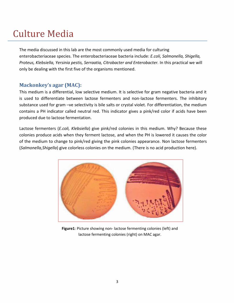
Lactose fermenters (E.coli, Klebsiella) give pink/red colonies in this medium. Why? Because these
colonies produce acids when they ferment lactose, and when the PH is lowered it causes the color
of the medium to change to pink/red giving the pink colonies appearance. Non lactose fermenters
(Salmonella,Shigella) give colorless colonies on the medium. (There is no acid production here).
Figure1: Picture showing non- lactose fermenting colonies (left) and
lactose fermenting colonies (right) on MAC agar.
4
Cystein Lactose Electrolyte Deficient Agar (CLED): This medium is a non-selective differential medium for lactose fermenters and non-lactose
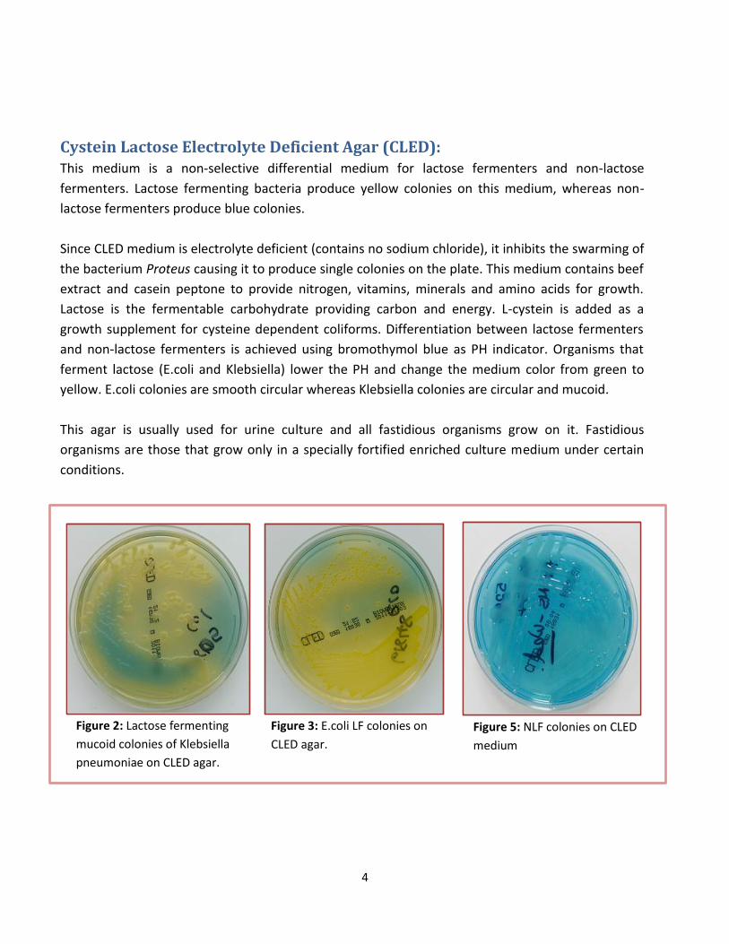
fermenters. Lactose fermenting bacteria produce yellow colonies on this medium, whereas non-
lactose fermenters produce blue colonies.
Since CLED medium is electrolyte deficient (contains no sodium chloride), it inhibits the swarming of
the bacterium Proteus causing it to produce single colonies on the plate. This medium contains beef
extract and casein peptone to provide nitrogen, vitamins, minerals and amino acids for growth.
Lactose is the fermentable carbohydrate providing carbon and energy. L-cystein is added as a
growth supplement for cysteine dependent coliforms. Differentiation between lactose fermenters
and non-lactose fermenters is achieved using bromothymol blue as PH indicator. Organisms that
ferment lactose (E.coli and Klebsiella) lower the PH and change the medium color from green to
yellow. E.coli colonies are smooth circular whereas Klebsiella colonies are circular and mucoid.
This agar is usually used for urine culture and all fastidious organisms grow on it. Fastidious
organisms are those that grow only in a specially fortified enriched culture medium under certain
conditions.
Figure 2: Lactose fermenting
mucoid colonies of Klebsiella
pneumoniae on CLED agar.
Figure 3: E.coli LF colonies on
CLED agar.
Figure 5: NLF colonies on CLED
medium
5
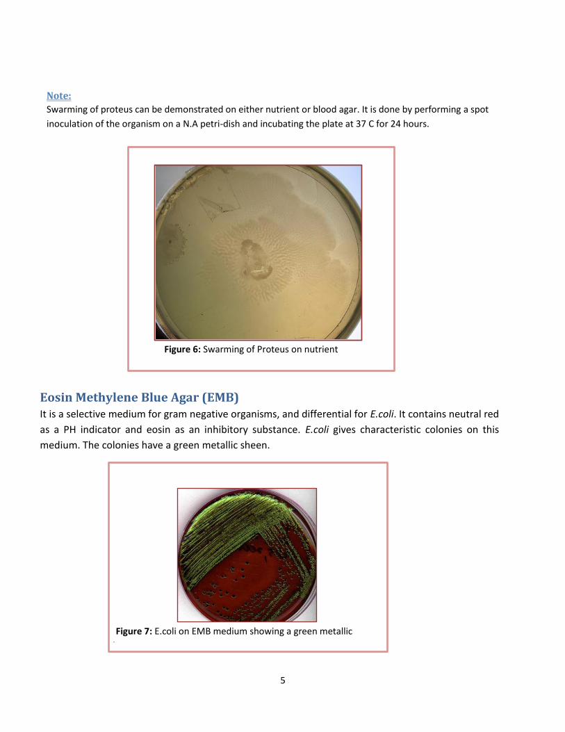
Note:
Swarming of proteus can be demonstrated on either nutrient or blood agar. It is done by performing a spot
inoculation of the organism on a N.A petri-dish and incubating the plate at 37 C for 24 hours.
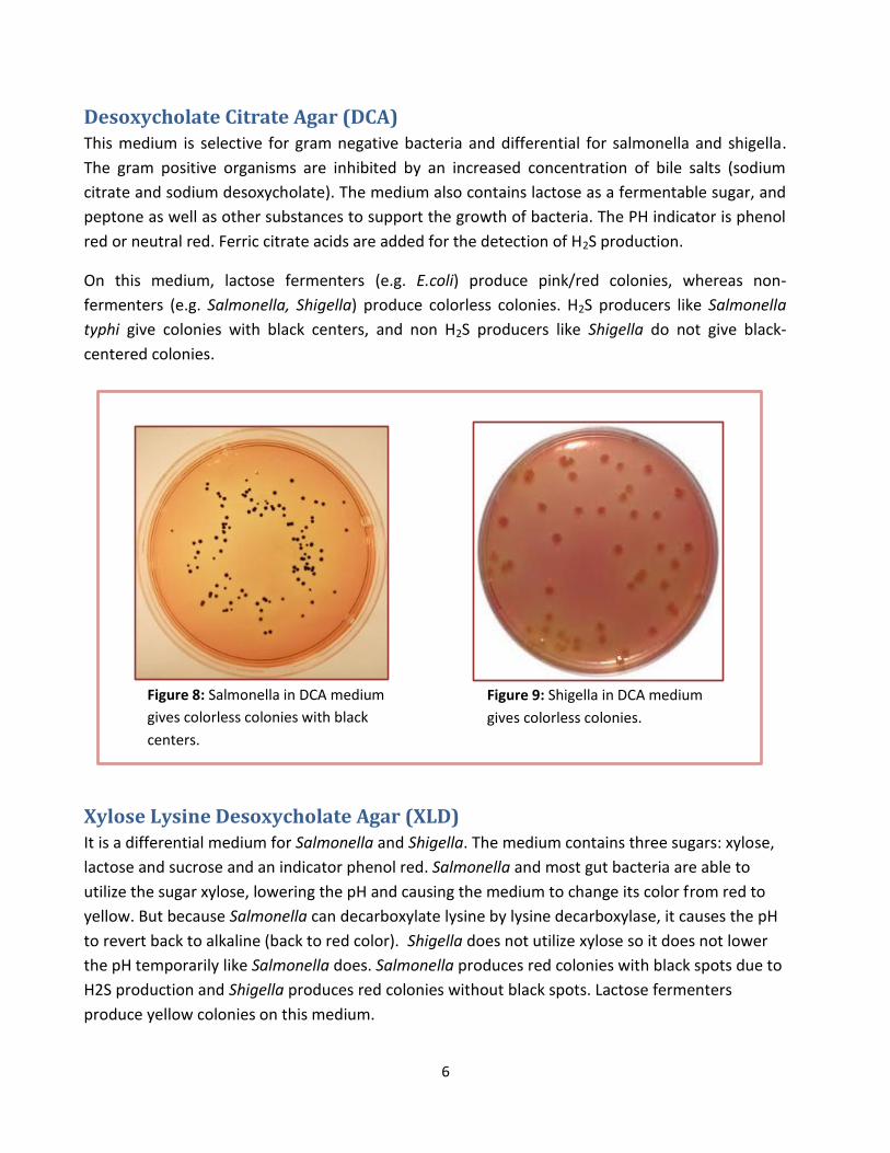
Eosin Methylene Blue Agar (EMB) It is a selective medium for gram negative organisms, and differential for E.coli. It contains neutral red
as a PH indicator and eosin as an inhibitory substance. E.coli gives characteristic colonies on this
medium. The colonies have a green metallic sheen.
Figure 7: E.coli on EMB medium showing a green metallic
sheen
Figure 6: Swarming of Proteus on nutrient
agar
6
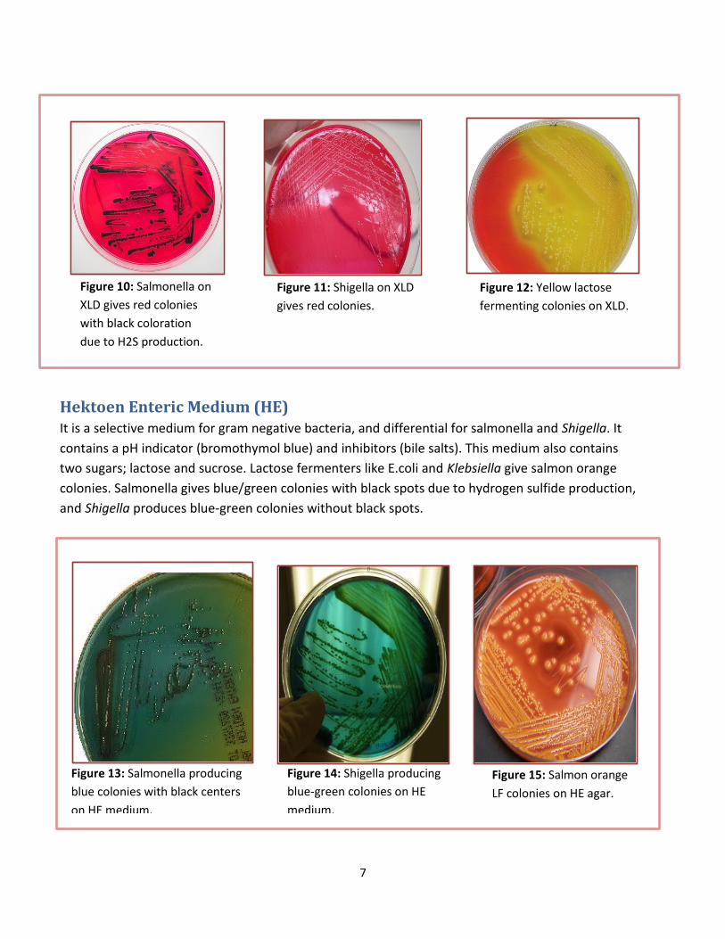
Desoxycholate Citrate Agar (DCA) This medium is selective for gram negative bacteria and differential for salmonella and shigella.
The gram positive organisms are inhibited by an increased concentration of bile salts (sodium
citrate and sodium desoxycholate). The medium also contains lactose as a fermentable sugar, and
peptone as well as other substances to support the growth of bacteria. The PH indicator is phenol
red or neutral red. Ferric citrate acids are added for the detection of H2S production.
On this medium, lactose fermenters (e.g. E.coli) produce pink/red colonies, whereas non-
fermenters (e.g. Salmonella, Shigella) produce colorless colonies. H2S producers like Salmonella
typhi give colonies with black centers, and non H2S producers like Shigella do not give black-
centered colonies.
Xylose Lysine Desoxycholate Agar (XLD) It is a differential medium for Salmonella and Shigella. The medium contains three sugars: xylose,
lactose and sucrose and an indicator phenol red. Salmonella and most gut bacteria are able to
utilize the sugar xylose, lowering the pH and causing the medium to change its color from red to
yellow. But because Salmonella can decarboxylate lysine by lysine decarboxylase, it causes the pH
to revert back to alkaline (back to red color). Shigella does not utilize xylose so it does not lower
the pH temporarily like Salmonella does. Salmonella produces red colonies with black spots due to
H2S production and Shigella produces red colonies without black spots. Lactose fermenters
produce yellow colonies on this medium.
Figure 8: Salmonella in DCA medium
gives colorless colonies with black
centers.
Figure 9: Shigella in DCA medium
gives colorless colonies.
7
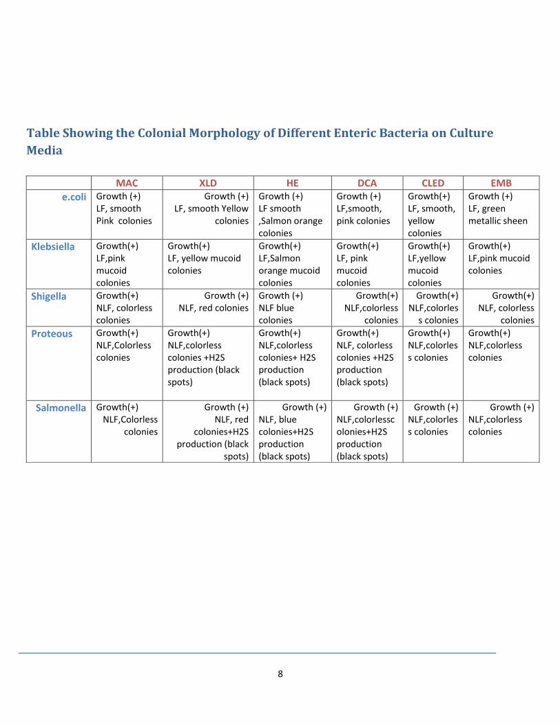
Hektoen Enteric Medium (HE) It is a selective medium for gram negative bacteria, and differential for salmonella and Shigella. It
contains a pH indicator (bromothymol blue) and inhibitors (bile salts). This medium also contains
two sugars; lactose and sucrose. Lactose fermenters like E.coli and Klebsiella give salmon orange
colonies. Salmonella gives blue/green colonies with black spots due to hydrogen sulfide production,
and Shigella produces blue-green colonies without black spots.
Figure 10: Salmonella on
XLD gives red colonies
with black coloration
due to H2S production.
Figure 11: Shigella on XLD
gives red colonies.
Figure 12: Yellow lactose
fermenting colonies on XLD.
Figure 13: Salmonella producing
blue colonies with black centers
on HE medium.
Figure 14: Shigella producing
blue-green colonies on HE
medium.
Figure 15: Salmon orange
LF colonies on HE agar.
8
Table Showing the Colonial Morphology of Different Enteric Bacteria on Culture
Media
EMB CLED DCA HE XLD MAC
Growth (+) LF, green metallic sheen
Growth(+) LF, smooth, yellow colonies
Growth (+) LF,smooth, pink colonies
Growth (+) LF smooth ,Salmon orange colonies
Growth (+) LF, smooth Yellow
colonies
Growth (+) LF, smooth Pink colonies
e.coli
Growth(+) LF,pink mucoid colonies
Growth(+) LF,yellow mucoid colonies
Growth(+) LF, pink mucoid colonies
Growth(+) LF,Salmon orange mucoid colonies
Growth(+) LF, yellow mucoid colonies
Growth(+) LF,pink mucoid colonies
Klebsiella
Growth(+) NLF, colorless
colonies
Growth(+) NLF,colorles
s colonies
Growth(+) NLF,colorless
colonies
Growth (+) NLF blue colonies
Growth (+) NLF, red colonies
Growth(+) NLF, colorless colonies
Shigella
Growth(+) NLF,colorless colonies
Growth(+) NLF,colorless colonies
Growth(+) NLF, colorless colonies +H2S production (black spots)
Growth(+) NLF,colorless colonies+ H2S production (black spots)
Growth(+) NLF,colorless colonies +H2S production (black spots)
Growth(+) NLF,Colorless colonies
Proteous
Growth (+) NLF,colorless colonies
Growth (+) NLF,colorless colonies
Growth (+) NLF,colorlesscolonies+H2S production (black spots)
Growth (+) NLF, blue colonies+H2S production (black spots)
Growth (+) NLF, red
colonies+H2S production (black
spots)
Growth(+) NLF,Colorless
colonies
Salmonella
9
Biochemical Reactions of Enterobacteriaceae
It is important to know that all members of the enterobacteriaceae are:
Catalase positive
Oxidase negative
Nitrite positive
Facultative anaerobes
Glucose fermenters
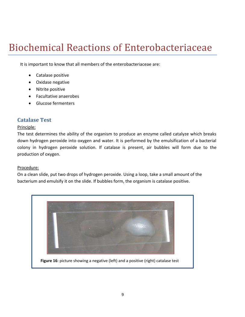
Catalase Test Principle:
The test determines the ability of the organism to produce an enzyme called catalyze which breaks
down hydrogen peroxide into oxygen and water. It is performed by the emulsification of a bacterial
colony in hydrogen peroxide solution. If catalase is present, air bubbles will form due to the
production of oxygen.
Procedure:
On a clean slide, put two drops of hydrogen peroxide. Using a loop, take a small amount of the
bacterium and emulsify it on the slide. If bubbles form, the organism is catalase positive.
Figure 16: picture showing a negative (left) and a positive (right) catalase test
result.
10
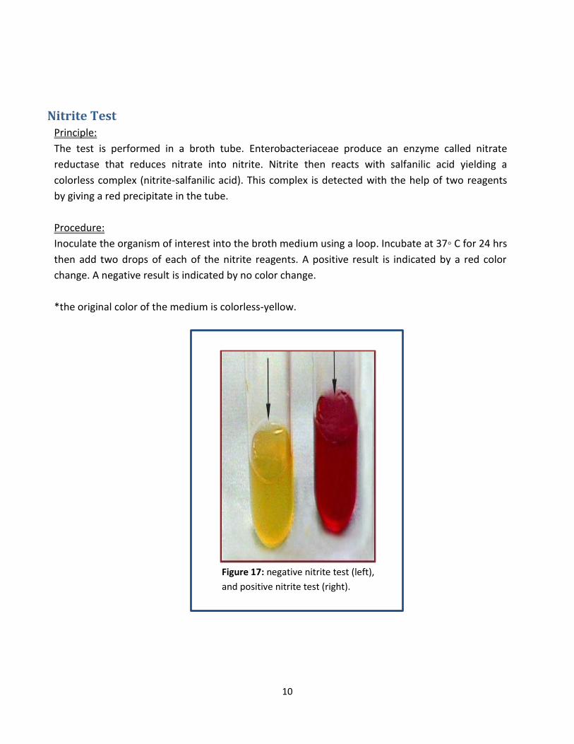
Nitrite Test Principle:
The test is performed in a broth tube. Enterobacteriaceae produce an enzyme called nitrate
reductase that reduces nitrate into nitrite. Nitrite then reacts with salfanilic acid yielding a
colorless complex (nitrite-salfanilic acid). This complex is detected with the help of two reagents
by giving a red precipitate in the tube.
Procedure:
Inoculate the organism of interest into the broth medium using a loop. Incubate at 37◦ C for 24 hrs
then add two drops of each of the nitrite reagents. A positive result is indicated by a red color
change. A negative result is indicated by no color change.
*the original color of the medium is colorless-yellow.
Figure 17: negative nitrite test (left),
and positive nitrite test (right).
11
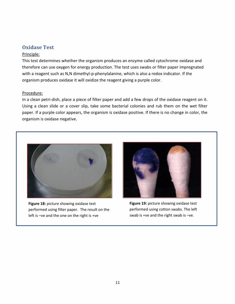
Oxidase Test Principle:
This test determines whether the organism produces an enzyme called cytochrome oxidase and
therefore can use oxygen for energy production. The test uses swabs or filter paper impregnated
with a reagent such as N,N dimethyl-p-phenylalanine, which is also a redox indicator. If the
organism produces oxidase it will oxidize the reagent giving a purple color.
Procedure:
In a clean petri-dish, place a piece of filter paper and add a few drops of the oxidase reagent on it.
Using a clean slide or a cover slip, take some bacterial colonies and rub them on the wet filter
paper. If a purple color appears, the organism is oxidase positive. If there is no change in color, the
organism is oxidase negative.
Figure 18: picture showing oxidase test
performed using filter paper. The result on the
left is –ve and the one on the right is +ve
Figure 19: picture showing oxidase test
performed using cotton swabs. The left
swab is +ve and the right swab is –ve.
12
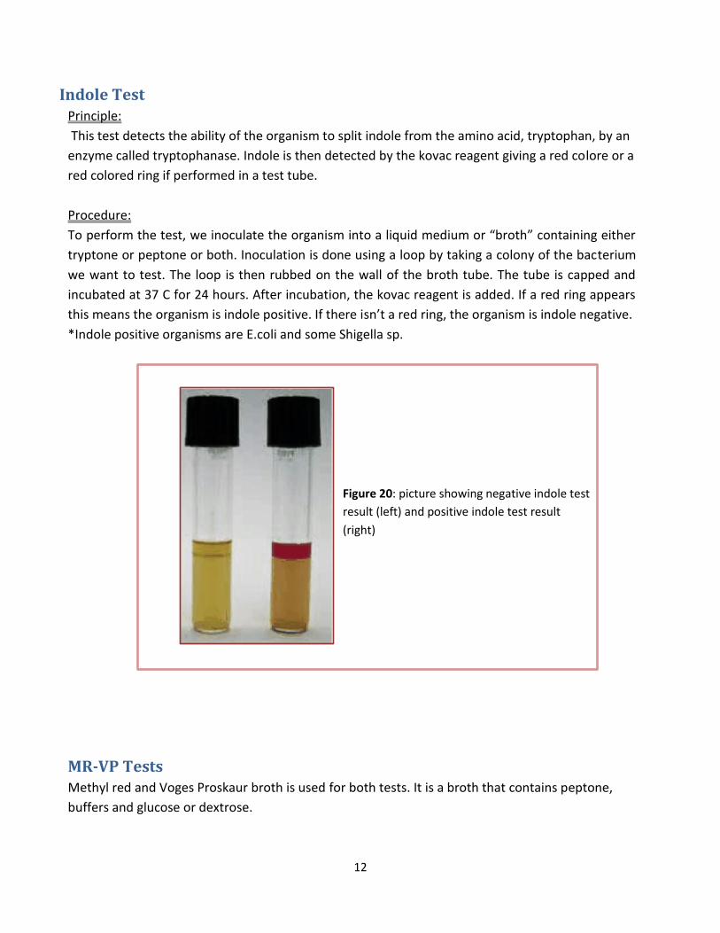
Indole Test Principle:
This test detects the ability of the organism to split indole from the amino acid, tryptophan, by an
enzyme called tryptophanase. Indole is then detected by the kovac reagent giving a red colore or a
red colored ring if performed in a test tube.
Procedure:
To perform the test, we inoculate the organism into a liquid medium or “broth” containing either
tryptone or peptone or both. Inoculation is done using a loop by taking a colony of the bacterium
we want to test. The loop is then rubbed on the wall of the broth tube. The tube is capped and
incubated at 37 C for 24 hours. After incubation, the kovac reagent is added. If a red ring appears
this means the organism is indole positive. If there isn’t a red ring, the organism is indole negative.
*Indole positive organisms are E.coli and some Shigella sp.
MR-VP Tests Methyl red and Voges Proskaur broth is used for both tests. It is a broth that contains peptone,
buffers and glucose or dextrose.
Figure 20: picture showing negative indole test
result (left) and positive indole test result
(right)
13
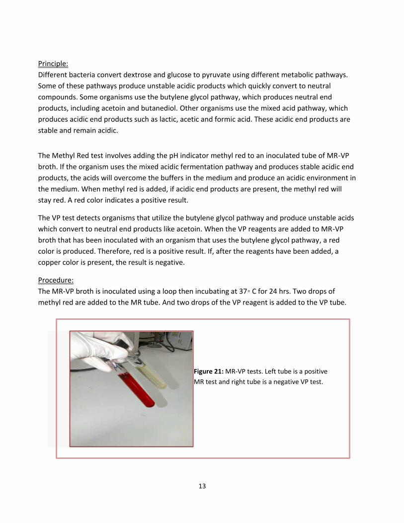
Principle:
Different bacteria convert dextrose and glucose to pyruvate using different metabolic pathways.
Some of these pathways produce unstable acidic products which quickly convert to neutral
compounds. Some organisms use the butylene glycol pathway, which produces neutral end
products, including acetoin and butanediol. Other organisms use the mixed acid pathway, which
produces acidic end products such as lactic, acetic and formic acid. These acidic end products are
stable and remain acidic.
The Methyl Red test involves adding the pH indicator methyl red to an inoculated tube of MR-VP
broth. If the organism uses the mixed acidic fermentation pathway and produces stable acidic end
products, the acids will overcome the buffers in the medium and produce an acidic environment in
the medium. When methyl red is added, if acidic end products are present, the methyl red will
stay red. A red color indicates a positive result.
The VP test detects organisms that utilize the butylene glycol pathway and produce unstable acids
which convert to neutral end products like acetoin. When the VP reagents are added to MR-VP
broth that has been inoculated with an organism that uses the butylene glycol pathway, a red
color is produced. Therefore, red is a positive result. If, after the reagents have been added, a
copper color is present, the result is negative.
Procedure:
The MR-VP broth is inoculated using a loop then incubating at 37◦ C for 24 hrs. Two drops of
methyl red are added to the MR tube. And two drops of the VP reagent is added to the VP tube.
Figure 21: MR-VP tests. Left tube is a positive
MR test and right tube is a negative VP test.
14
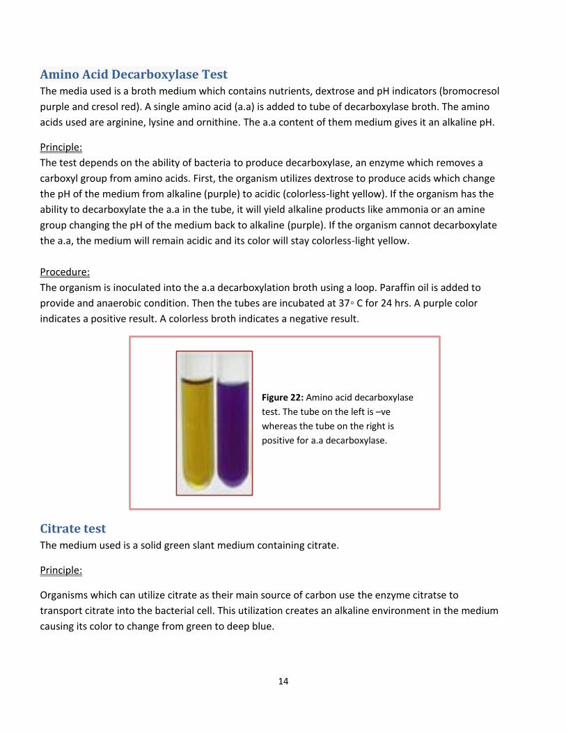
Amino Acid Decarboxylase Test The media used is a broth medium which contains nutrients, dextrose and pH indicators (bromocresol
purple and cresol red). A single amino acid (a.a) is added to tube of decarboxylase broth. The amino
acids used are arginine, lysine and ornithine. The a.a content of them medium gives it an alkaline pH.
Principle:
The test depends on the ability of bacteria to produce decarboxylase, an enzyme which removes a
carboxyl group from amino acids. First, the organism utilizes dextrose to produce acids which change
the pH of the medium from alkaline (purple) to acidic (colorless-light yellow). If the organism has the
ability to decarboxylate the a.a in the tube, it will yield alkaline products like ammonia or an amine
group changing the pH of the medium back to alkaline (purple). If the organism cannot decarboxylate
the a.a, the medium will remain acidic and its color will stay colorless-light yellow.
Procedure:
The organism is inoculated into the a.a decarboxylation broth using a loop. Paraffin oil is added to
provide and anaerobic condition. Then the tubes are incubated at 37◦ C for 24 hrs. A purple color
indicates a positive result. A colorless broth indicates a negative result.
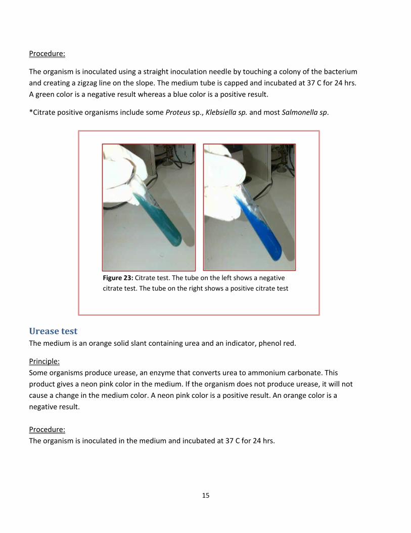
Citrate test The medium used is a solid green slant medium containing citrate.
Principle:
Organisms which can utilize citrate as their main source of carbon use the enzyme citratse to
transport citrate into the bacterial cell. This utilization creates an alkaline environment in the medium
causing its color to change from green to deep blue.
Figure 22: Amino acid decarboxylase
test. The tube on the left is –ve
whereas the tube on the right is
positive for a.a decarboxylase.
15
Procedure:
The organism is inoculated using a straight inoculation needle by touching a colony of the bacterium
and creating a zigzag line on the slope. The medium tube is capped and incubated at 37 C for 24 hrs.
A green color is a negative result whereas a blue color is a positive result.
*Citrate positive organisms include some Proteus sp., Klebsiella sp. and most Salmonella sp.
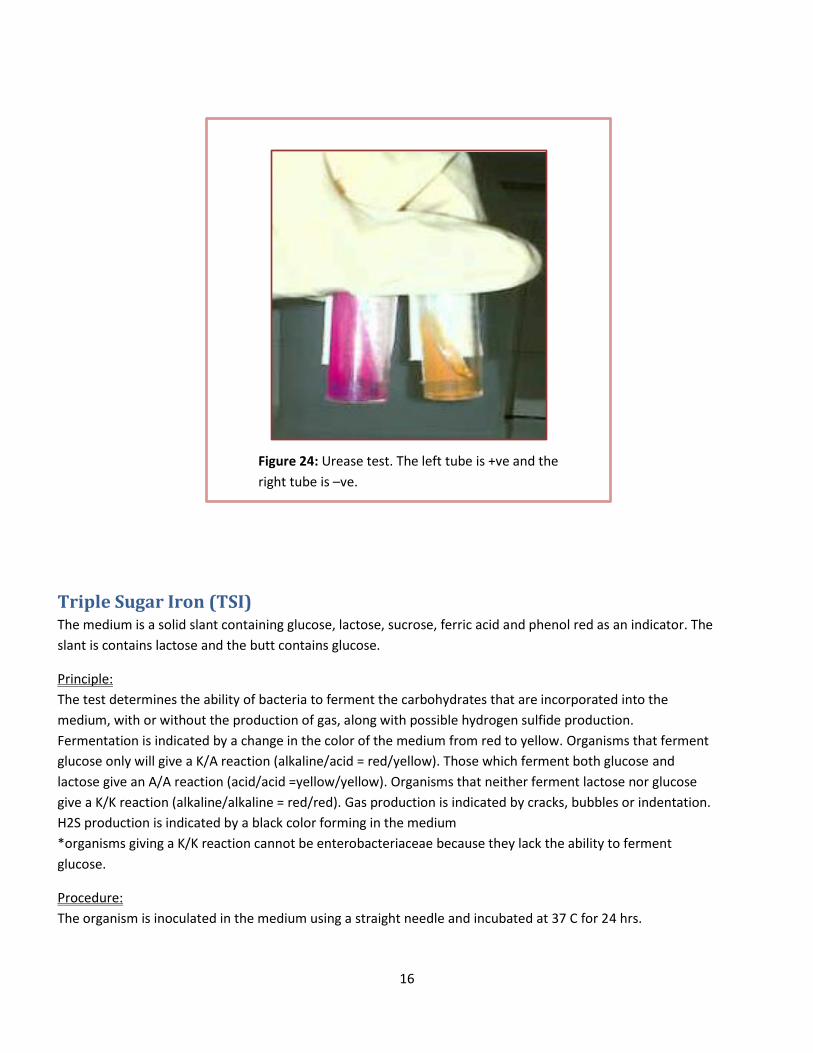
Urease test The medium is an orange solid slant containing urea and an indicator, phenol red.
Principle:
Some organisms produce urease, an enzyme that converts urea to ammonium carbonate. This
product gives a neon pink color in the medium. If the organism does not produce urease, it will not
cause a change in the medium color. A neon pink color is a positive result. An orange color is a
negative result.
Procedure:
The organism is inoculated in the medium and incubated at 37 C for 24 hrs.
Figure 23: Citrate test. The tube on the left shows a negative
citrate test. The tube on the right shows a positive citrate test
16
Triple Sugar Iron (TSI) The medium is a solid slant containing glucose, lactose, sucrose, ferric acid and phenol red as an indicator. The
slant is contains lactose and the butt contains glucose.
Principle:
The test determines the ability of bacteria to ferment the carbohydrates that are incorporated into the
medium, with or without the production of gas, along with possible hydrogen sulfide production.
Fermentation is indicated by a change in the color of the medium from red to yellow. Organisms that ferment
glucose only will give a K/A reaction (alkaline/acid = red/yellow). Those which ferment both glucose and
lactose give an A/A reaction (acid/acid =yellow/yellow). Organisms that neither ferment lactose nor glucose
give a K/K reaction (alkaline/alkaline = red/red). Gas production is indicated by cracks, bubbles or indentation.
H2S production is indicated by a black color forming in the medium
*organisms giving a K/K reaction cannot be enterobacteriaceae because they lack the ability to ferment
glucose.
Procedure:
The organism is inoculated in the medium using a straight needle and incubated at 37 C for 24 hrs.
Figure 24: Urease test. The left tube is +ve and the
right tube is –ve.
17
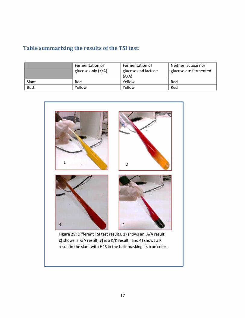
Table summarizing the results of the TSI test:
Fermentation of glucose only (K/A)
Fermentation of glucose and lactose (A/A)
Neither lactose nor glucose are fermented
Slant Red Yellow Red
Butt Yellow Yellow Red
1 2
3 4
Figure 25: Different TSI test results. 1) shows an A/A result,
2) shows a K/A result, 3) is a K/K result, and 4) shows a K
result in the slant with H2S in the butt masking its true color.
18
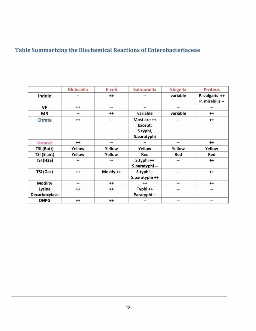
Table Summarizing the Biochemical Reactions of Enterobacteriaceae
Klebsiella E.coli Salmonella Shigella Proteus
Indole -- ++
-- variable P. valgaris ++ P. mirabilis --
VP ++ -- -- -- --
MR -- ++ variable variable ++
Citrate ++ -- Most are ++ Except: S.typhi,
S.paratyphi
-- ++
Urease ++ -- -- -- ++
TSI (Butt) Yellow Yellow Yellow Yellow Yellow
TSI (Slant) Yellow Yellow Red Red Red
TSI (H2S) -- -- S.typhi ++ S.paratyphi --
-- ++
TSI (Gas) ++ Mostly ++ S.typhi -- S.paratyphi ++
-- ++
Motility -- ++ ++ -- ++
Lysine Decarboxylase
++ ++ Typhi ++ Paratyphi --
-- --
ONPG ++ ++ -- -- --
19
Analytical Profile Index (API E 20) and ONPG
Analytical Profile Index API E 20 The analytical profile index E 20 or (API E 20) provides an easy way to inoculate and read tests
relevant to members of the family Enterobacteriaceae and associated organisms. It is
a classification of bacteria based on experiments, allowing fast identification. The test systems are
stored in 20 small reaction tubes, which contain the substrates. These small tubes are referred to
as wells or cupules.
Procedure:
1) Prepare a suspension of the bacteria in normal saline
Inoculate a large colony of the bacterium into the NaCl solution, making sure that
the suspension is homogenous and without clumps of floating bacteria.
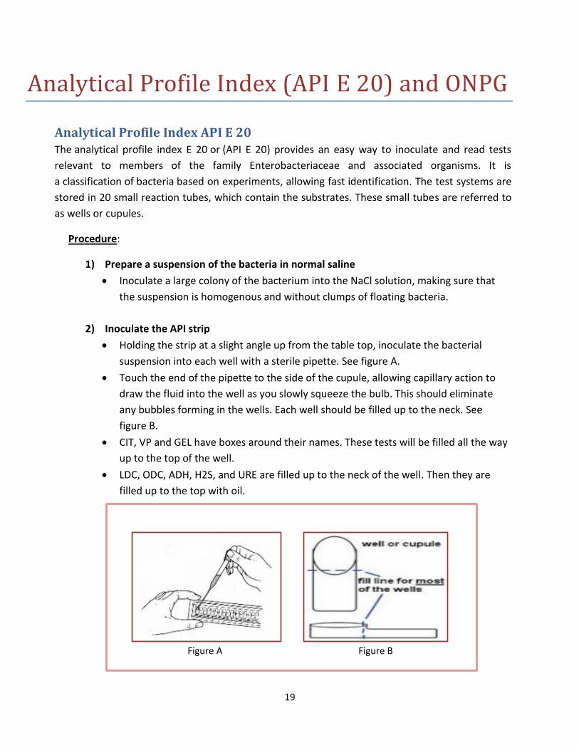
2) Inoculate the API strip
Holding the strip at a slight angle up from the table top, inoculate the bacterial
suspension into each well with a sterile pipette. See figure A.
Touch the end of the pipette to the side of the cupule, allowing capillary action to
draw the fluid into the well as you slowly squeeze the bulb. This should eliminate
any bubbles forming in the wells. Each well should be filled up to the neck. See
figure B.
CIT, VP and GEL have boxes around their names. These tests will be filled all the way
up to the top of the well.
LDC, ODC, ADH, H2S, and URE are filled up to the neck of the well. Then they are
filled up to the top with oil.
Figure A Figure B
20
3) Incubate the strip in its chamber
The bottom of the incubation chamber has small indented wells in the bottom: fill it with water just enough to fill these indentations.
Place the strip into this bottom. There should not be so much water that it slops onto the API strip.
Place the top of the incubation chamber over the bottom, and label it.
Place the strip at 37° C for 18-24 hours.
Ortho-Nitrophenyle-β –galactoside (ONPG) This test detects an enzyme called ortho-nitrophenyl galactosidase. The enzyme ferments
lactose and therefore, this test detects lactose fermentation. The substrate for the enzyme is
ONPG. Lactose fermenters produce two enzymes: 1) β-galactosidase which is an intracellular
enzyme and 2) permease, an extra cellular enzyme that regulates entry of lactose into the cell.
In the presence of ONPG, the enzyme β-galactosidase hydrolyzes ONPG to orthonitrophenol.
The enzyme will also ferment lactose giving a yellow color.
In organisms that lack the permease enzyme and have only β-galactosidase, lactose will enter
the cell through passive transport which leads to a slower fermentation of lactose. This is called
delayed lactose fermentation.
*Non lactose fermenters lack both enzymes and they do not ferment lactose.
*This test in incorporated into the API E 20 testing system. There is a specific cupule for this test
on the strip.
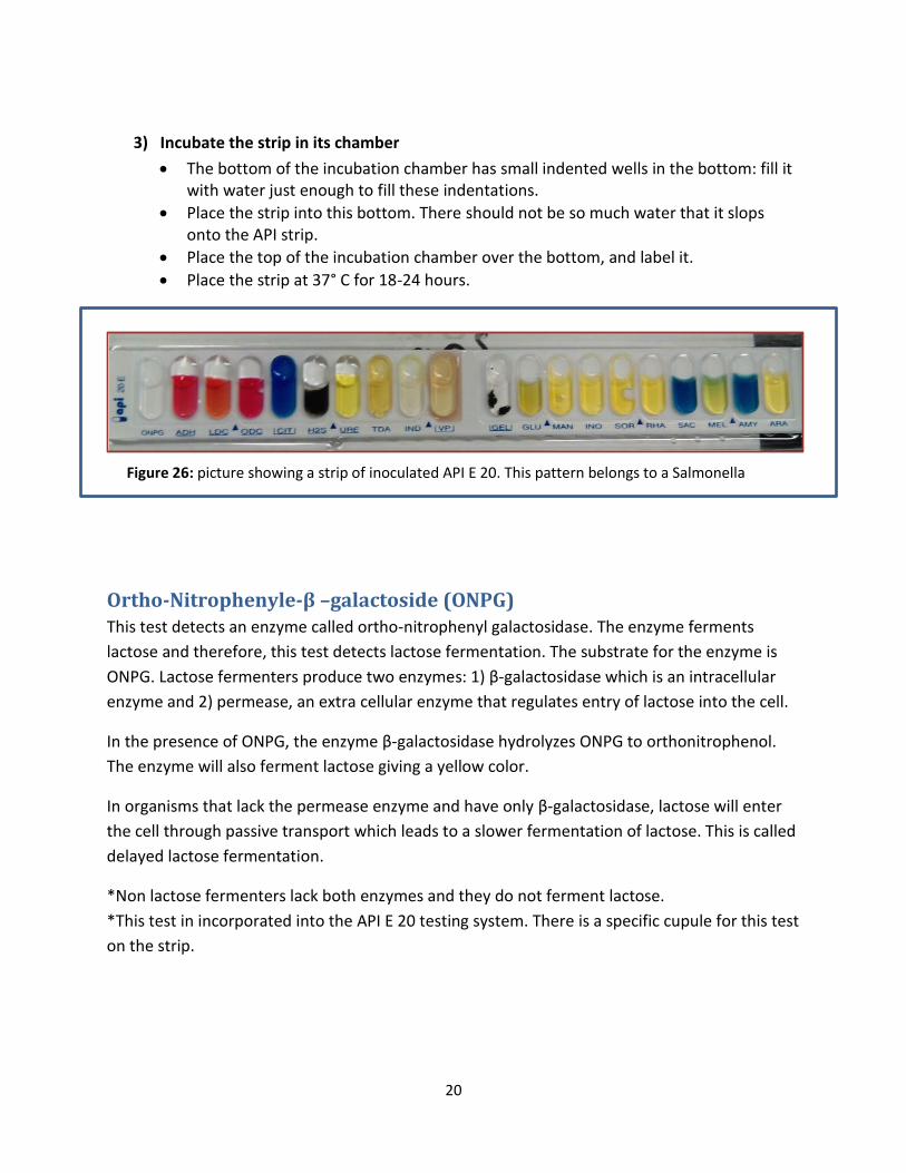
Figure 26: picture showing a strip of inoculated API E 20. This pattern belongs to a Salmonella
spp.
21
Sulfide-Indole-Motility Medium This is a semi solid medium containing ferrous sulfide. It detects H2S production, Indole and
motility. The test is done by inoculation the organism into the media using a straight needle. To
inoculate, take a bacterial colony using a straight needle and insert the needle vertically into
the medium letting it go all the way through to the bottom. Then pull the needle out slowly and
gently. Incubate the tube at 37° C for 24 hours
Motility is detected by diffused growth in the medium. If growth occurs only at the site of
inoculation, the organism is non-motile. H2S production is indicated by a black color. Indole is
detected by the formation of a red ring.
Note: In clinical labs, the abbreviation IMViC is used for the following group of tests: “I” for indole, “M” for
methyl red, “V” for Voges Proskauer and “iC” for citrate.
22
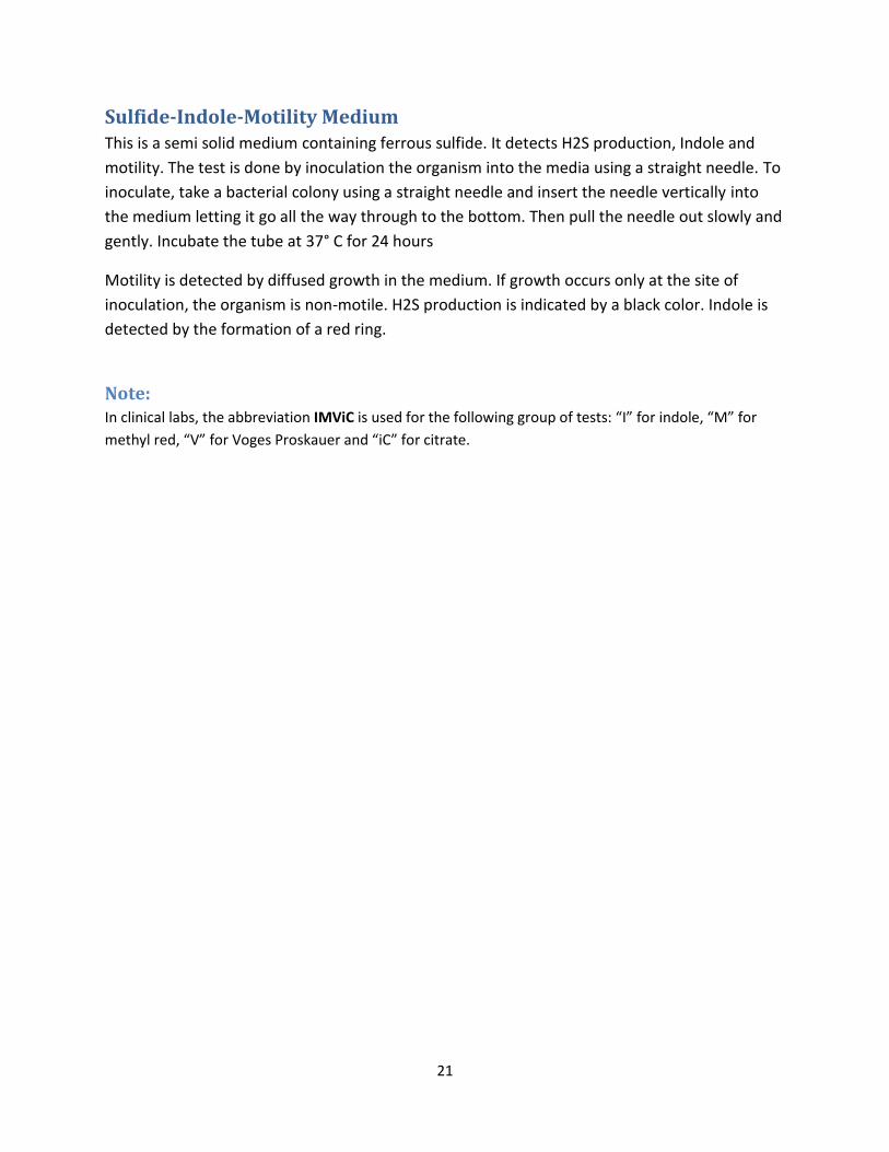
Pseudomonas
Pseudomonas is a gram negative pathogenic bacterium that doesn’t belong to the
Enterobacteriaceae family. It is highly resistant to antibiotics and causes various nosocomial
infections. It is oxidase positive and doesn’t ferment lactose or glucose.
Gram Smear and Culture
Gram stain: gram negative bacilli
Culture: Strict aerobe, Produces green pigments on nutrient agar and dark colonies on
blood agar due to pigmentation.
Figure 27: picture showing Pseudomonas pigment production on nutrient agar
23
Hemophilus Influenzae
Hemophilus species include; Hemophilus influenza, Hemophilus para influenza and Hemophilus
duchrii.
Gram Smear and Culture
Gram reaction: gram negative coccobacilli, pleomorphic in shape
Culture: fastidious organisms that need enrichment media (chocolate agar)
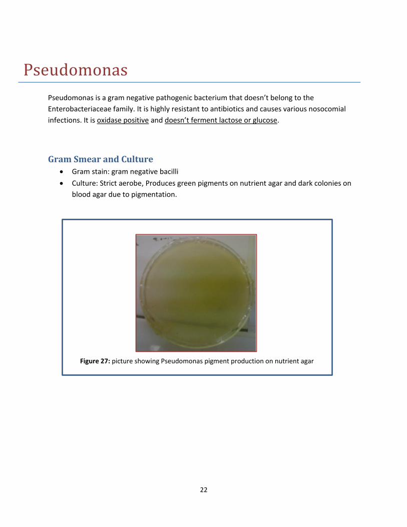
Hemophilus on Chocolate Agar Preparation of the agar:
Prepare blood base broth. Then add 10% of sheep blood. Let it cool down at 45 degrees Celsius
and put it in a water bath for 30 min at 75 degrees Celsius. This will cause the RBCs to lyse and
release the X factor (heamin) and the V factor (porphorin) which are essential for the growth of
Hemophilus Sp.
Colonial Morphology:
Hemophlius Sp. give grey moist colonies on chocolate agar.
Figure 28: picture showing H.influenzae
on chocolate agar. The colonies are grey
and moist.
24
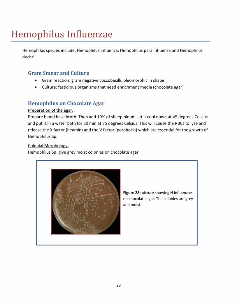
Hemophilus on Nutrient Agar On nutrient agar, V and X factor discs are added to support the growth of Hemophilus.
Hemophilus influenza requires both factors to grow whereas Hemophilus parainfluenzae
requires only the X factor.
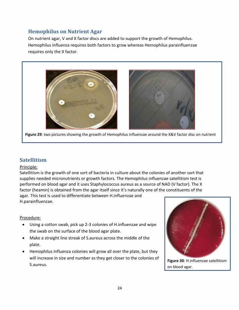
Satellitism Principle: Satellitism is the growth of one sort of bacteria in culture about the colonies of another sort that supplies needed micronutrients or growth factors. The Hemophilus influenzae satellitism test is performed on blood agar and it uses Staphylococcus aureus as a source of NAD (V factor). The X factor (heamin) is obtained from the agar itself since it’s naturally one of the constituents of the agar. This test is used to differentiate between H.influenzae and H.parainfluenzae.
Procedure:
Using a cotton swab, pick up 2-3 colonies of H.influenzae and wipe
the swab on the surface of the blood agar plate.
Make a straight line streak of S.aureus across the middle of the
plate.
Hemophilus influenza colonies will grow all over the plate, but they
will increase in size and number as they get closer to the colonies of
S.aureus.
Figure 29: two pictures showing the growth of Hemophilus influenzae around the X&V factor disc on nutrient
agar.
Figure 30: H.influenzae satellitism
on blood agar.
25
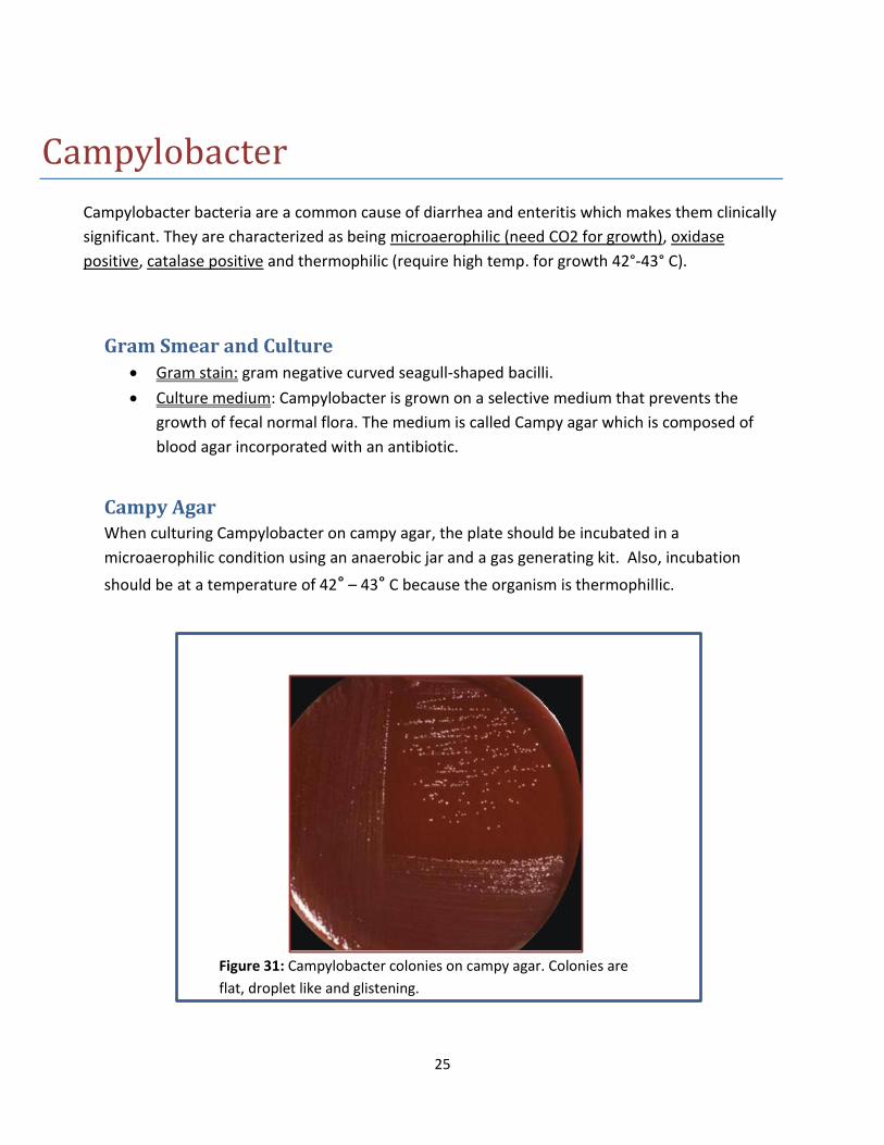
Campylobacter
Campylobacter bacteria are a common cause of diarrhea and enteritis which makes them clinically
significant. They are characterized as being microaerophilic (need CO2 for growth), oxidase
positive, catalase positive and thermophilic (require high temp. for growth 42°-43° C).
Gram Smear and Culture
Gram stain: gram negative curved seagull-shaped bacilli.
Culture medium: Campylobacter is grown on a selective medium that prevents the
growth of fecal normal flora. The medium is called Campy agar which is composed of
blood agar incorporated with an antibiotic.
Campy Agar When culturing Campylobacter on campy agar, the plate should be incubated in a
microaerophilic condition using an anaerobic jar and a gas generating kit. Also, incubation
should be at a temperature of 42° – 43° C because the organism is thermophillic.
Figure 31: Campylobacter colonies on campy agar. Colonies are
flat, droplet like and glistening.
26
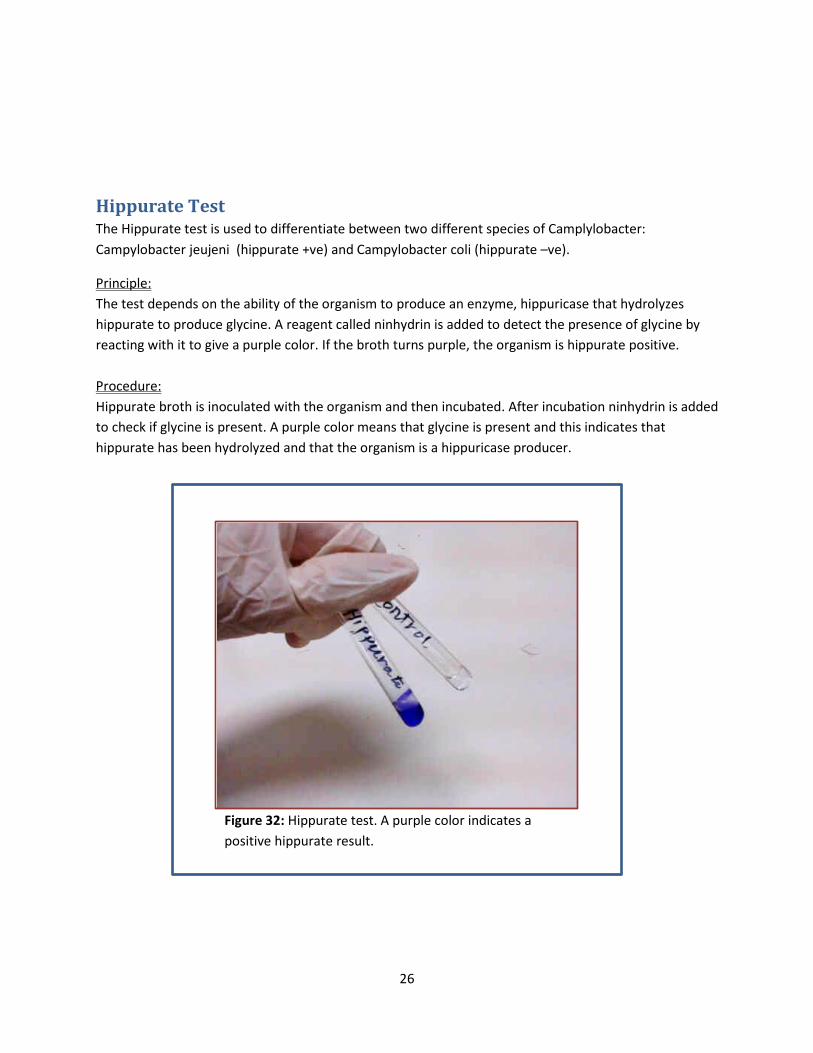
Hippurate Test The Hippurate test is used to differentiate between two different species of Camplylobacter:
Campylobacter jeujeni (hippurate +ve) and Campylobacter coli (hippurate –ve).
Principle:
The test depends on the ability of the organism to produce an enzyme, hippuricase that hydrolyzes
hippurate to produce glycine. A reagent called ninhydrin is added to detect the presence of glycine by
reacting with it to give a purple color. If the broth turns purple, the organism is hippurate positive.
Procedure:
Hippurate broth is inoculated with the organism and then incubated. After incubation ninhydrin is added
to check if glycine is present. A purple color means that glycine is present and this indicates that
hippurate has been hydrolyzed and that the organism is a hippuricase producer.
Figure 32: Hippurate test. A purple color indicates a
positive hippurate result.
27
Vibrio Species
There are many different species of Vibrio bacteria. In this lab we will discuss: Vibrio cholera and
Vibrio parahemolyticus. V.cholerae causes the well-known cholereae disease and V.parahemolyticus
causes food poisoning. Vibrio species are oxidase positive, facultative anaerobes, motile and can
grow at a wide range of temperatures (16◦ C- 40◦ C).
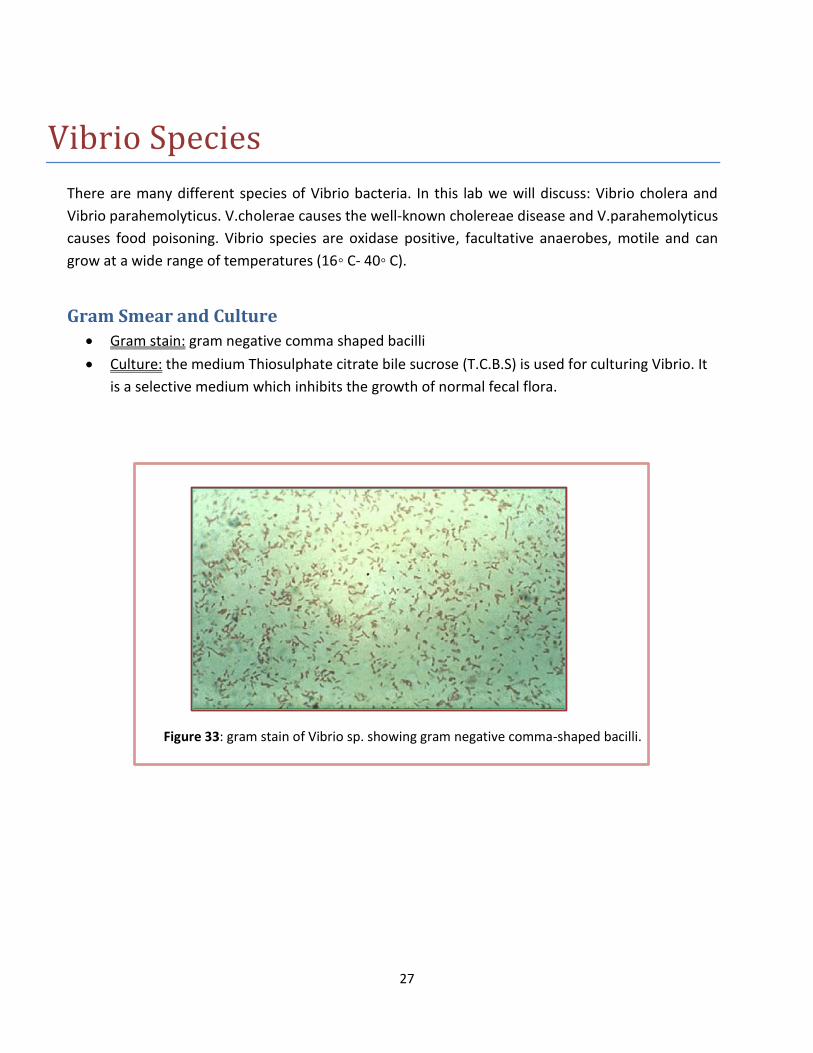
Gram Smear and Culture
Gram stain: gram negative comma shaped bacilli
Culture: the medium Thiosulphate citrate bile sucrose (T.C.B.S) is used for culturing Vibrio. It
is a selective medium which inhibits the growth of normal fecal flora.
Figure 33: gram stain of Vibrio sp. showing gram negative comma-shaped bacilli.
28
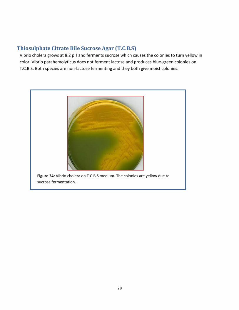
Thiosulphate Citrate Bile Sucrose Agar (T.C.B.S) Vibrio cholera grows at 8.2 pH and ferments sucrose which causes the colonies to turn yellow in
color. Vibrio parahemolyticus does not ferment lactose and produces blue-green colonies on
T.C.B.S. Both species are non-lactose fermenting and they both give moist colonies.
Figure 34: Vibrio cholera on T.C.B.S medium. The colonies are yellow due to
sucrose fermentation.
29
Anaerobes
Clostridia These organisms are spore forming anaerobic bacteria. Some are commensals of the human gut and others cause diseases such as gas gangrene, tetanus, botulism and food poisoning. The most common pathogens are: Clostridium perfringens, Clostridium tetani, Clostridium botulinum and Clostridium difficile.
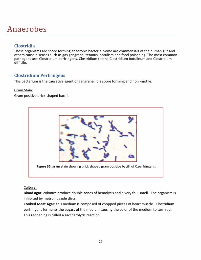
Clostridium Perfringens This bacterium is the causative agent of gangrene. It is spore forming and non -motile.
Gram Stain:
Gram positive brick-shaped bacilli.
Culture:
Blood agar: colonies produce double zones of hemolysis and a very foul smell. The organism is
inhibited by metronidazole discs.
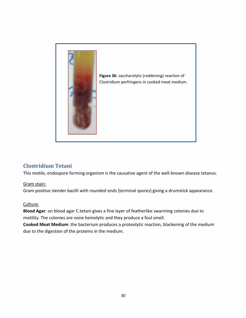
Cooked Meat Agar: this medium is composed of chopped pieces of heart muscle. Clostridium
perfringens ferments the sugars of the medium causing the color of the medium to turn red.
This reddening is called a saccharolytic reaction.
Figure 35: gram stain showing brick shaped gram positive bacilli of C.perfringens.
30
Clostridium Tetani This motile, endospore forming organism is the causative agent of the well-known disease tetanus.
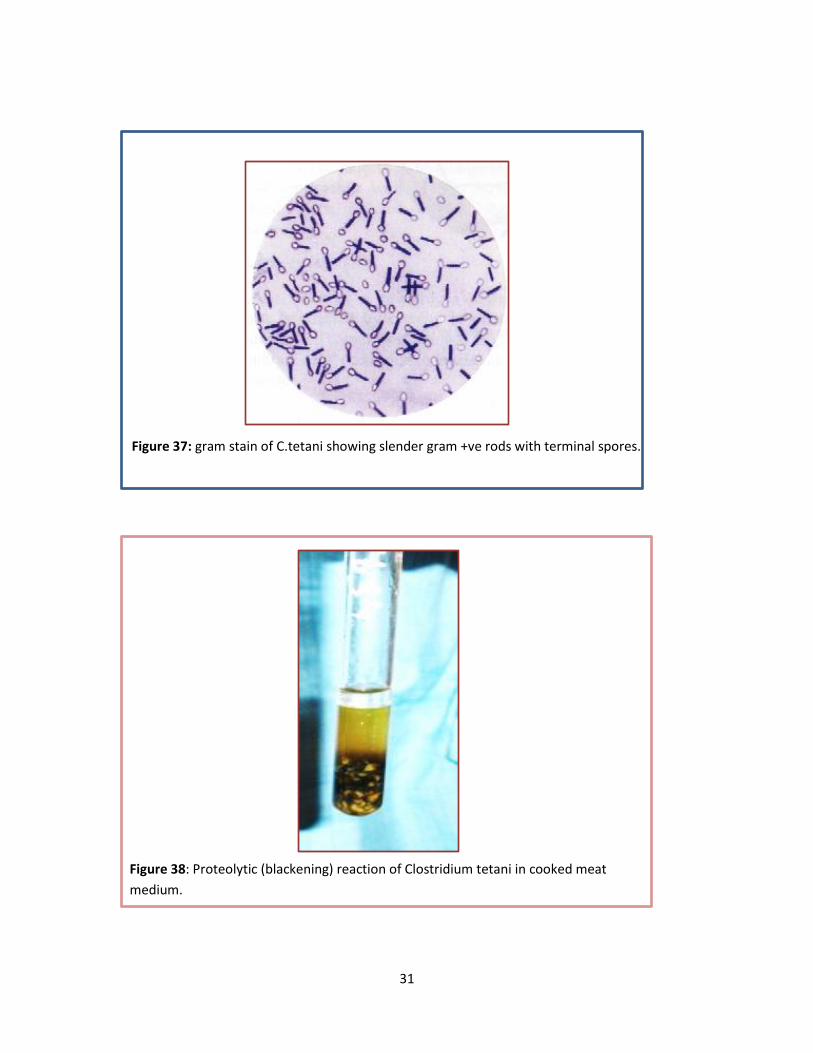
Gram stain:
Gram positive slender bacilli with rounded ends (terminal spores) giving a drumstick appearance.
Culture:
Blood Agar: on blood agar C.tetani gives a fine layer of featherlike swarming colonies due to
motility. The colonies are none hemolytic and they produce a foul smell.
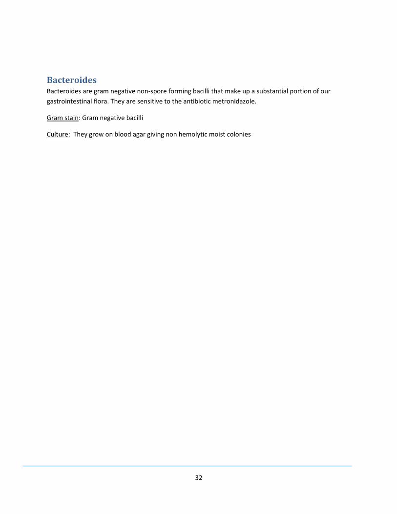
Cooked Meat Medium: the bacterium produces a proteolytic reaction, blackening of the medium
due to the digestion of the proteins in the medium.
Figure 36: saccharolytic (reddening) reaction of
Clostridium perfringens in cooked meat medium.
31
Figure 37: gram stain of C.tetani showing slender gram +ve rods with terminal spores.
Figure 38: Proteolytic (blackening) reaction of Clostridium tetani in cooked meat
medium.
32
Bacteroides Bacteroides are gram negative non-spore forming bacilli that make up a substantial portion of our
gastrointestinal flora. They are sensitive to the antibiotic metronidazole.
Gram stain: Gram negative bacilli
Culture: They grow on blood agar giving non hemolytic moist colonies
33
Spirochetes
Borrelia Normal oral flora
Gram stain: gram negative spirochetes
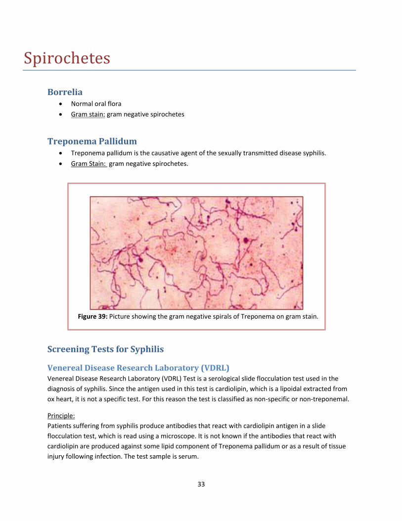
Treponema Pallidum Treponema pallidum is the causative agent of the sexually transmitted disease syphilis.
Gram Stain: gram negative spirochetes.
Screening Tests for Syphilis
Venereal Disease Research Laboratory (VDRL) Venereal Disease Research Laboratory (VDRL) Test is a serological slide flocculation test used in the
diagnosis of syphilis. Since the antigen used in this test is cardiolipin, which is a lipoidal extracted from
ox heart, it is not a specific test. For this reason the test is classified as non-specific or non-treponemal.
Principle:
Patients suffering from syphilis produce antibodies that react with cardiolipin antigen in a slide
flocculation test, which is read using a microscope. It is not known if the antibodies that react with
cardiolipin are produced against some lipid component of Treponema pallidum or as a result of tissue
injury following infection. The test sample is serum.
Figure 39: Picture showing the gram negative spirals of Treponema on gram stain.
34
Procedure:
Serum should be heated in a water bath to inactivate nonspecific inhibitors such as
complement.
A drop of inactivated serum is added into the well
Then a drop of cardiolipin Ag is added
The well is then rotated gently for 4 minutes manually or on a rotator.
After 4 minutes, the test is examined microscopically for flocculation.
If flocculation is present, the test is reactive (positive).
Rapid Plasma Reagin (RPR) This test shares the same principle of the VDRL test with some modifications. In the RPR test, the serum
does not require heating and the result can be viewed without a microscope. The cardiolipin Ag is mixed
with visible carbon particles which enable the test to be performed macroscopically.
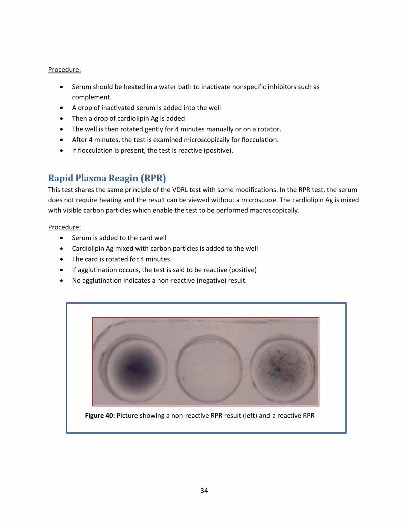
Procedure:
Serum is added to the card well
Cardiolipin Ag mixed with carbon particles is added to the well
The card is rotated for 4 minutes
If agglutination occurs, the test is said to be reactive (positive)
No agglutination indicates a non-reactive (negative) result.
Figure 40: Picture showing a non-reactive RPR result (left) and a reactive RPR
(right)

35
Picture References:
http://www.microbiologyinpictures.com/bacteria%20photos/haemophilus%20influenzae%20ph
otos/HAIN24.html
http://www.mansmed.net/forums/showthread.php?t=4800&page=136
http://www.uwyo.edu/molb2210_lab/info/biochemical_tests.htm
http://www.proprofs.com/flashcards/story.php?title=bio-150-microbiology-lab-packet-ii-review
http://www.biotech.univ.gda.pl/odl/biochem/api.html
http://www.jlindquist.net/generalmicro/102bactid2.html
http://www.microbiologyinpictures.com/bacteria%20photos/haemophilus%20influenzae%20ph
otos/HAIN2.html
http://www.ppdictionary.com/bacteria/gnbac/jejuni.htm
http://www.flickriver.com/photos/tags/practicle/interesting/
http://85.238.144.18/analytics/Micro_Manual/TEDISdata/prods/1_10263_0500.html
http://www.scribd.com/doc/53930320/Lecture-21-Vibrio-cholerae
http://intro.bio.umb.edu/111-112/OLLM/112s99/phyla/bacteria.htm
http://microblog.me.uk/119
http://commons.wikimedia.org/wiki/File:Robertsons_cooked_meat_medium.jpg
http://www.flickr.com/photos/nathanreading/sets/72157629070432847/detail/
http://geileteile.de/e-coli-2-on-c-l-e-d-agar/
http://www.retroscope.eu/wordpress/salmonella-spp/salmonellaapi20e/
http://www.kemitekskimya.com.tr/?desoxycholate-citrate-agar-(hynes)_m159-27--1
http://www.sigmaaldrich.com/technical-documents/articles/analytix/differentiation-of.html


