51
Preanalytical variables. POCT. Metabolic diseases Ivan Šebesta ÚLBLD 1.LF UK
Preanalytical variables. POCT. Metabolic diseases
Ivan Šebesta
ÚLBLD 1.LF UK

Circuit diagram of the clinical biochemistry process

How biochemical tests are used

• case history
• physical examination
INFORMATION ABOUT PATIENT
• imaging studies (x-ray, EEG,etc..)
• laboratory tests• clinical chemistry (60 – 70%)• haematology• microbiology• immunology

The results of laboratory tests are usefuland effective informations under followingconditions:
• proper indication
• rapid availability
• accurancy
• proper interpretation

The most useful is rapid information
Examination near the patient (POCT)
DRY CHEMISTRY
1) simple test – bed side testing
2) general practitioner’s office
3) primary health care laboratory

A portable bench analyser

• gives information about metabolic functions
• has wide range and high specifity
CLINICAL CHEMISTRY INFORMATION
• is there quantification
• is relatively easy available
• is relatively harmless to the patient
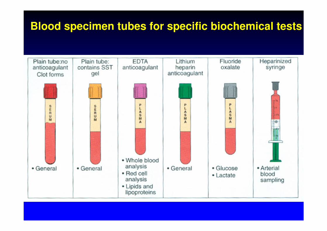
Blood specimen tubes for specific biochemical tests

Before considering diagnosis or treatment based on ananalytical results the clinicians should ask himself threequestions:
1) If it is the first time the estimation has been performed inthis patients, IS IT NORMAL OR ABNORMAL?
INTERPRETING RESULTS
this patients, IS IT NORMAL OR ABNORMAL?
2) If it is abnormal, IS THE ABNORMALITY OF DIAGNOSTICVALUE or is it a no-specific finding?
3) If it is one of a series of results, HAS THERE BEEN ACHANGE, AND IF SO, IS THIS CHANGE CLINICALLYSIGNIFICANT?

Several commonly asked questions may be answered, atleast in part, by laboratory testing.
1) Is the diagnosis correct?Proper selected laboratory tests may corroborate or refute a workingdiagnosis.
CHOOSING LABORATORY TESTS
2) What is the etiology of the disease?
3) How severe is the disease?
4) Has the patient’s condition improved or deteriorated?
5) Is the patient at risk for disease or is there a disease notclinically apparent?
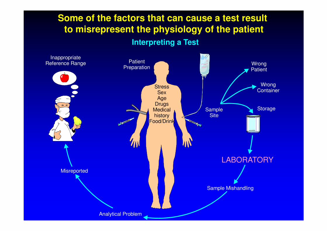
Some of the factors that can cause a test result to misrepresent the physiology of the patient
Interpreting a Test
Patient Preparation
StressSexAge
DrugsMedicalhistory
SampleSite
WrongPatient
WrongContainer
InappropriateReference Range
Storagehistory
Food/DrinkSite
Sample Mishandling
Analytical Problem
Misreported
LABORATORY

SchSchematicematic diagram ofdiagram oflipoprotein lipoprotein particleparticle:
ApolipoproteinApolipoprotein
Polar shellPhPhososphpholipidolipidss
Non-polar core
13
FreeFree cholesterolcholesterol
EsterEsterifiedified
cholesterolcholesterol
TriacylglycerolTriacylglycerolss

DeterminationDetermination of lof lipoproteinipoproteinss::
• An ultracentrifugation (to distinguish variousclasses according to the hydrated density):
VLDL, IDL, LDL, HDL
• Electrophoretically: α-lipoproteins,
14
• Electrophoretically: α-lipoproteins, pre-β-lipoproteins,
β-lipoproteins,chylomicrons
• Immunochemical methods:Apo A, Apo B, Apo C, Apo D, Apo E,

LLipoproteinipoprotein eelelecctrotrophphororesisesis
Lp(a)
α-lipoproteins
HDL
15
Pre-β-lipoproteins
VLDL
β-lipoproteins
IDL,LDL
chylomicrons

Basic Basic investigationsinvestigations of of lipid lipid metabolismmetabolism
•• Cholesterol Cholesterol 3.8 - 5.2 mmol/l
16
•• TTAAGG 0.9 - 1.7 mmol/l
•• HDL HDL > 0.9 mmol/l
•• LDLLDL < 4.5 mmol/l

TargetTarget valuesvalues of of CzechCzech Society Society forforAtherosclerosisAtherosclerosis
•• CholesterolCholesterol 4.5 – 5.0 mmol/l (at individuals with decreased risk to 6.0 mmol/l)
17
(at individuals with decreased risk to 6.0 mmol/l)•• HDL HDL > 0.9 mmol/l•• LDLLDL < 2.5 mmol/l at secondary prevention
< 3 – 3.5 mmol/l at increased risk < 4 – 4.5 mmol/l at decreased risk
•• TTAAG G < 2.3 mmol/l

AdditionalAdditional teststests
�calculation of LDL cholesterol after Friedewald formula :
(the formula cannot be used if the concentration of TAG › 4,5 mmol/l )LDL = total cholesterol - (HDL + TAG x 0.37)
[mmol/l]
18
[mmol/l]total cholesterol - HDL
atherogenic index AI = HDL
� investigation of apo A-I and apo B-100�electrophoresis of lipoproteins
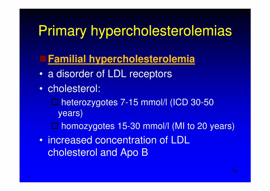
PrimPrimaryary hypercholesterolhypercholesteroleemimiasas
�Familial hypercholesterolemia• a disorder of LDL receptors• cholesterol:
19
� heterozygotes 7-15 mmol/l (ICD 30-50 years)
� homozygotes 15-30 mmol/l (MI to 20 years)
• increased concentration of LDL cholesterol and Apo B

• Familial defectiveApo B100
• a point mutation anda replacement of oneamino acid in
• Polygenichypercholesterolemia
• a combination of
PrimPrimaryary hypercholesterolhypercholesteroleemimiasas
20
amino acid in the position 3500 on the huge Apo B100molecule
• cholesterol: 7-10 mmol/l
• a combination of adverse genetic andexternal factors
• cholesterol: 8 mmol/lapproximately

CCombinombineded hyperlipidhyperlipideemimiasas
� Familial combinedhyperlipidemia
• an intensive Apo B synthesisin liver with a concomitantincreased production of
� Familialdysbetalipoproteinemia
21
increased production ofVLDL and LDL (highatherogenic particles)
• a frequent cause of ICD andMI to 60 years
• cholesterol 10 - 15 mmol/l TAG 2.3 - 5.7 mmol/l
• a defective gene for ApoE -pathological lipoprotein β-VLDL
• cholesterol 7.5 - 25 mmol/l
TAG 2 - 10(20) mmol/l

PrimPrimaarryy hypertriacylglycerolhypertriacylglyceroleemimiasas
� Familialhyperlipoproteinemiatype V
• rather uncommon disorder• more frequently in adults, obese,
with DM and with hyperuricemia
� Familialhyperchylomicronemia
• a deficit of lipoprotein lipaseor Apo CII
22
with DM and with hyperuricemia• an inductive factor: alcohol, drugs
containing estrogens, renal insufficiency
• increased in ELPHO:pre-β-lipoproteinsand chylomicrons
• cholesterol 7 - 13 mmol/lTAG 10 - 20 mmol/l
• TAG 20 - 120 mmol/l
• Treatment: fats containing FA with medium chains

� Familial hypertriacylglycerolemia• autosomal dominant transfer of disorder• increased concentration of VLDL• decreased concentration of HDL
PrimaryPrimary hyperliphyperlipoproteineoproteinemimiasas
23
• decreased concentration of HDL• non-insulin-dependent diabetes mellitus adds in
seniors• cholesterol normal• TAG to 6 mmol/l

HypolipoproteinemiHypolipoproteinemiasas
� Familialhypo-β-lipoproteinemia
• a longevity
� A-β-lipoproteinemia• a rare autosomal recessive
disorder• heterozygotes have
descreased LDL cholesterol• other lipids are in norm
24
• low values of LDL cholesterol
• a normal catabolism of LDL
• a reduced production of apo B
• other lipids are in norm• homozygotes have a total
deficit of lipoprotein particlescontaining apo B (malabsorption of fat, steatorrhea, retard grow, progressive degenerationof CNS, reduced visualsharpness, hemeralopia)

� Hypo-α-lipoproteinemia
• lower HDL levels• a defective apo A-I
(according to the location of
� An-α-lipoproteinemia(Tangier disease)
• absence of HDL in plasma
HypolipoproteinemiHypolipoproteinemiasas
25
(according to the location of the discribed case – Apo-A-I-Milano)
• HDL cannot be producedwithout apo A-I
• Apo C-II cannot betransported back into liver –relative deficiency of apo C-II
• an increased level of VLDL
• extremely low levels of apo A-I and apo A-II
• abnormally fast catabolismof HDL and apo A-I
SeSecocondndaryaryhyperlipoproteinhyperlipoproteineemimiasas
�Diabetes mellitus type I • insulin is an activator of lipoprotein lipase• if DM is decompensated
� ketoacidosis, hypertriglyceridemia andsometimes increased cholesterol as well
26
sometimes increased cholesterol as well
�Diabetes mellitus type II• a more intensive synthesis of VLDL in liver,
insulin resistence, HDL reduction, TAG rise• if DM is decompensated
� glycosylation of apo B

�Hypothyreoidism• thyroxine increases the biosynthesis of LDL
receptors in liver and an activity of lipoprotein lipase in adipocytes (by action of cAMP) as well
SeSecocondndaryary hyperlipoproteinhyperlipoproteineemimiasas
27
lipase in adipocytes (by action of cAMP) as well
�Nephrotic syndrome• hypoalbuminemia• a stimulation of lipoprotein synthesis.• increased cholesterol and TAG

Chronic renal failure• an inhibition of lipoprotein lipase in the plasma of
uremic patients• elevated TAG
Primary biliary cirrhosis
SeSecocondndaryary hyperlipoproteinhyperlipoproteineemimiaass
28
Primary biliary cirrhosis• hypercholesterolemia
�Obesity - TAG�Alcoholism - TAG Treatment with hormones and diuretic
drugs�Mental anorexia

To To HowHow to to recognizerecognize a a patientpatient withwith riskriskof of coronarycoronary diseasedisease ??
Cholestrol > 5,2 mmol/l Checkup after 5 years approximatelyNo
Binary decision:•Male?•Smoker? Cholesterol > 6,5 mmol/l
Yes
No
No
29
•Smoker?•Diabetic?•Hypertension?•Familial history of early MI ?•Angina pectoris?•Oral contraception?
Cholesterol > 6,5 mmol/lNo
Q cholesterol/ HDL > 5or TAG > 2,3 mmol/lor Lp(a) > 0,3 g/l
Yes
Yes •Dietary arrangement, monitoring•Secondary hyperlipidemia•Drug therapy
Diet and running checkups
Yes
No

Symptoms and findings referring to the presence of ahyperuricemia and indicating a serum uric acid determination:
• overweight• disturbance of carbohydrate tolerance• disturbances in lipid metabolism• urolithisasis• hypertonia
URIC ACID
• hypertonia• renal diseases• early severe atherosclerosis• fatty liver infiltration• family predisposition• hemoblastoses• cytostatic therapy and x-ray radiation• pre-eclampsia
A determination of the uric acid concentration is also to beundretaken with:
• clinical complaints, pointing to gout and with gouttherapy
URIC ACID
With regard to early gout recognition it is important to know,that ill-defined joint complaints and the presence of othermetabolic disturbances may point to a developing gout.
Obrázek nelze zobrazit. V počítači pravděpodobně není k dispozici dostatek paměti pro otevření obrázku nebo byl obrázek poškozen. Restartujte počítač a otevřete příslušný soubor znovu. Pokud se opět zobrazí červený křížek, bude nutné obrázek odstranit a vložit jej znovu.

Obrázek nelze zobrazit. V počítači pravděpodobně není k dispozici dostatek paměti pro otevření obrázku nebo byl obrázek poškozen. Restartujte počítač a otevřete příslušný soubor znovu. Pokud se opět zobrazí červený křížek, bude nutné obrázek odstranit a vložit jej znovu.
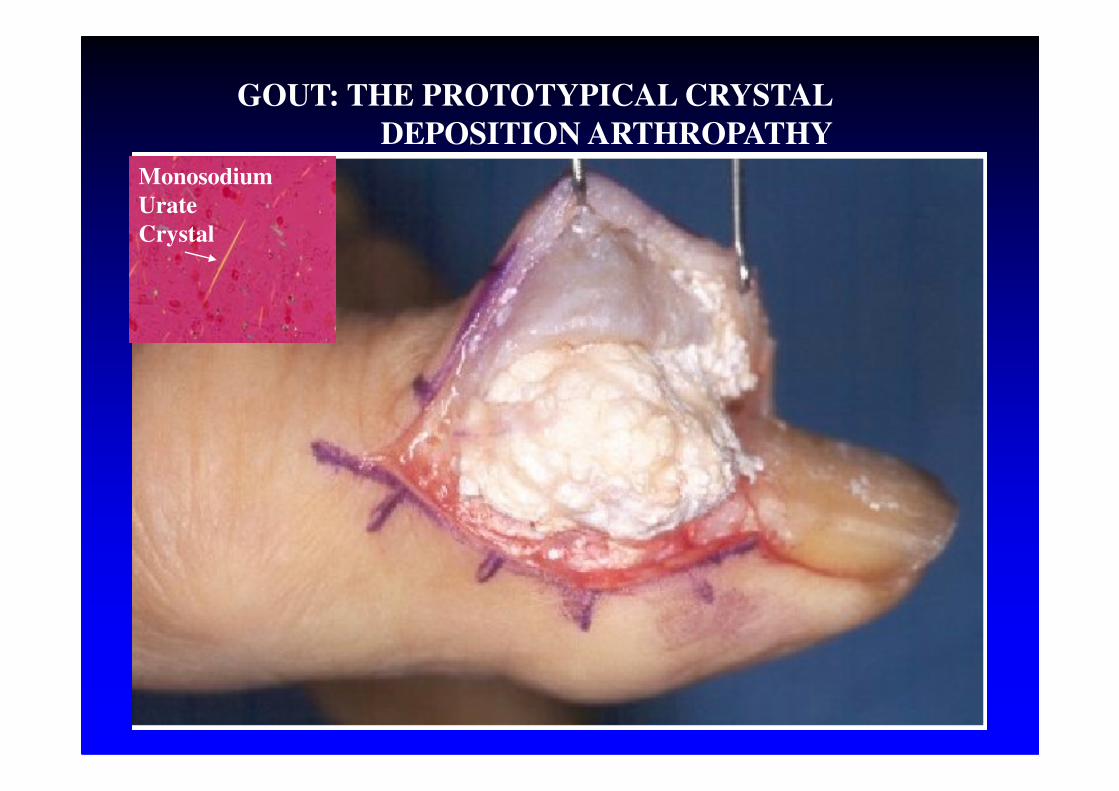
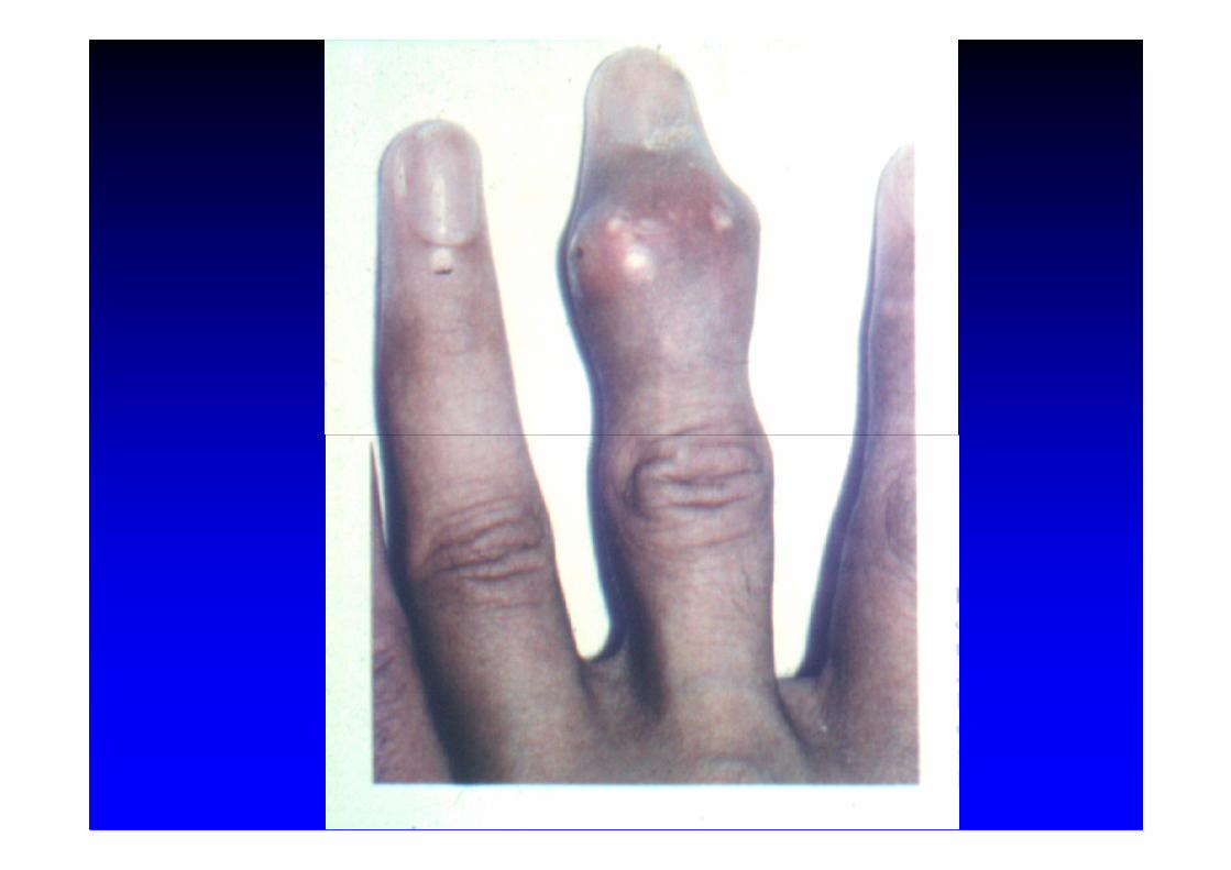
GOUT: THE PROTOTYPICAL CRYSTAL
DEPOSITION ARTHROPATHY
Monosodium
Urate
Crystal
7
6
3
1
2
4
93
5
8


Pla
sma
Uri
c A
cid
(µ
mo
l/l)
Gout
Low purine High purine
Diet
Pla
sma
Uri
c A
cid
(µ
mo
l/l)
Normal

Overview of the pathogenesis of gout
??
Choi, H. K. et. al. Ann Intern Med 2005;143:499-516

LOW PURINE DIET
Are allowed:
2000 mg of uric acid/week
Once a day a normal portion (cca 150 g.) of meat, milk and diary productsT
One glass of alcoholic drinks , coffee, tea
Is prohibited:
Organ meats , some fish species (lobsters, shrimps ), pulses,Larger quantities of alcohol

TYPES OF GOUT
primary gout:
renal hypoexcretion of uric acidexcessive intake of purines in the diet
hypoxanthine-guanine phosphoribosyltransferase (HPRT) T
deficiencycomplete –Lesch-Nyhan syndromepartial – Kelley Seegmiler syndrome
genetic defect increased activity of phophoribosylpyrophosphate synthetase(PRPPs)
genetic defect
familial juvenile hyperuricemic nephropathy
unknown pathogenesis
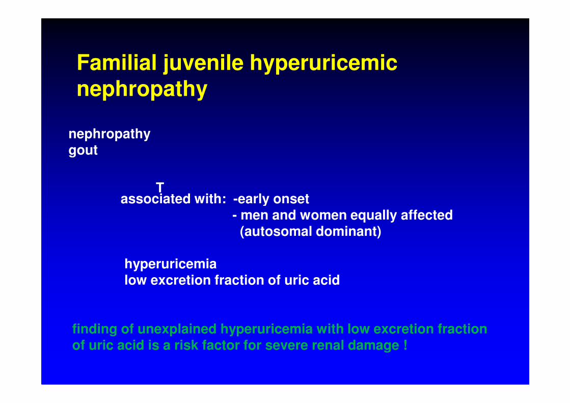
Familial juvenile hyperuricemicnephropathy
nephropathygout
associated with: -early onsetT
associated with: -early onset- men and women equally affected(autosomal dominant)
hyperuricemialow excretion fraction of uric acid
finding of unexplained hyperuricemia with low excretion fraction of uric acid is a risk factor for severe renal damage !

Flow chart for diferential diagnosis of gout
clinical - rheumatological examination(an important information - family history
↓↓
determination of serum and urinary uric acid
↓
detailed examination of purine metabolism
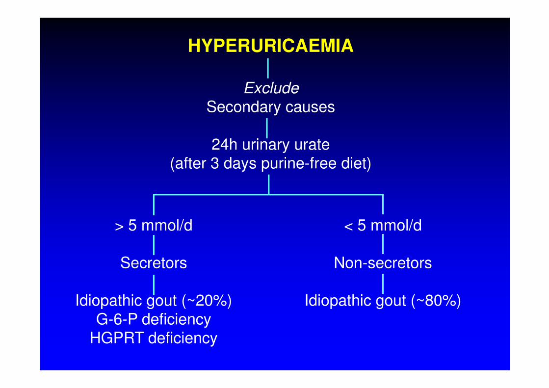
HYPERURICAEMIA
Exclude
Secondary causes
24h urinary urate(after 3 days purine-free diet)
> 5 mmol/d
Secretors
Idiopathic gout (~20%)G-6-P deficiency
HGPRT deficiency
< 5 mmol/d
Non-secretors
Idiopathic gout (~80%)

Detailed examination of purine metabolism.
1. Determination of serum and urinary uric acid* repeatedly* after diet (low purine)
2. Determination of purine metabolites using HPLC in* urine* plasma* CSF* CSF
3. Enzyme assays in ery, lymph.* hypoxanthin-phosphoribosyltransferase (HGPRT)* phosphoribosylpyrophosphatsynthetase (PRPPs)* adenin-phosphoribosyltransferase (APRT)* adenosine deaminase (ADA)* purine nucleosidphosphorilase (PNP)
4. DNA analýza URAT1 (SLC22A12), GLUT9

glomerular filtration 100%
reabsorption 99%
urate
4-component model of urate handling
secretion50%
reabsorption 99%
post-secretory reabsorption 40%
urine excretion
1%
51%
10%

• Enomoto, A., et al., Molecular identification of a renalurate anion exchanger that regulates blood urate levels.Nature, 2002. 417(6887): p. 447-52.
Urate transporterURAT 1-gene SLC22A12
• OMIM 607096, GeneID 116085• 11q13, 2 transcript variants (3206 and 2940 bp)553
amino acids• expressed in fetal and adult kidney
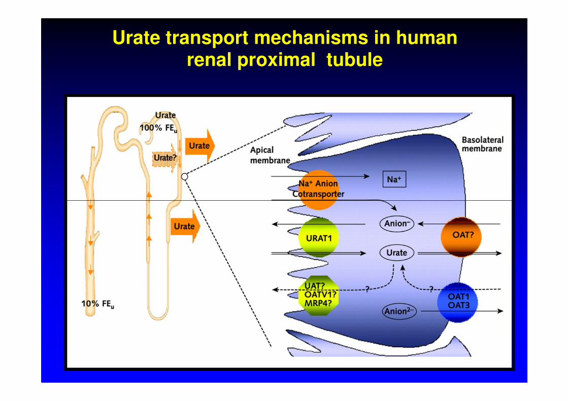
Urate transport mechanisms in human renal proximal tubule
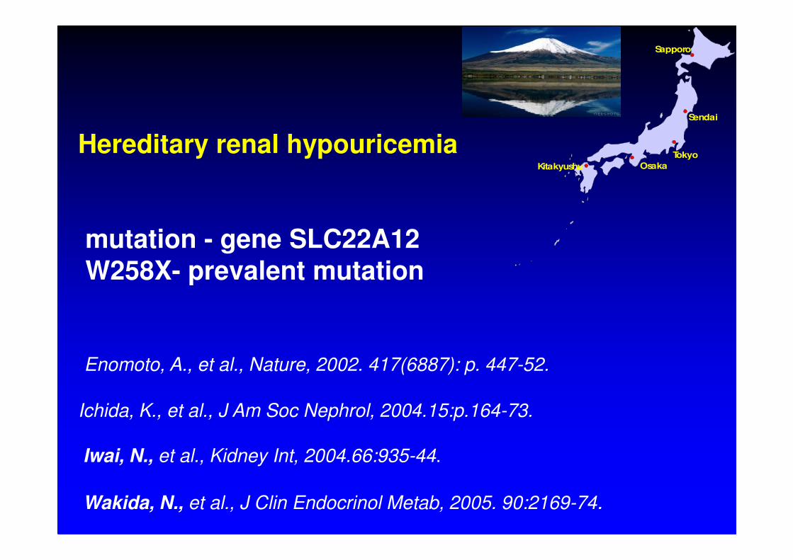
Hereditary renal hypouricemia
mutation - gene SLC22A12 W258X- prevalent mutation
Sendai
Sapporo
TokyoOsakaKitakyushu
W258X- prevalent mutation
Enomoto, A., et al., Nature, 2002. 417(6887): p. 447-52.
Ichida, K., et al., J Am Soc Nephrol, 2004.15:p.164-73.
Iwai, N., et al., Kidney Int, 2004.66:935-44.
Wakida, N., et al., J Clin Endocrinol Metab, 2005. 90:2169-74.

• new transport defect of purine metabolism
• biochemical markers
Hereditary renal hypouricemia, OMIM #220150
• biochemical markers– hypouricemia (SKM<120 µmol/l) – inceased excretion fraction of uric acid ( EFKM >10% )
• clinical features – urolithiasis– acute renal failure (exercise-induced)

1) If the stone is available → send it to the lab.
2) Exclude hypercalcemia and hyperuricaemia.
3) If the plasma calcium is normal collect a 24-hour
INVESTIGATION OF THE PATIENT WITH RENAL CALCULI
3) If the plasma calcium is normal collect a 24-hourspeciemn of urine for urinary calcium estimation.
4) If all these tests are negative and especially if there is afamily history of calculi → screen urine for cystine.If the qualitative test is positive the 24-hour excretion ofcystine should be estimated.


