Drug delivery in oncology: overviewCancer is one of the leading causes of death in the human population [1,2]. Systemic chemo-therapy is the most commonly used therapy, although several caveats exist relating to the fact that it is mostly based on pulsatile adminis-tration regimens using the maximum tolerated doses of cytotoxic drugs. The maximum toler-ated dose is the maximum amount of drug that can be administered before the toxicities exceed the potential benefits according to the formula: Therapeutic ratio = TD
50/ED
50, where TD
50
is the dose that is toxic to 50% of the receiv-ing patients and ED
50 is the dose efficacious in
50% of the patients. Being based on the aver-age toleration of the medication, and considering that anticancer drugs are more toxic than drugs belonging to other categories, this often results in high morbidity and mortality for the patients. As a result, pauses are inserted between the che-motherapy administrations to allow recovery from the side effects. Unfortunately, the long break periods between therapies that are neces-sary for the patients’ recovery from different tox-icities, in particular bone marrow suppression, allow drug-treated tumors to recover as well, and to become refractory to chemotherapy [3–8]. The therapeutic index of many drugs is very narrow and can be associated with intolerable systemic side effects that could, in particular, lead to potentially fatal hemorrhage and sepsis [9–11]. Moreover, systemic chemotherapy is often not efficient in delivering drugs to target sites at therapeutic concentrations, and maintaining
adequate drug levels within tumors is challeng-ing [12–14]. Delivering chemotherapy drugs at therapeutic doses within solid neoplasms is chal-lenging, since it is affected by several limiting factors, including [1,12–15]:nDrug transport to tissues via the blood circu-
latory system to tissues (this factor includes the issue of plasma binding proteins)
nInterstitial space
nDrug removal by capillaries
nTissue structure and composition with respect to drug distribution
The last point is a serious issue considering that all chemotherapy agents are hydrophilic in order to be administered as an infusion; con-versely, they are strongly lipophobic and enter the cell with difficulty (see the description for a specific anticancer agent used in electroche-motherapy [ECT] below). Therefore, only a small portion of the therapeutic dose reaches its target and, consequently, patients often receive insufficient doses at the tumor site that are inadequate for an effective therapy but still able to cause toxicity to healthy tissue [1,12–14]. Considering that more than 85% of human cancers are solid tumors [8], different strategies have been devised to solve these problems and to promote drug targeting (drug concentration in its area of effect). These approaches include intra-arterial chemotherapy [16], chemotherapy-impregnated implants [17–19] and polymeric drug delivery systems [8]. Despite the investigators’
Preclinical models in electrochemotherapy: the role of veterinary patients
Enrico Pierluigi Spugnini*1, Maurizio Fanciulli1, Gennaro Citro2 & Alfonso Baldi31SAFU Department, Regina Elena Cancer Institute, Via delle Messi d’Oro 156, Rome 00158, Italy2NSA, Ardea, Italy3Second University of Naples, Naples, Italy*Author for correspondence: Tel.: +39 065 266 2512 n Fax: +39 065 266 2505 n [email protected]
Electrochemotherapy is a tumor treatment that adapts the systemic or local delivery of anticancer drugs by the application of permeabilizing electric pulses with appropriate amplitude and waveforms. This allows the use of lipophobic drugs, which frequently have a narrow therapeutic index, with a decreased morbidity for the patient, while maintaining appropriate anticancer efficacy. Electrochemotherapy is used in humans for the treatment of cutaneous neoplasms or the palliation of skin tumor metastases, and a standard operating procedure has been devised. In veterinary oncology, the electrochemotherapy approach is gaining popularity, becoming a first-line treatment in consideration of its high efficacy and low toxicity. This review summarizes the state of the art in veterinary oncology as a preclinical model.
Keywords
n anthracenediones n bleomycin n cisplatin n companion animal n electroporation n mitoxantrone n therapeutic index
efforts, there has been minimum therapeutic gain [8,16–19]; therefore, localized delivery has become more and more popular in oncology. This strategy aims to maintain low systemic drug levels while ensuring effective drug target-ing [8,14]. Selective drug retention at target sites, with consequent decreased systemic drug expo-sure, is an important issue, in order to minimize toxicity and maximize efficacy [20]. This goal can be achieved through ECT, a cancer treatment that combines permeabilizing electric pulses that have appropriate waveforms and the local or sys-temic administration of chemotherapy agents [21,22]. In recent years, many cohorts of compan-ion animals affected by advanced spontaneous tumors have been recruited in ECT studies and compared with control groups treated with sur- control groups treated with sur-gery alone, obtaining high response rates and long-lasting effects [23–26]. Several drugs have been used with permeabilizing pulses; bleomy-cin, cisplatin and mitoxantrone. Below, a brief description of these drugs is provided.
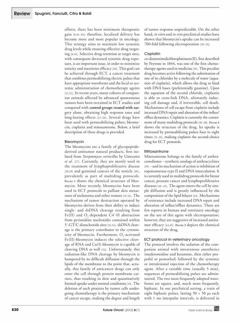
BleomycinThe bleomycins are a family of glycopeptide-derived antitumor natural products, first iso-lated from Streptomyces verticillus by Umezawa et al. [27]. Currently, they are mostly used in the treatment of lymphoproliferative disease [28,29] and germinal cancers of the testicle [30], prevalently as part of multidrug protocols. Figure 1 shows the chemical structure of bleo-mycin. More recently, bleomycins have been used in ECT protocols to palliate skin metas-tases of melanoma and other tumors [31,32]. The mechanism of tumor destruction operated by bleomycins derives from their ability to induce single- and dsDNA cleavage resulting from Fe(II) and O
2-dependent C4´-H abstraction
from pyrimidine nucleotides contained within 5́ -GT/C dinucleotide sites [33,34]. dsDNA cleav-age is the primary contributor to the cytotox-icity of bleomycin. Furthermore, O
2-activated
Fe(II)·bleomycin induces the selective cleav-age of RNA and Cu(I)·bleomycin is capable of cleaving DNA as well [34]. Unfortunately, this radiation-like DNA cleavage by bleomycin is hampered by its difficult diffusion through the lipids of the membrane to the point that, actu-ally, this family of anticancer drugs can only enter the cell through protein membrane car-riers, thus resulting in slow and quantitatively limited uptake under normal conditions [33]. The deletion of such proteins by tumor cells under-going chemotherapy is the primary mechanism of cancer escape, making the degree and length
of tumor response unpredictable. On the other hand, in vitro and in vivo preclinical studies have shown that bleomycin’s uptake can be increased 700-fold following electroporation [30–33].
Cisplatincis-diamminedichloroplatinum(II), first described by Peyrone in 1844, was one of the first chemo-therapy agents used in medicine [35]. This peculiar drug becomes active following the substitution of one of its chlorides by a molecule of water (aqua-tion of cisplatin), which allows the drug to bind with DNA bases (preferentially guanine). Upon the aquation of the second chloride, cisplastin is able to cross-link DNA, ultimately induc-ing cell damage and, if irreversible, cell death. Mechanisms of cell escape from cisplatin include increased DNA repair and alteration of the influx/efflux dynamics. Cisplatin is currently the corner-stone of many multidrug protocols [36–38]. Figure 2 shows the structure of the drug. Its uptake is increased by permeabilizing pulses four to eight times [31–33], making cisplatin the second-choice drug for ECT prototols.
MitoxantroneMitoxantrone belongs to the family of anthra-cenediones – synthetic analogs of anthracyclines [39] – and its mechanism of action is inhibition of topoisomerase type II and DNA intercalation. It is currently used in multidrug protocols for breast cancer, prostate cancer and lymphoproliferative diseases [40–42]. The agent enters the cell by sim-ple diffusion and is greatly influenced by the composition of the lipid bilayer [43]. Mechanisms of resistance include increased DNA repair and alteration of influx/efflux dynamics. There are few reports in human and veterinary oncology on the use of this agent with electroporation; however, they are suggestive of increased antitu-mor efficacy [44,45]. Figure 3 depicts the chemical structure of the drug.
ECT protocol in veterinary oncologyThe protocol involves the sedation of the com-panion animal with intravenous anesthetics (medetomidine and ketamine, then either pro-pofol or pentothal) followed by the systemic or intralesional injection of the chemotherapy agent. After a variable time (usually 5 min), sequences of permeabilizing pulses are admin-istered. The two most frequently adopted wave-forms are square, and, much more frequently, biphasic. In our preclinical setting, a train of eight biphasic pulses, lasting 50 + 50 µs each with 1 ms interpulse intervals, is delivered in
www.futuremedicine.com 831future science group
Preclinical models in electrochemotherapy: the role of veterinary patients Review
Fig
ure
1. B
leo
myc
in.
SO
O
OO
O
ON
N H
NN
N
S
S
N
S+
N H
O
NH
OH
NH
OO
H
NH
NH
O
ON
H2
NH
2
O
NH
NH
2O
NH
2
O
O
OH
OH
O
OH
OH
O OH
NH
2
O
OH
O
ON
N H
NN
N
S
S
N
S+
N H
O
NH
OH
NH
OO
H
NH
NH
O
ON
H2
NH
2
O
NH
NH
2O
NH
2
O
O
OH
OH
O
OH
OH
O OH
NH
2
O
OH
--
Future Oncol. (2012) 8(7)832 future science group
Review Spugnini, Fanciulli, Citro & Baldi
bursts of 1300 V/cm (800 V/cm for exposed lesions) at 1 Hz frequency, by means of caliper or needle-array electrodes. The biphasic pulses are adopted because they allow a smooth electrical transition; moreover, there is some evidence that they could be more effective at permeabilizing tumor cells that have a nonhomogeneous align-ment with regard to field polarity. Several com-mercial and in-house apparati have been used by different research groups to date. Adherence of the external electrodes to the lesion is maximized using an electroconductive gel. The treatment is repeated at 1- or 2-week intervals until a com-plete response is obtained or tumor recurrence occurs.
ECT in veterinary oncologyThe first in vivo study involved the use of ECT as a rescue protocol in cats with recurring soft tissue sarcoma after radiation therapy and sur-gical ablation [46]. In this clinical model, cats were randomized to receive bleomycin as a single agent or combined with an implant of cells secreting IL-2, followed by the delivery of eight single square pulses. A control group of untreated animals was included as well. The authors reported only one measurable response; however, they claimed a prolonged survival in the cohort of cats receiving ECT versus con-trols. This low response rate could be partially attributed to previous treatments, which could have elicited chemoresistance [20]. A few years after the first preliminary investigation in feline companion animals, two Phase I/II studies were conducted in companion animals; in the first, a cohort of dogs and cats was treated with intra-lesional cisplatin coupled with square electric pulses [47]. At the same time, a preliminary study, including different solid tumors, was conducted using trains of biphasic pulses associated with intralesional bleomycin [20]. The overall response rate of this preliminary investigation was 80% with a 40% rate of extended remissions. This work underlined two important issues; the need for dedicated electrodes for the therapy of soft tissue neoplasms, and the resistance to a smooth
permeabilization offered by connective tissue, especially in mesenchymal tumors [48]. After this preliminary phase, several studies have been performed involving direct attack on specific histotypes with ECT or evaluation of adjuvant and neoadjuvant ECT in advanced companion animal cancers.
Perianal tumorsIn 2005, Tozon et al. investigated the pos-sible use of cisplatin coupled with ECT in the treatment/palliation of large perianal tumors, obtaining a response rate of 82% (41% com-plete responses); a limit of this study was the lack of histopathological characterization, which did not allow discrimination between adenoma and carcinoma [49]. In 2007, Spugnini et al. treated a similar cohort of dogs with perianal tumors (eight adenoma and four carcinoma) using intralesional bleomycin, obtaining a 91% response rate (83% complete responses) and observing a tendency to resistance from car-cinomas [50]. As a result, except for adenomas, the current strategy for aggressive perineal neo-plasms involves marginal tumor resection fol-lowed by ECT [51]. This strategy allows good tumor control while preserving the anatomical and functional integrity of the anus.
Malignant melanomaDogs are prone to oral melanoma, although it has been seldom reported in the GI tract [23,52,53]. They are rapidly growing and chemoresistant neoplasms. A preliminary study using intra-lesional bleomycin followed by electropora-tion obtained a high response rate (80%, with 50% complete responders) and 40% long-term responders [23]. Interestingly, all the long-term responders presented vitiligo-like mucosal discolorations at the tumor site, suggesting recruitment of the immune system following the tumor’s ECT-induced apoptotic death. The efficacy of ECT for this tumor has been further substantiated by a case report in which cisplatin-based ECT allowed the successful pal-liation (3 months) of a dog with an unresectable perianal melanoma with distant metastases [53].
Soft tissue sarcomaA preclinical study involving cats with injection-site sarcomas evaluated the efficacy of intraop-erative and postoperative ECT [24]. Cats were assigned to three treatment groups: surgery (con-trol group), surgery plus intraoperative ECT and surgery plus postoperative ECT. The study clearly showed that ECT improved local control and
Pt2-
Cl
Cl NH2
NH2
Figure 2. Cisplatin.
www.futuremedicine.com 833future science group
Preclinical models in electrochemotherapy: the role of veterinary patients Review
survival compared with the control group. Time to recurrence was 12 and 19 months, respectively, for the two ECT cohorts, while the control group showed recurrence within 4 months. Compared with radiation therapy, ECT allows a replication of the treatment course in case of recurrence without the side effects of re-irradiated tissues [24]. A confirmatory study in 22 dogs with soft tissue sarcomas resulted in a median time to recurrence of 730 days with a 95% response rate [54]. A successful outcome has also been reported in a dog treated in a neoadjuvant fashion [55]. The side effects for veterinary patients treated with adjuvant ECT were confined to local inflamma-tion and occasional wound dehiscence [24,54]. These studies offer promising perspectives for the translation of these adjuvant and neoadjuvant protocols to humans, especially for soft tissue sarcomas of the limb, where oncologists strive to achieve local control while preserving function-ality with tolerable side effects for the patient [21]. The latest article published on this topic shows an original approach to the problem of local con-trol in sarcomas, investigating the use of a drug that is otherwise potentially lethal to cats when coupled with ECT [15]. Electroporation rapidly transfers the drug from the interstitial space to the cytoplasm, preventing systemic absorp-tion and the toxicities (fatal pulmonary edema) associated with the use of this drug in felines. The article reports a high response rate (70%) without systemic toxicites, the only side effects being limited to local inflammation. The use of electroporation to make a potentially lethal che-motherapy agent effective opens a new field of investigation for preclinical and clinical oncolo-gists, and may lead to the re-introduction of chemotherapy or ECT protocols of drugs previ-ously excluded due to their too-narrow therapeu-tic index. However, caution should be exercised when retreating patients with ECT who had pre-vious radiation therapy for solid tumors, since a case report describes, in a cat previously radiated for a soft tissue sarcoma, a severe radiation recall after ECT with cisplatin [56]. Radiation recall is a well-documented phenomenon involving a red-ness resembling severe sunburn that can happen within a previously radiated area following the administration of many chemotherapy agents. This observation suggests that these same drugs can elicit this complication even when delivered by electric pulses.
Mast cell tumorsMast cell tumor is a cancer entity that is peculiar to companion animals and consists of a solid
tumor made up entirely of neoplastic mast cells. The neoplasm has peculiar characteristics since the cells contain large amounts of vasoactive substances, including histamine, which makes the neoplasm extremely invasive and exposes the patient to the risk of tumor degranulation, which is associated with anaphylactic-like phenomena [57]. Concurrent with the studies in sarcoma, bleomycin with adjuvant ECT has been tested in a cohort of 28 dogs with mast cell sarcomas, resulting in a response rate of 85% and a mean time to recurrence of 52.7 ± 6.5 months [28]. The efficacy of ECT in dogs has been further explored by comparing treatment with intralesional cis-platin coupled with square electric pulses versus surgery [58]. The study showed improvement in terms of local control; however, has the limita-tion of lacking histopathological characteriza-tion of the treated tumors. This fact deprives the clinicians of crucial prognostic information such as tumor grade. Finally, a group of 37 dogs with incompletely excised grade II and III mast cell tumors was treated with ECT and cispla-tin, obtaining a 78% response rate and minimal side effects, broadening the spectrum of adju-vant protocols that could be translated to human patients [26].
Head & neck carcinomaAs for human patients, the local control of these neoplasms poses a significant challenge in veterinary patients as well. Aggressive sur-gery alone, or surgery combined with radia-tion therapy and systemic chemotherapy, have been advocated, obtaining variable degrees of success [59]. Recently, ECT using intralesional bleomycin has been used to treat advanced squamous cell carcinoma of the nasal pla-num in cats. The treatment resulted in a total
OH O NH
NH
NH
OH
NH
OHOH O
Figure 3. Mitoxantrone.
Future Oncol. (2012) 8(7)834 future science group
Review Spugnini, Fanciulli, Citro & Baldi
response rate (pooling together partial and complete responses) of 100%, with a com-plete response rate of 77%. Side effects were contained to erythema, swelling and focal inflammation. In a dog that failed surgery and two courses of adjuvant bleomycin or cisplatin for an aggressive apocrine gland carcinoma, systemic mitoxantrone, potentiated by trains of biphasic pulses, resulted in successful pal-liation, obtaining a complete remission that lasted in excess of 6 months [45]. An advantage of the increased drug uptake following elec-troporation is that the dog has been treated with the lowest dose of mitoxantrone within the therapeutic index, dramatically decreasing hematological and gastrointestinal side effects.
ConclusionECT has been proven to increase the therapeutic index of many chemotherapy drugs, resulting in better local control of solid neoplasms in com-panion animals. Moreover, its effectiveness is strengthened when it is used in an adjuvant fash-ion through the generation of trains of biphasic pulses [15,20–26]. This peculiar feature gives ECT
two unique advantages over currently adopted strategies aiming to increase local control (i.e., radiation therapy):nA lack of acute and delayed side effects such
as radiation-induced erythema, necrosis and pneumonia;
nThe possibility of repeating the treatments in case of tumor recurrence.
Finally, it must be emphasized that the adop-tion of ECT as a rescue protocol in patients who have failed radiation therapy could be associ-ated with consistent side effects; the only case report in the literature describes a severe radia-tion recall in a cat treated with cisplatin rather than bleomycin [60].
Future perspectiveECT is currently being investigated for differ-ent tumor histotypes in companion animals, displaying promising results and defining specific patterns of response as well as clinical [24,26] and histopathological prognostic factors [25]. Currently, multiple studies are ongoing at several institutions to evaluate new drugs and
Executive summary
Drug delivery in oncology: overviewn Delivering chemotherapy drugs at therapeutic doses within solid neoplasms is affected by several limiting factors including
pharmacokinetics and tumor environment.n The goal for clinicians is to deliver the maximum tolerated dose of chemotherapy agents.n The maximum tolerated dose is the maximum amount of drug that can be administered before the toxicities exceed the potential
benefits according to the formula: Therapeutic ratio = TD50
/ED50
, where TD50
is the dose that is toxic to 50% of the receiving patients and ED
50 is the dose efficacious in 50% of the patients.
n Electrochemotherapy (ECT) allows a greater drug delivery through the electropermeabilization of cancer cells.n Currently adopted drugs in veterinary ECT are bleomycin, cisplatin and mitoxantrone.
ECT protocol in veterinary oncologyn The protocol involves sedation of the animal.n Following that is the intralesional or systemic injection of the chemotherapy agent.n After a variable time, the animal undergoes electroporation with eight electric pulses per centimeter.
ECT in veterinary oncologyn The first protocols directly attacked bulky tumors with variable success.n Improved protocols were based on an adjuvant use of ECT, following tumor debulking.n In selected tumors, neoadjuvant ECT allowed the removal of surgically unresectable neoplasms.n The most frequently used drug has been bleomycin.n Cisplatin is a second-choice drug.n Mitoxantrone is currently under investigation.
Conclusionn ECT has proven to be a safe and efficacious therapy for the local control of solid neoplasms in companion animals, and its effectiveness
is especially strengthened when it is used in an adjuvant fashion through the generation of trains of biphasic pulses.
Future perspectiven Further studies are currently ongoing to evaluate new drugs and delivery systems.n ECT can play an important role in the reversal of chemoresistance.n ECT has shown great potential at implementing the uptake of biological agents.
www.futuremedicine.com 835future science group
Preclinical models in electrochemotherapy: the role of veterinary patients Review
humans has already been evaluated for lesions other than cutaneous neoplasms or skin lesion metastases [66]. We think that, in the near future, thanks to experience gained from pre-clinical models, as outlined in this article, sev-eral targeted therapy protocols in humans will be made possible or implemented through the adoption of electroporation.
ReferencesPapers of special note have been highlighted as:n of interestnn of considerable interest
1. De Souza R, Zahedi P, Allen CJ, Piquette-Miller M. Polymeric drug delivery systems for localized cancer chemotherapy. Drug Delivery 17, 365–375 (2010).
2. Ma X, Yu H. Global burden of cancer. Yale J. Biol. Med. 79, 85–94 (2006).
3. Kim JJ, Tannock IF. Repopulation of cancer cells during therapy: an important cause of treatment failure. Nat. Rev. Cancer 5, 516–525 (2005).
4. Stavreva NA, Stavrev PV, Warkentin B, Fallone BG. Investigating the effect of cell repopulation on the tumor response to fractionated external radiotherapy. Med. Phys. 30, 735–742 (2003).
5. Brade AM, Tannock IF. Scheduling of radiation and chemotherapy for limited-stage small-cell lung cancer: repopulation as a cause of treatment failure? J. Clin. Oncol. 24, 1020–1022 (2006).
6. Davis AJ, Tannock JF. Repopulation of tumour cells between cycles of chemotherapy: a neglected factor. Lancet Oncol. 1, 86–93 (2001).
7. Corry J, Rischin D. Strategies to overcome accelerated repopulation and hypoxia – what have we learned from clinical trials? Semin. Oncol. 31, 802–808 (2004).
8. Vassileva V, Allen CJ, Piquette-Miller M. Effects of sustained and intermittent paclitaxel therapy on tumor repopulation in ovarian cancer. Mol. Cancer Ther. 7, 630–637 (2008).
9. Ishikawa E, Yamamoto T, Sakamoto N et al. Low peripheral lymphocyte count before focal radiotherapy plus concomitant temozolomide predicts severe lymphopenia during malignant glioma treatment. Neurol. Med. Chir. (Tokyo) 50, 638–644 (2010).
10. Gasparini G. Metronomic scheduling: the future of chemotherapy? Lancet Oncol. 2, 733–740 (2001).
11. Scharovsky OG, Mainetti LE, Rozados VR. Metronomic chemotherapy: changing the paradigm that more is better. Curr. Oncol. 16, 91–99 (2009).
12. Au JL, Jang SH, Zheng J et al. Determinants of drug delivery and transport to solid tumors. J. Control. Release 74, 31–46 (2001).
13. Au JL, Jang SH, Wientjes MG. Clinical aspects of drug delivery to tumors. J. Control. Release 78, 81–95 (2002).
14. Jang SH, Wientjes MG, Lu D, Au JLS. Drug delivery and transport to solid tumors. Pharm. Res. 20, 1337–1350 (2003).
15. Spugnini EP, Renaud SM, Buglioni S et al. Electrochemotherapy with cisplatin enhances local control after surgical ablation of fibrosarcoma in cats: an approach to improve the therapeutic index of highly toxic
chemotherapy drugs. J. Transl Med. 9, 152 (2011).
nn First proof of concept that electrochemotherapy (ECT) can dramatically improve the therapeutic index of highly toxic chemotherapy agents.
16. Dizon DS, Kemeny NE. Intrahepatic arterial infusion of chemotherapy: clinical results. Semin. Oncol. 29, 126–135 (2002).
17. Withrow SJ, Liptak JM, Straw RC et al. Biodegradable cisplatin polymer in limb-sparing surgery for canine osteosarcoma. Ann. Surg. Oncol. 11, 705–713 (2004).
18. Liu J, Meisner D, Kwong E, Wu XY, Johnston MR. A novel trans-lymphatic drug delivery system: implantable gelatin sponge impregnated with PLGA–paclitaxel microspheres. Biomaterials 28, 3236–3244 (2007).
19. Olson JJ, McKenzie E, Skurski-Martin M, Zhang Z, Brat D, Phuphanich S. Phase I ana lysis of BCNU-impregnated biodegradable polymer wafers followed by systemic interferon alfa-2b in adults with recurrent glioblastoma multiforme. J. Neurooncol. 90, 293–299 (2008).
20. Spugnini EP, Porrello A. Potentiation of chemotherapy in companion animals with spontaneous large neoplasms by application of biphasic electric pulses. J. Exp. Clin. Cancer Res. 22, 571–580 (2003).
Financial & competing interests disclosureThis work has been supported by an Istituto Nazionale per l’Assicurazione contro gli Infortuni sul Lavoro (INAIL) grant, by ‘Grant 2011’, by an Italian Association for Cell Cultures (AiCC) grant and a PROJECT FIRB/MUR (RBIPO6LCA9-009) grant of the Italian Ministry of Health to EP Spugnini, as well as by a FUTURA-onlus grant to EP Spugnini and A Baldi and a Second University of Naples grant to A Baldi. The authors have no other relevant affiliations or financial involvement with any organization or entity with a financial interest in or financial conflict with the subject matter or materials discussed in the manuscript apart from those disclosed.
No writing assistance was utilized in the production of this manuscript.
delivery systems. Mitoxantrone is showing con-siderable promise [45]. An area in which the use of ECT might be implemented is that of reversal of chemoresistance. Preliminary findings, limited to three patients with refractory penile tumors, showed clinically relevant responses, with long duration [61]. More importantly, although expe-rience is currently limited to laboratory animals, ECT has shown great potential for increasing the uptake of biological agents such as oligoanti-sense, plasmids and vaccines. In our experience, electroporation markedly enhanced the uptake and efficacy of anti-Bcl-2 oligonucleotides in an in vivo melanoma xenograft model [62]. A frequently neglected field of clinical interven-tion in which ECT is showing promise is the palliation of paraneoplastic syndromes; more precisely, Bodles-Brakhop’s group has success-fully treated cancer-associated anemia in dogs, opening a whole new field of investigations [63].
It should be highlighted that the use of ECT for the treatment of cutaneous and subcutane-ous metastases in humans has been standard-ized, and is now widely adopted in many cancer centers worldwide [64,65]. Furthermore, ECT in
Future Oncol. (2012) 8(7)836 future science group
Review Spugnini, Fanciulli, Citro & Baldi
21. Spugnini EP, Citro G, D’Avino A, Baldi A. Potential role of electrochemotherapy for the treatment of soft tissue sarcoma: first insights from preclinical studies in animals. Int. J. Biochem. Cell. Biol. 40, 159–163 (2008).
22. Spugnini EP, Citro G, Baldi A. Adjuvant electrochemotherapy in veterinary patients: a model for the planning of future therapies in humans. J. Exp. Clin. Cancer Res. 28, 114 (2009).
23. Spugnini EP, Dragonetti E, Vincenzi B, Onori N, Citro G, Baldi A. Pulse-mediated chemotherapy enhances local control and survival in a spontaneous canine model of primary mucosal melanoma. Melanoma Res. 16, 23–27 (2006).
24. Spugnini EP, Baldi A, Vincenzi B et al. Intraoperative versus postoperative electrochemotherapy in high grade soft tissue sarcomas: a preliminary study in a spontaneous feline model. Cancer Chemother. Pharmacol. 59, 375–381 (2007).
nn First study identifying prognostic factors in ECT.
25. Spugnini EP, Baldi F, Mellone P et al. Patterns of tumor response in canine and feline cancer patients treated with electrochemotherapy: preclinical data for the standardization of this treatment in pets and humans. J. Transl Med. 5, 48 (2007).
nn First detailed ana lysis of the histopathology of ECT-treated neoplasms.
26. Spugnini EP, Vincenzi B, Citro G, Dotsinsky I, Mudrov T, Baldi A. Evaluation of cisplatin as an electrochemotherapy agent for the treatment of incompletely excised mast cell tumors in dogs. J. Vet. Intern. Med. 25, 407–411 (2011).
27. Umezawa H, Maeda K, Takeuchi T, Okami Y. New antibiotics, bleomycin A and B. J. Antibiot. (Tokyo) 19, 200–209 (1996).
28. Connors JM. State-of-the-art therapeutics: Hodgkin’s lymphoma. J. Clin. Oncol. 23, 6400–6408 (2005).
29. Reyes F, Lepage E, Ganem C et al. ACVBP versus CHOP plus radiotherapy for localized aggressive lymphoma. N. Engl. J. Med. 352, 1197–1205 (2005).
30. Einhorn LH. Curing metastatic testicular cancer. Proc. Natl Acad. Sci. USA 99, 4592–4595 (2002).
31. Gehl J, Geertsen PF. Efficient palliation of haemorrhaging malignant melanoma skin metastases by electrochemotherapy. Melanoma Res. 10, 585–589 (2000).
32. Matthiessen LW, Chalmers RL, Sainsbury DC. Management of cutaneous metastases using electrochemotherapy. Acta Oncol. 50, 621–629 (2011).
33. Gothelf A, Mir LM, Gehl J. Electrochemotherapy: results of cancer treatment using enhanced delivery of bleomycin by electroporation. Cancer Treat. Rev. 29, 371–387 (2003).
34. Goodwin KD, Lewis MA, Long EC, Georgiadis MM. Crystal structure of DNA-bound Co(III) bleomycin B2: insights on intercalation and minor groove binding. Proc. Natl Acad. Sci. USA. 105, 5052–5056 (2008).
36. Furuse J, Okusaka T, Bridgewater J et al. Lessons from the comparison of two randomized clinical trials using gemcitabine and cisplatin for advanced biliary tract cancer. Crit. Rev. Oncol. Hematol. 80, 31–39 (2011).
37. Ismaili N, Amzerin M, Flechon A. Chemotherapy in advanced bladder cancer: current status and future. J. Hematol. Oncol. 4, 35 (2011).
38. Otty Z, Skinner MB, Dass J et al. Efficacy and tolerability of weekly low-dose cisplatin concurrent with radiotherapy in head and neck cancer patients. Asia Pac. J. Clin. Oncol. 7, 287–292 (2011).
39. Murdock KC, Child RG, Fabio PF et al. Antitumor agents. 1. 1,4-bis[(aminoalkyl)amino]-9,10-anthracenediones. J. Med. Chem. 22, 1024–1030 (1979).
40. Kim J, Lee J, Chang E et al. Prognostic factors in patients with stage II/III breast cancer treated with adjuvant extension of neoadjuvant chemotherapy: a retrospective cohort study with ten-years of follow-up data. J. Breast Cancer 14, 39–45 (2011).
41. Yap TA, Zivi A, Omlin A, de Bono JS. The changing therapeutic landscape of castration-resistant prostate cancer. Nat. Rev. Clin. Oncol. 8, 597–610 (2011).
42. Parker C, Waters R, Leighton C et al. Effect of mitoxantrone on outcome of children with first relapse of acute lymphoblastic leukaemia (ALL R3): an open-label randomised trial. Lancet 376, 2009–2017 (2010).
43. Burns CP, Haugstad BN, Mossman CJ, North JA, Ingraham LM. Membrane lipid alteration: effect on cellular uptake of mitoxantrone. Lipids 23, 393–397 (1988).
nn Assessment of the impact of lipid composition on the uptake of chemotherapy drugs.
44. Trifonov D, Katev N, Daskalov I. Intra-arterial chemotherapy of breast cancer combined with electroporation. J. BUON 6, 331 (2001).
45. Spugnini EP, Dotsinsky I, Mudrov N et al. Successful rescue of an apocrine gland carcinoma metastatic to the cervical lymph nodes by mitoxantrone coupled with trains of permeabilizing electrical pulses (electrochemotherapy). In Vivo 22, 51–53 (2008).
nn First report of the use of mitoxantrone in veterinary ECT.
46. Mir LM, Devauchelle P, Quintin-Colonna F et al. First clinical trial of cat soft-tissue sarcomas treatment by electrochemotherapy. Br. J. Cancer 76, 1617–1622 (1997).
nn First clinical trial on veterinary cancer patients.
47. Tozon N, Sersa G, Cemazar M. Electrochemotherapy: potentiation of local antitumour effectiveness of cisplatin in dogs and cats. Anticancer Res. 21, 2483–2488 (2001).
48. Zaharoff DA, Barr RC, Li CY, Yuan F. Electromobility of plasmid DNA in tumor tissues during electric field-mediated gene delivery. Gene Ther. 9, 1286–1290 (2002).
49. Tozon N, Kodre V, Sersa G, Cemazar M. Effective treatment of perianal tumors in dogs with electrochemotherapy. Anticancer Res. 25, 839–845 (2005).
50. Spugnini EP, Dotsinsky I, Mudrov N et al. Biphasic pulses enhance bleomycin efficacy in a spontaneous canine perianal tumors model. J. Exp. Clin. Cancer Res. 26, 483–487 (2007).
51. Spugnini EP, Dotsinsky I, Mudrov N et al. Adjuvant electrochemotherapy for incompletely excised anal sac carcinoma in a dog. In Vivo 22, 47–49 (2008).
52. Spugnini EP, Dragonetti E, Murace R et al. Spontaneous intestinal melanoma in dogs. In Vivo 19, 1051–1054 (2005).
53. Spugnini EP, Filipponi M, Romani L et al. Local control and distant metastasis after electrochemotherapy of a canine anal melanoma. In Vivo 21, 897–899 (2007).
54. Spugnini EP, Vincenzi B, Citro G et al. Adjuvant electrochemotherapy for the treatment of incompletely excised spontaneous canine sarcomas. In Vivo 21, 819–822 (2007).
55. Spugnini EP, Vincenzi B, Betti G et al. Surgery and electrochemotherapy of a high-grade soft tissue sarcoma in a dog. Vet. Rec. 162, 186–188 (2008).
56. Spugnini EP, Dotsinsky I, Mudrov N et al. Electrochemotherapy-induced radiation recall in a cat. In Vivo 22, 751–753 (2008).
57. Spugnini EP, Vincenzi B, Baldi F, Citro G, Baldi A. Adjuvant electrochemotherapy for the treatment of incompletely resected canine mast cell tumors. Anticancer Res. 26, 4585–4589 (2006).
www.futuremedicine.com 837future science group
Preclinical models in electrochemotherapy: the role of veterinary patients Review
58. Kodre V, Cemazar M, Pecar J, Sersa G, Cor A, Tozon N. Electrochemotherapy compared with surgery for treatment of canine mast cell tumours. In Vivo 23, 55–62 (2009).
59. Spugnini EP, Vincenzi B, Citro G et al. Electrochemotherapy for the treatment of squamous cell carcinoma in cats: a preliminary report. Vet. J. 179, 117–120 (2009).
60. Azria D, Magnè N, Zouhair A et al. Radiation recall: a well recognized but neglected phenomenon. Cancer Treat. Rev. 31, 555–570 (2005).
61. Spugnini EP, Dotsinsky I, Mudrov N, Citro G, D’Avino A, Baldi A. Biphasic pulses enhance bleomycin efficacy in a spontaneous canine genital tumor model of
chemoresistance: Sticker sarcoma. J. Exp. Clin. Cancer Res. 27, 58 (2008).
62. Spugnini EP, Biroccio A, De Mori R et al. Electroporation increases antitumoral efficacy of the bcl-2 antisense G3139 and chemotherapy in a human melanoma xenograft. J. Transl Med. 9, 125 (2011).
63. Bodles-Brakhop AM, Brown PA, Pope MA, Draghia-Akli R. Double-blinded placebo controlled plasmid GHRH trial for cancer-associated anemia in dogs. Mol. Ther. 16, 862–870 (2008).
n First treatment of paraneoplastic syndromes through electroporation.
64. Marty M, Sersa G, Garbay JR et al. Electrochemotherapy – an easy highly effective and safe treatment of cutaneous and
subcutaneous metastases: results of ESOPE (European Standard Operating Procedures of Electrochemotherapy) study. Eur. J. Cancer Suppl. 4(11), 3–13 (2006).
65. Mir LM, Gehl J, Sersa G et al. Standard operating procedures of the electrochemotherapy: instructions for the use of bleomycin or cisplatin administered either systemically or locally and electric pulses delivered by the CliniporatorTM by means of invasive or non-invasive electrodes. Eur. J. Cancer Suppl. 4(11), 14–25 (2006).
66. Edhemovic I. Electrochemotherapy: a new technological approach in treatment of metastases in the liver. Technol. Cancer Res. Treat. 10(5), 475–485 (2011).