Prediction of cyclin-dependent kinase 2 inhibitorpotency using the fragment molecular orbitalmethodMichael P Mazanetz*, Osamu Ichihara, Richard J Law, Mark Whittaker
Abstract
Background: The reliable and robust estimation of ligand binding affinity continues to be a challenge in drug
design. Many current methods rely on molecular mechanics (MM) calculations which do not fully explain complex
molecular interactions. Full quantum mechanical (QM) computation of the electronic state of protein-ligandcomplexes has recently become possible by the latest advances in the development of linear-scaling QM methods
such as the ab initio fragment molecular orbital (FMO) method. This approximate molecular orbital method is
sufficiently fast that it can be incorporated into the development cycle during structure-based drug design for the
reliable estimation of ligand binding affinity. Additionally, the FMO method can be combined with approximations
for entropy and solvation to make it applicable for binding affinity prediction for a broad range of target and
chemotypes.
Results: We applied this method to examine the binding affinity for a series of published cyclin-dependent kinase
2 (CDK2) inhibitors. We calculated the binding affinity for 28 CDK2 inhibitors using the ab initio FMO method
based on a number of X-ray crystal structures. The sum of the pair interaction energies (PIE) was calculated and
used to explain the gas-phase enthalpic contribution to binding. The correlation of the ligand potencies to the
protein-ligand interaction energies gained from FMO was examined and was seen to give a good correlation
which outperformed three MM force field based scoring functions used to appoximate the free energy of binding.
Although the FMO calculation allows for the enthalpic component of binding interactions to be understood at the
quantum level, as it is an in vacuo single point calculation, the entropic component and solvation terms are
neglected. For this reason a more accurate and predictive estimate for binding free energy was desired. Therefore,
additional terms used to describe the protein-ligand interactions were then calculated to improve the correlation
of the FMO derived values to experimental free energies of binding. These terms were used to account for the
polar and non-polar solvation of the molecule estimated by the Poisson-Boltzmann equation and the solvent
accessible surface area (SASA), respectively, as well as a correction term for ligand entropy. A quantitative structure-
activity relationship (QSAR) model obtained by Partial Least Squares projection to latent structures (PLS) analysis of
the ligand potencies and the calculated terms showed a strong correlation (r 2 = 0.939, q2 = 0.896) for the 14
molecule test set which had a Pearson rank order correlation of 0.97. A training set of a further 14 molecules was
well predicted (r 2 = 0.842), and could be used to obtain meaningful estimations of the binding free energy.
Conclusions: Our results show that binding energies calculated with the FMO method correlate well with
published data. Analysis of the terms used to derive the FMO energies adds greater understanding to the bindinginteractions than can be gained by MM methods. Combining this information with additional terms and creating a
scaled model to describe the data results in more accurate predictions of ligand potencies than the absolute
BackgroundA major goal in computational structure-based drugdesign and virtual screening protocols is to accurately predict the free energy of ligand binding to a receptor ina timescale that is amenable to drug discovery [1]. Thisis attractive for reducing costs in the discovery processby replacing wet-lab experiments with computer simula-tion, accelerating the discovery process and assisting inlead optimisation [2]. A popular procedure to identify possible lead compounds is to run a virtual screeningcampaign by docking a large number of diverse com-pounds to a receptor binding site [3]. A score is thengiven to the docked pose based on a potential functionwhich relates the spatial orientation of a ligand in abinding site to the free energy of binding. The scoringfunctions are generally used in a qualitative manner torank ligand binding poses and in doing so estimate the
free energy of binding. Docking programmes are gener-ally recognised for making reasonably successful predic-tions of binding modes, however, the scoring functionsused to predict the binding affinity are less reliable[4-6]. There must be a balance between the attemptedaccuracy of the scoring function and the computationaltime required to perform that calculation. A compro-mise for improved accuracy at greater computationalexpense can result in overly complicated and slowerfunctions illsuited for the turn-around times requiredwithin a medicinal chemistry program. Methods todevelop a physically satisfying model to estimate the
free energy of ligand binding to a receptor accurately enough to be predictive and useful, in a reasonableamount of time, has proven challenging [7,8].
The most rigorous theoretical methods that have beendeveloped to estimate the free energy of binding from athermodynamic standpoint are based on free-energy perturbation (FEP), thermodynamic integration (TI) andsimilar methodologies [9]. These methods are still lim-ited by their use of MM force fields, and are furtherlimited by high computational expense and are best sui-ted to examining relative binding affinities of a smallnumber of similar ligands. A number of approximatemethods based on structural sampling, have been devel-
oped, to find appropriate stable structures and to coverenough conformational space for entropy estimations tobe possible. These methods include linear-responseapproximation (LRA), the semi-macroscopic version of the protein-dipole Langevin-dipole approach (PDLD/S-LRA), the linear interaction energy (LIE) and molecularmechanics Poisson-Boltzmann surface area (MM-PBSA)approaches [10-15]. Also, there has been some valida-tion for the use of a single molecular conformation,where the estimation of binding affinities is based oneither physical or statistical measures [8,16,17].
The physical methods mentioned are based on calcu-lations with a MM force field, enabling fast energy determination through the utilisation of extensive phasespace sampling [18,19]. Additionally, the system can beparameterised to account for solvation effects. However,the accuracy of the underlying force field underpins any estimation of binding free energies [20]. Conventionalforce fields are limited in that electronic effects are notaccounted for adequately. It is becoming increasingly apparent that there are numerous kinds of non-classicalintermolecular forces, such as cation-π [21,22], dipole-π[23], halogen-π [24], carbonyl n-π* [25], and so-called“non-conventional hydrogen bonds”, are playing animportant role in inter- and intra-molecular interactions.Implementation of QM chemical calculations can signif-icantly improve the accuracy of conventional force fieldsby accounting for charge transfer, polarisation effects,
dispersion and other bonding interactions with greaterrigor [26-28]. QM chemical calculations explicitly describe these non-classical interactions whereas they are not accounted for by MM force fields. Such QMmethods are typically based on either semi-empiricalcalculations [29] or ab initio methods using fractionalapproaches, e.g ., the fragment molecular orbital (FMO)method or the molecular fractionation with conjugatecaps (MFCC) and related methods [30,31]. The QM/MM method is another method that attempts to over-come the system size and sampling limitations of QMmethods. In QM/MM simulations, a region that requiresaccurate analysis is studied quantum-mechanically, andother regions are studied by classical force fieldcalculations.
Binding interaction energies can be studied in a new light using QM methods. The charge transfer and polar-isation effects are particularly important when studyinghydrogen bonding [32]. Many force fields treat hydrogenbond effects through their van der Waals (vdW) andfixed electrostatic contributions, however, hydrogenbonding interactions are complex. Hydrogen bonds arehighly directional. There are however varying amountsof charge transfer and polarisation energy componentsthat contribute to hydrogen bonding [33-35]. QM meth-
ods account for dispersion forces more adequately thanMM force fields because the electronic correlationeffects are taken into account appropriately [36]. Only one of these previous studies has been performed at alevel (MP2/6-311(+)G(2 d.p)) for which there is hopethat dispersion and polarisation effects are treated in abalanced and satisfactory way [37].
QM methods have begun to demonstrate their useful-ness as scoring functions for calculating ligand bindingfree energies. Semi-empirical methods have been used tobuild PLS models to describe protein-ligand interactions
Mazanetz et al . Journal of Cheminformatics 2011, 3:2http://www.jcheminf.com/content/3/1/2
Page 2 of 15
8/3/2019 Prediction of Cyclin-Dependent Kinase 2 Inhibitor
[38,39], and as computer power has increased ab initio
QM methods have also been used [30,40,41]. Historically QM methods were primarily limited to smaller systemsbecause of the computational expense, but these methodsare now tenable for larger systems because of the adventof fractional QM methods. However, in order to providereliable ligand-binding energies, additional terms account-ing for solvent effects, entropy, and sampling need tobe considered. Only recently has an estimate of ligand-binding energies with realistic QM methods, which con-sidered these factors, been published [42].
The FMO method is an attractive method for dealingwith large biomolecular systems quantum mechanically.In the FMO method, a large molecular system is dividedinto smaller fragments, and the conventional molecularorbital calculations are performed for each fragment andfragment pair. This QM method is gaining attention as
an accurate and fast method to correlate binding affinity to calculated values [43-46]. We compare our results toMM-based scoring functions and show the importanceof high level QM methods to obtain reasonable bindingenergy predictions. Using only the FMO methodresulted in values for the gas phase binding interactions,however protein-ligand interactions are more complexthan this, as illustrated in the thermodynamic cycleshown in Figure 1. An alternative approach was thentaken to account for all aspects of the binding phenom-enon at various levels of approximation. In an effort toaccount for solvation and entropic binding eventsfurther terms were included, together with the enthalpiccontribution of ligand binding calculated from the FMOmethod, to form a scoring function. The electrostaticinteractions between the ligand and the protein andbetween the solvent and the protein-ligand complex aredetermined by solving the Poisson-Boltzmann equation.An entropic term was derived from the number of rota-table bonds present in the ligand. These terms werethen used to build a PLS model as a scoring functionto estimate the free energy of binding. The resultsshow that consideration of other contributing terms
pertaining to the thermodynamic cycle greatly enhancesthe predictability of free energy binding models. Thiswas validated using a series of CDK2 inhibitors.
Computational and Experimental Details
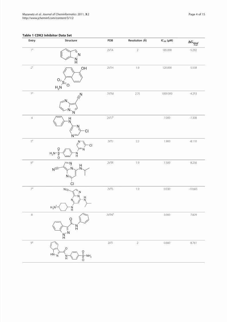
Data Set PreparationA database of 28 CDK2 inhibitors with experimentalbinding affinity available in the literature was compiled[47,48]. The reported IC50 (μM) values were convertedto -ln(IC50) values and the free energy of binding(ΔG bind ) was calculated according to the Eq. (1) at310 K.
Δ Δ ΔG RT T Sbind = − −1nIC =50 H (1)
The compounds with known X-ray structures wereselected as the training set to compare the various meth-
ods used to predict the free energy of binding. In orderto effectively validate the PLS model, compounds thatwere not included in the data set to obtain the modelwere placed into a separate test set to assess the predic-tive potential of the model. The distribution of the dataset into training and test sets is shown in Table 1.
Structure PreparationThe 14 X-ray structures, corresponding to the 14ligands in the training set were obtained from the PDB(Table 1). The remaining 14 ligands for which the X-ray structure data was not available were modelled into oneof the 14 reported PDB structures based on ligand
structural similarity (Table 1). The protein-ligand com-plexes were aligned in PyMOL [49]. Hydrogen atomswere added and the protonation state of the acidic andbasic amino acid residues were adjusted at pH 7 usingthe Protonate3 D tool within MOE [50]. An inclusionsphere with a 4.5 Å radius was projected around thebound ligands. This area defined the residues whichwere to be included in the QM and MM calculations.All water molecules were removed. The N-terminals of the residues were capped with acetyl groups and theC-terminal ends were N-methyl capped using the geo-metry of the cleaved neighbouring residue as a vector to
place the capping group. Partial charges were initially calculated to optimise the system using MM. The partialcharges for the ligand binding site were calculated usingthe MMFF94x force field and the ligand partial chargeswere calculated with AM1BCC charges [51,52]. The sys-tem was geometry optimized using MMFF94x forcefield in the presence of the Born continuous implicitwater model, with an internal dielectric constant of 3and an external dielectric constant of 80. The coordi-nates of the heavy atoms of the protein and the ligandwere held fixed and the protons were energy mini-mised using the other default settings in MOE. The 14
Figure 1 Schematic view of the thermodynamic cycle used toin the derivation of the binding affinity. The cycle calculates the
receptor (R), ligand (L), and complex (C) in vacuum and then
transfers them to solvent to find the solvation free energy.
Mazanetz et al . Journal of Cheminformatics 2011, 3:2http://www.jcheminf.com/content/3/1/2
Page 3 of 15
8/3/2019 Prediction of Cyclin-Dependent Kinase 2 Inhibitor
modelled ligands were built by manually modifying thereference ligand and then energy minimising the ligandwhilst keeping the reference heavy atoms fixed accord-ing to the method detailed above. Where appropriate,
charged amino acid residues were neutralised witheither a chloride anion or a lithium cation.
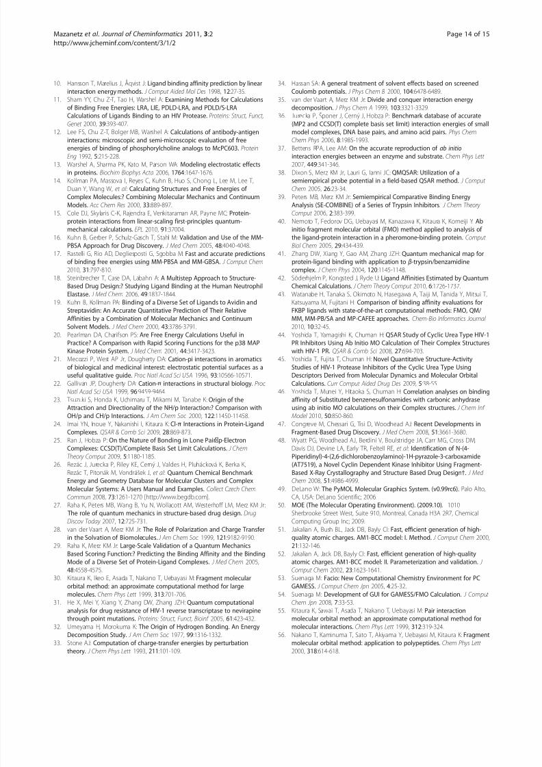
The FMO input files for GAMESS were preparedusing Facio (version 14.2.4) [53,54] following preproces-sing of the structure in MOE. An example of the pro-tein-ligand system is shown in Figure 2.
The FMO MethodThe FMO method has previously been thoroughly described [30,55-57]. Therefore, we provide only a shortsummary of the method. The FMO calculations wereused to perform high-level ab initio quantum chemical
calculations at MP2/6-31G* (6-31G 3df for Cl and S)theory level using the one-residue-per-fragment frag-mentation scheme. All calculations were run using theGAMESS implementation (either April 2008 or January 2009 version) [58-60].
The principle behind the FMO method is to divide alarge biomolecular system into a collection of small FMOfragments and then perform molecular orbital calculationsfor each fragment (called monomer) and fragment pair(called dimer). Generally the system is fragmented intoamino acids residues and ligands. It should be noted thatFMO fragments differ from the standard assignment for
amino acids residues. Here, amino acids are fragmentedalong the sp3 bond joining the C
acarbon to the peptide-
bond carbonyl carbon. This simple calculation scheme sig-nificantly reduces computational time. It may be possible
that this method impairs the computational accuracy because the covalent bonds are detached. However, in the
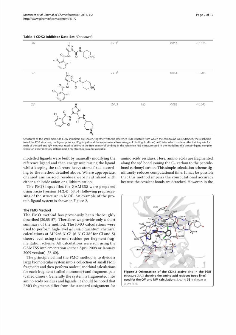
Table 1 CDK2 Inhibitor Data Set (Continued)
26
NH
O
NNH
NHO
F
O
NH2
+
2VTT b 0.052 -10.326
27
NH
O
NNH
NHO
F
Cl
NH2
+
2VTT b 0.063 -10.208
28a
N
H
O
N
NH
NH
O
NH2
+
Cl
Cl
2VU3 1.85 0.082 -10.045
Structures of the small molecule CDK2 inhibitors are shown, together with the reference PDB structure from which the compound was extracted, the resolution(Å) of the PDB structure, the ligand potency (IC50 in μM) and the experimental free energy of binding (kcal/mol). a) Entries which made up the training sets foreach of the MM and QM methods used to estimate the free energy of binding; b) the reference PDB structure used in the modelling the protein-ligand complexwhere an experimentally determined X-ray structure was not available.
Figure 2 Orientation of the CDK2 active site in the PDBstructure 2VU3 showing the amino acid residues (grey lines)used for the QM and MM calculations . Ligand 33 is shown as
grey sticks.
Mazanetz et al . Journal of Cheminformatics 2011, 3:2http://www.jcheminf.com/content/3/1/2
Page 7 of 15
8/3/2019 Prediction of Cyclin-Dependent Kinase 2 Inhibitor
FMO method, the accuracy is kept by employing projec-tion operators made from the sp3 hybrid orbital.
One of the great advantages of the FMO method isthat it can be combined with a number of currentquantum chemical techniques. Thus, an appropriatemethod for each system can be chosen. It was notedthat incorporation of vdW interactions is an importantconsideration in studying protein-ligand interactions.Here, the FMO method provides a useful calculationscheme for dealing with these effects. Dispersionenergy, which can dominant in vdW interactions, isnot considered by Hartree-Fock (HF) and poorly mod-elled by Density Functional Theory (DFT). To achieveaccurate dispersion energies correlated ab inito meth-ods are required. Here, the second-order Møller-Plesset (MP2) perturbation method was used as it isthe least expensive non-empirical approach. The MP2
method has been implemented in the FMO method(FMO-MP2).The molecular system is divided into a number of
monomer fragments and the ab inito molecular orbitalcalculations on the monomers are solved repeatedly atthe HF level until all monomer densities become self-consistent. Then the FMO-MP2 method begins withMP2 calculations of monomers, followed by MP2 calcu-lations of dimers. The results are used to calculate thetotal energy of a system following the formula:
E E E E EFMO I IJ I J I J
N
I
N = + − −( )
>∑∑ (2)
where I and J run over all the of the fragments, N . Theterm E I is the self-consistent field (SCF) energy of the I thfragment in the external Coulomb field of the other N -1 fragments. The E IJ term is the SCF energy of the I + J
dimer in the external Coulomb field of the other N - 2fragments. The FMO calculations can provide PIEs, alsoknown as inter-fragment interaction energies (IFIE),between fragments. The PIEs are used during the analysisof interaction between protein residues and the boundligand and is derived from the FMO calculation:
Δ ΔEIJ IJ I J IJ IJ E E E Tr = ′ − ′ − ′( ) + ( ) D V (3)
where Δ D IJ and V IJ are the difference density matrixand the environmental electrostatic potential for dim-mer IJ from other fragments, ′EI and ′EIJ are the mono-mer energy and the dimer energy without environmentalelectrostatic potential, respectively. The IFIE analysis canbe plotted in two-dimensions, referred to as the IFIEmap, to highlight hot-spots of protein-ligand interac-tions [40,61-63].
Scoring Function Used to Estimate Free Energy of BindingThe values of the free energy of binding in solvent(ΔG bind ) of each inhibitor were calculated according toEq. (4) following thermodynamic cycle shown in Figure 1.
Δ Δ Δ Δ ΔG G G G Gbind bind gas
solvcomplex
solvreceptor
solvligand= + − − (4)
Δ Δ ΔG H T Sbind gas
bind gas
bind gas= − (5)
ΔH ES EX DI CT mixbind gas = + + + + (6)
T S num rot bondsbind gasΔ = ( )_ (7)
Δ Δ ΔG G G solv psolv npsolv
= + (8)
ΔG SASA bnpsolv = +γ (9)
The solution-phase ΔGbind was decomposed into the
solvation free energy and the gas-phase interactionenergy. The free energy change on solvation is com-posed of terms for the desolvation of the receptor and
ligand − −( )Δ ΔG G solvreceptor
solvligand
and and the solvation of
the complex ΔG solvcomplex( ) . The gas phase free energy of
binding, ΔGbind gas , is the sum of the enthalpic contribu-
tions ΔH bind gas( ) from the electrostatic and nonpolar
interaction energies and the entropic term TΔSbind gas( )
for the degrees of freedom for each component of thesystem at a given temperature (310 K).
The enthalpic binding energy of interaction term isderived from the FMO method at the MP2/6-31G* level.The breakdown of this interaction energy can be expressedas relating to electrostatic interactions (ES), exchangerepulsion (EX), dispersion contributions (DI) and chargetransfer (CT) with higher order mixed terms, Eq. (6)[64,65]. Evaluation of the enthalpic ligand binding energy is
commonly performed by the supermolecule method. Here,the difference between the energy of the receptor-ligandcomplex and the sum of the energies of the apo-receptorand the isolated ligand is considered, Eq. (10).
Δ Δ Δ ΔH H H H bind gas complex receptor ligand= − +( ) (10)
Thus three separate calculations are required to obtainthe total ligand binding energy. However, in the FMO cal-culation, as all the PIEs between fragments are calculated
Mazanetz et al . Journal of Cheminformatics 2011, 3:2http://www.jcheminf.com/content/3/1/2
Page 8 of 15
8/3/2019 Prediction of Cyclin-Dependent Kinase 2 Inhibitor
by default, the ligand binding energy can be conveniently estimated by simply taking the sum of all the PIEs betweenthe ligand and receptor fragments. Although the sum of PIE does not include the effect of electron redistributionin the complex as a result of ligand-protein binding, it isknown that there is good qualitative agreement betweenthe binding energy calculated by the supermoleculemethod and the sum of PIE [44].
Entropy plays an important role in binding. Duringreceptor-ligand complex formation, there are changes inthe degrees of rotational, translational and conformationalfreedom, which make the process entropically unfavour-able [66-68]. The number of rotatable bonds in a ligand[69-71] or the receptor and ligand together [67,72] haspreviously been used as a measure for conformationalentropy. More complex measures of determining entropy using vibrational frequencies of a ligand when complexed
to a receptor have been shown to correlate well to thenumber of rotatable bonds [73]. However, vibrationalentropy is not a component of conformational entropy,and do not make significant contributions to the overallentropy of the system [74]. The calculation of the num-ber of rotatable bonds is also an attractive estimation forconformational ligand entropy as it is significantly lesscomputationally demanding than other methods. There-fore we chose this method and assigned a conformationalpenalty of 1 kcal/mol for each rotatable bond in theligand according to published work [73].
The other term of the binding free energy is the solva-tion free energy ΔG
solv( ). To account for the solvation
effects during receptor-ligand binding, the Born conti-nuum implicit solvation model was chosen as it hasbeen shown to appropriately describe solvent interac-tions [66]. The solvation free energy was described by apolar solvation term ΔGpsolv( ) and a nonpolar solvationterm ΔGnpsolv( ) , Eq. (8).
The polar solvation term estimated by solving thePoisson-Boltzmann (PB) equation using MOE [50]. Thesystem was parameterised as described in the StructurePreparation section. The nonpolar solvation term was esti-mated from the solvent-accessible surface area (SASA) of the molecule, Eq. (9). This was computed in MOE with a
solvent probe radius of 1.4 Å. The values taken for g andb were 5.0 cal/mol·Å2 and 0.86 kcal/mol, respectively, asdescribed in the literature [75,76]. In order to speed upthe calculation of the free energy of solvation we chose touse a single energy-minimised structure which has beenreported in the literature to be a reasonable estimation tomolecular dynamics simulations [16,17].
Multivariate AnalysisThe statistical program SIMCAP, version 11.0.0.0, fromUmetrics was used to build a PLS model [77,78]. TheX-variables originate from the components used to
derive the free energy of binding in solvent, see above.The dependent Y-variable was the experimental bindingaffinity in -ln(IC50), Eq. (1). The variables were mean-centred and scaled to unit variance. The non-cross-
validated variance coefficient (r 2) and the cross-validated variance coefficient (q 2) were used to describe how wella model can reproduce the data under analysis and thepredictive ability of the model. Cross-validation was per-formed by dividing the training sets into 7 groups anddeveloping a number of parallel models for the datadevoid of one group. The omitted group then becamethe test set for the reduced model and residuals for thetest set were calculated. A measure of the predictivity of the models, termed predictive residual sum of squareswas derived from the sum of squares of these differencesfor all parallel models. The q 2 value that resulted in theoptimum number of components and lowest predictive
residual sum of squares was used. The root mean squareerror of estimation (RMSEE) of the fit for observationsin the model and the root mean square error of predic-tion (RMSEP) were also calculated.
Results and DiscussionLigand and Protein PreparationA series of 14 X-ray crystal structures of CDK2-ligandcomplexes with known experimental binding affinitiesand with resolutions of better than 2.3 Å were down-loaded from the PDB [48]. A further 14 ligands fromthe same chemical series were manually docked toeither one of 4 known ligand X-ray structures whichhad the closest chemical similarity as indicated in Table1. Details of the proteins and the ligand structures, dataset clustering into training and test sets, the resolutionof the PDB structures and the experimental binding affi-nities are detailed in Table 1. The well resolved X-ray crystal structures meant that we were confident of theinitial conformation of the complexes. Our experimentsfocussed primarily in the binding pocket, which forCDK2 is well resolved, particularly the residues whichconstitute the gate keeper and the Hinge region (resi-dues Phe80 - Gln85 Figure 2). Our rationale for only using minimised X-ray structures as a single protein-
ligand structure in preference to the averaging over anumber of molecular dynamics snapshots is that thisconformation can be considered to contribute signifi-cantly to and thus dominate the Boltzmann-averagedpotentials for the free energy estimation. This is particu-larly true when the bound conformation of the ligandcorresponds to a particular stable conformation of theunbound ligand. Also, a good single point calculation ismore likely to be a good representation of the systemthan one from which the phase space is poorly sampled.Other studies have used MM/MD simulations ascertainan optimal system conformation before further analysis
Mazanetz et al . Journal of Cheminformatics 2011, 3:2http://www.jcheminf.com/content/3/1/2
Page 9 of 15
8/3/2019 Prediction of Cyclin-Dependent Kinase 2 Inhibitor
using FMO [79]. For the 14 X-ray structures examinedthe average local strain energy (the potential energy of the X-ray structure minus the value of the energy at anear local minimum) was 6 kcal/mol, which is an accep-tably reasonable energy [80]. Therefore, it can be arguedthat the energetic penalties coming from ligand confor-mational strain are minimal as the ligand is already in agood binding conformation [81]. Using a single-pointcalculation is also more amenable to virtual screening.The method is not only a comparably accurate alterna-tive to averaged snap shots over a molecular dynamicssimulation, but is less time-consuming to setup andcompute. It follows then that the remaining 14 ligandscan be modelled by slight modifications to the X-ray solved ligands whilst maintaining the geometry of thecommon chemical scaffold, followed by a minimisationstep (see Methods) would be a reasonable approxima-
tion of actual binding pose.Although the structural sampling was not performedin the FMO calculation of the enthalpic contributions tobinding free energy, a good correlation (r 2 of 0.68)was obtained to the experimental free energy of binding,Figure 3. The binding pocket in CDK2 is not exposed tosolvent and important hydrogen bonding interactionswithin the active site are of limited flexibility [ 82].Under these conditions, enthalpic binding contributessignificantly to the free energy of binding, and thistherefore accounts for the good correlation to the FMOsum of PIE. For this target it appears that structuralsampling is not crucial in order to obtain good correla-tions. However, the appropriate selection of atomiccoordinates is an important factor to obtain well corre-lated data. A consideration of the optimal binding posefor the modelled ligands was out of the scope of thiswork, and further validations regarding conformationalrefinement of docked or aligned poses using the FMOmethod are in progress.
Correlation Between MM-Based Scoring Functions andBiochemical ActivityThe performance of the FMO method was comparedwith that of several MM scoring functions implemented
in MOE, including the Generalized Born solvationmodel VI, London dG, Affinity dG, Alpha HB, and ASEscoring functions (Figure 3). In each of these MM meth-ods, the protein was parameterised using the MMFF94xforce field and ligand charges calculated using AM1-BCC. The 14 X-ray structures used to build the PLSmodel were used to compare the 6 scoring functions.The FMO method clearly outperformed three of thescoring functions and was similar to the London dGand the Alpha HB score. A good correlation wasobserved (r 2 of 0.68) for the FMO sum of PIE and thebest performing MM scoring function was the ASE
score (r 2 of 0.75). The ASE score has terms for the over-lap of the ligand pose with alpha spheres and theoverlap between ligand and receptor atom volumesapproximated by Gaussians, and therefore can bethought of as mimicking dispersion interactions of ligand binding. As the CDK2 binding pocket is very hydrophobic this generalisation may be sufficient to geta good correlation to experimental binding energy. TheGeneralized Born solvation model VI failed to correlatethe data (r 2 of 0.03). The Affinity dG scoring functiononly considers enthalpy of ligand binding in a simplisticfashion (r 2 of 0.31). This function is improved by termsto account for hydrogen bonding in the Alpha HB func-tion (r 2 of 0.61). The London dG scoring function hasfurther improvements, adding rotational and transla-tional entropy and a desolvation term which resulted ina good estimation of binding free energy (r 2 of 0.73).
The two methods that yield free energy binding predic-tions close to the actual values are the ASE and theLondon dG scoring functions.
The main purpose of a scoring function though is torank binding poses, and here the MOE scoring functionsare effective. A Pearson rank order analysis for LondondG, Alpha HB and ASE score all gave a value of 0.76,the FMO method performing less well with a Pearson
value of 0.64. However, an important consideration indrug development is the identification of active com-pounds, thus good correlations to experimental bindingfree energy is of more value than the rank ordering of compounds. To effectively account for other compo-nents pertaining to binding additional terms were intro-duced, the results of which are detailed below.
Data PreparationThe four X-variables used to build and test the PLSmodel were derived from the sum of the enthalpic con-tributions ΔH bind
gas( ) calculated by the FMO method,the polar solvation term (ΔG psolv), the nonpolar solva-tion term (ΔG npsolv), and the entropic term TΔSbind
gas( ) .These descriptors were mean-centred and normalisedfor the model generation. The PLS model was trainedon the 14 X-ray structures obtained from the literature
using the experimental reported inhibitory data as theY-variable [48]. The PLS model was tested against the14 modelled complexes.
PLS Analysis ResultsThe optimum number of components in the PLS modelwas two which gave a very high q 2 of 0.896, and theRMSEE of the fit for observations was 0.632. The r 2
value was 0.939 for this model. The 4 X-variables con-tributed similarly to the model, and there were no out-liers in the observations used to build the model. Themodel rank orders the compounds extremely well, with
Mazanetz et al . Journal of Cheminformatics 2011, 3:2http://www.jcheminf.com/content/3/1/2
Page 10 of 15
8/3/2019 Prediction of Cyclin-Dependent Kinase 2 Inhibitor
a Pearson correlation of 0.97. This robust model pre-dicted the test set well, the r 2 of the test set was 0.824,and the RMSEP was 1.005. The plot of computed andexperimentally determined binding free energies isshown in Figure 3 and together with the residual
differences between these values for each of the ligandsis shown in Table 2. The majority of the data (Figure 3)lies within the two yellow-dashed lines, indicating errorsof less than 1 order of magnitude. There are twoligands, 12 and 15 that fall well outside this boundary.
Figure 3 Calculated versus observed free energy of binding for 14 CDK2 inhibitors assessed using seven different methods . Methods
include a) FMO (green diamonds), r 2 = 0.68; b) GBVI (orange diamonds), r 2 = 0.03; c) London dG (purple diamonds), r 2 = 0.73; d) Affinity dG
(green triangles), r 2 = 0.31; e) Alpha HB (red diamonds), r
2 = 0.61; f) ASE (black diamonds) r 2 = 0.75; and g) QM-based scoring function (red
squares) together with the 14 compound test set (blue diamonds) for the QM-scoring function, and h) the calculated versus the experimental
pIC50 values for the QM-scoring function, r 2 = 0.94. For graphs a-f, and g, the line of best fit is shown in black. Graph h shows the line of best fit
as a dotted red line and the two dotted yellow lines correspond to 1 log unit boundaries.
Mazanetz et al . Journal of Cheminformatics 2011, 3:2http://www.jcheminf.com/content/3/1/2
Page 11 of 15
8/3/2019 Prediction of Cyclin-Dependent Kinase 2 Inhibitor
An examination of the 4 components which make upthe free energy term does not reveal any strong trendresulting in the large residual values. All the ligandsused to train and test the model were well within the
95% confidence intervals for the predicted Y-values andthere were no observations that deviated significantly from the model in X-space.
QMbased Scoring FunctionFMO has been used previously to generate a chargetransfer term for a quantitative structure-activity rela-tionship (QSAR) model [44]. Here, we aimed at produ-cing a QM-based scoring function which would takeinto consideration complex binding interactions, solva-tion effects and ligand binding entropy on a timescaleamenable to drug discovery. The FMO methods allows
for accurate treatment of charge transfer and polarisa-tion effects. It has been noted previously that the major-ity of polarisation energy is within 5 Å of a ligand [ 83].This observation justifies the 4.5 Å residue inclusion
radius used to describe the binding pocket and allowsfor this polarisation to be incorporated into the enthalpy of binding energy term. The contribution of chargetransfer effects on ligand binding have already beenmentioned, and represent an important addition to ascoring function particularly when examining particularligand-residue interactions [44]. However, the contribu-tion of charge transfer on the enthaplic binding term isdependent on the wave function used. The FMO contri-bution to the binding free energy has a very broad range(-28 to -178), this may be a result of using the MP2method which is known to overestimate charge transfer
Table 2 Estimation of Free Energy of Ligand Binding
free energy of binding and the associated terms used to derive the scoring function including, see the text
for more information. All the energy terms are in kcal/mol. The residual differences between the calculated and the experimental free energies of binding areshown. a) indicates an entry which was used to train the PLS QSAR model.
Mazanetz et al . Journal of Cheminformatics 2011, 3:2http://www.jcheminf.com/content/3/1/2
Page 12 of 15
8/3/2019 Prediction of Cyclin-Dependent Kinase 2 Inhibitor
interactions [33]. Energy decomposition analysis fromthe FMO calculation reveals that the majority of theenergy comes from the charge transfer contribution of charged atoms. The approximations for other terms inthe scoring function make this overestimation less sig-nificant compared to the absolute binding energy deter-mined by the FMO method when used in isolation.
The binding free energy is a combination of enthalpicand entropic terms. Indeed, a thorough understandingof enthalpy/entropy compensation is needed to accu-rately predict binding energies [84,85]. Ligand confor-mational entropy contributions are also significant, andneglecting this will adversely affect binding energy predic-tions [86]. As a very simplistic method to account for thiswe chose to examine how the number of rotationalbonds in the ligand would influence the predicted bind-ing free energy. The good correlation obtained with our
data, in this test case, indicates that this extremely fastmethod is adequate for this purpose. More detailed stu-dies of entropy could be performed by normal mode ana-lysis of molecular dynamics simulations. The lack of anadequate protein entropy term can result in an overesti-mation of binding free energy, and more work is neededto examine the effect of this on such calculations.
The solvation free energy is divided into polar and non-polar terms. The nonpolar term is dependent upon thesize of the ligand, which is scaled by the two constants g
and b. This scaling makes the nonpolar term small andnegative, allowing the polar terms to dominate the solva-tion free energy of binding. Recently, the polarisable conti-nuum model (PCM) implemented in the GAMESSprogram was used to calculate solvation energies and werecompared to those obtained with PB+SASA [42]. It wasfound that PCM exaggerated the nonpolar contributionsubstantially, and therefore a QM treatment of solvationwas not advantageous. Solvation calculations with thePCM have been implemented with the FMO method [87]and although this does increase computational time, moreaccurate treatment of solvation from a single point calcu-lation would be possible. Solvation effects have also beendeveloped for FMO using the PB equation to account forion concentrations [88]. Further studies need to be per-
formed to assess the effects these advances in treating sol- vation effects brings to the determination of binding freeenergy and the computational cost of the method.
ConclusionsThe results show that a single point QM calculationusing the FMO method gives a good correlation toexperimentally determined free energies of ligand bind-ing calculated from ligand potencies. The FMO methodoutperformed 3 other methods used to estimate the freeenergy of binding by MM-based methods. We concludethat the additional terms which treat charge transfer,
polarisation and dispersion effects during ligand bindingin this QM method significantly improves the estimationof ligand potency compared to MM-based procedures.Methods were then introduced to further improve uponthe initial estimates. This paper presents the firstattempt to calculate ligand-binding free energies using acombination of high-level ab inito FMO methodstogether with PBSA techniques to derive reasonable esti-mations of enthalpy, entropy and solvation energies. Weused a PLS QSAR model to correlate the 4 componentsof our scoring function to build a model which was very robust and highly predictive. The data set was composedof ligands from a lead development program thatresulted in a clinical candidate against CDK2, thus test-ing the QSAR model against a range of ligand potencies.The need to run a PLS model stems from the poorabsolute prediction of free binding energies and there-
fore the need to adjust the data. This QM-based scoringfunction represents a new protocol to estimate ligandpotencies in a congeneric series of compounds whereby single point changes can be performed on a knownX-ray crystal structure to guide medicinal chemistry.
Acknowledgements The authors acknowledge Dmitri Fedorov for his support in implementing
the FMO method.
Authors’ contributionsMPM and OI ran the FMO calculations and MPM wrote the scripts for FMO
analysis and developed the QSAR method. MPM and OI tested the
presented methods and prepared the manuscript for this publication. RJL
supervised the project. All authors have read and approved of the finalmanuscript.
Competing interests The authors declare that they have no competing interests.
Received: 2 November 2010 Accepted: 10 January 2011Published: 10 January 2011
References1. Jorgensen WL: The Many Roles of Computation in Drug Discovery.
Science 2004, 303:1813-1818.2. Shoichet BK, McGovern SL, Wei B, Irwin JJ: Lead discovery using molecular
docking. Curr Opin Chem Biol 2002, 6:439-446.3 . Good A: Structure-based virtual screening protocols. Curr Opin Drug
Discov Devel 2001, 4:301-307.
4. Kontoyianni M, McClellan LM, Sokol GS: Evaluation of Docking Performance:Comparative Data on Docking Algorithms.J Med Chem 2004, 47:558-565.
5. Stahl M, Rarey M: Detailed Analysis of Scoring Functions for Virtual
10. Hansson T, Marelius J, Åqvist J: Ligand binding affinity prediction by linear
interaction energy methods. J Comput Aided Mol Des 1998, 12:27-35.11. Sham YY, Chu Z-T, Tao H, Warshel A: Examining Methods for Calculations
of Binding Free Energies: LRA, LIE, PDLD-LRA, and PDLD/S-LRACalculations of Ligands Binding to an HIV Protease. Proteins: Struct, Funct,
Genet 2000, 39:393-407.
12. Lee FS, Chu Z-T, Bolger MB, Warshel A: Calculations of antibody-antigeninteractions: microscopic and semi-microscopic evaluation of freeenergies of binding of phosphorylcholine analogs to McPC603. Protein
Eng 1992, 5:215-228.13. Warshel A, Sharma PK, Kato M, Parson WA: Modeling electrostatic effects
in proteins. Biochim Biophys Acta 2006, 1764:1647-1676.14. Kollman PA, Massova I, Reyes C, Kuhn B, Huo S, Chong L, Lee M, Lee T,
Duan Y, Wang W, et al : Calculating Structures and Free Energies of
Complex Molecules:? Combining Molecular Mechanics and Continuum
Models. Acc Chem Res 2000, 33:889-897.15. Cole DJ, Skylaris C-K, Rajendra E, Venkitaraman AR, Payne MC: Protein-
protein interactions from linear-scaling first-principles quantum-
mechanical calculations. EPL 2010, 91:37004.16. Kuhn B, Gerber P, Schulz-Gasch T, Stahl M: Validation and Use of the MM-
PBSA Approach for Drug Discovery. J Med Chem 2005, 48:4040-4048.17. Rastelli G, Rio AD, Degliesposti G, Sgobba M: Fast and accurate predictions
of binding free energies using MM-PBSA and MM-GBSA. J Comput Chem
2010, 31:797-810.18. Steinbrecher T, Case DA, Labahn A: A Multistep Approach to Structure-
Based Drug Design:? Studying Ligand Binding at the Human Neutrophil
Elastase. J Med Chem 2006, 49:1837-1844.19. Kuhn B, Kollman PA: Binding of a Diverse Set of Ligands to Avidin and
Streptavidin: An Accurate Quantitative Prediction of Their Relative
Affinities by a Combination of Molecular Mechanics and Continuum
Solvent Models. J Med Chem 2000, 43:3786-3791.20. Pearlman DA, Charifson PS: Are Free Energy Calculations Useful in
Practice? A Comparison with Rapid Scoring Functions for the p38 MAP
Kinase Protein System. J Med Chem 2001, 44:3417-3423.21. Mecozzi P, West AP Jr, Dougherty DA: Cation-pi interactions in aromatics
of biological and medicinal interest: electrostatic potential surfaces as auseful qualitative guide. Proc Natl Acad Sci USA 1996, 93:10566-10571.
22. Gallivan JP, Dougherty DA: Cation-π interactions in structural biology. Proc
Natl Acad Sci USA 1999, 96:9459-9464.
23. Tsuzuki S, Honda K, Uchimaru T, Mikami M, Tanabe K: Origin of theAttraction and Directionality of the NH/p Interaction:? Comparison with
OH/p and CH/p Interactions. J Am Chem Soc 2000, 122:11450-11458.24. Imai YN, Inoue Y, Nakanishi I, Kitaura K: Cl-π Interactions in Protein-Ligand
Complexes. QSAR & Comb Sci 2009, 28:869-873.25. Ran J, Hobza P: On the Nature of Bonding in Lone Pair⋯p-Electron
Complexes: CCSD(T)/Complete Basis Set Limit Calculations. J Chem
Theory Comput 2009, 5:1180-1185.26. Rezác J, Jurecka P, Riley KE, Cerný J, Valdes H, Pluhácková K, Berka K,
Rezác T, Pitonák M, Vondrášek J, et al : Quantum Chemical Benchmark Energy and Geometry Database for Molecular Clusters and Complex
Molecular Systems: A Users Manual and Examples. Collect Czech Chem
Commun 2008, 73:1261-1270 [http://www.begdb.com].27. Raha K, Peters MB, Wang B, Yu N, Wollacott AM, Westerhoff LM, Merz KM Jr:
The role of quantum mechanics in structure-based drug design. Drug
Discov Today 2007, 12:725-731.28. van der Vaart A, Merz KM Jr: The Role of Polarization and Charge Transfer
in the Solvation of Biomolecules. J Am Chem Soc 1999, 121:9182-9190.29. Raha K, Merz KM Jr: Large-Scale Validation of a Quantum Mechanics
Based Scoring Function:? Predicting the Binding Affinity and the Binding
Mode of a Diverse Set of Protein-Ligand Complexes. J Med Chem 2005,48:4558-4575.
30. Kitaura K, Ikeo E, Asada T, Nakano T, Uebayasi M: Fragment molecular
orbital method: an approximate computational method for large
molecules. Chem Phys Lett 1999, 313:701-706.31. He X, Mei Y, Xiang Y, Zhang DW, Zhang JZH: Quantum computational
analysis for drug resistance of HIV-1 reverse transcriptase to nevirapinethrough point mutations. Proteins: Struct, Funct, Bioinf 2005, 61:423-432.
32. Umeyama H, Morokuma K: The Origin of Hydrogen Bonding. An Energy
Decomposition Study. J Am Chem Soc 1977, 99:1316-1332.33. Stone AJ: Computation of charge-transfer energies by perturbation
theory. J Chem Phys Lett 1993, 211:101-109.
34. Hassan SA: A general treatment of solvent effects based on screened
Coulomb potentials. J Phys Chem B 2000, 104:6478-6489.35. van der Vaart A, Merz KM Jr: Divide and conquer interaction energy
decomposition. J Phys Chem A 1999, 103:3321-3329.36. Jurecka P, Šponer J, Cerný J, Hobza P: Benchmark database of accurate
(MP2 and CCSD(T) complete basis set limit) interaction energies of small
model complexes, DNA base pairs, and amino acid pairs. Phys ChemChem Phys 2006, 8:1985-1993.37. Bettens RPA, Lee AM: On the accurate reproduction of ab initio
interaction energies between an enzyme and substrate. Chem Phys Lett
2007, 449:341-346.38. Dixon S, Merz KM Jr, Lauri G, Ianni JC: QMQSAR: Utilization of a
semiempirical probe potential in a field-based QSAR method. J Comput
Chem 2005, 26:23-34.39. Peters MB, Merz KM Jr: Semiempirical Comparative Binding Energy
Analysis (SE-COMBINE) of a Series of Trypsin Inhibitors. J Chem Theory
Comput 2006, 2:383-399.40. Nemoto T, Fedorov DG, Uebayasi M, Kanazawa K, Kitaura K, Komeiji Y: Ab
initio fragment molecular orbital (FMO) method applied to analysis of
the ligand-protein interaction in a pheromone-binding protein. Comput
Biol Chem 2005, 29:434-439.41. Zhang DW, Xiang Y, Gao AM, Zhang JZH: Quantum mechanical map for
protein-ligand binding with application to β-trypsin/benzamidine
50. MOE (The Molecular Operating Environment). (2009.10). 1010Sherbrooke Street West, Suite 910, Montreal, Canada H3A 2R7, ChemicalComputing Group Inc; 2009.
51. Jakalian A, Bush BL, Jack DB, Bayly CI: Fast, efficient generation of high-
Comput Chem 2007, 28:222-237.65. Kitaura K, Morokuma K: A new energy decomposition scheme for
molecular interactions within the Hartree-Fock approximation. Int J
Quantum Chem 1976, 10:325-340.66. Schwarzl SM, Tschopp TB, Smith JC, Fischer S: Can the calculation of
ligand binding free energies be improved with continuum solvent
electrostatics and an ideal-gas entropy correction? J Comput Chem 2002,23:1143-1149.
67. Murray CW, Verdonk ML: The consequences of translational androtational entropy lost by small molecules on binding to proteins. J
Comput Aided Mol Des 2002, 16:741-753.68. Tirado-Rives J, Jorgensen WL: Contribution of Conformer Focusing to the
Uncertainty in Predicting Free Energies for Protein-Ligand Binding. J Med Chem 2006, 49:5880-5884.69. Eldridge MD, Murray CW, Auton TR, Paolini GV, Mee RP: Empirical scoring
functions: I. The development of a fast empirical scoring function to
estimate the binding affinity of ligands in receptor complexes. J Comput
Aided Mol Des 1997, 11:425-445.70. Böhm H-J: The development of a simple empirical scoring function to
estimate the binding constant for a protein-ligand complex of known
three-dimensional structure. J Comput Aided Mol Des 1994, 8:243-256.71. Ishchenko AV, Shakhnovich EI: SMall Molecule Growth 2001 (SMoG2001):
An Improved Knowledge-Based Scoring Function for Protein-Ligand
Interactions. J Med Chem 2002, 45:2770-2780.72. Raha K, Merz KM Jr: A Quantum Mechanics-Based Scoring Function:?
Study of Zinc Ion-Mediated Ligand Binding. J Am Chem Soc 2004,126:1020-1021.
73. Hayik SA, Dunbrack R Jr, Merz KM Jr: Mixed Quantum Mechanics/
Molecular Mechanics Scoring Function To Predict Protein-Ligand
Binding Affinity. J Chem Theory Comput 2010, 6:3079-3091.74. Mammen M, Shakhnovich EI, Whitesides GM: Using a Convenient,
Quantitative Model for Torsional Entropy To Establish Qualitative Trends
for Molecular Processes That Restrict Conformational Freedom. J Org
Chem 1998, 63:3168-3175.75. Hermann RB: Theory of hydrophobic bonding. II. Correlation of
hydrocarbon solubility in water with solvent cavity surface area. J Phys
Chem 1972, 76:2754-2759.76. Honig B, Sharp K, Yang AS: Macroscopic models of aqueous solutions:
biological and chemical applications. J Phys Chem 1993, 97:1101-1109.77. SIMCAP (11.0.0.0). Box 7960, SE90719 Umeå, Sweden, Umetrics AB; 2005.78. Wold S, Sjöström M, Eriksson L: PLS-regression: a basic tool of
chemometrics. Chemom Intell Lab Syst 2001, 58:109-130.79. Hitaoka S, Harada M, Yoshida T, Chuman H: Correlation Analyses on
Binding Affinity of Sialic Acid Analogues with Influenza Virus
Neuraminidase-1 Using ab Initio MO Calculations on Their Complex
Structures. J Chem Inf Model 2010, 50:1796-1805.80. Perola E, Charifson PS: Conformational Analysis of Drug-Like Molecules
Bound to Proteins:? An Extensive Study of Ligand Reorganization upon
Binding. J Med Chem 2004, 47:2499-2510.81. Gilson MK, Zhou H-X: Calculation of Protein-Ligand Binding Affinities.
Annu Rev Biophys Biomol Struct 2007, 36:21-42.82. Mazanetz MP, Withers IM, Laughton CA, Fischer PM: A Study of CDK2
Inhibitors Using a Novel 3D-QSAR Method Exploiting Receptor
effect of MM polarization on the QM/MM transition state stabilization:
application to chorismate mutase. Mol Phys 2008, 106:1511-1515.84. Lafont V, Armstrong AA, Ohtaka H, Kiso Y, Amzel LM, Freire E:
Compensating Enthalpic and Entropic Changes Hinder Binding Affinity
Optimization. Chem Biol Drug Des 2007, 69:413-422.85. Freire E: Do enthalpy and entropy distinguish first in class from best in
class? Drug Discov Today 2008, 13:869-874.86. Chang CA, Chen W, Gilson MK : Ligand configurational entropy and
protein binding. Proc Natl Acad Sci USA 2007, 104:1534-1539.87. Fedorov DG, Kitaura K, Li H, Jensen JH, Gordon MS: The polarizable
continuum model (PCM) interfaced with the fragment molecular orbital
method (FMO). J Comput Chem 2006, 27:976-985.
88. Watanabe H, Okiyama Y, Nakano T, Tanaka S: Incorporation of solvationeffects into the fragment molecular orbital calculations with the
Poisson-Boltzmann equation. Chem Phys Lett 2010, 500:116-119.
doi:10.1186/1758-2946-3-2Cite this article as: Mazanetz et al .: Prediction of cyclin-dependentkinase 2 inhibitor potency using the fragment molecular orbitalmethod. Journal of Cheminformatics 2011 3:2.
Open access provides opportunities to our
colleagues in other parts of the globe, by allowing
anyone to view the content free of charge.
Publish with ChemistryCentral and every
scientist can read your work free of charge
W. Jeffery Hurst, The Hershey Company.
available free of charge to the entire scientific community
peer reviewed and published immediately upon acceptance
cited in PubMed and archived on PubMed Central
yours you keep the copyright
Submit your manuscript here:
http://www.chemistrycentral.com/manuscript/
Mazanetz et al . Journal of Cheminformatics 2011, 3:2http://www.jcheminf.com/content/3/1/2