Page 1
Prehospital treatment of sepsis
Christopher W. Seymour, MD MSc
The CRISMA Center
Assistant Professor of Critical Care Medicine & Emergency Medicine
University of Pittsburgh School of Medicine
Page 2
• Received funding from:
NIH NIGMS, SCCM, AHA, MedicOne, SIS
• Consulting fees from Beckman Coulter, Edwards Inc.
• Member, Surviving Sepsis Campaign, ATS representative
• Member, 2016 Third International Sepsis Definitions Task
Force
Disclosures
Page 3
• I am not an EMS clinician
• Involved in prehospital sepsis treatment trial (CIHR, PI:
Scales, PITSTOP) planning to enroll in 2018
• Intensivist at UPMC-Mercy in Pittsburgh, PA
Caveats
Page 4
• What did we discuss last lecture • Definitions, criteria
• What are the main tenets of sepsis treatment
• What can we do NOW during prehospital care
• What will we do in the FUTURE
• Questions
Objectives
Page 5
2
million US cases each
year
5
percent of US
healthcare spending
Gaieski et al. Crit Care Med, 2014
Singer et al., JAMA, 2016
Sepsis is everywhere.
Page 6
Singer et al., JAMA, 2016
• Infection
• Organ dysfunction
• Life threatening
• Dysregulated host response
Sepsis defined
Page 7
• Infection plus 2 or more SOFA points above baseline
Prompt to consider sepsis outside the
ICU
• Infection plus 2 or more qSOFA points
Clinical criteria for sepsis
Page 8
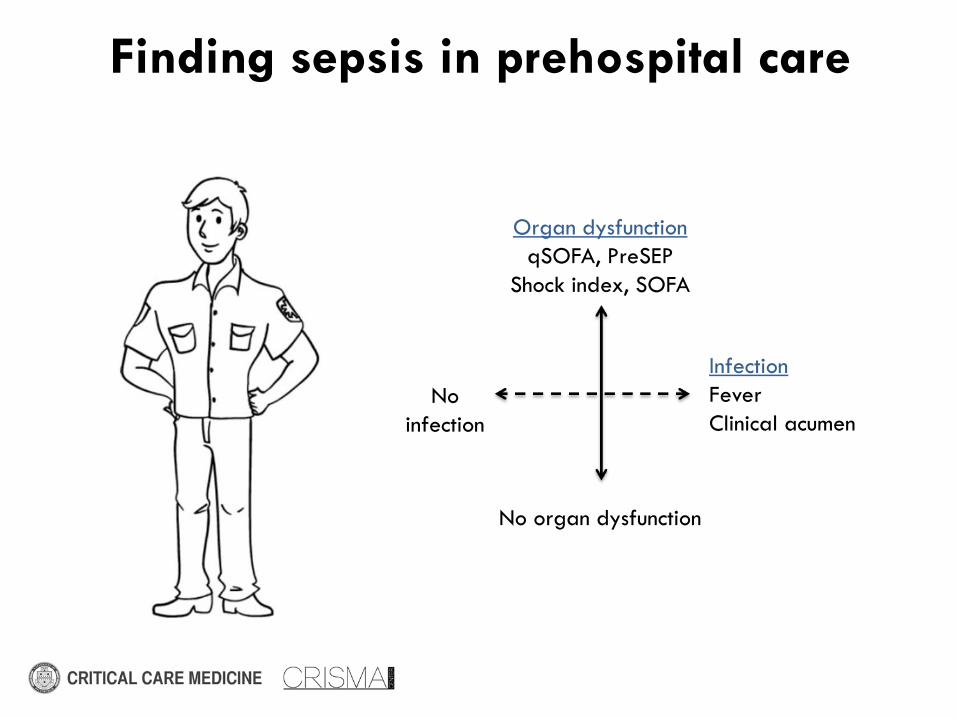
No organ dysfunction
Organ dysfunction
qSOFA, PreSEP
Shock index, SOFA
Infection
Fever
Clinical acumen
No
infection
Finding sepsis in prehospital care
Page 9
• Sepsis is an enormous pubic health problem
• New sepsis definitions released in 2016
• Clinical suspicion for infection remains a challenge
• New tools such as qSOFA may be prompts but are not adequately
sensitive
• New and old biomarkers – good for research – not yet ready for
prime time
Conclusions – last time
Page 10
I’ve found a septic patient, what can
we do…
So now what?
Page 11
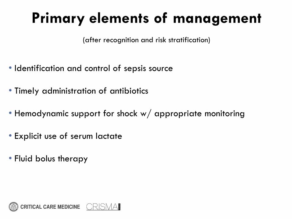
(after recognition and risk stratification)
Primary elements of management
• Identification and control of sepsis source
• Timely administration of antibiotics
• Hemodynamic support for shock w/ appropriate monitoring
• Explicit use of serum lactate
• Fluid bolus therapy
Page 12
All those physical measures used to control a focus of invasive
infection and to restore the optimal function of the affected area.
John Marshall
• Drainage of closed space infection, liquid
• Debridement or physical removal of infected tissue/device
• Abdomen, chest, skin, soft tissue
Marshall et al. Crit Care Clin, 2009
Source control
Page 13
Martinez et al. Crit Care Med, 2016
99 Medical – surgical ICUs
3,663 patients severe sepsis, septic
shock
2011 – 2013
OR for source control: 0.81 (95%CI:
0.65, 0.99, p=0.04)None Source control
0
10
20
30
In h
osp
ital m
ort
alt
iy (
%)
Source control, 2
Page 14
Dellinger et al., Crit Care Med, 2012
Source control,3
Page 15
Clinical practice guidelines and CMS
Rhodes, Evans et al., Crit Care Med, 2017
Timely administration of antibiotics
Page 16
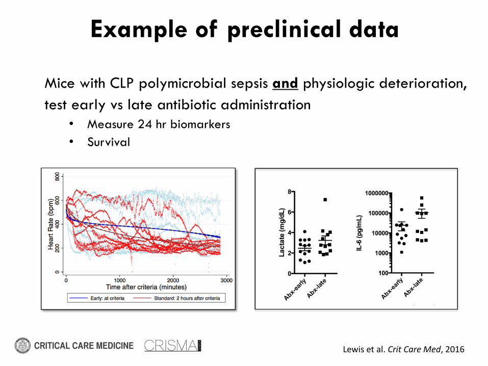
Mice with CLP polymicrobial sepsis and physiologic deterioration,
test early vs late antibiotic administration
• Measure 24 hr biomarkers
• Survival
Lewis et al. Crit Care Med, 2016
Example of preclinical data
Page 17
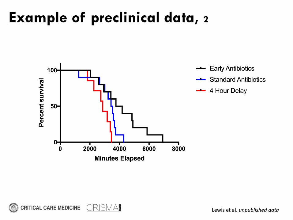
Lewis et al. unpublished data
Example of preclinical data, 2
Page 18
• No benefit from antibiotics administered with 3 hours of ED
arrival
• Unintended consequences?• Adverse effects
• Burden on clinical team
• Over-use, resistance
• No randomized clinical trial
Sterling et al., Crit Care Med, 2015
Meta analysis not so fast
Page 20
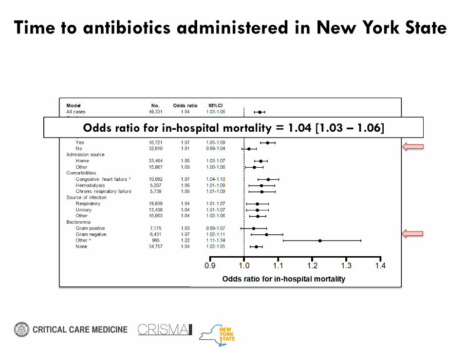
Time to antibiotics administered in New York State
Page 21
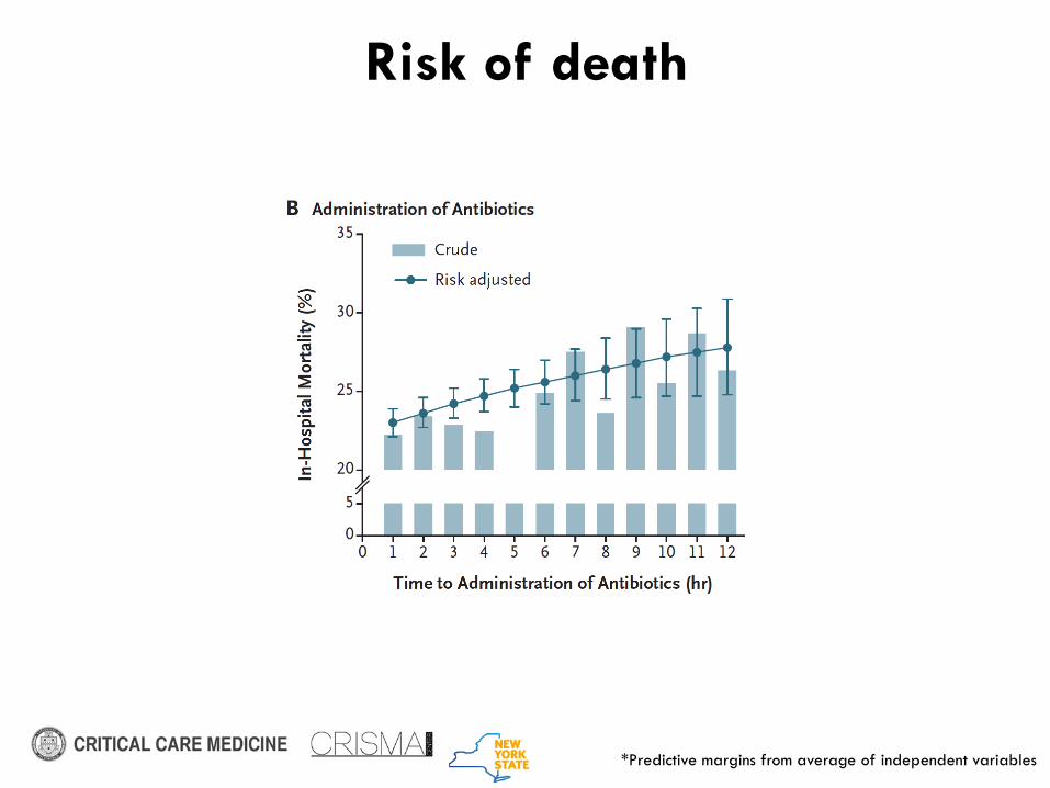
*Predictive margins from average of independent variables
Risk of death
Page 22
Guideline Severe Sepsis Septic shock
Surviving Sepsis
Campaign, 2012 *
1 hr of recognition 1 hr of recognition
CMS SEP1 bundle 3 hr of recognition 3 hr of recognition
* Strong recommendation, moderate quality of evidence
Dellinger et al. Crit Care Med, 2011https://www.acep.org/content.aspx?id=104615
Recommendations
Page 23
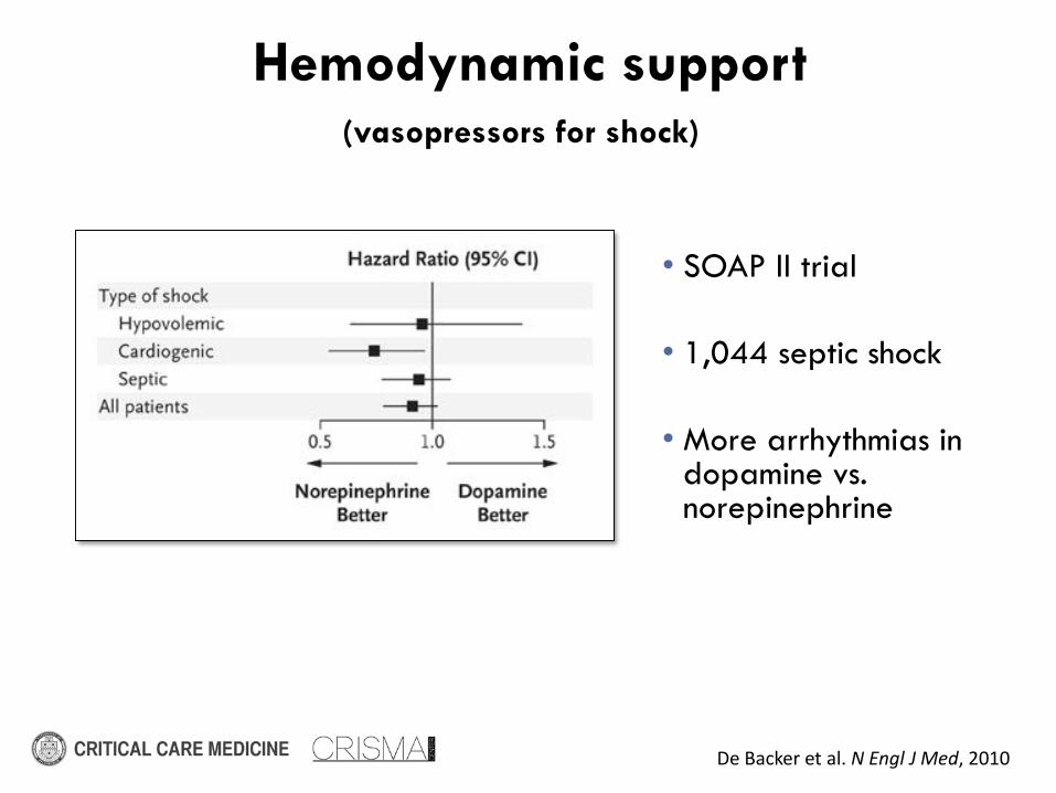
(vasopressors for shock)
Hemodynamic support
• SOAP II trial
• 1,044 septic shock
• More arrhythmias in dopamine vs. norepinephrine
De Backer et al. N Engl J Med, 2010
Page 24
Dellinger et al. Crit Care Med, 2012
Hemodynamic support, 2
•Not specified in CMS SEP1 bundle
•Appropriate for patients with septic shock (defined?) who are not responsive to initial fluid challenge
Page 25
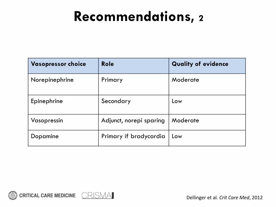
Vasopressor choice Role Quality of evidence
Norepinephrine Primary Moderate
Epinephrine Secondary Low
Vasopressin Adjunct, norepi sparing Moderate
Dopamine Primary if bradycardia Low
Dellinger et al. Crit Care Med, 2012
Recommendations, 2
Page 26
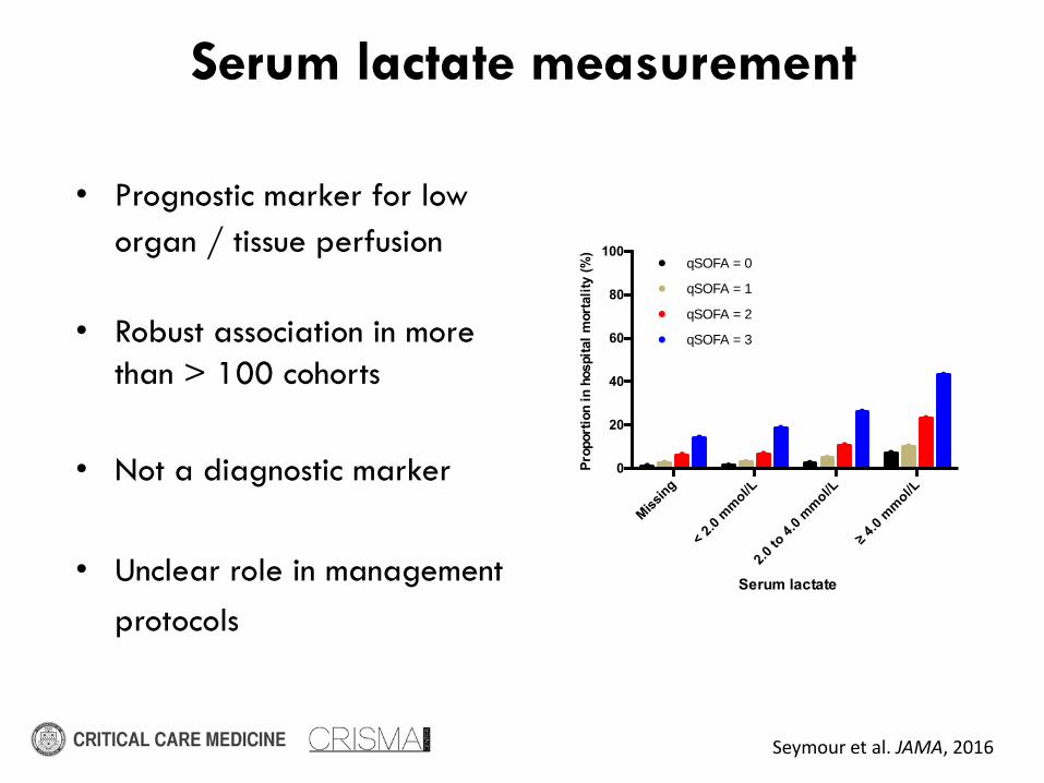
• Prognostic marker for low
organ / tissue perfusion
• Robust association in more
than > 100 cohorts
• Not a diagnostic marker
• Unclear role in management
protocols
Mis
sing
< 2.0
mm
ol/L
2.0
to 4
.0 m
mol/L
≥ 4.
0 m
mol/L
0
20
40
60
80
100
Pro
port
ion i
n h
ospit
al
mort
ali
ty (
%)
qSOFA = 0
qSOFA = 1
qSOFA = 2
qSOFA = 3
Serum lactate
Seymour et al. JAMA, 2016
Serum lactate measurement
Page 27
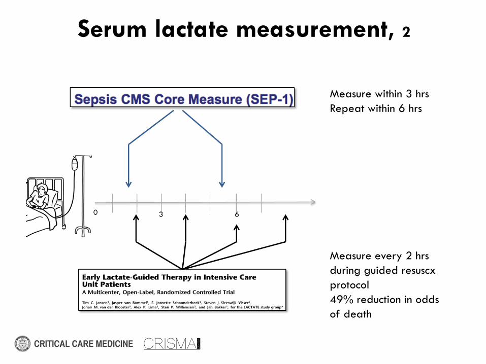
Measure within 3 hrs
Repeat within 6 hrs
Measure every 2 hrs
during guided resuscx
protocol
49% reduction in odds
of death
Serum lactate measurement, 2
Page 28
Lactate
measurement
Purpose Timing Recommended by..
First
measurement
Help determine
if shock present
or not
Triage or
immediate at
sepsis recognition
SSC – dx criteria
SEP1, mandated
Repeat
measure
Response to
initial
resuscitation
Minimum- 2 hrs
Max – 6 hrs
SSC, low quality
SEP1, mandated
RCTs, improve
mortality
Recommendations, 3
Page 29
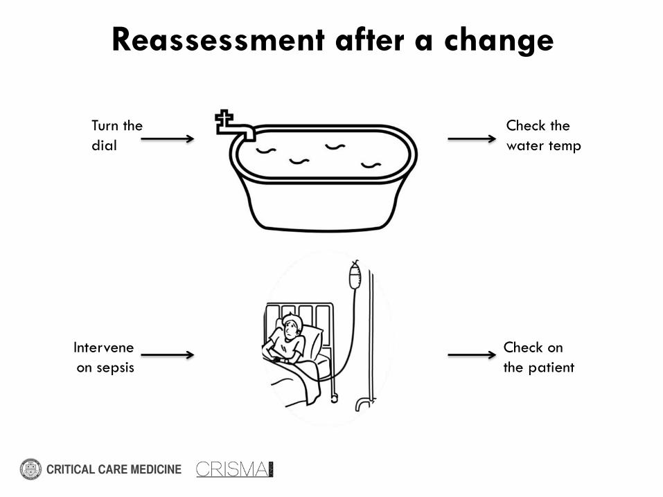
Turn the
dial
Check the
water temp
Intervene
on sepsis
Check on
the patient
Reassessment after a change
Page 30
Source Recommendation Evidence
CMS SEP1 bundle Assessment of volume
status, tissue perfusion
“Best practice”
Reassessment after a change, 2
Page 31
• Ubiquitous intervention in acute medicine
• Drug like any other from pharmacy
• Millions of unit administered to patients each day
• Hypovolemic shock
• Dehydration
• Many others
Intravenous fluids
Page 32
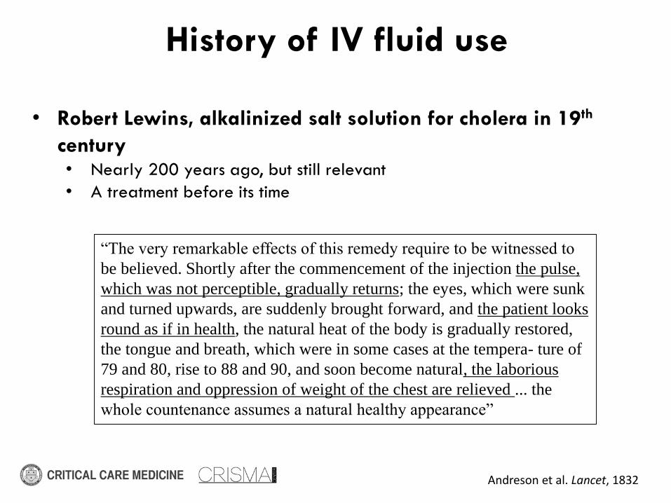
“The very remarkable effects of this remedy require to be witnessed to
be believed. Shortly after the commencement of the injection the pulse,
which was not perceptible, gradually returns; the eyes, which were sunk
and turned upwards, are suddenly brought forward, and the patient looks
round as if in health, the natural heat of the body is gradually restored,
the tongue and breath, which were in some cases at the tempera- ture of
79 and 80, rise to 88 and 90, and soon become natural, the laborious
respiration and oppression of weight of the chest are relieved ... the
whole countenance assumes a natural healthy appearance”
Andreson et al. Lancet, 1832
History of IV fluid use
• Robert Lewins, alkalinized salt solution for cholera in 19th
century• Nearly 200 years ago, but still relevant
• A treatment before its time
Page 33
• Altered membrane permeability in critically ill patients
• Endothelial glycocalx loses integrity
• Increased interstitial edema
• Particularly in surgical trauma and sepsis
Myburgh et al. NEJM, 2013
Physiology of fluid resuscitation
Page 34
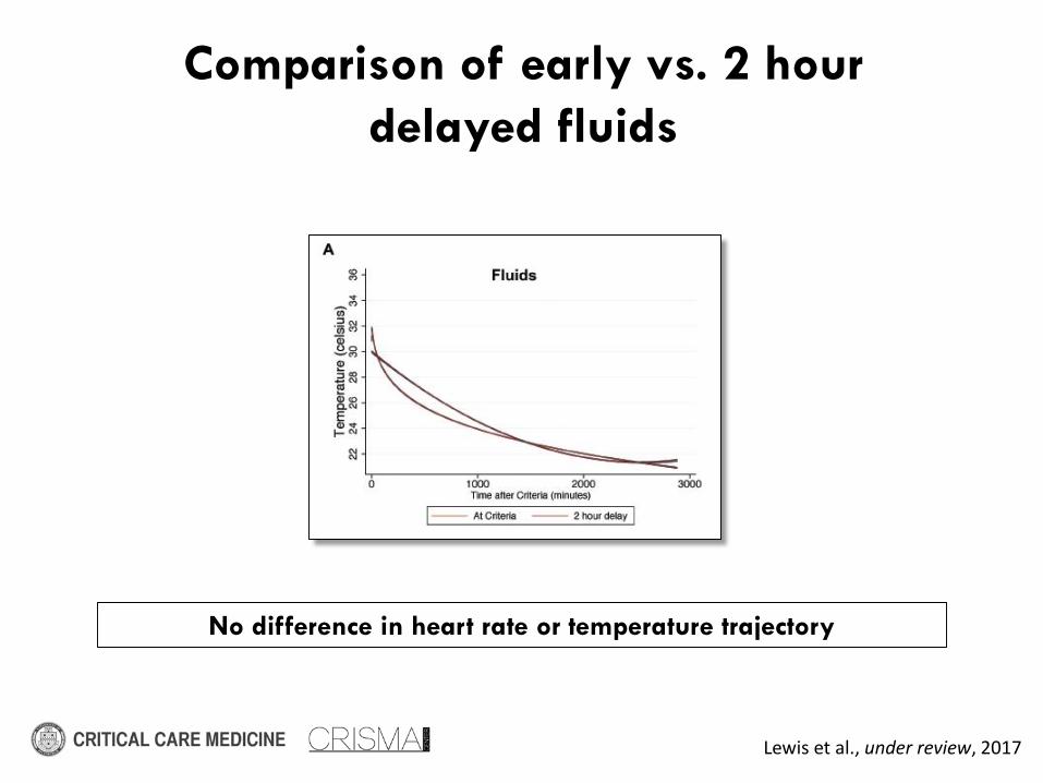
No difference in heart rate or temperature trajectory
Lewis et al., under review, 2017
Comparison of early vs. 2 hour
delayed fluids
Page 35
No significant difference in pH, base excess, or lactate with earlier
fluids
Lewis et al., under review, 2017
Comparison of early vs. 2 hour
delayed fluids, 2
Page 36
No significant difference in survival
Lewis et al., under review, 2017
Comparison of early vs. 2 hour
delayed fluids, 3
Page 37
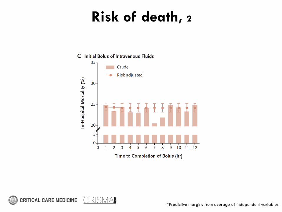
*Predictive margins from average of independent variables
Risk of death, 2
Page 38
• Centers for Medicare and Medicaid bundle for sepsis (SEP1)
• All severe sepsis or septic shock patients must receive a fluid bolus of
30cc/kg of crystalloid fluids
• Hospitals must report all cases, compliance with fluid bolus completion
• Controversial
• No exclusions for ESRD
• No exclusions for CHF
Recent national policies reinforce fluids
Page 39
What for prehospital?
Page 40
Advanced notification• Modeled after STEMI and stroke alert systems
• Mostly small before / after studies testing activation of sepsis teams
• No large cluster RCT
• Proposed to speed process measures at the hospital• Source control
• Antibiotic administration
• Hospital fluids
Page 41
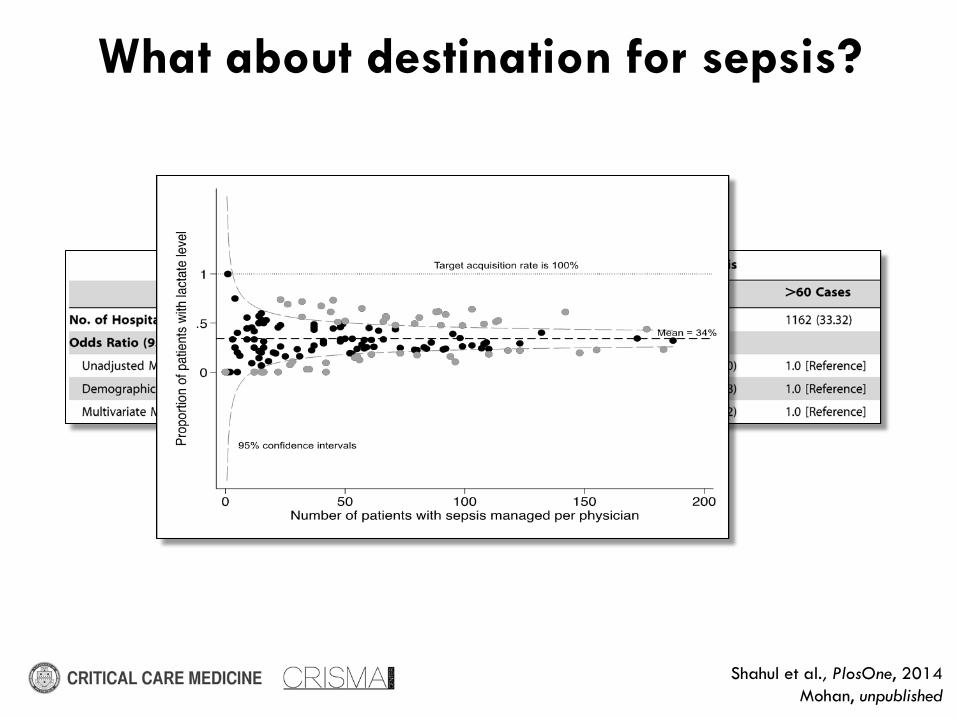
Shahul et al., PlosOne, 2014
Mohan, unpublished
What about destination for sepsis?
Page 42
Direct treatment with fluids
• Prehospital fluids
• No RCTs yet
• Observational studies in large cohorts
Page 44
Other treatments?
Page 45
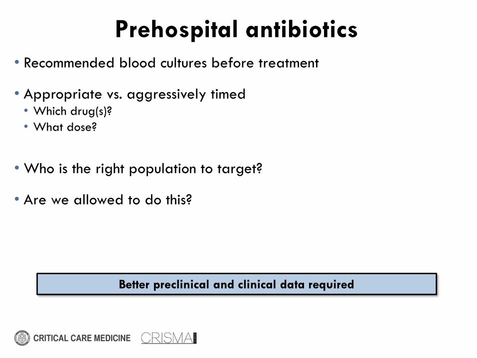
Better preclinical and clinical data required
Prehospital antibiotics
• Recommended blood cultures before treatment
• Appropriate vs. aggressively timed • Which drug(s)?
• What dose?
• Who is the right population to target?
• Are we allowed to do this?
Page 46
Walchuk et al, Prehosp Emerg Med, 2016
Demonstration project in EMS
Page 47
Randomized trial in Europe
Page 48
• Awareness and recognition is most important
• Consider advanced notification, don’t be shy
• Follow existing protocols for fluids (shock)
• No role for antibiotics (for now)
European, Canadian, and US trials either funded or under review to
generate a larger evidence base
So now what? Take home…