JOURNAL OF BACTERioLOGY, Oct. 1974, p. 131-138 Copyright @ 1974 American Society for Microbiology Vol. 120, No. 1 Printed in U.SA. Preliminary Analysis of Lipids and Fatty Acids of Green Bacteria and Chloroflexus aurantiacus CHRISTINE N. KENYON AND ALANE M. GRAY Department of Bacteriology and Immunology, University of California, Berkeley, California 94720 Received for publication 6 May 1974 The complex lipids and fatty acids of the seven type species of green bacteria and three strains of Chloroflexus aurantiacus were analyzed. The green bacteria contained lipids that behaved as cardiolipin and phosphatidylglycerol on thin-layer chromatography. They did not contain phosphatidylethanolamine or phosphatidylserine. Similarly, Chloroflexus contained lipids that behaved as phosphatidylglycerol and phosphatidylinositol on thin-layer chromatography and did not contain phosphatidylethanolamine or phosphatidylserine. The green bacteria contained glycolipids I and II of Constantopoulos and Bloch (monoga- lactosyldiglyceride and a galactose- and rhamnose-containing diglyceride). Chloroflexus exhibited galactose-containing glycolipids that behaved identically with the mono- and digalactosyldiglycerides of spinach on thin-layer chromatog- raphy, and each contained galactose as well as at least one other sugar. The fatty acids of both groups of bacteria consisted entirely of saturated and monounsatu- rated fatty acids. In the green bacteria, myristic, palmitic, and hexadecenoic acids predominated. In Chloroflexus, palmitic, stearic, and oleic acids predomi- nated. The positions of the double bonds in the monounsaturated fatty acids of Chloroflexus indicated synthesis by the anaerobic pathway. The lipid analyses suggest a close relationship between the green bacteria and Chloroflexus and further suggest that these groups of photosynthetic bacteria are more closely related to the blue-green algae than are the purple bacteria. The lipids of the nonsulfur purple bacteria have been rather extensively described (11). Phosphatidylethanolamine (PE) and phos- phatidylglycerol (PG) were found in all of the strains examined, whereas phosphatidylcholine (PC), cardiolipin (CL), and ornithine-contain- ing phospholipids were found in some strains. The galactolipids characteristic of the chloro- plasts of eukaryotic photosynthetic organisms are never found in the nonsulfur purple bacteria with the exception of a trace amount of a compound with the behavior of monogalactosyl- diglyceride (MGDG) found in Rhodospirillum molischianum (8). In contrast, the lipids of only one strain of sulfur purple bacteria have been described. Chromatium, strain D, contains PE, PG, CL, and glycolipids which may be mono-, di-, and triglycosyldiglycerides (27). The phospholipids of the green bacteria have not been described. Three strains of green bacteria were found to contain monogalactosyldiglyceride and a rham- nose- and galactose-containing glycolipid (10). The fatty acids of the nonsulfur purple bacte- ria have been found to consist of saturated and monounsaturated fatty acids as well as, in one species, hydroxy-fatty acids, cyclopropane group-containing fatty acids, and branched- chain fatty acids (8, 21, 24, 30). The fatty acids of pure cultures of green bacteria have not been examined. The other major group of photosynthetic prokaryotic organisms is the blue-green algae. Nichols et al. (20) reported the presence of PG, MGDG, digalactosyldiglyceride (DGDG), and sulfoquinovosyldiglyceride (SQDG) in two strains of blue-green algae. They found that PC, PE, and phosphatidylinositol were absent. We have previously reported the presence in 11 strains of unicellular blue-green algae and 13 strains of filamentous blue-green algae of glyco- lipids which when analyzed by thin-layer chro- matography exhibit the R, and staining behav- ior of MGDG and DGDG. Other glycolipids are also present in these strains, as are possibly SQDG and PC. PE was not found in any of the strains (14, 15; C. N. Kenyon, unpublished observations). Recently B. K. Pierson and R. W. Castenholz (submitted for publication) described a photo- trophic gliding filamentous bacterium of hot springs which they have named Chloroflexus 131 on July 10, 2018 by guest http://jb.asm.org/ Downloaded from
Transcript
JOURNAL OF BACTERioLOGY, Oct. 1974, p. 131-138Copyright @ 1974 American Society for Microbiology
Vol. 120, No. 1Printed in U.SA.
Preliminary Analysis of Lipids and Fatty Acids of GreenBacteria and Chloroflexus aurantiacus
CHRISTINE N. KENYON AND ALANE M. GRAYDepartment of Bacteriology and Immunology, University of California, Berkeley, California 94720
Received for publication 6 May 1974
The complex lipids and fatty acids of the seven type species of green bacteriaand three strains of Chloroflexus aurantiacus were analyzed. The green bacteriacontained lipids that behaved as cardiolipin and phosphatidylglycerol onthin-layer chromatography. They did not contain phosphatidylethanolamine orphosphatidylserine. Similarly, Chloroflexus contained lipids that behaved asphosphatidylglycerol and phosphatidylinositol on thin-layer chromatographyand did not contain phosphatidylethanolamine or phosphatidylserine. The greenbacteria contained glycolipids I and II of Constantopoulos and Bloch (monoga-lactosyldiglyceride and a galactose- and rhamnose-containing diglyceride).Chloroflexus exhibited galactose-containing glycolipids that behaved identicallywith the mono- and digalactosyldiglycerides of spinach on thin-layer chromatog-raphy, and each contained galactose as well as at least one other sugar. The fattyacids of both groups of bacteria consisted entirely of saturated and monounsatu-rated fatty acids. In the green bacteria, myristic, palmitic, and hexadecenoicacids predominated. In Chloroflexus, palmitic, stearic, and oleic acids predomi-nated. The positions of the double bonds in the monounsaturated fatty acids ofChloroflexus indicated synthesis by the anaerobic pathway. The lipid analysessuggest a close relationship between the green bacteria and Chloroflexus andfurther suggest that these groups of photosynthetic bacteria are more closelyrelated to the blue-green algae than are the purple bacteria.
The lipids of the nonsulfur purple bacteriahave been rather extensively described (11).Phosphatidylethanolamine (PE) and phos-phatidylglycerol (PG) were found in all of thestrains examined, whereas phosphatidylcholine(PC), cardiolipin (CL), and ornithine-contain-ing phospholipids were found in some strains.The galactolipids characteristic of the chloro-plasts of eukaryotic photosynthetic organismsare never found in the nonsulfur purple bacteriawith the exception of a trace amount of acompound with the behavior of monogalactosyl-diglyceride (MGDG) found in Rhodospirillummolischianum (8).In contrast, the lipids of only one strain of
sulfur purple bacteria have been described.Chromatium, strain D, contains PE, PG, CL,and glycolipids which may be mono-, di-, andtriglycosyldiglycerides (27). The phospholipidsof the green bacteria have not been described.Three strains of green bacteria were found tocontain monogalactosyldiglyceride and a rham-nose- and galactose-containing glycolipid (10).The fatty acids of the nonsulfur purple bacte-
ria have been found to consist of saturated andmonounsaturated fatty acids as well as, in one
species, hydroxy-fatty acids, cyclopropanegroup-containing fatty acids, and branched-chain fatty acids (8, 21, 24, 30). The fatty acidsof pure cultures of green bacteria have not beenexamined.The other major group of photosynthetic
prokaryotic organisms is the blue-green algae.Nichols et al. (20) reported the presence of PG,MGDG, digalactosyldiglyceride (DGDG), andsulfoquinovosyldiglyceride (SQDG) in twostrains of blue-green algae. They found that PC,PE, and phosphatidylinositol were absent. Wehave previously reported the presence in 11strains of unicellular blue-green algae and 13strains of filamentous blue-green algae of glyco-lipids which when analyzed by thin-layer chro-matography exhibit the R, and staining behav-ior of MGDG and DGDG. Other glycolipids arealso present in these strains, as are possiblySQDG and PC. PE was not found in any of thestrains (14, 15; C. N. Kenyon, unpublishedobservations).
Recently B. K. Pierson and R. W. Castenholz(submitted for publication) described a photo-trophic gliding filamentous bacterium of hotsprings which they have named Chloroflexus
aurantiacus. This organism is related taxonomi-cally to the green bacteria and, to a somewhatlesser extent, to the nonsulfur purple bacteriaand to the blue-green algae. Because fatty acidand lipid compositions have been of considera-ble usefulness in describing the taxonomic posi-tion of other prokaryotic organisms, particularlythose capable of photosynthesis (11, 14, 15), wedecided to examine the lipids and fatty acids ofthree strains of C. aurantiacus and of each ofthe seven type species of green bacteria. Thispaper reports the results of a preliminary com-parative study of the complex lipids and fattyacids of these organisms.
MATERIALS AND METHODS
Strains and conditions of cultivation. Typestrains of all the species of green sulfur bacteria weregrown in the laboratory of N. Pfennig (University ofGottingen) in the medium described by Pfennig (24)at pH 6.8 with 0.090 g of Na2S .9H20, 0.04 g ofammonium acetate, 0.033 g of NH4Cl, and 2 gg ofvitamin B12 per 100 ml of medium. The strains wereChlorobium limicola, strain 6330; C. limicola formathiosulfatophilum, strain 6230; vibrioforme, strain6030; C. vibrioforme forma thiosulfatophilum, strain1930; C. phaeobacteriodes, strain 2430; C. phaeovi-broides, strain 2631; and Pelodictyon luteolum, strain2530. The cell material from 500 ml of culture wasconcentrated by centrifugationr resuspended in asmall amount of culture medium, and stored at 4 Cuntil analyzed.
Three strains of Chloroflexus aurantiacus (J-10-fl,OK-70-fl, and OH-64-fl) were grown in the laboratoryof R. Castenholz (University of Oregon). The cultureswere grown in screw-cap Roux bottles in Van Baalen'smedium Cg-10 (28) or medium D (6) supplementedwith 0.2% yeast extract, at 45 C, with 170 f-c ofillumination from a tungsten lamp, semi-aerobically,to a density of about 100 Klett units. Growth of allthree strains was photoheterotrophic. Cell material ofChloroflexus was collected by centrifugation, washedthoroughly, and either analyzed directly or lyophi-lized and stored at - 10 C until analyzed. The analy-ses of Chloroflexus aurantiacus were performed on twoseparate cultures of each strain with identical results.
Lipid extraction. Whole cells of green bacteria orChloroflexus were sonicated for 1.5 or 4 min, respec-tively, in a 60-W MSE (MSE, Ltd., London) sonicdisintegrator. The total lipids were extracted by themethod of Bligh and Dyer as described by Ames (1).
Phosphate content of lipids. The phosphate con-tent of the lipids was determined by the method ofBartlett (2).
Thin-layer chromatography of lipids. Lipidswere analyzed by thin-layer chromatography on com-mercially prepared silica gel plates (Brinkmann In-struments, Inc., or Analtech, Inc.). Plates were acti-vated at 100 C for 30 min before use. The solventsystems used for unidirectional development were (i)chloroform-methanol-acetic acid-water (85:15:10:3.5,
vol/vol) (20); (ii) chloroform-methanol-7 N ammonia(60:35:5, vol/vol) (19); and (iii) chloroform-methanol-acetic acid (65:25:8, vol/vol) (19).The solvent systems used for two-dimensional thin-
layer chromatography were, in the first direction,chloroform-methanol-water (65: 25:4, vol/vol), and inthe second direction, diisobutylketone-acetic acid-water (80:55:15, vol/vol) (modified from Lepage [17]).The plates were dried for 30 min in air betweendevelopments. After both developments, the plateswere dried thoroughly under vacuum.
All lipids were visualized by exposure of each plateto iodine vapor. After removal of the iodine, thefollowing stains were used. PE and phosphatidylser-ine (PS) were visualized with ninhydrin (26). Glyco-lipids were visualized with diphenylamine (13). Phos-pholipids were visualized with the molybdate spraydescribed by Vaskovsky and Kostetsky (29). PC wasvisualized with the Dragendorff reagent (4). Thepresence of PG was confirmed by staining with theacetylacetone spray of Schwartz (25).The lipids of the green bacteria were identified by
comparison of their R, values in all four of the abovesolvent systems with the R, values of known stan-dards. Standards were run on each one-dimensionalplate. For two-dimensional thin-layer chromatogra-phy, parallel plates were spotted with mixtures ofstandards and were developed at the same time as theplates of unknown lipids. In some cases, known andunknown lipids were co-chromatographed. The plateswere routinely stained with iodine, ninhydrin, anddiphenylamine, or with iodine, ninhydrin, and themolybdate spray, in that order.The total lipids of Chloroflexus were identified by
comparison of their R, values in solvent system (i)with those of known standards run on the same plateand by their behavior on staining the plate succes-sively with iodine, ninhydrin, diphenylamine, and themolybdate spray.The standard phospholipids used were PE, PC, PS,
and phosphatidylinositol (Applied Science Laborato-ries, Inc.); PG (Supelco., Inc.); and CL (PierceChemical Co.).The MGDG and DGDG of spinach and of Chlorella
vulgaris were used as glycolipid standards. The totallipids of these tissues were extracted with chloroform-methanol (2: 1, vol/vol) at room temperature forseveral hours. No attempt was made to quantitate theamounts of the cellular lipids.
Fatty acid content of lipids. The fatty acidcontent of the lipids was determined as describedpreviously (14, 16). The Chloroflexus strains weresonicated before saponification in one of the twoanalyses performed. This did not alter the resultsobtained. Confirmation of the chain length of thefatty acids was made by hydrogenation of the fattyacid methyl esters and rechromatography, as de-scribed previously (14). The position of the doublebond in the fatty acids was determined by thin-layerchromatography on silver nitrate-impregnated silicagel thin-layer plates for which development was intoluene at - 15 C (9).
Glycolipid analysis. For analysis of the sugars inthe glycolipids of the green bacteria, approximately 3
mg of the total lipids of each strain was applied to athin-layer plate and the lipids were separated bytwo-dimensional thin-layer chromatography as de-scribed above. The lipids were visualized with iodine,and the two glycolipids were eluted from the silica gelwith methanol-chloroform (2:1, vol/vol). The solventwas evaporated and the glycolipids were hydrolyzedas described by Constantopoulos and Bloch (8). Theresulting sugars were identified by thin-layer chroma-tography on acetate-impregnated silica gel thin-layerplates using known sugars as references (8).The glycolipids of Chloroflexus were isolated by
thin-layer chromatography in solvent system (iii) andfurther treated as for the glycolipids of the greenbacteria. In addition, the sugars were identified byspraying the acetate-impregnated silica gel plateswith Glucostat and Galactostat (Worthington Bio-chemical Corp.). The identification of the sugars wasconfirmed by thin-layer chromatography on boricacid-impregnated silica gel thin-layer plates (22).
RESULTSPhospholipids of green bacteria. The phos-
phate contents of the total lipids of each of thestrains of green bacteria are presented in Table1. Although the purity of the lipid samples wasnot determined, it is clear that all strains, withthe possible exception of strain 2430, containedsimilar levels of phospholipids.
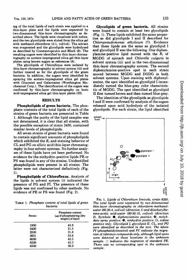
All seven strains of green bacteria were foundto contain significant amounts of phospholipidswhich exhibited the R, and staining behavior ofCL and PG on silicic acid thin-layer chromatog-raphy in four solvent systems. No further analy-ses of these lipids have yet been performed. Noevidence for the ninhydrin-positive lipids PE orPS was found in any of the strains. Unidentifiedphospholipids were present in all strains. Thelatter were not characterized definitively (Fig.1).Phospholipids of Chloroflexus. Analysis of
the lipids in solvent system (i) indicated thepresence of PG and PI. The presence of theselipids was not confirmed by other methods. Noevidence of PE or PS was found (Fig. 2).
TABLE 1. Phosphate content of total lipids of greenbacteria
Glycolipids of green bacteria. All strainswere found to contain at least two glycolipids(Fig. 1). These lipids exhibited the same proper-ties as did glycolipids I and II described forChloropseudomonas ethylicum (7). Evidencethat these lipids are the same as glycolipid Iand glycolipid II was the following. One diphen-ylamine-positive lipid moved with the R, ofMGDG of spinach and Chlorella vulgaris insolvent system (iii) and in the two-dimensionalthin-layer chromatography system. The seconddiphenylamine-positive spot in each strainmoved between MGDG and DGDG in bothsolvent systems. Upon staining with diphenyl-amine, the spot identified as glycolipid I imme-diately turned the blue-gray color characteris-tic of MGDG. The spot identified as glycolipidII first turned brown and then turned blue-gray.The identities of the glycolipids as glycolipids
I and II were confirmed by analysis of the sugarsreleased upon acid hydrolysis of the isolatedglycolipids. For each strain, the lipid identified
solventfront
FIG. 1. Lipids of Chlorobium limicola, strain 6330.The total lipids were separated by two-dimensionalthin-layer chromatography in chloroform-methanol-water (65:25:4, vol/vol) (direction 1) and diisobutylke-tone-acetic acid-water (80:55:15, vol/vol) (direction2). Symbols: 0, diphenylamine positive; O, molyb-date spray positive; O, ninhydrin positive; 0, iodinepositive only. Glycolipid I, glycolipid II, CL, and PGwere identified as described in the text. The labelsPI (phosphatidylinositol) and PC indicate the migra-tion of reference compounds only, although lipid spotswere detected at those locations in the unknownsample. indicates the migration of standard PE.There was no corresponding spot in the unknownsample.
as glycolipid I was found to yield a single sugarwhich stained blue-gray with napthoresorcinolas did galactose, and which had the same Rt(0.09) as did galactose. The second glycolipid,identified as glycolipid II, was found to yieldone sugar which behaved as galactose (by stain-ing and R,) and a second sugar which had thesame Rt (0.34) and staining behavior withnaphthoresorcinol (blue-violet) as did rham-nose.
The presence of SQDG in the lipids of eachstrain was suggested by the presence of a
ninhydrin-, molybdate-, and diphenylamine-negative lipid with the R, of SQDG in all threeone-dimensional thin-layer systems. One or
more unknown ninhydrin- and molybdate-nega-tive lipids were also present in each strain (Fig.1).
Glycolipids of Chloroflexus. All threestrains of Chloroflexus were found to containglycolipids which exhibited the same R, andstaining behavior with diphenylamine as didthe MGDG and DGDG of spinach. The pres-
ence of SQDG in each strain was suggested bythe presence of a ninhydrin-, molybdate-, anddiphenylamine-negative lipid with the Rt ofSQDG (Fig. 2).
Hydrolysis of the two glycolipids of each
strain of Chloroflexus revealed that both glyco-lipids contained galactose. Galactose was iden-tified on the basis of R, in two solvent systemsand staining with napthoresorcinol and Galac-tostat. In addition, both glycolipid fractionsyielded a sugar with the R, of mannose which isGalactostat positive and Glucostat negative.This could be a galactose derivative. A smallamount of a Glucostat-positive sugar with theRt and staining properties of glucose was alsodetected in the diglycolipid fraction (Fig. 3).Fatty acids of green bacteria. The fatty acid
composition of the extractable lipids of six ofthe strains of green bacteria is presented inTable 2. All strains contained large amounts ofmyristic acid, palmitic acid, and hexadecenoicacid.Thin-layer chromatography of the fatty acid
methyl esters of strain 6330 on silver nitrate-impregnated silicic acid thin-layer platesshowed that the unsaturated fatty acids of thisstrain migrated with oleic acid rather than withcis-vaccenic acid. This suggests that the doublebond of 16:1 is between carbons 9 and 10.The fatty acid identified as 17cy co-chro-
matographs with 9,10-methylene hexadecenoicacid from Escherichia coli on 10% diethylene-glycol succinate gas chromatography columns,
FIG. 3. Sugars of the glycolipids of Chloroflexus aurantiacus. The hydrolysates of the glycolipids of strainOH-70-fl were analyzed by thin-layer chromatography on acetate-impregnated silica gel. Symbols: 0,Galactostat-positive spots; 0, Glucostat-positive spots.
is resistant to mild catalytic hydrogenation,and is sensitive to bromination (5). This analy-sis was made for strains 2530 and 6230 only,since they are the only strains found to containsignificant amounts of this acid.Fatty acids of Chloroflexus aurantiacus.
The fatty acid composition of the three strainsof Chloroflexus is presented in Table 3.
Separation of the total fatty acids of eachstrain on AgNO,,-impregnated plates revealedthat fatty acids with double bonds at positions
(7, 8), (9, 10), and (11, 12) were present (Fig. 4).Argentation chromatography of 16:1, 17:1, 18:1,19;1, and 20:1 collected from the gas chromato-graph showed the fatty acid isomers which were
present in each strain (Table 4). Each fatty acidisomer separated by argentation chromatogra-phy was rechromatographed on 10% diethylene-glycol succinate to check its purity. It was thenhydrogenated in order to confirm the chainlength.
DISCUSSIONThe phosphate contents of the total lipids of
the green bacteria reported here are all withinthe same range as the values reported byCruden and Stanier (10) for strain 6230. Thepresence ofPG and CL in the green bacteria is aproperty shared with other photosynthetic bac-teria. Likewise, the blue-green algae also con-
tain PG. However, the absence of PE distin-guishes the lipids of the green bacteria and ofChloroflexus from those of the other photosyn-thetic bacteria, whereas it is a property sharedwith the blue-green algae (14, 15, 30).
FIG. 4. Fatty acids of Chloroflexus aurantiacus strain OK-70-fl. The total fatty acid methyl esters wereseparated by thin-layer chromatography on silver nitrate-impregnated plates. (1) Cis-vaccenic acid; (2)palmitoleic acid; (3) fatty acids of strain OK-70-fl alone; (4) 3 + oleic acids; (5) 3 + cis-vaccenic acid; (6) 3 +palmitoleic acid; (7) palmitoleic acid + cis-vaccenic acid; (8) oleic acid, linoleic acid, a-linolenic acid.Visualization 2, 7-dichlorofluorescein, photographed under ultraviolet light.
The occurrence ofPE in the bacteria has beendiscussed by Goldfine (11), who points out thatPE is nearly a universal component of the lipidsof the bacteria, although the distribution oflipids derived from PE, including PC, is lesswidespread. Our findings suggest that the greenbacteria and Chloroflexus are taxonomically orevolutionarily more closely related to each otherand to the oxygen-evolving blue-green algaethan to other bacteria.The properties of glycolipids obtained from
all seven type strains of the green bacteria arethe same as those of Chlorobium thiosulfato-philum, strains 6130 and 6230, and Chlorobiumlimicola, strain 1230, previously described (10).These glycolipids were originally characterizedby Constantopoulos and Bloch (8) after theirisolation from Chloropseudomonas ethylicum.However, that "strain" has subsequently beenshown to be a two-membered culture of C.limicola and another nonphotosynthetic bac-terium (12). Thus, the source of the glycolipids
TABLnE 4. Position of double bonds in fatty acids ofChloroflexus
Double bond positionFatty acid
7,8a 9,10 11,12
16:1 + -
17:1 _ + _18:1 4 + 4
19:1 - + +20:1 - +
a This double bond position is inferred.h +, Present; -, absent.
was uncertain in the analyses by Constanto-poulos and Bloch.The occurrence of the galactose-containing
glycolipid I in the green bacteria distinguishesthis group from all the other photosyntheticbacteria so far examined, and indeed from mostother gram-negative bacteria (11). Only in thesulfur purple bacterium Chromatium strain Dhas a monoglycolipid (glucosyldiglyceride) beenfound in significant quantities. Moreover, thepresence of a lipid with similar properties inChloroflexus supports the above conclusion thatthe green bacteria and Chloroflexus are taxo-nomically rather close to each other and to theblue-green algae.
In extending these observations, although thestructure of the glycolipids of Chloroflexus hasnot been examined, the presence of a galactose-containing glycolipid with the chromatographicproperties of DGDG as found in the blue-greenalgae and in photosynthetic eukaryotic cellswould suggest that Chloroflexus is more closelyrelated to these higher forms than are any of theother photosynthetic bacteria so far described.These conclusions, which are based entirely onthe complex lipid patterns of the organisms inquestion, are borne out by comparison of otherproperties of the groups (Pierson and Casten-holz, submitted for publication).The cellular localization of the various glyco-
lipids described for photosynthetic microorga-nisms is known. Glycolipid I, which may beMGDG, is localized in the chlorobium vesicles,whereas glycolipid II is in the cell membrane ofgreen bacteria (10). MGDG and DGDG areknown to be localized in the photosyntheticlamellae of a blue-green alga (Anabaenavariabilis) and in the chloroplasts of eukaryoticphotosynthetic organisms (18). The occurrenceof other sugars in the glycolipids of a eukaryoticphotosynthetic microorganism has been re-ported, although their cellular localization isunknown (C. N. Kenyon, Ph.D. thesis, Harvard
University, Cambridge, Mass., 1967). It wouldbe interesting to know the cellular localizationof the galactose-containing glycolipid found inChloroflexus which exhibits a polarity similar tothat of DGDG. It would also be of interest toknow the structures of the glycolipids ofChloroflexus. Are they indeed MGDG andDGDG, perhaps with one galactose occurring asa derivative?The function of the galactolipids in the photo-
synthetic organelles is not known. What isknown is that the monogalactolipid and chloro-phyll contents of the cell are related, suggestingthat this lipid has an important structural rolein the process of photosynthesis (3). It is strik-ing that this lipid occurs in only those photosyn-thetic bacteria in which photosynthesis takesplace in chlorobium vesicles. Finally, the greenbacteria, Chloroflexus, the blue-green algae,and eukaryotic photosynthetic organisms are allcharacterized by chlorophylls with ring struc-tures on the same oxidation level (7; Piersonand Castenholz, submitted for publication).Examination of the fatty acids of the green
bacteria and Chloroflexus revealed that bothgroups contain only the saturated and monoun-saturated fatty acids characteristic of the bacte-ria in general and of some strains of blue-greenalgae (11, 14, 15). The trace of 18:2 found inChloroflexus was probably derived from theyeast extract in the medium used for growth,since saponification and extraction of yeastextract yielded 18:2.Any bacterium capable of anaerobic growth
which always contains unsaturated fatty acidswould be expected to utilize the anaerobicpathway for synthesis of unsaturated fattyacids. The presence of several different isomersof each unsaturated fatty acid as found inChloroflexus is indicative of this pathway.The presence of 17cy in the green bacteria is
rather unusual since these fatty acids are notoften found in gram-negative bacteria not be-longing to the order Eubacteriales (11). Anotherunusual feature of the fatty acids ofChloroflexus is the presence of saturated andmonounsaturated odd-chain-length fatty acids.These fatty acids did not co-chromatographwith any of the known common branched-chainfatty acids.
ACKNOWLEDGMENTS
We are grateful to N. Pfennig and B. Pierson for supplyingthe biological material for these experiments. We also wish tothank R. Y. Stanier and B. Pierson for suggesting that theseanalyses be undertaken.
This research was supported by Public Health Service
grant AM 13492 from the National Institute of Arthritis,Metabolism and Digestive Diseases.
LITERATURE CITED1. Ames. G. 1968. Lipids of Salmonella typhimurium and
Escherichia coli: structure and metabolism. J.Bacteriol. 95:833-843.
2. Bartlett, G. R. 1959. Phosphorus assay in column chro-matography. J. Biol. Chem. 234:466-468.
3. Bloch, K., G. Constantopoulos, C. Kenyon, and J. Nagai.1967. Lipid metabolism of algae in the light and in thedark, p. 197-211. In T. W. Goodwin (ed.), Biochemistryof chloroplasts, vol. 2. Academic Press Inc., Londonand New York.
4. Bregoff, H. M., E. Roberts, and C. C. Delwiche. 1953.Paper chromatography of quaternary ammonium basesand related compounds. J. Biol. Chem. 205:565-574.
5. Brian, B. L., and E. W. Gardner. 1968. A simpleprocedure for detecting the presence of cyclopropanefatty acids in bacterial lipids. J. Bacteriol. 16:549-552.
6. Castenholz, R. W. 1969. Thermophilic blue-green algaeand the thermal environment. Bacteriol. Rev.33:476-504.
7. Cohen-Bazire, G. 1971. The photosynthetic apparatus ofprocaryotic organisms, p. 65-90. In P. J. Harris (ed.),Biological ultrastructure: the origin of cell organelles.Proc. 13th Annu. Biol. Coll. Oregon State UniversityPress, Corvallis.
8. Constantopoulos, G., and K. Bloch. 1967. Isolation andcharacterization of glycolipids from some photosyn-thetic bacteria. J. Bacteriol. 93:1788-1793.
9. Cronan, J. E. 1967. The unsaturated fatty acids ofEscherichia coli. Biochim. Biophys. Acta 144:695-697.
10. Cruden, D. L., and R. Y. Stanier. 1970. The characteriza-tion of chlorobium vesicles and membranes isolatedfrom green bacteria. Arch. Mikrobiol. 72:115-134.
11. Goldfine, H. 1972. Comparative aspects of bacteriallipids. Advan. Microbiol. Physiol. 8:4-58.
12. Gray, B. H., C. F. Fowler, N. A. Nugent, N. Rigopoulos,and R. C. Fuller. 1973. Reevaluation of Chloro-pseudomonas ethylica strain 2-K. Int. J. Syst. Bac-teriol. 23:256-264.
13. Harris, G., and I. C. MacWilliam. 1954. A dippingtechnique for revealing sugars on paper chromato-grams. Chem. Ind., p. 249.
14. Kenyon, C. N. 1972. Fatty acid composition of unicellularstrains of blue-green algae. J. Bacteriol. 109:827-834.
15. Kenyon, C. N., R. Rippka, and R. Y. Stanier. 1972. Fattyacid composition and physiological properties of somefilamentous blue-green algae. Arch. Mikrobiol.83:216-236.
16. Kenyon, C. N., and R. Y. Stanier. 1970. Possible evolu-tionary significance of polyunsaturated fatty acids inblue-green algae. Nature (London) 227:1164-1166.
17. Lepage, M. 1964. The separation and identification ofplant phospholipids and glycolipids by two-dimen-sional thin-layer chromatography. J. Chromatogr.13:99-103.
18. Levin, E. C., W. J. Lennarz, and K. Bloch. 1964.Occurrence and localization of a-linolenic acid contain-ing galactolipids in the photosynthetic apparatus ofAnabaena variabilis. Biochim. Biophys. Acta84:471-474.
19. Nichols, B. W. 1963. Separation of the lipids of photosyn-thetic tissues: improvement in analysis by thin layerchromatography. Biochim. Biophys. Acta 70:417-422.
20. Nichols, B. W., R. V. Harris, and A. T. James. 1965. Thelipid metabolism of blue-green algae. Biochem. Bio-phys. Res. Commun. 20:256-262.
21. Park, C.-E., and L. R. Berger. 1967. Fatty acids ofextractable and bound lipids of Rhodomicrobiumvannielii. J. Bacteriol. 93:230-236.
22. Pastuska, G. 1961. Untersuchungen Uber die qualitativeund quantitative Bestimmung der Zucker mit Hilfe derKieselgelschiche-Chromatographie. II. Mitteilung. Z.Anal. Chem. 179:427-429.
23. Pfennig, N. 1965. Anreicherungskulturen fir rote undgriine Schwefelbakterien. Zentrabl. Bakteriol. Parasi-tenk. Infektionskr. Hyg. I Abt. Orig. l(Suppl.):179-189, 503-504.
24. Schr6der, J., M. Biedermann, and G. Drews. 1969. DieFettsauren in ganzen. Zellen, Thylakoiden und Lipo-polysacchariden von Rhodospirillum rubrum und Rho-dopseudomonas capsulata. Arch. Mikrobiol.66:273-280.
25. Schwartz, D. P. 1958. Specific identification of hydrox-yamino acids on paper cb1romatograms of proteinhydrolysates. Anal. Chem. 30:1855-1856.
26. Stahl, E. (ed.) 1965. Thin-layer chromatography, p. 496.Academic Press Inc., New York.
27. Steiner, S., S. F. Conti, and R. L. Lester. 1969. Separa-tion and identification of the polar lipids ofChromatium strain 0. J. Bacteriol. 98:10-15.
28. van Baalen, C. 1967. Further observations on growth ofsingle cells of coccoid blue-green algae. J. Phycol.3:154-157.
29. Vaskovsky, V. E., and E. Y. Kostetsky. 1968. Modifiedspray for the detection of phospholipids on thin layerchromatograms. J. Lipid Res. 9:396.
30. Wood, B. J. B., B. W. Nichols, and A. T. James. 1965.The lipids and fatty acid metabolism of photosyntheticbacteria. Biochim. Biophys. Acta 106:261-273.