30
Pediatric Surgery Prem Puri Michael E. Höllwarth Editors 123 Springer Surgery Atlas Series Series Editors: J. S. P. Lumley · James R. Howe Second Edition

Pediatric Surgery
Prem PuriMichael E. Höllwarth Editors
123
Springer Surgery Atlas Series Series Editors: J. S. P. Lumley · James R. Howe
Second Edition
Springer Surgery Atlas Series
Series editorsJ. S. P. LumleyJames R. Howe
More information about this series at http://www.springer.com/series/4484

Prem Puri • Michael E. HöllwarthEditors
Pediatric SurgerySecond Edition

EditorsPrem PuriUniversity College DublinNational Children’s Research Centre Our Lady’s Children HospitalDublinIreland
Michael E. HöllwarthMedical UniversityUniversity Clinic for Pediatric and Adolescent SurgeryGrazAustria
Springer Surgery Atlas SeriesISBN 978-3-662-56280-2 ISBN 978-3-662-56282-6 (eBook)https://doi.org/10.1007/978-3-662-56282-6
Library of Congress Control Number: 2018968448
© Springer-Verlag GmbH Germany, part of Springer Nature 2006, 2019This work is subject to copyright. All rights are reserved by the Publisher, whether the whole or part of the material is concerned, specifically the rights of translation, reprinting, reuse of illustrations, recitation, broadcasting, reproduction on microfilms or in any other physical way, and transmission or information storage and retrieval, electronic adaptation, computer software, or by similar or dissimilar methodology now known or hereafter developed.The use of general descriptive names, registered names, trademarks, service marks, etc. in this publication does not imply, even in the absence of a specific statement, that such names are exempt from the relevant protective laws and regulations and therefore free for general use.The publisher, the authors and the editors are safe to assume that the advice and information in this book are believed to be true and accurate at the date of publication. Neither the publisher nor the authors or the editors give a warranty, express or implied, with respect to the material contained herein or for any errors or omissions that may have been made. The publisher remains neutral with regard to jurisdictional claims in published maps and institutional affiliations.
This Springer imprint is published by the registered company Springer-Verlag GmbH, DE part of Springer Nature.The registered company address is: Heidelberger Platz 3, 14197 Berlin, Germany

To our families for their love and patience.

vii
It is 13 years since the first edition of this book was published in 2006. In the intervening years, tremendous advances have been made in the management of children with surgical disorders. Minimally invasive surgical techniques are now routinely employed for most intracavity pro-cedures in infants and children. Robot-assisted laparoscopy is evolving rapidly in the pediatric surgical field, and robotic technology is now available in many major children’s hospitals around the world.
The second edition of Pediatric Surgery has been thoroughly revised and updated. It con-tains 74 chapters from 110 contributors from five continents. This edition contains 15 new chapters on key topics including thyroidectomy, empyema, gynecomastia, laparoscopy, stomas for small and large bowel, rectal biopsy, rectal prolapse, bariatric surgery, portal hypertension, liver transplantation, soft tissue tumors, ovarian tumors, laparoscopic and robotic urology, and dialysis. Each chapter has been written by internationally renowned leaders in their respective fields. Many younger surgeons were selected as coauthors, who will become the next genera-tion of leaders in pediatric surgery and pediatric urology.
The main aim of the new edition, as with the first edition, is to provide a comprehensive description of operative techniques for various surgical conditions in infants and children. The most unique feature of the book is the generous use of high-quality color illustrations to clarify and simplify various operative techniques. The text is organized in a systematic manner pro-viding a step-by-step, detailed, practical guide to operative approaches in the management of congenital and acquired surgical conditions in children. The book is intended for pediatric surgeons, pediatric urologists, trainees in pediatric surgery and pediatric urology, and general surgeons with interest in pediatric surgery.
The first edition of the book was very successful, accepted worldwide as a reference book for the operative management of childhood surgical disorders, and was translated into multiple languages including Chinese, Russian, and Turkish. We hope that the substantially revised and updated second edition of the book will continue to act as a reference book for pediatric sur-geons all over the world.
We wish to thank most sincerely all the contributors for their outstanding work in the prepa-ration of this innovative operative Pediatric Surgery atlas. We wish to express our gratitude to the editorial staff of Springer, for all their help during preparation and publication of this book. We wish to thank Dr. Hiroki Nakamura for help with the galley proofs of the book.
Dublin, Ireland Prem Puri Graz, Austria Michael E Höllwarth
Preface to the Second Edition

ix
Contents
1 Thyroglossal Duct Cyst . . . . . . . . . . . . . . . . . . . . . . . . . . . . . . . . . . . . . . . . . . . . . . . 1Michael E. Höllwarth
2 Branchial Cysts and Sinuses . . . . . . . . . . . . . . . . . . . . . . . . . . . . . . . . . . . . . . . . . . 5Michael E. Höllwarth
3 Lymphatic Malformations . . . . . . . . . . . . . . . . . . . . . . . . . . . . . . . . . . . . . . . . . . . . 9James K. Wall and Craig T. Albanese
4 Tracheostomy . . . . . . . . . . . . . . . . . . . . . . . . . . . . . . . . . . . . . . . . . . . . . . . . . . . . . . 15Andrew Dias, Sheila Hayes, Siobhan Fitzgerald, and John Russell
5 Thyroidectomy in Children . . . . . . . . . . . . . . . . . . . . . . . . . . . . . . . . . . . . . . . . . . . 21Tom R. Kurzawinski and Paolo De Coppi
6 Oesophageal Atresia . . . . . . . . . . . . . . . . . . . . . . . . . . . . . . . . . . . . . . . . . . . . . . . . . 29Michael E. Höllwarth and Paola Zaupa
7 Gastro-oesophageal Reflux and Hiatus Hernia . . . . . . . . . . . . . . . . . . . . . . . . . . . 41Keith E. Georgeson and Michael E. Höllwarth
8 Achalasia . . . . . . . . . . . . . . . . . . . . . . . . . . . . . . . . . . . . . . . . . . . . . . . . . . . . . . . . . . 51Paul K. H. Tam and Patrick H. Y. Chung
9 Colonic Replacement of the Oesophagus . . . . . . . . . . . . . . . . . . . . . . . . . . . . . . . . 59Devendra K. Gupta and Shilpa Sharma
10 Gastric Transposition for Oesophageal Replacement . . . . . . . . . . . . . . . . . . . . . . 67Lewis Spitz and Arnold Coran
11 Thoracoscopy . . . . . . . . . . . . . . . . . . . . . . . . . . . . . . . . . . . . . . . . . . . . . . . . . . . . . . . 73Bethany J. Slater and Steven S. Rothenberg
12 Repair of Pectus Excavatum . . . . . . . . . . . . . . . . . . . . . . . . . . . . . . . . . . . . . . . . . . 79Robert C. Shamberger
13 Pulmonary Malformations . . . . . . . . . . . . . . . . . . . . . . . . . . . . . . . . . . . . . . . . . . . . 89Henry L. Chang and Keith T. Oldham
14 Congenital Diaphragmatic Hernia and Eventration . . . . . . . . . . . . . . . . . . . . . . . 95Prem Puri
15 Empyema . . . . . . . . . . . . . . . . . . . . . . . . . . . . . . . . . . . . . . . . . . . . . . . . . . . . . . . . . . 101Michael Singh and Dakshesh Parikh
16 Gynecomastia . . . . . . . . . . . . . . . . . . . . . . . . . . . . . . . . . . . . . . . . . . . . . . . . . . . . . . 107Laura A. Monson and Mary L. Brandt
17 Extracorporeal Membrane Oxygenation . . . . . . . . . . . . . . . . . . . . . . . . . . . . . . . . 113Jason S. Frischer, Charles J. H. Stolar, and Ronald B. Hirschl
x
18 Pediatric Laparoscopic Surgery . . . . . . . . . . . . . . . . . . . . . . . . . . . . . . . . . . . . . . . 121Amulya K. Saxena
19 Hernias: Inguinal, Femoral, Umbilical, Epigastric, and Hydrocele . . . . . . . . . . . 141Juan A. Tovar and Leopoldo Martinez
20 Omphalocele . . . . . . . . . . . . . . . . . . . . . . . . . . . . . . . . . . . . . . . . . . . . . . . . . . . . . . . 153Stig Sømme and Jacob C. Langer
21 Gastroschisis . . . . . . . . . . . . . . . . . . . . . . . . . . . . . . . . . . . . . . . . . . . . . . . . . . . . . . . 161Marshall Z. Schwartz
22 Hypertrophic Pyloric Stenosis . . . . . . . . . . . . . . . . . . . . . . . . . . . . . . . . . . . . . . . . . 169Takao Fujimoto
23 Gastrostomy . . . . . . . . . . . . . . . . . . . . . . . . . . . . . . . . . . . . . . . . . . . . . . . . . . . . . . . . 175Michael W. L. Gauderer
24 Malrotation . . . . . . . . . . . . . . . . . . . . . . . . . . . . . . . . . . . . . . . . . . . . . . . . . . . . . . . . 185Augusto Zani and Agostino Pierro
25 Duodenal Obstruction . . . . . . . . . . . . . . . . . . . . . . . . . . . . . . . . . . . . . . . . . . . . . . . 191Yechiel Sweed and Arcady Vachyan
26 Jejunoileal Atresia . . . . . . . . . . . . . . . . . . . . . . . . . . . . . . . . . . . . . . . . . . . . . . . . . . . 201Alastair J. W. Millar and Alp Numanoglu
27 Meconium Ileus . . . . . . . . . . . . . . . . . . . . . . . . . . . . . . . . . . . . . . . . . . . . . . . . . . . . . 213Guido Ciprandi and Massimo Rivosecchi
28 Gastrointestinal Duplications . . . . . . . . . . . . . . . . . . . . . . . . . . . . . . . . . . . . . . . . . 221Mark D. Stringer
29 Short Bowel Syndrome . . . . . . . . . . . . . . . . . . . . . . . . . . . . . . . . . . . . . . . . . . . . . . . 237Michael E. Höllwarth
30 Hirschsprung’s Disease . . . . . . . . . . . . . . . . . . . . . . . . . . . . . . . . . . . . . . . . . . . . . . . 249Prem Puri and David Coyle
31 Anorectal Anomalies . . . . . . . . . . . . . . . . . . . . . . . . . . . . . . . . . . . . . . . . . . . . . . . . . 261Alberto Peña, Andrea Bischoff, and Marc A. Levitt
32 Intussusception . . . . . . . . . . . . . . . . . . . . . . . . . . . . . . . . . . . . . . . . . . . . . . . . . . . . . 279Holger Till and Erich Sorantin
33 Appendectomy . . . . . . . . . . . . . . . . . . . . . . . . . . . . . . . . . . . . . . . . . . . . . . . . . . . . . . 287Girolamo Mattioli
34 Omphalomesenteric Duct Remnants . . . . . . . . . . . . . . . . . . . . . . . . . . . . . . . . . . . . 293Kenneth K. Y. Wong and Paul K. H. Tam
35 Ulcerative Colitis . . . . . . . . . . . . . . . . . . . . . . . . . . . . . . . . . . . . . . . . . . . . . . . . . . . . 301Risto Rintala
36 Crohn’s Disease . . . . . . . . . . . . . . . . . . . . . . . . . . . . . . . . . . . . . . . . . . . . . . . . . . . . . 313Risto Rintala
37 Stomas for Large and Small Bowel . . . . . . . . . . . . . . . . . . . . . . . . . . . . . . . . . . . . . 319Andrea Bischoff and Alberto Peña
38 Rectal Biopsy . . . . . . . . . . . . . . . . . . . . . . . . . . . . . . . . . . . . . . . . . . . . . . . . . . . . . . . 331Augusto Zani
Contents

xi
39 Rectal Prolapse in Children . . . . . . . . . . . . . . . . . . . . . . . . . . . . . . . . . . . . . . . . . . . 335Paolo De Coppi
40 Bariatric Surgery . . . . . . . . . . . . . . . . . . . . . . . . . . . . . . . . . . . . . . . . . . . . . . . . . . . 341Brian Dalton and Thomas H. Inge
41 Biliary Atresia . . . . . . . . . . . . . . . . . . . . . . . . . . . . . . . . . . . . . . . . . . . . . . . . . . . . . . 349Masaki Nio, Hideyuki Sasaki, Hiromu Tanaka, and Ryoji Ohi
42 Choledochal Cyst . . . . . . . . . . . . . . . . . . . . . . . . . . . . . . . . . . . . . . . . . . . . . . . . . . . 359Atsuyuki Yamataka, Joel Cazares, and Hiroyuki Koga
43 Cholecystectomy . . . . . . . . . . . . . . . . . . . . . . . . . . . . . . . . . . . . . . . . . . . . . . . . . . . . 375Alan Mortell and Farhan Tareen
44 Congenital Hyperinsulinism of Infancy . . . . . . . . . . . . . . . . . . . . . . . . . . . . . . . . . 381Agostino Pierro, Augusto Zani, and Lewis Spitz
45 Splenectomy . . . . . . . . . . . . . . . . . . . . . . . . . . . . . . . . . . . . . . . . . . . . . . . . . . . . . . . . 389Peter Borzi
46 Portal Hypertension . . . . . . . . . . . . . . . . . . . . . . . . . . . . . . . . . . . . . . . . . . . . . . . . . 395Mark Davenport
47 Liver Transplantation . . . . . . . . . . . . . . . . . . . . . . . . . . . . . . . . . . . . . . . . . . . . . . . . 405Alastair J. W. Millar
48 Spina Bifida . . . . . . . . . . . . . . . . . . . . . . . . . . . . . . . . . . . . . . . . . . . . . . . . . . . . . . . . 425Jonathan R. Ellenbogen and Conor L. Mallucci
49 Hydrocephalus . . . . . . . . . . . . . . . . . . . . . . . . . . . . . . . . . . . . . . . . . . . . . . . . . . . . . . 429Kai Arnell and Tomas Wester
50 Dermal Sinus . . . . . . . . . . . . . . . . . . . . . . . . . . . . . . . . . . . . . . . . . . . . . . . . . . . . . . . 435Jonathan R. Ellenbogen and Conor L. Mallucci
51 Sacrococcygeal Teratoma . . . . . . . . . . . . . . . . . . . . . . . . . . . . . . . . . . . . . . . . . . . . . 439Kevin C. Pringle and Hiroaki Kitagawa
52 Neuroblastoma . . . . . . . . . . . . . . . . . . . . . . . . . . . . . . . . . . . . . . . . . . . . . . . . . . . . . 445Edward Kiely and Michael E. Höllwarth
53 Wilms Tumour . . . . . . . . . . . . . . . . . . . . . . . . . . . . . . . . . . . . . . . . . . . . . . . . . . . . . . 451Philip Hammond and Robert Carachi
54 Liver Tumours . . . . . . . . . . . . . . . . . . . . . . . . . . . . . . . . . . . . . . . . . . . . . . . . . . . . . . 457Irene Isabel P. Lim and Michael P. La Quaglia
55 Testicular Tumours . . . . . . . . . . . . . . . . . . . . . . . . . . . . . . . . . . . . . . . . . . . . . . . . . . 471Jonathan Ross
56 Soft Tissue Sarcomas . . . . . . . . . . . . . . . . . . . . . . . . . . . . . . . . . . . . . . . . . . . . . . . . 475Amos Loh Hong Pheng and Bhaskar Rao
57 Ovarian Tumors . . . . . . . . . . . . . . . . . . . . . . . . . . . . . . . . . . . . . . . . . . . . . . . . . . . . 487Daniel von Allmen and Mary E. Fallat
58 Laparoscopic and Robotic Urology . . . . . . . . . . . . . . . . . . . . . . . . . . . . . . . . . . . . . 499Andrew C. Strine and Paul H. Noh
59 Pyeloplasty . . . . . . . . . . . . . . . . . . . . . . . . . . . . . . . . . . . . . . . . . . . . . . . . . . . . . . . . . 507Boris Chertin and Prem Puri
Contents

xii
60 Endoscopic Treatment of Vesicoureteral Reflux . . . . . . . . . . . . . . . . . . . . . . . . . . 513Prem Puri
61 Vesicoureteral Reflux: Surgical Treatment . . . . . . . . . . . . . . . . . . . . . . . . . . . . . . . 519Jack S. Elder
62 Ureteric Duplication . . . . . . . . . . . . . . . . . . . . . . . . . . . . . . . . . . . . . . . . . . . . . . . . . 535Angela M. Arlen and Andrew J. Kirsch
63 Posterior Urethral Valves . . . . . . . . . . . . . . . . . . . . . . . . . . . . . . . . . . . . . . . . . . . . . 543Abhishek Seth, Chester J. Koh, and David A. Diamond
64 Hypospadias. . . . . . . . . . . . . . . . . . . . . . . . . . . . . . . . . . . . . . . . . . . . . . . . . . . . . . . . 549Mariette Renaux-Petel, Pierre-Yves Mure, Daniela- Brindusa Gorduza, and Pierre Mouriquand
65 Phimosis and Buried Penis . . . . . . . . . . . . . . . . . . . . . . . . . . . . . . . . . . . . . . . . . . . . 561Navroop Johal and Peter Cuckow
66 Orchidopexy . . . . . . . . . . . . . . . . . . . . . . . . . . . . . . . . . . . . . . . . . . . . . . . . . . . . . . . 569John M. Hutson
67 Varicocele . . . . . . . . . . . . . . . . . . . . . . . . . . . . . . . . . . . . . . . . . . . . . . . . . . . . . . . . . . 579Michael E. Höllwarth
68 Genitoplasty for Congenital Adrenal Hyperplasia. . . . . . . . . . . . . . . . . . . . . . . . . 585Martin A. Koyle and Richard S. Hurwitz
69 Bladder Exstrophy and Epispadias . . . . . . . . . . . . . . . . . . . . . . . . . . . . . . . . . . . . . 599Ezekiel E. Young and John P. Gearhart
70 Cloacal Exstrophy . . . . . . . . . . . . . . . . . . . . . . . . . . . . . . . . . . . . . . . . . . . . . . . . . . . 615Vijaya M. Vemulakonda and Duncan T. Wilcox
71 Augmentation Cystoplasty and Appendicovesicostomy (Mitrofanoff Principle) . . . . . . . . . . . . . . . . . . . . . . . . . . . . . . . . . . . . . . . . . . . . . . . 621Boris Chertin
72 The MACE (Malone Antegrade Continence Enema) Procedure . . . . . . . . . . . . . 629Frank J. Penna and Martin A. Koyle
73 Hydrometrocolpos . . . . . . . . . . . . . . . . . . . . . . . . . . . . . . . . . . . . . . . . . . . . . . . . . . . 637Devendra K. Gupta and Shilpa Sharma
74 Venous and Peritoneal Access in Renal Failure . . . . . . . . . . . . . . . . . . . . . . . . . . . 651Marcus D. Jarboe and Ronald B. Hirschl
Contents

xiii
Craig T. Albanese, MD NewYork-Presbyterian/Morgan Stanley Children’s Hospital and Sloane Hospital for Women, New York, NY, USA
Angela M. Arlen, MD Department of Urology, Yale School of Medicine, New Haven, CT, USA
Kai Arnell, MD, PhD Department of Women’s and Children’s Health, Uppsala University, Uppsala, Sweden
Andrea Bischoff, MD Department of Pediatric Surgery, International Center for Colorectal and Urogenital care, Children’s Hospital Colorado, Aurora, CO, USA
Peter Borzi, FRCS, FRACS Department of Paediatric Surgery and Urology, Lady Cilento Children’s Hospital, Brisbane, QLD, Australia
Paediatrics and Child Health, University of Queensland, Brisbane, QLD, Australia
Mary L. Brandt, MD Division of Pediatric Surgery, Michael E. DeBakey Department of Surgery, Baylor College of Medicine and Texas Children’s Hospital, Houston, TX, USA
Robert Carachi, MD, PhD Surgical Pediatrics, College of Medical, Veterinary and Life Sciences, The Royal Hospital for Sick Children, University of Glasgow School of Medicine, Glasgow, Scotland, UK
Joel Cazares, MD Department of Pediatric Surgery, Hospital Regional De Alta Especialidad Materno Infantil, Monterrey, Mexico
Henry L. Chang, MD Division of Pediatric Surgery, Medical College of Wisconsin, Milwaukee, WI, USA
Boris Chertin, MD Department of Pediatric Urology, Shaare Zedek Medical Center, Jerusalem, Israel
Faculty of Medicine, Hebrew University, Jerusalem, Israel
Patrick H. Y. Chung, MBBS, FRCS (Paed), FCSHK, FHKAM Department of Surgery, Queen Mary Hospital, Pokfulam, Hong Kong, China
Guido Ciprandi, MD Division of Plastic and Maxillofacial Surgery, Department of Surgery, Bambino Gesù Children’s Hospital, IRCCS, Rome, Italy
Arnold Coran, MD Department of Pediatric Surgery, C.S. Mott Children’s Hospital, Ann Arbor, MI, USA
David Coyle, MD National Children’s Research Centre, Our Lady’s Children’s Hospital, Dublin, Ireland
Peter Cuckow, FRCS (Paeds) Department of Paediatric Urology, Great Ormond Street Hospital, London, UK
Contributors
xiv
Brian Dalton, MD Department of Thoracic Oncology, Fox Chase Cancer Center, Philadelphia, PA, USA
Mark Davenport, ChM FRCPS (Glas) FRCS (Eng) Department of Paediatric Surgery, King’s College Hospital NHS Foundation Trust, London, UK
Paolo De Coppi, MD, PhD Stem Cells and Regenerative Medicine Section, Developmental Biology and Cancer Programme, University College of London Great Ormond Street Institute of Child Health, London, UK
David A. Diamond, MD Department of Urology, Children’s Hospital Boston and Harvard Medical School, Boston, MA, USA
Andrew Dias, MRCS National Children’s Research Centre, Our Lady’s Children’s Hospital, Crumlin, Dublin, Ireland
Jack S. Elder, MD Division of Pediatric Urology, Massachusetts General Hospital for Children, Harvard Medical School, Boston, MA, USA
Jonathan R. Ellenbogen, FRCS (Neuro Surg) Department of Neurosurgery, Alder Hey Children’s NHS Foundation Trust, Liverpool, Merseyside, UK
Mary E. Fallat, MD, FACS, FAAP Hiram C. Polk Jr. Department of Surgery, University of Louisville School of Medicine, Louisville, KY, USA
Siobhan Fitzgerald National Children’s Research Centre, Our Lady’s Children’s Hospital, Crumlin, Dublin, Ireland
Jason S. Frischer, MD Division of Pediatric General and Thoracic Surgery, Cincinnati Children’s Hospital Medical Center, College of Medicine, University of Cincinnati, Cincinnati, OH, USA
Takao Fujimoto, MD, PhD Department of Pediatric Surgery and Urology, Fujimoto Children’s Clinic, Tokyo, Japan
Michael W. L. Gauderer, MD, FACS, FAAP University of South Carolina Greenville, Greenville, SC, USA
John P. Gearhart, MD Department of Pediatric Urology, The James Buchanan Brady Urological Institute, Johns Hopkins University Hospital, Baltimore, MD, USA
Keith E. Georgeson, MD Providence Pediatric Surgery Center, Spokane, WA, USA
Daniela-Brindusa Gorduza, MD Department of Paediatric Urology, Hôpital Mére-Enfant—Groupe Hospitalier Est, Université Claude-Bernard, Bron, France
Devendra K. Gupta, MS, MCh, FRCS (Edin) Department of Pediatric Surgery, All India Institute of Medical Sciences, New Delhi, India
Philip Hammond, MBChB FRCS (Paed Surg) Department of Paediatric Surgery, The Royal Hospital for Sick Children, Edinburgh, Scotland, UK
Sheila Hayes National Children’s Research Centre, Our Lady’s Children’s Hospital, Crumlin, Dublin, Ireland
Ronald B. Hirschl, MD Section of Pediatric Surgery, C.S. Mott Children’s Hospital, University of Michigan, Ann Arbor, MI, USA
Michael E. Höllwarth, MD Medical University, University Clinic for Pediatric and Adolescent Surgery, Graz, Austria
Richard S. Hurwitz, MD Department of Pediatric Urology, Kaiser Permanente Medical Center, Los Angeles, CA, USA
Contributors

xv
John M. Hutson, BS, MD, DSc, FRACS, FAAP (Hon) The Royal Children’s Hospital, Parkville, VIC, Australia
Department of Paediatrics, University of Melbourne, Parkville, VIC, Australia
Surgical Research Unit, Murdoch Children’s Research Institute, Parkville, VIC, Australia
Thomas H. Inge, MD, PhD, FACS, FAAP Division of Pediatric Surgery, Adolescent Metabolic and Bariatric Surgery, Children’s Hospital Colorado, Aurora, CO, USA
Marcus D. Jarboe, MD Section of Pediatric Surgery, C.S. Mott Children’s Hospital, University of Michigan, Ann Arbor, MI, USA
Division of Interventional Radiology, University of Michigan, Ann Arbor, MI, USA
Navroop Johal, PhD, FRCS (Paeds) Department of Paediatric Urology, Great Ormond Street Hospital, London, UK
Edward Kiely, MD Department of Paediatric Surgery, Gold Coast University Hospital, Gold Coast, Australia
Andrew J. Kirsch, MD, FAAP, FACS Division of Pediatric Urology, Georgia Pediatric Urology, Children’s Healthcare of Atlanta, Emory University School of Medicine, Atlanta, GA, USA
Hiroaki Kitagawa, MD, PhD Department of Pediatric Surgery, St. Marianna University School of Medicine, Kawasaki, Japan
Hiroyuki Koga, MD, PhD Department of Pediatric Surgery, Juntendo University School of Medicine, Tokyo, Japan
Chester J. Koh, MD Division of Urology, Department of Surgery; and Scott Department of Urology, Texas Children’s Hospital and Baylor College of Medicine, Houston, TX, USA
Martin A. Koyle, MD, MSc, FAAP, FACS, FRCSC Department of Surgery, Institute of Policy, Management, and Evaluation, University of Toronto School of Medicine, Toronto, ON, Canada
Division of Pediatric Urology, Hospital for Sick Children, Toronto, ON, Canada
Tom R. Kurzawinski, PhD, FRCS Centre for Endocrine Surgery, University College and Great Ormond Street Hospitals, London, UK
Michael P. La Quaglia, MD Pediatric Service, Department of Surgery, Memorial Sloan Kettering Cancer Center, New York, NY, USA
Jacob C. Langer, MD Hospital for Sick Children, University of Toronto, Toronto, ON, Canada
Marc A. Levitt, MD Center for Colorectal and Pelvic Reconstruction, Nationwide Children’s Hospital, The Ohio State University School of Medicine, Columbus, OH, USA
Irene Isabel P. Lim, MD Pediatric Service, Department of Surgery, Memorial Sloan Kettering Cancer Center, New York, NY, USA
Conor L. Mallucci, FRCS (SN) Department of Neurosurgery, Alder Hey Children’s NHS Foundation Trust, Liverpool, UK
Leopoldo Martinez, MD, PhD Department of Pediatric Surgery, Hospital Universitario La Paz, Madrid, Spain
Girolamo Mattioli Department of surgery, DINOGMI, Giannina Gaslini Institute and University of Genova, Genova, Italy
Alastair J. W. Millar, FRCS, FRACS, FCS(SA), DCH Division of Paediatric Surgery, Red Cross War Memorial Children’s Hospital, University of Cape Town, Cape Town, South Africa
Contributors

xvi
Laura A. Monson, MD Division of Plastic Surgery, Department of Surgery, Michael E. DeBakey Department of Surgery, Baylor College of Medicine and Texas Children’s Hospital, Houston, TX, USA
Alan Mortell, MD Our Lady’s Children’s Hospital, Dublin, Ireland
Pierre Mouriquand, MD, PhD Department of Paediatric Urology, Hôpital Mére-Enfant—Groupe Hospitalier Est, Université Claude-Bernard Lyon 1, Bron, France
Pierre-Yves Mure, MD Department of Paediatric Urology, Hôpital Mére-Enfant—Groupe Hospitalier Est, Université Claude-Bernard Lyon 1, Bron, France
Masaki Nio, MD, PhD Department of Pediatric Surgery, Tohoku University Graduate School of Medicine, Sendai, Japan
Paul H. Noh, MD, FACS, FAAP Division of Pediatric Urology, Cincinnati Children’s Hospital Medical Center, Cincinnati, OH, USA
Alp Numanoglu, FCS (SA) Division of Paediatric Surgery, Red Cross Children’s Hospital, University of Cape Town, Cape Town, South Africa
Ryoji Ohi, MD Department of Pediatric Surgery, Tohoku University School of Medicine, Sendai, Japan
Keith T. Oldham, MD Division of Pediatric Surgery, Medical College of Wisconsin, Children’s Hospital of Wisconsin, Milwaukee, WI, USA
Dakshesh Parikh, MBBS, MS, FRCS (Peds), MD Department of Paediatric Surgery and Urology, Birmingham Women’s and Children’s Hospital NHS FT, Birmingham, UK
Alberto Peña, MD, FAAP, FACS, FRCS International Center for Colorectal Care, Children’s Hospital Colorado, Aurora, CO, USA
Frank J. Penna, MD Dartmouth-Hitchcock Medical Center, Children’s Hospital at Dartmouth, Lebanon, NH, USA
Amos Loh Hong Pheng, MBBS, MCRSEd, MMed, FAMS Department of Paediatric Surgery, KK Women’s and Children’s Hospital, Singapore, Singapore
Agostino Pierro, OBE, MD, FRCS (Engl) Division of General and Thoracic Surgery, Department of Surgery, The Hospital for Sick Children, University of Toronto, Toronto, ON, Canada
Kevin C. Pringle, MB, ChB, FRACS, ONZM Department of Obstetrics and Gynaecology, University of Otago, Wellington, Wellington, New Zealand
Prem Puri, FRCS, FACS, FAAP(Hon), D.Sc.(Hon) National Children’s Research Centre, Our Lady’s Children’s Hospital, Dublin, Ireland
University College Dublin, Dublin, Ireland
UCD Conway Institute of Biomolecular and Biomedical Research, Dublin, Ireland
Beacon Hospital, Dublin, Ireland
Bhaskar Rao, MD Surgical Oncology Division, Department of Surgery, St. Jude Children’s Research Hospital, Memphis, TN, USA
Mariette Renaux-Petel, MD Department of Paediatric Urology, Hôpital Mére-Enfant–Groupe Hospitalier Est, Université Claude-Bernard Lyon 1, Bron, France
Risto Rintala, MD, PhD Paediatric Surgery, Children’s Hospital, Helsinki University Central Hospital, Helsinki, Finland
Contributors

xvii
Massimo Rivosecchi, MD Division of Surgery, Bambino Gesù Children’s Hospital, IRCCS, Rome, Italy
Jonathan Ross, MD Division of Pediatric Urology, University Hospitals Rainbow Babies and Children’s Hospital, Cleveland, OH, USA
Steven S. Rothenberg, MD Department of Pediatric Surgery, Rocky Mountain Hospital for Children, Denver, CO, USA
John Russell, FRCS (ORL) Our Lady’s Children’s Hospital Crumlin, Dublin, Ireland
Hideyuki Sasaki, MD Department of Pediatric Surgery, Tohoku University Graduate School of Medicine, Sendai, Japan
Amulya K. Saxena, MD, PhD, DSc (hon), FRCS (Glasg) Department of Pediatric Surgery, Chelsea Children’s Hospital, Chelsea and Westminster NHS Foundation Trust, Imperial College London, London, UK
Marshall Z. Schwartz, MD, FACS, FRCS-Eng (Hon) Drexel University College of Medicine, St. Christopher’s Hospital for Children, Philadelphia, PA, USA
Abhishek Seth, MD Division of Urology, Department of Surgery; and Scott Department of Urology, Texas Children’s Hospital and Baylor College of Medicine, Houston, TX, USA
Robert C. Shamberger, MD Department of Surgery, Boston Children’s Hospital, Boston, MA, USA
Shilpa Sharma, MS, MCh, National Board, PhD Department of Pediatric Surgery, All India Institute of Medical Sciences, New Delhi, India
Michael Singh, MBBS, FRCSEd (Paed Surg) Department of Paediatric Surgery, Birmingham Women’s and Children’s Hospital NHS FT, Birmingham, UK
Bethany J. Slater, MD Department of Pediatric Surgery, Rocky Mountain Hospital for Children, Denver, CO, USA
Stig Sømme, MD, MPH Department of Pediatric Surgery, Children’s Hospital Colorado, Aurora, CO, USA
University of Colorado School of Medicine, Aurora, CO, USA
Erich Sorantin Division of Pediatric Radiology, Department of Radiology, Medical University Graz, Graz, Austria
Lewis Spitz, PhD, FRCS, FRCPCH, FAAP, FACS Institute of Child Health, University College, London, London, UK
Great Ormond Street Hospital, London, London, UK
Charles J. H. Stolar, MD California Pediatric Surgery Group, Santa Barbara, CA, USA
Andrew C. Strine, MD Division of Pediatric Urology, Cincinnati Children’s Hospital Medical Center, Cincinnati, OH, USA
Mark D. Stringer, MS, FRCP, FRCS, FRCSEd, FRACS Department of Paediatric Surgery, Wellington Hospital and Department of Paediatrics and Child Health, University of Otago, Wellington, New Zealand
Yechiel Sweed, MD Department of Pediatric Surgery, Galilee Medical Center, Nahariya, Bar Ilan University, Safed, Israel
Paul K. H. Tam, MBBS, ChM, FRCS, FHKAM Department of Surgery, Queen Mary Hospital, The University of Hong Kong, Pokfulam, Hong Kong SAR, China
Contributors

xviii
Hiromu Tanaka, MD Department of Pediatric Surgery, Tohoku University Graduate School of Medicine, Sendai, Japan
Farhan Tareen, FCPS, FRCSI, FEBPS, FRCS Department of Paediatric Surgery, Our Lady’s Children’s Hospital, Crumlin, Dublin, Ireland
Holger Till, MD, PhD University Clinic of Paediatric and Adolescent Surgery, Medical University of Graz, Graz, Austria
Juan A. Tovar, MD Department of Pediatric Surgery, Hospital Universitario La Paz, Madrid, Spain
Vijaya M. Vemulakonda, MD, JD Department of Pediatric Urology, Children’s Hospital of Colorado, Aurora, CO, USA
Division of Urology, Department of Surgery, University of Colorado School of Medicine, Aurora, CO, USA
Daniel von Allmen, MD Department of Pediatric Surgery, Cincinnati Children’s Hospital, Cincinnati, OH, USA
James K. Wall, MD Department of Pediatric Surgery, Lucile Packard Children’s Hospital Stanford, Stanford, CA, USA
Tomas Wester, MD, PhD Unit of Pediatric Surgery, Karolinska University Hospital, Karolinska Institutet, Stockholm, Sweden
Duncan T. Wilcox, MBBS, MD Children’s Hospital Colorado, Aurora, CO, USA
Kenneth K. Y. Wong, PhD, FRCSEd, FHKAM Department of Surgery, Queen Mary Hospital, The University of Hong Kong, Pokfulam, Hong Kong SAR
Atsuyuki Yamataka, MD, PhD Department of Pediatric Surgery, Juntendo University School of Medicine, Tokyo, Japan
Ezekiel E. Young, MD Department of Urology, Health Sciences Center, Stony Brook University School of Medicine, Stony Brook, NY, USA
Alon Yulevich, MD Department of Pediatric Surgery, Galilee Medical Center, Nahariya, Bar Ilan University, Safed, Israel
Augusto Zani, MD, PhD Division of General and Thoracic Surgery, Department of Surgery, The Hospital for Sick Children, University of Toronto, Toronto, ON, Canada
Paola Zaupa, MD University Clinic of Pediatric Adolescent Surgery, Medical University of Graz, Graz, Austria
Contributors

1© Springer-Verlag GmbH Germany, part of Springer Nature 2019P. Puri, M. E. Höllwarth (eds.), Pediatric Surgery, Springer Surgery Atlas Series, https://doi.org/10.1007/978-3-662-56282-6_1
Thyroglossal Duct Cyst
Michael E. Höllwarth
The median cervical cyst is a remnant of the thyroglossus duct, which runs from the pyramidal lobe of the thyroid gland to the foramen caecum in the dorsal part of the tongue. Embryologically, the thyroid diverticulum devel-ops in a caudal direction from the foramen caecum after formation of the tongue. The thyroid gland descends to the neck in the same period of gestation when the hyoid bone develops from the second branchial arch. The thyroglossal duct may pass in front, behind, or through the body of the hyoid bone in the middle of the neck, and islands of thyroid tissue may be found scattered along the tract. At no time during embryogenesis does the thyroglossal duct contact the body surface; the original cysts thus never open to the skin. A fistula can only develop secondarily, such as fol-lowing spontaneous perforation or surgical incision of an infected cyst.
Thyroglossal duct remnants are slightly more common than branchial cleft anomalies (55% vs 45%) and are the
most common tumors of the anterior cervical region. They are usually located in the midline at the level of the hyoid bone or somewhat below it. Because of its connection with the foramen caecum of the tongue, the lesion typically moves upward with swallowing, like the thyroid gland, but unlike the thyroid, it also moves upward with tongue protrusion. In contrast, dermoid cysts or lymph nodes from the same loca-tion do not change their position with either act. In rare cases, the duct or cysts may contain either papillary cancer or squa-mous cell carcinoma. Thus, histologic evaluation is always indicated.
Ultrasound examination may be helpful, both to ascertain the presence of a normally situated, normal-size thyroid gland and to confirm the cystic nature of the mass under con-sideration. In cases of a suppurative infection, the appropri-ate treatment is incision and drainage in combination with antibiotics, followed by excision once the acute inflamma-tion has settled (Figs. 1.1, 1.2, 1.3 and 1.4).
1
M. E. Höllwarth Medical University, University Clinic for Pediatric and Adolescent Surgery, Graz, Austriae-mail: [email protected]

2
Fig. 1.1 Following induction of general anaesthesia with endotracheal intubation, the neck is hyperextended by placing a sandbag or towel roll beneath the shoulders. A horizontal skin incision is made over the cyst. In case of a fistula, the cutaneous orifice is circumcised in a horizontally oriented, elliptical fashion. Subcutaneous tissue, platysma, and cervical fascia are divided, exposing the capsule of the cyst. In cases with a previous history of inflammation, these layers may be fibrose and lack a clear demarcation from each other and from the cyst wall. The cyst is carefully separated from the surrounding tissue by blunt and sharp dissection
Fig. 1.2 The duct is attached to the cyst, running in a cephalic direc-tion between the sternohyoid muscles to the body of the hyoid bone. It is usually not possible to recognize whether the duct perforates the hyoid body or passes across its anterior or posterior surface. The central part of the hyoid bone is freed from the muscles attached to its upper and lower margin. The thyrohyoid membrane is carefully dissected off the posterior aspect with scissors
M. E. Höllwarth

3
Fig. 1.4 If the duct extends beyond the posterior aspect of the hyoid bone, it is followed upward and divided close to the base of the tongue with a 5/0 absorbable transfixation ligature. If the floor of the mouth is entered accidentally, the mucosa of the tongue is closed with inter-rupted plain absorbable sutures. Often, however, no duct structures are found behind the hyoid bone, in which case some of the midline con-nective tissue is excised in the cranial direction to make sure that no duct epithelium is left behind. The lateral segments of the hyoid bone are left separated, but the anterior neck muscles are approximated in the midline with absorbable 4/0 sutures. Platysma and subcutaneous fat are closed with absorbable 5/0 sutures, and the skin is closed with either interrupted subcuticular absorbable 6/0 stitches or a continuous subcu-ticular nonabsorbable 4/0 suture, which can be removed 3–4 days later. A drain is usually not necessary, except in cases requiring extensive dissection, as may occur if the cyst is previously infected or recurrent
Fig. 1.3 The exposed hyoid bone is then stabilized with strong Kocher forceps on one side, clearly lateral to the median line, and the central segment is excised with strong Mayo scissors
1 Thyroglossal Duct Cyst

4
1.1 Results and Conclusions
Complete excision of the thyroglossal cyst consists of removal of the cyst, the entire tract, and the midportion of the hyoid bone through which the tract passes. If this principle is followed, recurrence is extremely unlikely, but if the central part of the hyoid bone is untouched, the cyst will recur. Though the procedure is easily performed in native tissue, dissection of a previously infected cyst is much more diffi-cult, so postponement of the surgical procedure is not recom-mended once the diagnosis has been made.
Suggested Reading
El Gohary Y, Gittes G. Congenital cysts and sinuses of the neck. In: Puri P, editor. Newborn surgery. 3rd ed. London: Hodder Arnold; 2011.
Foley DS, Fallat ME. Thyroglossal duct and other congenital midline cervical anomalies. Semin Pediatr Surg. 2006;15:70–5.
Horisawa M, Niiomi N, Ito T. Anatomical reconstruction of the thyro-glossal duct. J Pediatr Surg. 1991;26:766–9.
Peretz A, Leibermann E, Kapelushnik J, et al. Thyroglossal duct car-cinoma in children: case presentation and review of the literature. Thyroid. 2004;14(9):777–85.
Waldhausen JHT, Tapper D. Head and neck sinuses and masses. In: Ashcraft KW, editor. Pediatric surgery. Philadelphia: WB Saunders; 2000. p. 987–99.
M. E. Höllwarth

5© Springer-Verlag GmbH Germany, part of Springer Nature 2019P. Puri, M. E. Höllwarth (eds.), Pediatric Surgery, Springer Surgery Atlas Series, https://doi.org/10.1007/978-3-662-56282-6_2
Branchial Cysts and Sinuses
Michael E. Höllwarth
During the fourth to eighth weeks of gestation, four pairs of branchial arches and their intervening clefts and pouches are formed. Congenital branchial cysts and sinuses are remnants of these embryonic structures that have failed to regress completely. Treatment of branchial remnants requires knowl-edge of the related embryology. The first arch, cleft, and pouch form the mandible, the maxillary process of the upper jaw, the external ear, parts of the Eustachian tube, and the tympanic cavity. Anomalies of the first branchial pouch are rare. Sinuses typically have their external orifice inferior to the ramus of the mandible. They may traverse the parotid gland, and run in close vicinity to the facial nerve in the external auditory canal. Cysts are located anterior or poste-rior to the ear or in the submandibular region. They must be distinguished from preauricular cysts and sinuses, which are ectodermal remnants from an aberrant development of the auditory tubercles, tend to be bilateral, and are localized anterior to the tragus of the ear. These sinuses are blind, end-ing in close vicinity of the external auditory meatus.
The most common branchial cysts and sinuses derive from the second branchial pouch, which forms the tonsillar fossa and the palatine tonsils. The external orifice of the sinus can be located anywhere along the middle to lower third of the anterior border of the sternocleidomastoid mus-cle. The sinus penetrates the platysma and runs parallel to the common carotid artery, crosses through its bifurcation, and most commonly exits internally in the posterior tonsillar
fossa. A complete sinus may discharge clear saliva. A cyst, as a remnant of the second branchial pouch, presents as a soft mass deep to the upper third of the sternocleidomastoid mus-cle. The depth distinguishes it from cystic hygromas, which are located in the subcutaneous plane.
The third arch forms the inferior parathyroid glands and the thymus, whereas the fourth arch migrates less far down and develops into the superior parathyroid glands. Sinuses of the third arch open externally in the same region as those of the second one, but run upward behind the carotid artery to the piriform fossa. Cystic remnants may compress the tra-chea and cause stridor. Sinuses and cysts of the fourth bran-chial arch and cleft are extremely rare. Remnants of both the third and fourth arches most commonly present as inflamma-tory, lateral neck masses, more often on the left side. The cyst may evoke a false impression of acute thyroiditis. CT scans of the neck help to identify the origin of such lesions. In an acute suppurative phase, external pressure onto the mass may result in laryngoscopically visible evacuation of pus into the piriform fossa.
Cystic remnants present commonly in adolescence and adulthood, whereas sinuses and fistulas are usually seen in infancy and early childhood. In principle, regardless of the patient’s age, clinical manifestation should be taken as an indication for elective excision before complications—mainly of an inflammatory nature—supervene (Figs. 2.1, 2.2, 2.3, 2.4 and 2.5).
2
M. E. Höllwarth Medical University, University Clinic for Pediatric and Adolescent Surgery, Graz, Austriae-mail: [email protected]

6
Fig. 2.1 For excision of the most common second branchial pouch remnant, the patient is placed in a supine position. Following induction of general anaesthesia with endotracheal intubation, the head is turned to the side. A sandbag is placed beneath the shoulders to expose the affected side. Instillation of methylene blue into the orifice aids identi-fication of the sinus during dissection. Some surgeons introduce a lacri-mal duct probe into the orifice to guide dissection of the tract
Hypoglossal nerve
Carotid bifurcation
Fig. 2.2 In patients with a branchial cyst, the incision is made over the cyst along the Langer’s lines. An elliptical incision is made around the sinus. A traction suture is applied to it just underneath the skin for manipulation during further dissection
Fig. 2.3 Subcutaneous tissue and platysma are divided until the sinus tract is reached; the tract is easily palpable when the traction suture is gently tensed. Mobilization of the sinus continues in a cephalad direction as far as possible with gentle traction. The operation can usually be done through a single elliptical incision by keeping traction on the sinus tract; the anaesthe-tist places a gloved finger to push the tonsillar fossa downwards. Dissection then continues through the carotid bifurcation to the tonsillar fossa. Close contact with the sinus is obligatory to avoid any injury to the arteries or the hypoglossal nerve. Close to the tonsillar fossa, the sinus is ligated with a 5/0 absorbable transfixation suture and is divided
Fig. 2.4 In adolescents, a second transverse (stepladder) incision, made approximately 4–5 cm above the first, may be necessary to com-pletely excise the sinus tract. Both incisions are closed with absorbable interrupted fine subcutaneous (5/0) and subcuticular (6/0) sutures
M. E. Höllwarth

7
Facial nerve
Fig. 2.5 For first branchial pouch remnants, the opening of the fistula is circumcised with an elliptical skin incision. Careful dissection liber-ates the subcutaneous layer of the embryological remnant, which is now transfixed with a stay suture. This suture is used for traction on the duct, which facilitates its identification on subsequent dissection into the depth towards the auditory canal. Because the tract is in intimate con-tact with the parotid gland and may be very close to the facial nerve, dissection must stay close to the tract, and electrocoagulation—exclu-sively bipolar—must be used sparingly. A neurosurgical nerve stimula-tor may be employed to identify and preserve fine nerve fibers. The opening of the fistula to the external ear canal should be included into the resection to avoid recurrence. The subcutaneous tissue is approxi-mated using 5/0 absorbable sutures, followed by interrupted subcuticu-lar absorbable 6/0 sutures
2.1 Results and Conclusions
Recurrences are most likely due to proliferation of residual epithelium from cysts or sinuses. The surgical procedure should thus be performed electively soon after diagnosis. Infected cysts and sinuses are treated with antibiotics until the inflammatory signs subside, unless abscess formation mandates incision and drainage. Repeated infections render identification of the tissue layers much more difficult. Surgery after infections of remnants of the first branchial pouch carries an increased risk of facial nerve injury. To avoid damage to vital vascular and nerve structures, it is important to keep dissection close to the sinus tract.
Suggested Reading
El Gohary Y, Gittes G. Congenital cysts and sinuses of the neck. In: Puri P, editor. Newborn Surgery. 3rd ed. London: Hodder Arnold; 2011.
Magdy EA, Ashram YA. First branchial cleft anomalies: presen-tation, variability and safe surgical management. Eur Arch Otorhinolaryngol. 2013;270:1917–25.
Nicoucar K, Giger R, Jaecklin T, Pope HG Jr, Dulguerov P. Management of congenital third branchial arch anomalies: a systematic review. Otolaryngol Head Neck Surg. 2010;142:21–28.e2.
Nicoucar K, Giger R, Pope HG Jr, Jaecklin T, Dulguerov P, et al. Management of congenital fourth branchial arch anomalies: a review and analysis of published cases. J Pediatr Surg. 2009;44:1432–9.
Sadler TW. Langman’s medical embryology. 12th ed. Philadelphia: Lippincott Williams & Wilkins; 2006.
Waldhausen JHT. Branchial cleft and arch anomalies in children. Semin Pediatr Surg. 2006;15:64–9.
2 Branchial Cysts and Sinuses

9© Springer-Verlag GmbH Germany, part of Springer Nature 2019P. Puri, M. E. Höllwarth (eds.), Pediatric Surgery, Springer Surgery Atlas Series, https://doi.org/10.1007/978-3-662-56282-6_3
Lymphatic Malformations
James K. Wall and Craig T. Albanese
Lymphatic malformations describe a broad range of lym-phatic lesions, including common cystic lymphatic lesions, lymphangiomatoses, lymphangiectasias, and lymphedema. Cystic lymphatic malformations are congenital lesions that result in a complex of multiple cysts lined with lymphatic vascular endothelium. The incidence is estimated at 1 in 5000 live births. Lymphatic malformations have been reported in almost every type of tissue in the body with the notable exception of the central nervous system. Most are found in areas rich in lymphatic channels, including the head, neck, and axillary regions. Traditionally, vascular malformations involving the lymphatic vessels have been called by a variety of terms, including cystic hygroma, lymphangioma, or hemangio-lymphangioma. Given the benign nature of these anomalies and their wide-ranging manifestations, the preferred all-encompassing term is “lymphatic malformations.”
3.1 Classification and Diagnosis
Cystic lymphatic malformations are the most common lym-phatic malformation. They are benign and tend to enlarge slowly over time. They typically present as a soft, spongy, non-tender mass noticeable in infancy. Rapid enlargement is associ-ated with infection, hemorrhage, or trauma. Modern classification is focused on characterizing the cystic compo-nent as macrocystic, microcystic, or mixed, and the extent as superficial versus deep and localized versus diffuse. Both char-acteristics are clinically useful in determining management.
Diagnosis is made based on a combination of history, phys-ical exam, and diagnostic imaging. The best imaging modali-ties for lymphatic malformations are ultrasonography, MRI, and contrast lymphography. Ultrasound is valuable in differ-entiating lymphatic malformations from other vascular mal-formations, which contain blood flow. Ultrasound is further able to characterizing the size and extent of superficial local-ized lesions. MRI offers an additional advantage of axial imaging, which can further identify the extent of large, diffuse lesions and highlight the precise relationship between malfor-mations and adjacent anatomic structures. Lymphography with both conventional contrast and specialized MRI sequences has been described. These modalities are most helpful in looking for a source of lymphatic leak in cases of ongoing chylous output from cysts, effusions, or ascites.
3
J. K. Wall Department of Pediatric Surgery, Lucile Packard Children’s Hospital Stanford, Stanford, CA, USAe-mail: [email protected]
C. T. Albanese (*) NewYork-Presbyterian/Morgan Stanley Children’s Hospital and Sloane Hospital for Women, New York, NY, USAe-mail: [email protected]; [email protected]

10
3.2 Treatment Goals and Options
Large lymphatic malformations involving the head and neck identified in utero have the potential to create airway obstruc-tion upon delivery. Very large cervical lesions that deviate and partially occlude the fetal airway have been managed by elective ceasarean delivery and either intubation or resection while the fetus is still attached to the placenta, called the ex utero, intrapartum treatment (EXIT) procedure. Unlike head and neck teratomas, lymphatic malformations are typically soft and less likely to significantly compress surrounding structures. The prenatal tracheoesophageal displacement index may be useful in predicting the need for surgical air-way management at birth.
The morbidity of lymphatic malformations after birth is associated with their type – macrocystic or microcystic. While the macrocystic lymphangiomas usually displace sur-rounding tissues, the microcystic forms growth within the nearby structures, especially within muscles. Thus the local extent and effects on surrounding anatomic structures is dif-ferent. Macroglossia is commonly seen in microcystic lymphangiomas and can have profound effects on ventilation and feeding, the teeth and the upper and lower jawbones causing craniofacial disfigurement. Proptosis can result in permanent vision loss in up to 40% of cases. Over time, cys-tic lymphatic lesions can develop bleeding, infections, and cutaneous vesicles. Recurrent infections and chronic wounds are common with superficial lesions.
Goals of treatment for lymphatic malformations in child-hood are to minimize symptoms and accomplish realistic cosmetic goals. These benefits must be weighed against the risks of disfigurement and disability.
Lymphatic malformations are amenable to multiple thera-peutic modalities, including sclerotherapy, surgery, and med-ical therapy. The symptoms, location, extent, and characteristics of the lesion determine the best individual therapy or combination of therapies. Sclerotherapy is ideally suited for superficial macrocystic lesions, but deeper macro-cystic lesions in the chest and abdomen have also been treated successfully with sclerotherapy. The preferred approach to sclerotherapy utilizes ultrasound guidance with aspiration followed by injection of a sclerosing agent. Multiple agents have been reported, including ethanol, doxy-cycline, bleomycin, OK-432, and sodium tetradecyl sulfate. There is no level I or II evidence to guide the use of specific agent(s), dwell times, or size criteria for sclerotherapy. Complications associated with sclerotherapy include nerve damage, systemic toxicity, and skin necrosis. Macrocystic lesions tend to spread along fascial planes and around neuro-vascular structures. Surgical resection is possible due to the fact that they growth circumscript and are well bordered, but injuries to surrounding structures and nerves, especially in
the neck and supraclavicular region, must be carefully avoided. Sclerotherapy is recommended for small recurrent cysts.
3.3 Surgical Resection
Surgical resection is typically required for the treatment of microcystic lesions. These lesions can encase major struc-tures, and great care must be taken to avoid significant vascu-lar and nerve damage. Involvement of neck structures, jawbones, the mouth floor muscles and the tongue are very difficult to operate without mutilating local structures. The lesions can be well vascularized and transfusion may be required. Staged resections for large tumors have been advo-cated but bring additional risks associated with a scarred resection bed. Because this is not a malignant lesion, it is seldom necessary to sacrifice essential local structures.
Since microvascular lesions tend to infiltrate tissue planes, they are more likely to bleed, and have a high rate of recur-rence. Any residual cystic tissue will increase the likelihood of recurrence. Recurrence is reported to range from 15 to 50% after surgical resection. Recurrence is associated with incomplete resection, often due to preservation of critical structures. Intraoperative rupture decreases the likelihood of complete resection, which averages 50%.
Complicated microvascular lymphangiomas in the head and neck region have limited surgical options. Recently, Sirolimus, a mammalian target of Rapamycin, has shown to be efficacious and well tolerated in children with these malformations.
3.3.1 Preoperative Planning
General anaesthesia is used and blood is made available if the lesion appears vascular on preoperative screening. If lesions are close to important motor nerves, one may use a nerve stimulator and interdict use of musculoskeletal block-ing agents. Preoperative planning will usually demonstrate a safe plane of attack and may set expectations with regard to a complete excision or a debulking operation. Loupe magni-fication is often helpful, as is a bipolar cautery when working close to nerves or vital structures. A first-generation cephalo-sporin is used perioperatively.
3.3.2 Operative Procedure
The series of intraoperative surgical steps illustrated in Figs. 3.1, 3.2, 3.3, 3.4, 3.5, and 3.6 demonstrate the resection of a microcystic cervical lesion that is not amenable to sclerotherapy.
J. K. Wall and C. T. Albanese

11
Fig. 3.1 For the most common (cervical) lesions, a transverse skin crease incision extending the length of the mass is placed in Langer’s lines
Fig. 3.2 If the lymphatic malformation demonstrates dermal infiltra-tion, an ellipse of skin is removed. Otherwise, generous sub-platysmal skin flaps are raised. The external jugular vein and ansa cervicalis are not considered essential and may be sacrificed
Mandibular branchof the facial nerve
Facial artery and vein
Fig. 3.3 Dissection of cervical lesions begins at the superior margin of the mass, near the ramus of the mandible. Upward reflection of the facial artery and vein allow the precise visualization necessary to pre-serve the marginal mandibular branch of the facial nerve. Bipolar cau-tery may be used and optical magnification is often helpful
Vagus nerve
Fig. 3.4 The dissection proceeds medially, lifting the cyst from the surrounding alveolar tissue. It may be necessary to divide the middle thyroid vein and artery as the carotid sheath is approached. Deep dis-section frequently involves the contents of the carotid sheath and some-times the following nerves: vagus, spinal accessory, hypoglossal, sympathetic trunk, phrenic, and the brachial plexus
3 Lymphatic Malformations
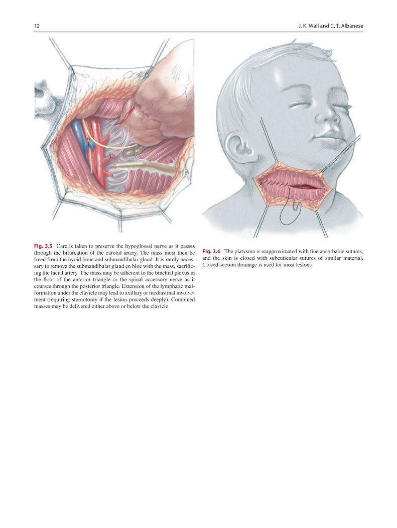
12
Fig. 3.5 Care is taken to preserve the hypoglossal nerve as it passes through the bifurcation of the carotid artery. The mass must then be freed from the hyoid bone and submandibular gland. It is rarely neces-sary to remove the submandibular gland en bloc with the mass, sacrific-ing the facial artery. The mass may be adherent to the brachial plexus in the floor of the anterior triangle or the spinal accessory nerve as it courses through the posterior triangle. Extension of the lymphatic mal-formation under the clavicle may lead to axillary or mediastinal involve-ment (requiring sternotomy if the lesion proceeds deeply). Combined masses may be delivered either above or below the clavicle
Fig. 3.6 The platysma is reapproximated with fine absorbable sutures, and the skin is closed with subcuticular sutures of similar material. Closed suction drainage is used for most lesions
J. K. Wall and C. T. Albanese

13
3.4 Postoperative Care and Complications
Feeding resumes when the infant is awake and alert. Extensive intraoral dissection may temporarily impair swal-lowing and delay the onset of oral feeds. Drain removal may take days or weeks and is dictated by the daily drainage vol-ume. Antibiotics are administered daily for 1 to 3 days.
In cases of partial resection, recurrence typically occurs within a year of surgery. Lymph leaks and nerve injuries are minimized by the use of bipolar diathermy. Rarely, lymph leaks may require re-exploration when drains are inadequate or are removed early.
Suggested Reading
Adams DM, Trenor III CC, Hammill AM, et al. Efficacy and safety of Sirolimus in the treatment of complicated vascular anomalies. Pediatrics 2016;137:e20153257
Azizkhan RG. Complex vascular anomalies. Pediatr Surg Int. 2013;29:1023–38.
Charabi B, Bretlau P, Bille M, Holmelund M. Cystic hygroma of the head and neck--long-term follow up of 44 cases. Acta Otolaryngol Suppl. 2000;543:248–50.
Lackner H, Karastaneva A, Schwinger W, et al. Sirolimus for the treat-ment of children with various complicated vascular anomalies. Eur J Pediatr 2015;174:1579–1584
Lazar DA, Olutoye OO, Moise KJ Jr, Ivey RT, Johnson A, Ayres N, et al. Ex-utero intrapartum treatment procedure for giant neck masses--fetal and maternal outcomes. J Pediatr Surg. 2011;46:817–22.
Nehra D, Jacobson L, Barnes P, Mallory B, Albanese CT, Sylvester KG. Doxycycline sclerotherapy as primary treatment of head and neck lymphatic malformations in children. J Pediatr Surg. 2008;43:451–60.
Swetman GL, Berk DR, Vasanawala SS, Feinstein JA, Lane AT, Bruckner AL. Sildenafil for severe lymphatic malformations [Case Reports Letter]. N Engl J Med. 2012;366:384–6.
3 Lymphatic Malformations

15© Springer-Verlag GmbH Germany, part of Springer Nature 2019P. Puri, M. E. Höllwarth (eds.), Pediatric Surgery, Springer Surgery Atlas Series, https://doi.org/10.1007/978-3-662-56282-6_4
Tracheostomy
Andrew Dias, Sheila Hayes, Siobhan Fitzgerald, and John Russell
4.1 Indications and Anatomic Considerations
The indications for paediatric tracheostomies are divided into three main categories: upper airway obstruction, assisted ventilation, and pulmonary toilet. The most common indica-tions have changed over the years, from upper airway obstruction secondary to infectious disorders to the current most common indication of prolonged ventilation. Paediatric tracheostomies place a significant amount of stress on the child and the carers, making social and verbal development more challenging, so alternatives to tracheostomy should be explored first. Currently, the indication to tracheotomise a child is generally ruled by the anticipation of long-term car-diopulmonary compromise or by the presence of a fixed upper airway obstruction that is unlikely to resolve for a sig-nificant period. The goal is an orderly, well-timed procedure with an experienced surgeon and the best personnel and equipment possible.
Basic anatomic differences from an adult neck should be borne in mind. The dome of the pleura extends into the neck and is thus vulnerable to injury. The trachea is pliable and can be difficult to palpate. It can be easily retracted to a great extent with little pull, and care must be taken to distinguish it from the carotid vessels. The paediatric neck is short, so the operative field is confined. Finally, the cricoid can be injured if it is not correctly identified.
4.2 Preoperative Planning
A key element of planning for tracheostomy is selection of the appropriate tube size. The tube should be small enough to allow the child to verbalise but not so small that insufflation leaks cause hypoventilation. Both diameter and length should be considered. Using a tube with too large a diameter may injure the tracheal mucosa by compromising its vascular supply. The tube should be long enough to allow adequate air entry with easy suctioning and clearance of secretions. A tube that is too short may result in accidental decannulation or formation of a false passage. If the tube is too long, the end may damage the carina or lie within the right main bron-chus, thereby occluding the left bronchus. Paediatric trache-ostomy tubes are cuffless except in larger children and adolescents. The diameter of the tracheostomy tube can be estimated on the basis of the size (corresponding to the inner diameter) of the child’s endotracheal tube.
Most tracheostomies are performed under general anaes-thesia (except in an absolute emergency), with an intubated patient. The patient’s neck is extended with a shoulder roll, and the head is stabilized with a ring under the occiput.
4
A. Dias · S. Hayes · S. Fitzgerald National Children’s Research Centre, Our Lady’s Children’s Hospital, Crumlin, Dublin, Irelande-mail: [email protected]
J. Russell (*) Our Lady’s Children’s Hospital, Crumlin, Dublin, Ireland

16
4.3 Operative Procedure
Figures 4.1, 4.2, 4.3, 4.4, 4.5, and 4.6 illustrate the steps in performing a successful paediatric tracheostomy.
Fig. 4.1 The tracheostomy begins with a horizontal incision after local anaesthetic with adrenaline for infiltration is administered midway between the cricoid cartilage and the sternal notch. The incision is extended to the subcutaneous tissues and platysma muscle (superficial cervical fascia). Excess subcutaneous fat is removed with bipolar cau-tery. Right-angled retractors better expose the operative site. Two atrau-matic forceps are used to grasp the investing layer of deep cervical fascia on either side of the midline, which is opened vertically. Next, the strap muscles enclosed in the muscular part of the pretracheal layer of deep cervical fascia are divided along the midline raphe. The visceral part of the pretracheal layer of deep cervical fascia is exposed and divided with bipolar cautery to expose the thyroid isthmus. The isthmus is either divided with bipolar cautery or, if possible, retracted superi-orly. It is imperative to keep the operative field dry at all times to avoid complications
Fig. 4.2 The trachea is now exposed. The proposed tracheostomy can-nula should be opened and its outer diameter visually checked against the exposed trachea. The pretracheal fascia is scored vertically with bipolar cautery to coagulate any vessels on the trachea in the midline. A suture of 4/0 nonabsorbable monofilament or its equivalent is placed on either side of the scored midline anterior trachea. Each suture incorpo-rates one or two tracheal rings. These sutures are not tied to the tracheal wall but at their ends; they are left 6–8 cm in length. On completion of the case, these sutures are labelled and taped securely to the anterior chest wall, so they can be used to locate the tracheal incision in the event of tracheal cannula dislodgment. These sutures have the added benefit of holding open the edges of the tracheal incision for ease of placement of the tracheostomy cannula at operation
A. Dias et al.


