This case study describes the management of a prenatally diagnosed lethal skeletal dysplasia, the perinatal care rendered, in conjunction with a review of literature.
Case reportA 39 year old Gravida 5 Para 3013 presented to our sonogram unit
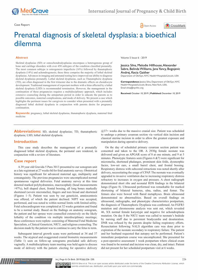
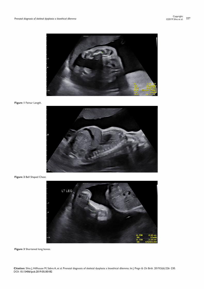
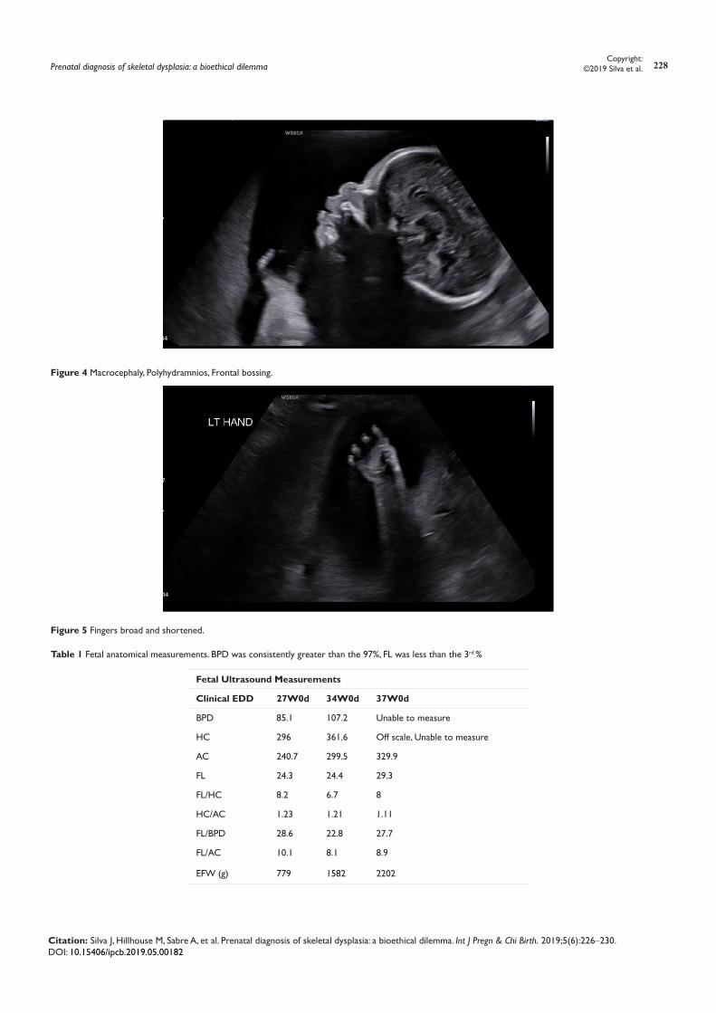
as a late registrant at 27 weeks 0d for fetal anatomic survey. Obstetrical history was significant for advanced maternal age, multiparity and consanguinity. The previous pregnancies were uncomplicated normal spontaneous vaginal deliveries. Fetal anatomy survey at this time denoted marked polyhydramnios, macrocephaly (head measurements >97%), bell shaped chest, frontal bossing, all long bones markedly shortened (severe micromelia), fingers and toes broad and shortened (Figures 1–5). Patient was seen by our geneticist, amniocentesis was offered, of which the patient declined. NIPT was accepted, performed, and was noted to within normal limits with limited utility. Fetal echocardiogram was completed at 32 weeks gestation and found to be a normal study. Based on the findings on anatomy sonogram, the patient and her spouse were counselled extensively on the likely lethality of the condition via multiple interdisciplinary meetings; these conferences were replete- consisting of Maternal Fetal Medicine Specialists, a Clinical Geneticist, NICU, and religious counseling. The decision made by the patient was to continue to carry the fetus to term.
Subsequent interval growth scans were performed at 34 and 37 weeks. The atypical and exaggerated cranial biometric measurements (Table 1) seen on follow-up sonograms precluded safe delivery vaginally. A multidisciplinary team meeting was held again to discuss mode of delivery with the patient- electing for operative delivery
@37+ weeks due to the massive cranial size. Patient was scheduled to undergo a primary cesarean section via vertical skin incision and classical uterine incision in order to allow the greatest exposure and manipulation during operative delivery.
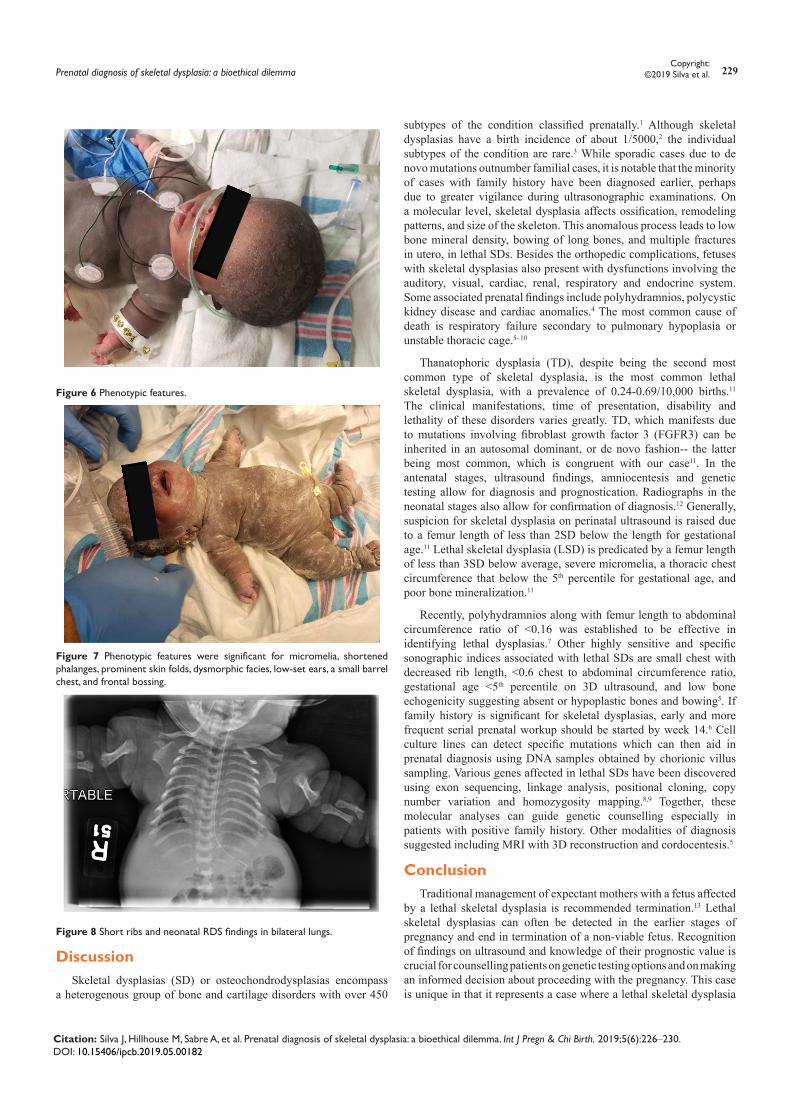
On the day of scheduled primary cesarean section patient was consented and taken to the OR. A 2930g female neonate was delivered and given an APGAR score of 8 at one minute, and 9 at 5 minutes. Phenotypic features seen (Figures 6 &7) were significant for micromelia, shortened phalanges, prominent skin folds, dysmorphic facies, low-set ears, a small barrel chest, and frontal bossing. Respiratory distress with subcostal retractions was noted shortly after delivery, necessitating the usage of CPAP. The neonate was eventually upgraded to invasive ventilation due to increasing respiratory distress refractory to increases in oxygen and pressure. A chest radiograph demonstrated short ribs and neonatal RDS findings in the bilateral lungs (Figure 8). Ultrasound performed was remarkable for marked shortening of bilateral humerus, ulna, radius, and femur. The femurs also were bowed with flared metaphyses. Brain ultrasound demonstrated no abnormalities. Based on overall findings on ultrasound, radiographs, and phenotypic characteristics postpartum, the diagnosis of Thanotophoric Dysplasia was confirmed. An FGFR3 mutation and chromosome analysis was sent out, which revealed 46, XX normal female karyotype and negative for achondroplasia mutation. On day 8 the NICU team was called to neonate’s bedside by nursing staff due to persistent bradycardia and desaturation. DNR was refused by the parents despite failing respiratory efforts. Resuscitation following NALS algorithm care was done prior to expiration of the neonate secondary to respiratory failure. The patient and her husband requested that autopsy not be performed. Patient’s immediate postpartum course was uncomplicated. She presented for a post-operative assessment 1 week postpartum where clinical exam was found to be normal and incision was clean, dry, and intact. Patient did not present for her scheduled postpartum visit at 6 weeks.
Prenatal diagnosis of skeletal dysplasia: a bioethical dilemma
Volume 5 Issue 6 - 2019
Jessica Silva, Melodie Hillhouse, Alexander Sabre, Belinda Williams, Jana Yancy, Bogojevic Andrej, Kecia GaitherDepartment of Ob/Gyn, NYC Health+Hospitals/Lincoln, USA
Correspondence: Jessica Silva, Department of Ob/Gyn, NYC Health+Hospitals/Lincoln, Bronx, New York, USA, Email
Received: October 10, 2019 | Published: December 10, 2019
Abstract
Skeletal dysplasias (SD) or osteochondrodysplasias encompass a heterogenous group of bone and cartilage disorders with over 450 subtypes of the condition classified prenatally. The most common subtype is osteogenesis imperfecta (16%) followed by thanatophoric dysplasia (14%) and achondrogenesis; these three comprise the majority of lethal skeletal dysplasias. Advances in imaging and antenatal testing have improved our ability to diagnose skeletal dysplasias prenatally. Lethal skeletal dysplasias, such as Thanatophoric dysplasia (TD), are often diagnosed in the first trimester due to the dramatic effects on chondrocyte development. Traditional management of expectant mothers with a fetus affected by a lethal skeletal dysplasia (LSD) is recommended termination. However, the management in the continuation of these pregnancies requires a multidisciplinary approach, which includes extensive counseling during the antepartum period in order to educate the parents as to possible outcomes, maternal complications, and mode of delivery. We present a case which highlights the pertinent issues for caregivers to consider when presented with a prenatally diagnosed lethal skeletal dysplasia in conjunction with parents desire for pregnancy continuation.
Keywords: pregnancy, lethal skeletal dysplasias, thanatophoric dysplasia, maternal fetal medicine
Citation: Silva J, Hillhouse M, Sabre A, et al. Prenatal diagnosis of skeletal dysplasia: a bioethical dilemma. Int J Pregn & Chi Birth. 2019;5(6):226‒230. DOI: 10.15406/ipcb.2019.05.00182
Citation: Silva J, Hillhouse M, Sabre A, et al. Prenatal diagnosis of skeletal dysplasia: a bioethical dilemma. Int J Pregn & Chi Birth. 2019;5(6):226‒230. DOI: 10.15406/ipcb.2019.05.00182
Citation: Silva J, Hillhouse M, Sabre A, et al. Prenatal diagnosis of skeletal dysplasia: a bioethical dilemma. Int J Pregn & Chi Birth. 2019;5(6):226‒230. DOI: 10.15406/ipcb.2019.05.00182
Figure 6 Phenotypic features.
Figure 7 Phenotypic features were significant for micromelia, shortened phalanges, prominent skin folds, dysmorphic facies, low-set ears, a small barrel chest, and frontal bossing.
Figure 8 Short ribs and neonatal RDS findings in bilateral lungs.
DiscussionSkeletal dysplasias (SD) or osteochondrodysplasias encompass
a heterogenous group of bone and cartilage disorders with over 450
subtypes of the condition classified prenatally.1 Although skeletal dysplasias have a birth incidence of about 1/5000,2 the individual subtypes of the condition are rare.3 While sporadic cases due to de novo mutations outnumber familial cases, it is notable that the minority of cases with family history have been diagnosed earlier, perhaps due to greater vigilance during ultrasonographic examinations. On a molecular level, skeletal dysplasia affects ossification, remodeling patterns, and size of the skeleton. This anomalous process leads to low bone mineral density, bowing of long bones, and multiple fractures in utero, in lethal SDs. Besides the orthopedic complications, fetuses with skeletal dysplasias also present with dysfunctions involving the auditory, visual, cardiac, renal, respiratory and endocrine system. Some associated prenatal findings include polyhydramnios, polycystic kidney disease and cardiac anomalies.4 The most common cause of death is respiratory failure secondary to pulmonary hypoplasia or unstable thoracic cage.5–10
Thanatophoric dysplasia (TD), despite being the second most common type of skeletal dysplasia, is the most common lethal skeletal dysplasia, with a prevalence of 0.24-0.69/10,000 births.11 The clinical manifestations, time of presentation, disability and lethality of these disorders varies greatly. TD, which manifests due to mutations involving fibroblast growth factor 3 (FGFR3) can be inherited in an autosomal dominant, or de novo fashion-- the latter being most common, which is congruent with our case11. In the antenatal stages, ultrasound findings, amniocentesis and genetic testing allow for diagnosis and prognostication. Radiographs in the neonatal stages also allow for confirmation of diagnosis.12 Generally, suspicion for skeletal dysplasia on perinatal ultrasound is raised due to a femur length of less than 2SD below the length for gestational age.11 Lethal skeletal dysplasia (LSD) is predicated by a femur length of less than 3SD below average, severe micromelia, a thoracic chest circumference that below the 5th percentile for gestational age, and poor bone mineralization.11
Recently, polyhydramnios along with femur length to abdominal circumference ratio of <0.16 was established to be effective in identifying lethal dysplasias.7 Other highly sensitive and specific sonographic indices associated with lethal SDs are small chest with decreased rib length, <0.6 chest to abdominal circumference ratio, gestational age <5th percentile on 3D ultrasound, and low bone echogenicity suggesting absent or hypoplastic bones and bowing5. If family history is significant for skeletal dysplasias, early and more frequent serial prenatal workup should be started by week 14.6 Cell culture lines can detect specific mutations which can then aid in prenatal diagnosis using DNA samples obtained by chorionic villus sampling. Various genes affected in lethal SDs have been discovered using exon sequencing, linkage analysis, positional cloning, copy number variation and homozygosity mapping.8,9 Together, these molecular analyses can guide genetic counselling especially in patients with positive family history. Other modalities of diagnosis suggested including MRI with 3D reconstruction and cordocentesis.5
ConclusionTraditional management of expectant mothers with a fetus affected
by a lethal skeletal dysplasia is recommended termination.13 Lethal skeletal dysplasias can often be detected in the earlier stages of pregnancy and end in termination of a non-viable fetus. Recognition of findings on ultrasound and knowledge of their prognostic value is crucial for counselling patients on genetic testing options and on making an informed decision about proceeding with the pregnancy. This case is unique in that it represents a case where a lethal skeletal dysplasia
Citation: Silva J, Hillhouse M, Sabre A, et al. Prenatal diagnosis of skeletal dysplasia: a bioethical dilemma. Int J Pregn & Chi Birth. 2019;5(6):226‒230. DOI: 10.15406/ipcb.2019.05.00182
was brought to term and delivered due to the decision of patient and her spouse. In cases such as these, continuous assessment to verify viability, monitoring for maternal respiratory compromise ( due to massive polyhydramnios) and performing a cesarean delivery at a stage that affords a lessened likelihood of operative complications are of paramount importance.13 A straightforward discussion concerning expectations for a child born with a lethal skeletal dysplasia must be initiated early on and in a setting with multiple specialties in order to allow for an adequate and comprehensive informed decision-making process. Careful consideration of explanations of the disorder, initiation of plans for the neonate , as care transfers from one specialty to another, and any possible risk factors for future pregnancies must be held with the patient-- in preferred language, and in terms the patient can comprehend. It is incumbent on providers to be knowledgeable in the nuances of care for patients so affected by this clinical conundrum.
AcknowledgementsNone.
Conflicts of interestAuthor declares there are no conflicts of interest.
FundingNone.
References1. Orioli IM, Castilla EE, Barbosa-Neto JG. The birth prevalence rates for
the skeletal dysplasias. J Med Genet. 1986;23(4):32–32.
2. Liangcheng Wang, Yasushi Takai, Kazunori Baba, et al. Can biparietal diameter-to-femur length ratio be a useful sonographic marker for screening thanatophoric dysplasia since the first trimester? A literature review of case reports and a retrospective study based on 10,293 routine fetal biometry measurements. Taiwan J Obstet Gynecol. 2017;56(3):374–378.
3. Krakow D, Rimoin DL. The skeletal dysplasias. Genetics in Medicine. 2010;12:327–341.
4. Savoldi A, Villar M, Machado H, et al. Fetal Skeletal Lethal Dysplasia: Case Report. Rev Bras Ginecol Obstet. 2017;39(10):576–582.
5. Milks KS, Hill LM, Hosseinzadeh K. Evaluating skeletal dysplasias on prenatal ultrasound: an emphasis on predicting lethality. Pediatr Radiol. 2017;47(2):134–145.
6. Sharony R, Browne C, Lachman RS, et al. Prenatal diagnosis of the skeletal dysplasias. Am J Obstet Gynecol. 1993;169(3):668–675.
7. Nelson DB, Dashe JS, McIntire DD, et al. Fetal Skeletal Dysplasias-sonographic indices associated with adverse outcomes. J Ultrasound Med. 2014;33(6):1085–1090.
8. Chitty LS, Mason S, Barrett AN, et al. Non-invasive prenatal diagnosis of achondroplasia and thanatophoric dysplasia: next-generation sequencing allows for a safer, more accurate, and comprehensive approach. Prenatal Diagnosis. 2015;35(7):656–662.
9. Dan S, Yuan Y, Wang Y, et al. Non-Invasive Prenatal Diagnosis of Lethal Skeletal Dysplasia by Targeted Capture Sequencing of Maternal Plasma. Plos One. 2016;11(7).
10. Bonafe L, Cormier-Daire V, Hall C, et al. Nosology and classification of genetic skeletal disorders: 2015 revision. Am J Med Genet A. 2015;167A:2869–2892.
11. Sharma M, Jyoti, Jain R, et al. Thanatophoric Dysplasia: A Case Report. Journal of clinical and diagnostic research. 2015;9(11):QD01–3.
12. Panda A, Gamanagatti S, Jana M, et al. Skeletal dysplasias: A radiographic approach and review of common non-lethal skeletal dysplasia. World J Radiol. 2014;6(10):808–825.
13. Karczeski B, Cutting GR. Thanatophoric Dysplasia. Seattle (WA): University of Washington, Seattle; 1993–2019.