22
Go to Section: Chapter 36 – The Skeletal, Muscular and Integumentary Systems Section 35-1

Go to Section:
Chapter 36 –
The Skeletal, Muscular and Integumentary Systems
Section 35-1

Go to Section:
36–1 The Skeletal System
A. The Skeleton
B. Structure of Bones
C. Development of Bones
D. Types of Joints
1. Immovable Joints
2. Slightly Movable Joints
3. Freely Movable Joints
E. Structure of Joints
F. Skeletal System Disorders
Section Outline
Section 36-1

Go to Section:
Skull
Sternum
Ribs
Vertebral
column
Metatarsals
Metacarpals
Phalanges
Clavicle
Scapula
Humerus
Radius Pelvis
Ulna
Carpals
Femur
Patella
Fibula
Tibia
Tarsals
Phalanges
The Skeletal System
Section 36-1
Axial
Skeleton
Appendicular
Skeleton
Go to Section:
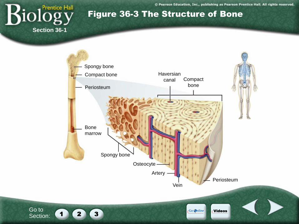
Spongy bone
Compact bone
Periosteum
Bone
marrow
Haversian
canal Compact
bone
Spongy bone
Osteocyte
Artery
Vein Periosteum
Figure 36-3 The Structure of Bone
Section 36-1
Go to Section:
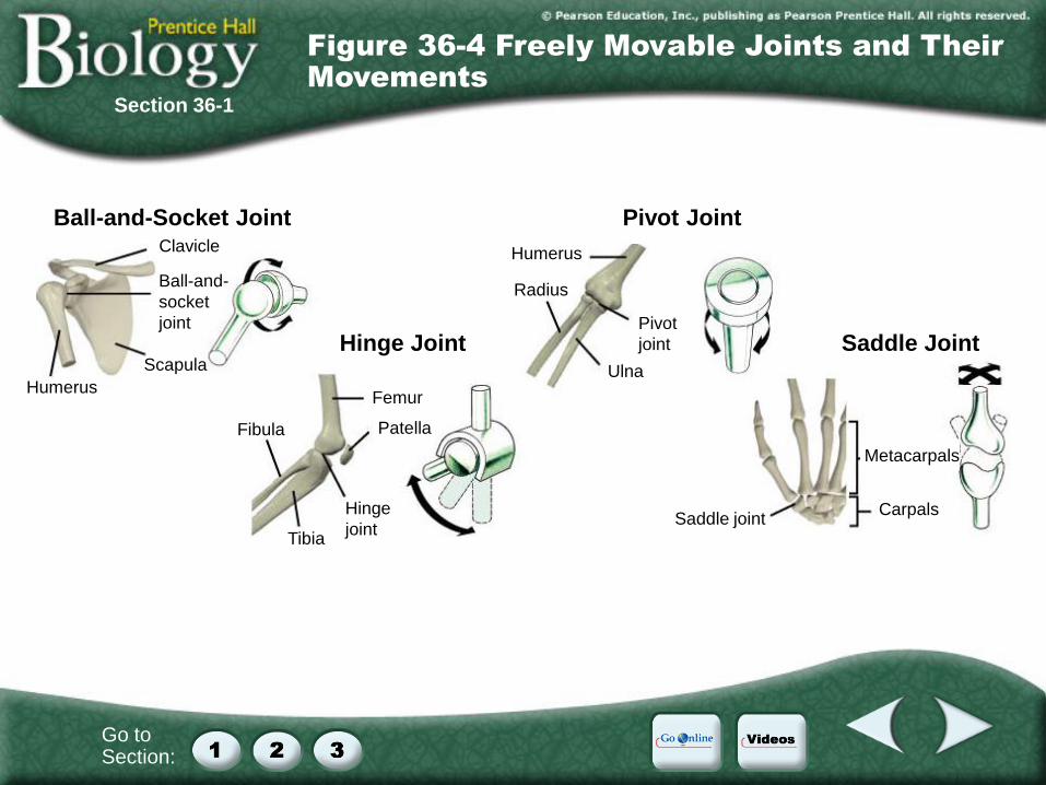
Ball-and-Socket Joint
Hinge Joint
Pivot Joint
Saddle Joint
Clavicle
Ball-and-
socket
joint
Scapula
Humerus Femur
Patella
Hinge
joint Tibia
Fibula
Humerus
Radius
Pivot
joint
Ulna
Metacarpals
Carpals Saddle joint
Figure 36-4 Freely Movable Joints and Their
Movements
Section 36-1

Go to Section:
Muscle
Tendon
Femur
Patella
Bursa
Ligament
Synovial fluid
Cartilage
Fat
Fibula
Tibia
Figure 36-5 Knee Joint
Section 36-1

Go to Section:
36–2 The Muscular System
A. Types of Muscle Tissue
1. Skeletal Muscles
2. Smooth Muscles
3. Cardiac Muscle
B. Muscle Contraction
C. Control of Muscle Contraction
D. How Muscles and Bones Interact
E. Exercise and Health
Section Outline
Section 36-2

Go to Section:
Actin pulled Cross-bridge
releases actic
Cross-bridge
changes
shape
Myosin returns
to original
shape
Myosin forms
cross-bridge
with actin
2
1
3 4
5
Cycle Diagram
Section 36-2

Go to Section:
Figure 36-7 Skeletal Muscle Structure
Section 36-2
Go to Section:
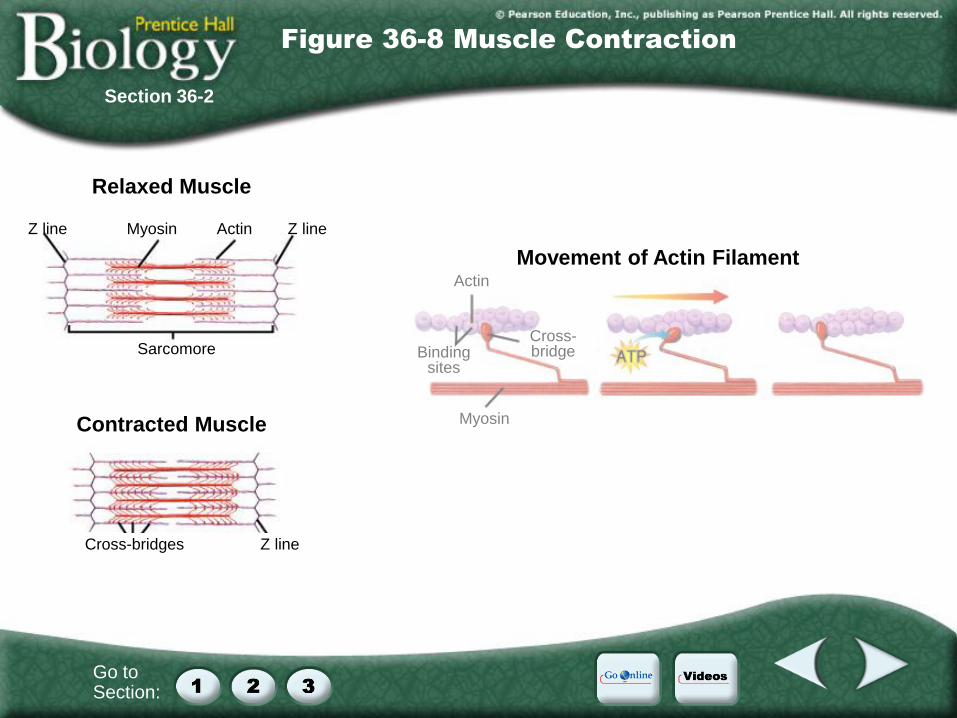
Relaxed Muscle
Contracted Muscle
Z line Myosin Actin Z line
Sarcomore
Cross-bridges Z line
Movement of Actin Filament Actin
Binding sites
Cross-bridge
Myosin
Figure 36-8 Muscle Contraction
Section 36-2

Go to Section:
Relaxed Muscle
Contracted Muscle
Z line Myosin Actin Z line
Sarcomore
Cross-bridges Z line
Movement of Actin Filament Actin
Binding sites
Cross-bridge
Myosin
Figure 36-8 Muscle Contraction
Section 36-2
During muscle contraction, the knoblike head of a myosin filament attaches to a binding site on actin, forming a cross-bridge.
Powered by ATP, the myosin cross-bridge changes shape and pulls the actin filament toward the center of the sarcomere.
The cross-bridge is broken, the myosin binds to another site on the actin filament, and the cycle begins again.

Go to Section:
Movement Movement
Biceps (relaxed)
Triceps (relaxed)
Biceps (contracted)
Triceps (relaxed)
Figure 36-11 Opposing Muscle Pairs
Section 36-2

Go to Section:
36–3 The Integumentary System
A. The Skin
1. Epidermis
2. Dermis
3. Skin Cancer
B. Hair and Nails
1. Hair
2. Nails
Section Outline
Section 36-3

Go to Section:
Skin
Barrier to
infection
Regulator of
body
temperature
Remover of
waste products
Protector
against UV
radiation
Epidermis Dermis
Outer layer Inner layer
functions
as a
is made
up of the
which
is the
which
is the
Concept Map
Section 36-3

Go to Section:
Figure 36-13 The Structure of Skin
Section 36-3

Video Contents
Videos
Click a hyperlink to choose a video.
Muscle Contraction, Part 1
Muscle Contraction, Part 2

Video 1
Click the image to play the video segment.
Video 1
Muscle Contraction, Part 1

Video 2
Click the image to play the video segment.
Video 2
Muscle Contraction, Part 2

Internet
Go Online
Links from the authors on making artificial skin
Interactive test
For links on bones and joints, go to www.SciLinks.org and enter the Web Code as follows: cbn-0361.
For links on muscle contraction, go to www.SciLinks.org and enter the Web Code as follows: cbn-0362.

End of Custom Shows
This slide is intentionally blank.


