Journal of Metals, Materials and Minerals. Vol. 12 No. 1 pp. 39-50 , 2002. Heat Effect on Viscosity and Curing of Light-Cured Dental Resin and Mechanical Strength of Conventional Dental Composites Piyanart EKWORAPOJ 1 , Rathanawan MAGARAPHAN 1* and David C. MARTIN 2 1 The Petroleum and Petrochemical College, Chulalongkorn University. 2 Department of Material Science and Engineering, University of Michigan, USA. Abstract Dimethacrylate polymers and silica are most commonly used as matrix resins and fillers for dental composites. Bis-GMA, UDMA are used as base monomers and TEGDMA as a diluent monomer for dental resin preparation. Pure UDMA, mixtures of UDMA:TEGDMA and Bis-GMA:TEGDMA 50:50 and 75:25 were mixed with silanized silica at compositions of monomer:fillers 30:70 and 40:60 (weight ratio). Heat curing of the monomers was studied by DSC and showed that temperature did not have an affect on curing from room temperature to 80°C. The viscosity of the monomers decreased as temperature increased; i.e. by the change of 50°C viscosity reduced to 3-5% of original values at 30°C. There was no significant difference in diametral tensile strength (DTS) of the dental composites having a monomer mixture of 50:50 and 75:25. Stiff monomer (Bis-GMA) shows a higher DTS and Vicker hardness than the flexible one. However, filler content is more prominent in controlling DTS and Vicker hardness than monomer type and ratio. The prepared dental composites showed lower DTS and hardness than the commercial ones. Keywords : Dental Material, Dental Composites, Silanized Silica, Hardness *To whom correspondence should be addressed 39
Transcript
Journal of Metals, Materials and Minerals. Vol. 12 No. 1 pp. 39-50 , 2002.
Heat Effect on Viscosity and Curing of Light-Cured Dental Resin and Mechanical Strength of Conventional Dental Composites
Piyanart EKWORAPOJ1, Rathanawan MAGARAPHAN1* and David C. MARTIN2 1The Petroleum and Petrochemical College, Chulalongkorn University.
2Department of Material Science and Engineering, University of Michigan, USA.
Abstract
Dimethacrylate polymers and silica are most commonly used as matrix resins and fillers for dental composites. Bis-GMA, UDMA are used as base monomers and TEGDMA as a diluent monomer for dental resin preparation. Pure UDMA, mixtures of UDMA:TEGDMA and Bis-GMA:TEGDMA 50:50 and 75:25 were mixed with silanized silica at compositions of monomer:fillers 30:70 and 40:60 (weight ratio). Heat curing of the monomers was studied by DSC and showed that temperature did not have an affect on curing from room temperature to 80°C. The viscosity of the monomers decreased as temperature increased; i.e. by the change of 50°C viscosity reduced to 3-5% of original values at 30°C. There was no significant difference in diametral tensile strength (DTS) of the dental composites having a monomer mixture of 50:50 and 75:25. Stiff monomer (Bis-GMA) shows a higher DTS and Vicker hardness than the flexible one. However, filler content is more prominent in controlling DTS and Vicker hardness than monomer type and ratio. The prepared dental composites showed lower DTS and hardness than the commercial ones. Keywords : Dental Material, Dental Composites, Silanized Silica, Hardness *To whom correspondence should be addressed
39
Heat Effect on Viscosity and Curing of Light-Cured Dental Resin and Mechanical Strength
Plastic filling materials or dental resin composites become the materials of choice selected by dentists for replacing the lost hard tissue of teeth. Dental composites are widely used as a restorative material in the substitution of metallic filling materials such as amalgam. Dental composites are composed of a polymer or matrix resin and a rigid mineral filler. There are several requirements for filling materials; for example, strong compressive and flexural strength, low shrinkage, good aesthetics, and good resistance to temperature and pH. The conventional dental composites or macrofilled dental composites usually consist of polymer binders (which are typically derived from visible light photopolymerization of dimethacrylate monomer), glass or ceramic fillers, and adhesive or coupling agents. The ceramic filler is used to give a high load bearing (strength) and varied around 60-80 %wt or 60-70 %volume. This material matches the color of the tooth but has less strength than metallic filling materials such as amalgam (Leinfelder, 1989). Other disadvantages are high polymerization shrinkage, low resistance to abrasion, and low adhesion to tooth structures. These lead to clinical failure. The filler particles incorporated in traditional or conventional dental composites are typically macrofillers with are mechanically ground or crushed from larger pieces of purely inorganic materials such as quartz, glass, borosilicate, or other ceramics (with irregular shape and sizes ranging from 1-100 µm) to smaller, softer and more rounded macrofillers with sizes ranging from 1-5 µm. Other types of fillers are microfillers (average size 0.04 µm or 0.05 µm) and hybrids or blended fillers (typically colloidal silica and larger fillers). Roulet, et al. (1978) reported that hybrids contain particles with an average size of 0.8-1.0 µm. To induce matrix/filler interaction, the filler is usually impregnated in the surface with a bifunctional coupling agent, e.g. silane coupling agents (Arcis, et al. 2002).
Polymer binders, used for filler embedding, are usually epoxy or acrylate types. These particular polymers are good adhesives which provide crosslinking to enhance strength and polarity
compatible with ceramic fillers. Rigid chemical structures of a monomer often possesses high viscosity in such a way that handling is very difficult. Practically, more than one type of monomer, e.g. one high viscosity and one low viscosity, is mixed at various ratios to bring about suitable viscosity (easy to use). The effect of monomer type and ratio on polymer conversion (and thus on strength of the composite) were reported by several scientists such as Stanbury, et al. (2001ab), Vaidyanathan, et al. (1992), McCabe (1985), Imazato (2001), and Asmussen, et al. (1998). The usable range of conversion found in commercial dental composites is 55-75%. The conversion increases with less content of the high viscosity monomer. Different monomer exhibit different reactivity and the lower viscosity one tends to be more reactive. Photopolymerization kinetics depends on the viscosity of the dental base monomers. From the study of Stanbury and Dickens (2001ab) Bis-GMA (bisphenol A bis [2-hydroxy-3-methacryloxypropyl]ether) showed the maximum polymerization rate at 10% conversion. However, UDMA/TEGDMA (urethane dimethacrylate/triethylene-glycol dimethacrylate or UT) resin reached a higher rate of polymerization at a higher degree of conversion than that of the Bis-GMA resin. This is due to a lower monomeric glass transition temperature (-81.7, -41.7 and -6.6°C for TEGDMA, UDMA and Bis-GMA, respectively) and a lower viscosity of the uncured UT resin than those of the Bis-GMA/TEGDMA (BT) resin. Nevertheless, Stanbury and Dickens (2001b) found that resin temperature was raised after light curing. The heat possibly has an affect on the curing condition. Moreover, most work has been emphasized on the effect of monomer type and ratio on the conversion but not many on the final mechanical properties. Zhao, et al. (1997), and Johnston, et al. (1994) found brittle failure for dental composites; however, the fracture toughness of the composites (75-84 %wt mixed filler) was larger than that of the pure resin. This depended on the notching type, test method, and sample shape. The mechanism of the failure is thought to involve the viscoelasticity of the resin, increased fracture surface area and crack length to enhance the fracture toughness but shorten the particle distance
Heat Effect on Viscosity and Curing of Light-Cured Dental Resin and Mechanical Strength
of Conventional Dental Composites
41
inhibiting plastic deformation and lower fracture strength.
In this work, the factors which influence the mechanical properties of dental composites were studied, e.g. monomer handling and filler properties. The conventional composites were made of a macrofiller such as silanized silica (silane treatment on silica surface) and acrylate-polymers. There were three parts to the work. First, the heat effect on the monomer that may contribute to enhance the degree of curing and mixing so thus mechanical strength can be improved. The viscosity of mixed monomers was determined at various temperatures to investigate the heating effect that could influence additional curing. Second, characterization of silanized silica was investigated for its size, chemistry, and morphological structure. Third, this study focuses on the effect of monomer type (Bis-GMA, UDMA, and TEGDMA) and ratio (50:50 and 75:25 %wt) and filler content of 60-70 %wt on the mechanical properties (diametral tensile strength and hardness) of the dental composites made at a fixed curing condition. The mechanical properties of the
prepared conventional composites were also compared with the commercial available ones.
Materials and Methods
The monomers employed in this study were Bisphenol A bis(2-hydroxy-3-methacryloxypropyl)ether (Bis-GMA) and urethanedimethacrylate (UDMA or UD) were supplied by Esstech Co. (Essington, PA) and were used as received. Triethyleneglycol dimethacrylate (TEGDMA), camphorquinone, dimethylaminoethyl methacrylate (DMAEMA) were purchased from Fluka, Switzerland. A commercial silanated Lantan-Alumino-Silicate glass ceramic with a high radiopacity (4.4 mm Al-eq.-thick) for dental applications, glass powder number GM31684, were obtained from Schott Glass, Germany and used as received. It has a high density of 2.89 g/cm3, water stability in class 1, low thermal expansion coefficient from 30 to 250°C of 0 ppm/K, and refractive index 1.579. Commercial dental composites, Amelogen® and Aelite fil®, were manufactured by Bisco (USA) and supplied by Nudent Co., Ltd. Thailand. The structure of the monomer is shown in Figure 1.
Base monomer
Bis-GMA
UDMA Diluent monomer
TEGDMA
Figure 1 Structure of monomers used
EKWORAPOJ, P. et al.
42
Dental resin preparation
Dental resins were prepared by mixing the base monomer and comonomer in the ratio of 75/25 and 50/50 by %wt using a four-twisted blade turbine on ALC-sL mixer head (variable speed upto 1500 rpm). They were activated for visible light photopolymerization by the addition of 0.2 %wt camphorquinone and 0.8 %wt dimethylaminoethyl methacrylate (initiator weight ratio = 1:4). Prepolymerized dental resin was blended with silanized silica by the mixer at 500 rpm for 20 minutes at room temperature.
Viscosity measurement
The various ratios of dental resins were poured into the sample chamber of the Brookfield viscometer model RVDV- III, with a small sample adapter (SSA 21/13 R) and a water bath. The measurement was performed from 30°C to 80°C by controlling the temperature in the water bath. The sample was left until reaching the required temperature then the viscosity value was read within 3 minutes.
Curing temperature measurement
The curing temperature of dental resins with initiators (by heat only) was determined by Perkins Elmer Different Scanning Calorimeter. Samples of 1-3 mg were put in aluminium sample pans and the heating rate used was 1°C /min from 30°C to 90°C with a nitrogen gas purge at 20 ml/min.
Microstructure studies
Filler particles were studied with a Cu K-α X-ray diffractometer Rigaku D/MAX-2000 series at 40 kV/30 mA. The standard sample holders were applied to the powder sample compared to commercial silica. The experiment was operated in the 2θ range of 5-70 degrees at a scan speed 5 degrees/min with a 0.02 degree 2θ-stepwise increment. The microstructural analyses were performed by using a JEOL 520 scanning electron microscope (SEM) at a voltage
of 10 kV and 500x magnifications. The morphology of the prepared dental composites was investigated also by SEM. Particle size was measured by Mastersizer X ver. 2.15, Malvern with a range lens of 45 mm. Fourier transform infrared (FTIR) spectroscopy was performed to obtain the chemical structure of silanized silica compared to commercial fumed silica. The samples were prepared by a pellet technique and the spectra of these fillers were determined in the wave number range of 4,000-400 cm-1 using a Bruker Equinox 55/s FTIR spectrometer.
Diametral tensile strength
The general tensile strength test method (ASTM D638) is not suitable for measuring the tensile strength of brittle materials. An alternative method, the diametral tensile strength (DTS) test was therefore utilized. DTS test specimens were prepared in a cylindrical stainless steel mold, then loaded to failure with a crosshead speed of 10 mm/min, using a 25 kN load cell. DTS was determined by the following standard formula (1)
DTS = 2f/(πdl) (1) Where f = load at failure; d = specimen diameter; l = specimen thickness
The specimens were prepared by pouring the unpolymerized dental resin into a stainless steel mold. Resin tablets (3 mm thick by 6 mm diameter) were photopolymerized between a glass slide and translucent polycarbonate strip in a dental curing unit (3M Curing Light XL3000) with a light intensity of approximately 300 mW/cm2 at approximately 470 nm measured by a radiometer for 40 seconds of each side. They were subjected to test in a Universal Testing Machine following the procedure in the American Dental Association-Specification No. 27 for Direct Filling Resins. Five specimens for each sample type were tested and their mean values were determined.
Heat Effect on Viscosity and Curing of Light-Cured Dental Resin and Mechanical Strength
of Conventional Dental Composites
43
Microhardness test
The microhardness of the sample was measured using a FM-700e Digital Microhardness tester (Future-tech Corp. Japan) with 500 gram load, dwell time 15 sec and 136° pyramid diamond indenter (Vicker Hardness scale). The specimens were prepared in the same way as the DTS specimen followed by polishing the surface with 0.3 µm slurry alpha alumina (Imptech, South Africa). The pyramidal shaped indenter applied the surface of specimens within dwell time then the force was removed. In the Vickers method, the indentation length of the vertical and horizontal axis is measured and averaged. The experiment was repeated five times for each sample set of parameters. The impression length was measured microscopically and the test load was used to calculate a hardness value.
Results and Discussion
The results in Table 1 and Figure 2 show that when the temperature increases, the viscosity of all dental resin systems decreases. The viscosity of UDMA resin is much less than that of Bis-GMA resin and when adding a diluent monomer TEGDMA, this mixture becomes less viscous. The viscosity of Bis-GMA/TEGDMA is more than that of UDMA/TEGDMA at the same monomer ratio. The activation energies incurred from the slopes of the plot of reciprocal Table 1 Viscosity properties of a prepolymerized dental resin.
aMeasure at the same speed = 80 rpm. Sheare rate = 27 sec -1
bEssetech, Certification of product analysis. UDMA viscosity = 7850 cps and Bis-GMA = 1,976,000 cps. (Brookfield Instrument)
10000
Figure 2 Plot of viscosity vs. reciprocal of temperature for UDMA and monomer mixtures of temperature vs. log viscosity for all samples are quite closed, especially those of BT50/50 (50 %wt Bis-GMA/50 %wt TEGDMA), UT75/25, and UT50/50 (21.9, 22.6, and 22.8 kJ/mole respectively). The activation energies of UDMA and BT75/25 are more close to each other (27.6 and 25 kJ/mole) and not much higher than those of the other three samples. This indicates that the viscosities of these mixtures reduce as temperature increases by almost the same manner. According to slopes in Figure 2, a change in viscosity of pure UDMA with temperature is larger than those of the mixtures of UDMA and TEGDMA. Thus UDMA is easier to handle at high temperatures.
In general, viscosity affects the quality of mixing or filler dispersion/distribution and generates heat. High viscosity can be reduced by heating, shearing (due to shear thinning effect), and mixing with low viscosity components. Most fillers show poor dispersion in the viscous materials. The higher viscosity of the Bis-GMA base resin makes itself difficult to incorporate the filler into the resin compared with the lower viscosity ones. When its viscosity is reduced by the addition of TEGDMA
1/T (°C)
UT75/25 UT50/50 UDMA
Log
Vis
cosi
ty (c
ps)
UT Regr BT75/25 1000 BT50/50 Regr
100
10
1.0030 .0028 .0032 .0034
EKWORAPOJ, P. et al.
44
or by increasing the temperature (due to irradiation), more filler content being incorporated into dental resin should be achieved. Furthermore, the polymerization reaction controlled by diffusion or chain flexibility mechanisms can occur more easily to get a higher conversion and conversion rate. Both good filler dispersion and a high conversion orhigher crosslinking of the polymer phase are expected to bring about better mechanical properties. UT75/25, BT50/50 and UT50/50 have a relatively low viscosity so their mechanical properties should be improved.
Figure 3 DSC thermogram of unfilled dental resin
For this experiment, camphorquinone is the curing agent that needs light at a wavelength of about 470 nm to initiate the polymerization. Light curing can be determined by exothermic heat at room temperature using DSC Vaidyanathan, et al. (1992), McCabe (1985), and Imazato, et al. (2001). The maximum rate of curing is affected by irradiation power. Higher power leads to a higher conversion but the relationship may not be linear. However, exposure to light can generate heat that may change the curing since irradiation heating (light induced thermal rise to 65°C) was found by Stanbury and Dickens (2001b). Moreover, heating may occur due to viscous shear during mixing. Thus, constant heating programs from room temperature to 200°C was applied to the samples. The DSC thermograms of the five dental resins in Figure 3 show no significant peak of energy change for any reaction or transition with increasing
temperature. This confirms that the dental resin cannot be thermally cured, at least over this temperature range. Thus the irradiation and viscous heat can only cause reduced viscosity but not for additional curing Silikas and Watts (1999). However, when viscosity is reduced, the monomer mobility increases (away from glass transition temperature of the polymerizing materials) such that the conversion can be enhanced McCabe (1985). This shows indirect effect of heating on curing. Besides, this information has the benefit of ensuring that the preparation of dental composite (before light curing) can be done at an elevated temperature (to reduce monomer viscosity) such that better filler uptaking (or better dispersion) is gained without premature curing.
Structure and Morphology of Silanized Silica
Silanized silica is silica that is treated with a silane coupling agent to induce a matrix/filler interaction to provide a better mechanical strength. The obtained silanized silica is characterized for its basic physical properties to ensure good adhesion and strength. The silanized silica morphology observed by SEM is shown in Figure 4. The particles are irregular in shape having a mean size
Figure 4 SEM micrograph of silanized silica of 4.23 µm as measured by a particle size analyzer which is in accordance with the size seen in the SEM micrograph. The chemical structure of the silanized silica is revealed by FTIR in Figure 5. The hydrophilic peak of hydroxyl group is typically
Heat Effect on Viscosity and Curing of Light-Cured Dental Resin and Mechanical Strength
of Conventional
0
0.2
0.4
0.6
0.8
1
1.2
1.4
1.6
1.8
2
1000 1500 2000 2500 3000 3500 4000
wave number (cm-1)
Abs
orba
nce
Uni
ts Dental Composites
45
Figures 5a FTIR of silanized silica powder (1000-4000 cm-1)
Figures 5b FTIR of silanized silica powder (1000-1300 cm-1) found on the silica surface Figure 5a is diminished with little peaks of symmetric and asymmetric -CH2 stretching in the region of 2800-3000 cm-1. Moreover, there are complex peaks, prominent in the region of 1000-1100 cm-1 Figure 5b, corresponding to silane groups or Si-O-R bonds. This indicated that there is a hydrophobic group present on the silica surface. In other words, the hydrophilic surface of the silica is modified tohave better hydrophobic properties (silanized silica). In Figure 6 shows x-ray diffraction patterns of commercial fumed and precipitated silica in comparison with silanized silica. This reveals that the commercial silicas (without surface modification) are amorphous glass while the highly radiopaque silanized silica has a crystal structure with the strongest peak at 2θ = 25.66 degrees corresponding to silica composition. The crystal structure should also bring about improved
mechanical strength which will be seen in the following section.
Si-O-R
sym-CH2 and asym-CH2
stretching
F
Fig
DiameCompo
reporteis expeet al. (200 sGMA/T(2001bBisGM
igure 6a XRD patter of silanized silica
ure 6b XRD patter of precipitated silica
Figure 6c XRD patter of fume silica
tral Tensile Strength (DTS) of Dental sites
Although the degree of conversion is not d here, the conversion of our samples cted to be within the workable range. Lovell, 1999ab) reported that using 3.0 mW/cm2 for econds, the conversion of 50/50 Bis-EGDMA was 0.60. Stanbury and Dickens
) found that a 70:30 mass ratio of A:TEGDMA gave a conversion of 0. 673 at
EKWORAPOJ, P. et al.
46
3 min after a light cure and increased to 0.689 by NIR at the seventh day after exposure to light cure for 60 seconds at an irradiation power of 65 mW/cm2. If the monomer ratio changes to 50:50, more conversion to 0.739 and 0.772, at 3 min and seventh day after light curing respectively was obtained. Chowdhury, et al. (1995) reported that dental restorative materials containing 60:40 %wtBis-GMA:TEGDMA yield have a high degree of polymerization while Shajii and Santerre (1999) reported the composition for optimal conversion was 75:25 %wt. We then selected to use a monomer ratio of 75:25 and 50:50 for a base monomer:diluent monomer and irradiated the uncured composites by a high power light source (300 mW/cm2) so a shorter cure time of 40 seconds could bring about the appropriate level of conversion.
Factors affecting the DTS such as monomer ratio, monomer type, and filler content were studied. The sample abbreviations are, for example, BT7246, UT5537, and so on. Base monomer and diluent monomer (denoted by two letters) ratios of 75:25 and 50:50 %wt are denoted by the first two numbers; i.e. 72 and 55 while the monomer:filler weight ratios were denoted by the last two numbers; i.e. 37 = 30:70 and 46 = 40:60. The volume fractions of silanized silica at 60 and 70 %wt are 0.36 and 0.465 respectively (according to the manufacturers, monomer and filler densities are about 1.08 and 2.89 g/cm3 respectively).
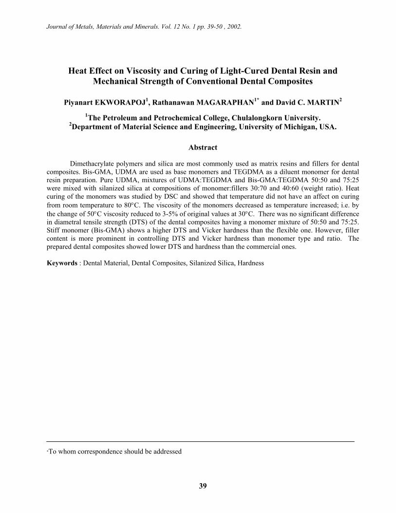
First, in the case of no filler, the monomer type and ratio (indicating different viscosity in the following sequence UDMA>BT75/25>UT75/25>BT50/50>UT50/50 clearly affect DTS as seen in Figure 7. The nature of the monomer in terms of molecular stiffness affects molecular mobility, conversion and final strength. In addition to good mixing, the combination of the effects of better diffusion-control conversion, molecular rigidity, and the capability to form networks (functionality) are all important for high strength materials. From the results, UT gives better DTS than BT due to better reactivity of UDMA than Bis-GMA since UDMA is more mobile and less rigid than
Bis-GMA. Mobility (known by low viscosity) influences diffusion-control conversion (important Figure 7 DTS of silanized silica reinforced
dental composites
at high conversion where viscosity builds up abruptly) such that it is more noticeable in UDMA than in Bis-GMA and thus leading to higher conversion. Less diluent content leads to higher DTS because TEGDMA is relatively soft or flexible and has less functionality than UDMA and Bis-GMA Stanbury and Dickens (2001a) and thus poorer network. Diametral tensile strengths of cured/unfilled dental resins are in the order of UT75/25>UT50/50>BT75/25>BT50/50. UT50/50 and BT75/25 are good samples of balance of the three effects so that they show not much different DTS. The results also indicate that monomer type has less influence on strength than monomer ratio (or viscosity). The DTS of UT75/25 is about half of the commercial ones (40-50 MPa).
However, when a filler is added at a relatively high content (60-70 %wt but still less than 50 %volume fraction), the effects of monomer type are more prominent than monomer ratio and DTS of the dental conventional composites with Bis-GMA is higher than that of UDMA. Although UT75/25 base monomer gives the highest DTS but its composites show DTS values that are not significantly different from BT75/25 composites. This implies a strong effect of the filler on the curing of monomers. Moreover, DTS is strongly
TEGDMA Content
50 BT
20 30 40 50 60 70
Dia
met
ral T
ensi
le S
tren
gth
10
20
30
40
BT30BT40UTUT30UT40AmelogenAelite
75/25
50/50
47 Heat Effect on Viscosity and Curing of Light-Cured Dental Resin and Mechanical Strength
of Conventional Dental Composites
dependent on filler content (or resin content). The filler clearly enhances DTS indicating filler reinforcement and good adhesion between the polymer and silanized silica. In general, the enhancement of diametral tensile strength can be achieved if the particles are well bonded to the resin matrix. The polymer matrix can spread on silane molecules that are attached to the silica surface via a condensation reaction between a silanol group and hydrolyzed silane (Lim, et al. 2002). But their DTS values are still less than the commercial ones. This is possibly a consequence of more complex components in the commercial ones, e.g. with colloidal silica or mixed fillers, than our samples that contain only macrofiller (Zhao, et al. 1997).
The prepared composites showed better strength at a lower filler content (60 %wt filler content) or higher resin content. Generally, it is expected that dental resin should function as a reactive binder for ceramic powders. The ceramic powder gives the strength due to an increase in elastic modulus. However, adding more filler content turns to lower DTS for all samples. This is in contrast to the previous works by Htang, et al. (1995), and Johnston, et al. (1994) for dental composites with both macrofillers and microfillers showing high mechanical properties (e.g. DTS, fracture toughness, flexural modulus) with filler loading (loading about 60-84 %wt). On the other hand, Maiti and Jeyakumar (1990) reported that
Figure 8 DTS of silanized silica reinforced in UDMA dental composites increasing the calcium carbonate filler content (10-60 %wt) beyond the optimum point (35 %wt) resulted in reduced breaking strength. Johnson, et al. (1993) also demonstrated that too high filler loading beyond a specific volume fraction of filler
resulted in a shorter distance between particles and thus more possible interaction of crack front and reduced fracture surface energy. The composites made of pure UDMA (having the highest viscosity over those of four monomer mixtures) for uniform monomer reactivity also show decreased DTS with a high filler content as in Figure 8. This reveals that 60%wt relates to the critical volume fraction of these monomer based thermosets for reinforcement effect.
(a) (b)
(c) (d)
Figures 9ad SEM micrograph of cryo-crack surface for UD37 at (a) 500x (b) 2000x and for UD46 at (c) 500x (d) 2000x magnifications
According to the SEM results of the cryo-crack surfaces of UD37 and UD46 in Figures 9a-d, the micrographs reveal a rather uniform filler distribution in both systems with more filler density in UD37 than in UD46 due to a higher filler content. This implies that silanized silica sufficiently provides hydrophobicity for good filler dispersion. However, by the observation of the crack surface in the holes (representing the location of filler particles), the failure mechanism can be analyzed. The most prominent evidence for the failure mechanism is shown in Figures 9cd. They shows different mode of failure; one (UD37) shows
EKWORAPOJ, P. et al.
48
brittle failure (fillers detached out leaving the smooth surface holes where the resin-filler interfaces were) (Johnston et al., 1994; and Lovell, et al. 1999a) and another (UD46) shows plastic deformation or ductile failure that consumes more crack energy contributing to higher strength. The brittle failure is associated with a short particle distance (due to a higher filler density) causing a high strain rate in this region because the particles are rigid and assumed to be non-deformable under stretching. Since the particle distance is short and fast extension, the deformation of the resin occurs in the elastic region but the crack is simultaneously initiated in the resin-filler interface then propagates into the resin region such that the crack occurs earlier at high tensile force (brittle failure). The ductile failure exhibits plastic deformation on the crack surface. This can happen due to a longer inter-particle distance causing lower strain rate; upon stretching, the resin is extended beyond its yield point allowing plastic deformation before breaking. So this transition from ductile to brittle failure limiting by inter-particle distance can confine the optimum filler loading for optimum load bearing capacity or strength. At higher filler content, brittle failure of mixed monomer may occur at a lower strength than in pure UDMA composite because of TEGDMA flexibility. Microhardness of Dental Composites
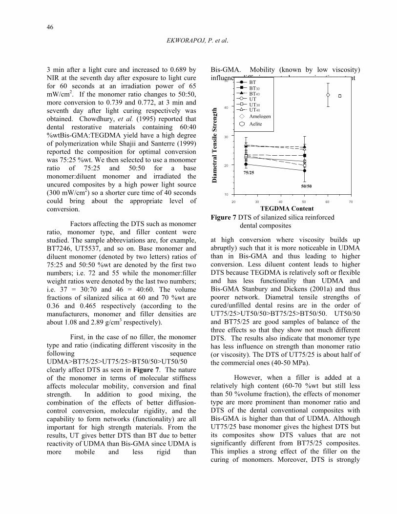
The hardness of the conventional composites is important as an indicator for wear or erosion resistance. It was studied as a function of filler content, monomer type and ratio. Mean value and standard deviations of Vicker hardness for the conventional composites are presented in Figure 10. The hardness values of BT composites are higher than those of UT composites. This is clearly due to monomer type; i.e. more molecular rigidity of Bis-GMA than that of UDMA and TEGDMA, respectively. Moreover, microhardness for samples with 60 %wt filler increases with increasing TEGDMA (but not for 70 %wt filler). Therefore the hardness value difference between filler loading 60 and 70%wt of BT72 (25% TEGDMA) is larger than that of BT55 (50% TEDGMA). The same result is also found for filled UT composites. This can be explained by 5-10
times lower viscosities of monomer mixtures with 50% TEDGMA than those with 25% TEDGMA so
Figu
thatdistsomfillerigidcom60-7Figuresicomdecr
conpuredensoftTEGhigh
re 10 VHN of BT and UT dental composites
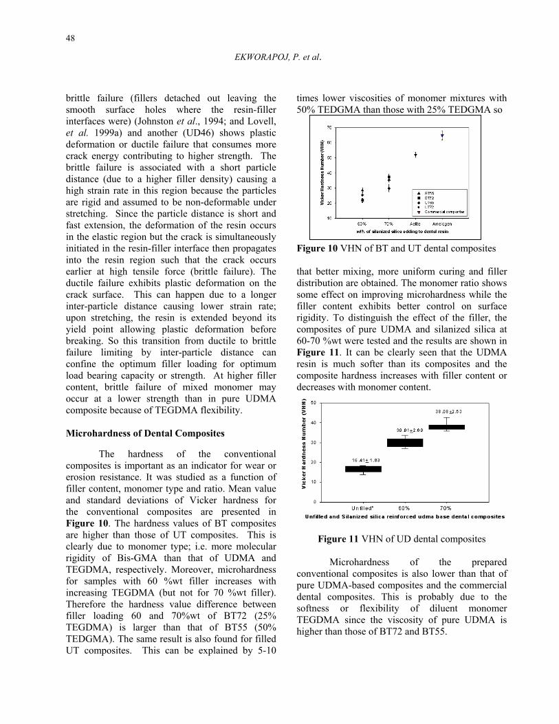
better mixing, more uniform curing and filler ribution are obtained. The monomer ratio shows e effect on improving microhardness while the r content exhibits better control on surface ity. To distinguish the effect of the filler, the posites of pure UDMA and silanized silica at 0 %wt were tested and the results are shown in re 11. It can be clearly seen that the UDMA
n is much softer than its composites and the posite hardness increases with filler content or eases with monomer content.
Figure 11 VHN of UD dental composites
Microhardness of the prepared ventional composites is also lower than that of UDMA-based composites and the commercial
tal composites. This is probably due to the ness or flexibility of diluent monomer DMA since the viscosity of pure UDMA is er than those of BT72 and BT55.
Heat Effect on Viscosity and Curing of Light-Cured Dental Resin and Mechanical Strength
of Conventional Dental Composites
49
The work able hardness and DTS are about 50-70 VHN and 40-50 MPa as indicated by the hardness and DTS of the commercial ones. These values cannot be achieved yet by only silanized silica filled in mixed polymer binders. Although hardness can be increased by adding more filler, the mechanical strength is also limited by filler content. Therefore, further development of new fillers or new monomers or new mixing methods should be carried out.
This work also shows the balance between stiff monomer structure and high viscosity v.s. flexible monomer structure and low viscosity. The former gives strength by structure but is less easy to cure and the later has a poorer structure but a greater chance of curing. It is likely that a low viscosity monomer provides better strength but when filler is added, the monomer type having a stiff structure shows higher strength along as well as filler content that alters strength significantly. However, overall DTS and VHN are not much different; i.e within 20-30 MPa and 20-40 VHN, respectively and these properties depend prominently on filler content and monomer type rather than monomer ratio.
Conclusions Heating reduces the resin viscosity with the
activation energy ranging from 21.9-27.6 kJ/mole. No evidence of heat curing occurs in the photocuring system. Monomer ratio plays a more important role on the diametral tensile strength of dental resin. The base monomer UT exhibits higher DTS than BT and the composition for the highest DTS is UT75/25 which has moderate viscosity. However, the cured composites with silanized silica show more dependence on monomer type (stiffness of monomer) for both DTS and VHS (i.e., the BT composites show higher properties than UT composites) rather than monomer ratio (viscosity adjustment). Furthermore, the strength and hardness of the conventional composites are varied more clearly with filler content compared to monomer type and monomer ratio. The optimum ratio of filler and mixed monomer resin is 60:40 to provide the optimum DTS. At 70 %wt filled UDMA, brittle failure is found. This corresponds with the reduced
DTS. The higher the filler content in resin, the higher the surface hardness is found.
Acknowledgements This work was supported by Grant for the
Development of a New Faculty and Researcher, Chulalongkorn University. The authors gratefully thank Dr. Allan D. Johnston, General Manager and Chief Technical Officer of ESSTECH, for supporting the base monomers, Mr. Phillip Lim, Schott Electronic Packaging Asia Pte Ltd., for silanized silica and Nudent Co., Ltd. (Thailand) for commercial dental composite.Inevitably, the authors are grateful to Dr. Suchit Poolthong, Faculty of Dentistry, Chulalongkorn University for his suggestion and allowance to use the Vicker hardness testing machine.
References Arcis, R. W., Lopez-Macipe, A., Toledano, M.,
Osorio, E., Rodriguez-clemete, R., Fanovich, M.A. and Pascual, C.D. 2002. Mechanical properties of visible light-cured resins reinforce with hydroxyapatite for dental restorative. Dent. Mater. 18 : 49- 57.
Asmussen, E. and Peutzfeldt, A. 1998. Influence of UEDMA, BisGMA and TEGDMA on selected mechanical properties of experimental resin composites. Dent. Mater 14(1) : 51-56.
Bowen, R. L. 1962. Dental filling material comprising vinyl silane treated fused silica and a binder consisting of a reaction product of bisphenol and glycidyl Acrylate. U.S.Patent, 3,006,112. Novemeber.
Chowdhury, N. A., Wakasa, K., Priyawan, R. and Yamaki, M. 1995. Effect of filler content on mechanical strength in bis-GMA base resins with urethane linkages. J. Mater. Sci. 6 : 400-403.
Council on Dental Materials and Device. 1997. New American Dental Association Specification No. 27 for direct filling resins. J. Am. Dent. Assoc. 94 : 1191-1194.
Htang, A., Ohasawa, M. and Matsumoto, H. 1995. Fatique resistance of composite
EKWORAPOJ, P. et al.
50
restorations: effect of filler content. Dent. Mater. 18 : 7-13.
Imazato, S., McCabe, J. F., Tarumi, H., Ehara, A. and Ebisu, S. 2001. Degree of conversion of composites measured by DTA and FTIR. Dent. Mater. 17 : 178-183.
Johnson, W. W., Dhuru, V. B. and Brantley, W. A. 1993. Composite microfiller content and its effect on fracture toughness and diametral tensile strength. Dent. Mater. 9 : 95-98.
Johnston, A. D., Dickens-Venz, S. H., Takagi, S., Chow, L. C., Bowen, R. L. and Dicken, B. 1994. Physical and chemical properties of resin-reinforced calcium phosphate cements. Dent. Mater. 10 : 100-106.
Leinfelder, K. F. 1989. Composite resins : properties and clinical performance, In : O’Brien, W.J., (ed.). Dental Material: Properties and Selection. Chicago, Quintessence Publishing.
Leinfelder, K. F. 1991. Using composite resin as posterior restoration material. J. Am. Dent. Assoc. 122 : 65.
Lim, B. S., Feranceane, J. L., Condon, J. R. and Adey, J. D. 2002. Effect of filler fraction and filler surface treatment on wear of microfilled composites. Dent. Mater. 18 : 1-11.
Lovell, L. G., Newman, S. M. and Bowman, C. N. 1999a. The effect of light intensity, temperature, and comonomer composition on the polymerization behavior of dimethacrylate dental resins. J. Dent. Rest. 78(8) : 1469-1476.
Lovell, L. G., Stanbury, J. W., Syrpes, D. C. and Bowman, C. N. 1999b. Effect of composition and reactivity on the reaction kinetics of dimethacrylate/dimethacrylate coipolymerizations. Macromolecules. 32 : 3913-3921.
Lutz, F. and Phillips, R. W. 1983. A classification and evaluation of composite resin system. J. Prosthet. Dent. 50 : 480.
McCabe, J. F. 1985. Cure performance of light-activated composites by differential thermal analysis (DTA). Dent. Mater. 1 : 231-234.
Maiti, S. N. and Jeyakumar, R. 1990. Mechanical and melt rheological properties of CaCO3-filled polyethylene. J. Polym. Mater. 7 : 29-34.
Phillips, R. W. and Moore, B. K. 1994. Elements of Dental Materials for Dental Hygienists and Dental Assistants. Philadelphia, W.B. Saunders.
Roulet, J. F. 1987. Degradation of Dental Polymers. New York, Karger.Shajii, L. and Santerre, J. P. 1999. Effect of filler content on the profile of released biodegradation products in micro-filled bis-GMA/TEGDMA dental composite resins. Biomaterials. 20 : 1897-1908.
Shernoy, A. V. 1999. Rheology of Filled Polymer Systems. Dordrecht, Kluwer Academic Publishers.
Silikas, N. and Watts, D. C. 1999. Rheology of urethane dimethacrylate and diluent formulations. Dent Mater. 15 : 257-261.
Stansbury, J. W. and Dickens, H. S. 2001a. Network formation and compositional drift during photo-initiated copolymerization of dimethacrylate monomers. Polymer. 42 : 6363-6369.
Stanbury, J. W. and Dickens, H. S. 2001b. Determination of double bond conversion in dental resins by near infrared spectroscopy. Dent Mater. 17 : 71-79.
Vaidyanathan, J., Vaisyanathan, T. K., Wang, Y. and Viswanadhan, T. 1992. Thermoanalytical characterization of visible light cure dental composites. J. Rehabil. 19 : 49-64.
Zhao, D., Botsis, J. and Drummond, J. L. 1997. Fracture studies of selected dental restorative composites. Dent Mater. 13 : 198-207.