California State University, San Bernardino California State University, San Bernardino CSUSB ScholarWorks CSUSB ScholarWorks Theses Digitization Project John M. Pfau Library 1995 Preparation of a site-specific lymphotoxin- mutant to be used in Preparation of a site-specific lymphotoxin- mutant to be used in protein characterization and receptor binding studies protein characterization and receptor binding studies Derek Andrew Knight Follow this and additional works at: https://scholarworks.lib.csusb.edu/etd-project Part of the Cell Biology Commons Recommended Citation Recommended Citation Knight, Derek Andrew, "Preparation of a site-specific lymphotoxin- mutant to be used in protein characterization and receptor binding studies" (1995). Theses Digitization Project. 987. https://scholarworks.lib.csusb.edu/etd-project/987 This Thesis is brought to you for free and open access by the John M. Pfau Library at CSUSB ScholarWorks. It has been accepted for inclusion in Theses Digitization Project by an authorized administrator of CSUSB ScholarWorks. For more information, please contact [email protected].
Transcript
California State University, San Bernardino California State University, San Bernardino
CSUSB ScholarWorks CSUSB ScholarWorks
Theses Digitization Project John M. Pfau Library
1995
Preparation of a site-specific lymphotoxin- mutant to be used in Preparation of a site-specific lymphotoxin- mutant to be used in
protein characterization and receptor binding studies protein characterization and receptor binding studies
Derek Andrew Knight
Follow this and additional works at: https://scholarworks.lib.csusb.edu/etd-project
Part of the Cell Biology Commons
Recommended Citation Recommended Citation Knight, Derek Andrew, "Preparation of a site-specific lymphotoxin- mutant to be used in protein characterization and receptor binding studies" (1995). Theses Digitization Project. 987. https://scholarworks.lib.csusb.edu/etd-project/987
This Thesis is brought to you for free and open access by the John M. Pfau Library at CSUSB ScholarWorks. It has been accepted for inclusion in Theses Digitization Project by an authorized administrator of CSUSB ScholarWorks. For more information, please contact [email protected].
were maintained ip.Dulbecco's Modified Eagles Medium (DMEM)
supplemented with 10% fetal calf serum,, 2 mM glutamine,
penicillin and streptomycin (100 ug/ml each) in 10% CO2/90%
ambient atmosphere at 37°C. The cells were passaged every 3-4
days upon reaching approximately 80-90% confluence.
GOS-7 cell trahsfection. 2.5 x 10^ cells were plated into
60 mm Culture dishes the day prior to transfection and grown
in DMEM as above.: The cells were rinsed twice with 5 ml warm
phosphate buffered saline (PBS) and then with 5 ml of serum
free DMEM. A DNA/Liposome solution was prepared containing 1
|j,g DNA, 2 ml serum free DMEM and 10 |il LipofectACE (GIBCO) in
a 5 ml polystyrene tube. Tubes were prepared containing
16
LT-a, LT-p, LT-p mutant and control vector (Cav.Not) DNA. The
solution from each tube was added to a plate and the plates
incubated at 37°C in 10% CO2/90% ambient atmosphere for 5
hours. 2 ml of DMEM supplemented with 20% fetal calf serum
was then added and the plates incubated at 37°C overnight.
The media was then replaced with 3 ml of DMEM containing 10%
fetal calf serum and incubated an additional 48 hours.
Biosyn'bhe'biG labeling of COS-7 calls. The monolayer of
transfected cells was washed twice with warm PBS. To each
dish, 1 ml of cys/met-free DMEM containing 10% dialyzed fetal
calf serum and glutamine was added and the dish incubated 5
minutes at room temperature. A 150 |a.Ci/ml ^^S^cys/met label
(ICN Trans label ~10 |j,Ci/ml) was then added and the plates
incubated at 37°C for 3 hours with occasional rocking. The
supernatant was then removed and transferred to a microfuge
tube. The monolayers were lysed with 1 ml/dish ice-cold
lysis buffer (20 mM Tris-HCl, pH 8.0, 150 mM NaCl, 2% Nonidet
P-40 (NP-40), 1 mM EDTA, 40 mM iodoacetamide and 2mM
phenylmethylsulfonyl flouride) for 10 minutes at 4°C. The
lysates were then transferred to a microfuge tube and the
tubes centrifuges at 13,000 rpm for 10 minutes at 4°C. Each
sample was then ttansferred to a fresh microfuge tube for
immunoprecipitation. Each tube was precleared by adding 1 \ig
of mouse IgG and 40 (xl protein G-Sepharose (GammaBind G-
Sepharose, Pharmacia). The tubes were capped tightly and
17
rocked 1 hour at 4°G, then centrifuged briefly to pellet the
protein Q beads and the supernatant was then removed and
placed in a new tube. A mixture of 1 m-S each of three
monoclonal anti-LT-p antibodies (B27, B9 and C37)(BIOGEN) was
added along with 40 |il/tube of protein G-Sepharose to the
tubes containing the Cav.Not control, LT-p and the LT—p
mutant. One microgram of an anti-LT-a monoclonal antibody
(NC2)(Biogen) along with the protein G beads was added to the
LT-a transfection control tube. The tubes were capped
tightly and rocked 1 hour at 4°C, then centrifuged briefly to
pellet the protein G beads. The samples were aspirated and
the beads washed three times with wash buffer containing 20
mM Tris (Ph 8,0), 0.5% NP-40, 150 mM NaCl, 0.5% deoxycholate
and 0.05% sodium dodecyl sulfate (SDS) and once with PBS
azide (N3) then resuspended in 50 jil 2x SDS-PAGE sample buffer
containing 100 mM Tris-Cl (pH 6.8), 200 mM dithiothreitol, 4%
SDS, 0.2% bromophenol blue, 20% glycerol plus 4% 2-mercapto
ethanol and heated for 5 minutes at 100°C.
SDS^Polyacrylimide gel electrophoresis (SDS-PAGE).
The samples were then loaded on a 12% SDS-polyacrylamide gel
and run overnight at 10 mA constant current to separate
proteins. The gel was then soaked for 30 minutes in 1 M
sodium salicylate (fluor) and transferred to Whatman 3MM
filter paper and dried at 75°C for 1 hour. The dried gel was
18
exposed to X-Omat AR film (Eastman Kodak) and a Cronex
intensifying screen, (Du Pont) for 24 hours at -70°C and
developed.
19
RESULTS
Prepara'tion of 5' and 3' PGR fragments containing the
mutation. Through the use of the two sets of primers
discussed previously, the expected fragments were obtained.
Agarose gel eletrophoresis of the PGR products showed bands
at approximately 710 bp and 107 bp respectively (Figure 9).
The optimization of reagent concentrations and cycling
parameters resulted in the amplification of only the desired
fragments while avoiding the generation of non-specific and
unwanted fragments.
joining of fragi^en'ts by PGR Overlap Extension yields
full-length mutant. Application of the overlap extension
technique using the two fragments generated above yielded a
760 bp fragment (Figure 9) which represents the full length
mutated LT-p cDNA. The PGR reaction yielded approximately 1
|ig of DNA. Optimization of reagent and cycling parameters
eliminated hon-specific fragments.
Sequence confirmation of mutation. The use of the
Sequenase PGR Product Sequencing kit allowed for rapid
sequence analysis of the PGR product. Both the 5' and 3' DNA
strands contained the desired mutation (figure 10) and no
Other random mutations were observed within 100 bp either
side of the site-directed mutation on either strand.
20
Cloning of mutant DNA in pcDNAl-amp plasmid.
Restriction digests of the vector and the PCR product
resulted in the creation of compatible cohesive ends that
allowed for specific ligation of the insert in the proper
orientation into the vector while avoiding religation of
vector alone. No transformants were noted in the vector only
control plate, while the 2:1 and 3:1 plates both contained 20
transformed colonies. Miniprep DNA from these colonies were
analyzed by restriction digests and all analyzed colonies
contained the expected 760 bp insert (figure 11).
Maxiprep of pcDNAl-amp/LT-p mutant DNA. A culture that
tested positive for the insert in the miniprep was used to
grow a large culture for the maxiprep. Approximately 250|j.g
of pcDNAl-amp/LT-p mutant DNA was obtained from a 200 ml
culture.
COS-7 cell transfection, biosynthetic labeling and
SDS-'PAGE. Immunoprecipitation of cell lysates using
monoclonal antibodies specific for LT-a or LT-p confirmed that
COS-7 cells were successfully transfected. The
autoradiograph of the labeled precipitates showed a band at
26 kDa representing the LT-a sample and indicates a
successful transfection. However, no bands were evident in
samples transfected with either LT-p or mutant LT-p.
Precipitation of these samples was attempted using three
21
different monoclonal antibodies to ensure the epitope had not
been lost during the mutagenesis, however none of the
monoclonal antibodies precipitated confirming proteins in
either LT-p sample.
22
C#nt«m«r« T«l#onwf »' 2ttb
B144 LT-P tnF UT-a
-|Hf|III HI
Ml 11 m CTIiiB mil i uiil
* .*! ? * * n« n X R RR R
' I > 1 _i "'' I " '
J L
!
f f ■ X NH HK <Mbp J- -J 1— 1 I I M
Figure 1. Schematic diagram of the region of chromosome 6 containing the LT/TNF locus. TNF, LT-a and LT-p are linked within 10 kb in the MHC on human chromosome 6. The exon^intron arrangements for each gene are similar. LT-p is coded for in an opposite direction from TNF and hT-tx. Restriction map shows sites for EcoRI (E), Xhol (X), Hindlll (H), Bglll (B), Kpnl (k), PstI (P) and Ncol (N). (From Browning et al., 1993)
23
SurfaceSecreted LT-a LT-p/LT-a Complex
Plasma OUT
Membrane
Secretory vesicle N
Golgi
LT-p
LT-a
Figure 2. A working model for LT biosynthesis and secretion. Newly formed LT-a and LT-p monomers can associate within the lumen of the endoplasmic reticulum to form trimers composed of a3, a2pi or alp3 subunits. These subunits are then processed within the Golgi and transported to the cell surface for expression as mLT (a2pi or aip2) or secreted LT (a3). (Adapted from Androlewicz et ai., 1992)
24
TNF Ugand Family
TKf LT. LTf LT*/t CCWOL CD30L C027L
148 150151157 154 156
ffiTmijffiTmr ^|m.
. 'Trimeric
i TNF/NGF RECEPTOR FAMILY
[] (] CO40 I3
TtfFfics , 4ieaU {] [] 13 (] 1] {]
C3 U3 (3" I Li! [3
Figure 3. Members of the TNF ligand and receptor family. For the ligands, an open box indicates the region of homology in the extracellular domain where the receptor binding sites are located, while the filled boxes indicate the cytoplasmic, transmembrane and extracellular stalk regions. The number of residues in each region are indicated. The receptor cys-rich repeat homology regions are shown as open boxes. Stripped boxes in the cytoplasmic region indicate homology. N or O indicate likely sites of glycosylation and P represents sites of phosphorylation (Ware, unpublished data).
25
LT-aP Complexes
a a
a
a a
P
a
15
P
(x3 a2pi alP2 P3
ReceptorBinding Interfaces
b/a b/a
b/x
z/a
2^
2/a
b/x
z/x
Figure 4. Proposed model of LT-aP complexes. The four potential trimers that are proposed for LT and the possible receptor binding interfaces for each. The a3 trimer could bind up to three TNFR which are specific for the a-a cleft. The aZpi trimer could potentially bind only one TNFR and one LT-pR, which are specific for the P-P cleft. The aip2 could not bind a TNFR but could bind up to two LT-pR. The P3, which has not been observed to occur in nature, could bind three LT—PR. The cleft formed at a-p subunit interfaces is thought to bind an as yet unidentified receptor (Ware, unpublished data).
26
%■
LT-P LT-a.
Figure 5. Molecular model of LT-a/LT-p interface. The LT-p subunit contains transmembrane and cytoplasmic domains not present on the LT-a subunit. LT-p also contains an N-linked glycosylation site very near the proposed binding region for the TNFR in the interface cleft. It is possible that this N-linked glycosylation site positions a carbohydrate that stearically blocks TNFR binding to mLT. The dark blue and yellow areas indicate regions of homology and the green area is a conserved proline thought to function in subunit association (Ware, unpublished data) .
27
Ouliside Primers:
5'pCDM8/LT-p
5'CGACTCACTATAGGGAGACC3' Melting Temp. 54°C
3'LT-p/Not-1
5'CGCGGCCGCACTCGGACCACGC3' MeIting Temp. 80°C
Inside Primers:
5'LT-pN-SOl
5'GTGACTGATGCTGACGTAGAC3' Melting Temp. 64°C
3'LT-pN-SOl
5'GTGTACGTCAGCATCAGTCAC3' Melting Temp. 64°C
5'Sequencing Primer:
5'LT-p/Seq
5'GAGACGGTGACTCCAGTG3' Melting Temp. 58°C
Figure 6. Primers utilized in PGR and sequencing reactions. Primers used in the PGR reactions include the
outside primers, 5'pGDM8/LT-p and 3'LT-p/Not-1. These primers allow for amplification of the entire LT-p gene and the 3'LTp/Not-1 also inserts a JSIot-l site in the 3' untranslated region. Inside primers include 5'LT-pN-SOl and 3'LT-pN-S02. These primers were used to insert the desired single base substitution (underlined) into the two fragments generated for use in the overlap extension procedure, changing the sequence to code for serine instead of asparagine. 5'LT-p/Seq was used to generate the 5' strand used in sequencing, this primer annealed approximately 100 bp upstream from the mutation site. A 3' strand was also generated for sequencing using the 3'LT-p/Not^l primer.
28
Creation ofLT-p Mutant Overlap Extension PGR
TargetedpCDMS/
SequenceLT-P
Denature
" Wutated sequence r
I tCombine
Denature y
t
Figure 7. Exsunple of sife-dlrected mufagenesis using overlap extension PGR. Specific base changes can be made using the overlap extension technique. Two separate PGR fragments are prepared, utilizing 5' and 3' non-mutated outside primers and complimentary internal primers that contain the desired base changes. These two fragments are amplified in separate PGR procedures, then the purified products combined, denatured and annealed and extended to form a full length template containing the mutation. The 5' and 3' outside primers are then added and another full PGR cycle run to amplify a full length mutant sequence.
29
fl* caojoo y) ox:q.03xi
XCQCQUJIUCQZXCOZX
c^^oo^ osz ca
Hpal
Pvu 1 cD
48Kb
Sfil
Nhe!
Figure 8. Map of the pcDNAl-amp plasmid used as a shuttle vector. The pcDNAl/Amp plasmid contains a strong promoter for eukaryotic expression, a ColEl sequence for high copy replication in E. Coll and ampicillin resistance in E, Coli for positive selection (Map supplied by Invitrogen).
30
-•««!rrr ;-2e>iu-»-.. ^S(r«_T-- -^as:
rSii S/? r'wv-T'iiiSS^C
Ee5iaassaSE«4HS3^-<^^
ii5« S5?:
»C£*
Figure 9. Agarose gel showing fragments made during PGR site directed mutagenesis procedure. Lane 1 contains molecular weight markers (BioMarker Low). Bands are at 1000, 700^ 525, 500, 400, 300, 200, 100 and 50 bp. Lane 2 shows the 5' fragment at approximately 710 bp. Lane 3 contains the 3' fragment of approximately 107 bp. Lane 4 shows the overlap extension product obtained by combining the fragments from lanes 2 and 3. This fragment represents the full length mutated LT-p gene of 760 bp and contains restriction enzyme sites for Hind III at the 5' end and Not-1 at the 3' end.
10. Sequence confirmation of desired mutation. The presence of the desired sequence was confirmed using dideoxy-DNA sequencing as seen on the 3' sequence shown in this autoradiograph. The site^specific mutation was the single base substitution underlined and in bold. This substitution results in an amino acid change from asparagine to serine, leading to a loss of the N-linked glycosylation site in the protein product.
32
Figure 11. Restriction digest of clone from pcDNA-amp. Following transfection, DNAminipreps were performed to confirm insertion of the cloned DNA. Restriction digests of the miniprep DNA with Not-1 and Hind III yielded a 760 bp fragment, indicating the mutated gene has been successfully inserted into the pcDNAl-amp plasmid. The left lane contains known molecular weight markers of Lambda DNA cut with the restriction enzyme Bst EII,
Figure 12. Autoradiograph of protein immunoprecipitation. Transfection of COS-7 cells with LT-a, LT-p and LT-p mutant followed by immunoprecipitation with monoclonal antibodies yielded a band at 26 kD for LT-a indicating a successful transfection. Known weight markers are at 200, 97, 69, 46, 30 and 14 kDa. No bands are present for either LT-p sample. The lack of bands may indicate LT-p is being rapidly degraded when no LT-a is present.
34
CoatrciOKA CcntroiONA
♦ ir-a. OHA
'■! 1' ■ 10 100 1000 4000 10 100 1000 4000
LT-OONA ^ LJ^OHk*
3 LT^ONA
19 100 1000 4000 10 100 1000 4000
RtlotivoFluoresceneo
Figure 13. Flow cytometry data showing surface expression of LT—a/p on COS-7 cells. The relative flourescence created by staining LTa/p cotransefected COS-7 cells with anti'-LT-a is shown as a solid line and the background flourescence due to control IgG staining is shown as a dashed line. Note no relative change when LT-a is expressed alone, this is because the LT-a is not retained on the cell surface. LT-p when expressed alone is not stained with the anti-LT-a, but when expressed with LT-ain a cotransfection experiment it causes LT-a to be targeted to the cell surface and retained, where it is stained by the antibody. (From Browning et ai., 1993)
35
DISCUSSION
The preparation of a site-specific DNA mutant of LT-p
was shown to be possible using the PGR overlap extension
technique. Although this technique was first demonstrated to
be a practical and efficient method of site-specific
mutagenesis several years ago, when utilized with the LT-p
gene several modifications were required. The LT—p gene is
very C-G rich in its 5' end, resulting in excessively high
melting temperatures and rapid reannealing of the two DNA
strands. This made the generation of a 5' mutant DNA
fragment very difficult and impractical using standard PGR
protocols. After trying a wide variety of reaction
conditions without success, the reactions were attempted in a
vessel containing 10% DMSO. Although not normally used in
PGR protocols utilizing Taq polymerase, DMSO was used
previously in Klenow-mediated PGR reactions (Scharf at al.,
1986). Although DMSO is known to reduce the effectiveness of
Taq polymerase by approximately 50%, its use in these
reactions allowed for the generation of mutant 5' fragments
at relatively good levels, yielding approximately 1 ng of
mutant DNA per 100 |j.l reaction. It is uncertain which
parameters are affected by inclusion of 10% DMSO, but DMSO
may affect the melting temperature of the primers, the
thermal activity profile of the Taq polymerase and/or the
degree of product strand separation during denaturation.
Even at denaturation temperatures of 99°G, reactions involving
36
the 5' end of this gene attempted without DMSO were not
successful/ while addition of 10% DMSO allowed reactions to
take place with denaturation temperatures of 95°C. Inclusion
of 10% DMSO was also required in the subsequent overlap
extension protocols to create full length mutant LT-p cDNA.
Once the full length cDNA was produced, it was sequenced
in the region surrounding the mutation site to confirm the
presence of the mutation and the absence of other random
mutations. Full length sequencing will be necessary
eventually since random mutations can be created while using
Taq polymerase at a rate approaching 1/4000 base pairs (Ho et
al., 1989). Since sequencing is time consuming and tedious,
full length sequericing will be performed only if changes in
binding characteristics, cytotoxicity or trimer formation
using the mutant DNA are observed in future assays. The use
of the Sequenase PGR sequencing kit with the 5'Seq and
3'LT-p7Not-l primers allowed for direct sequencing of both
strands of the PGR product a distance of 100 bp either side
of the desired mutation. No mutations other than the desired
mutation were observed and the sample was then digested with
Not 1 and Hind III arid cloned into the pcDNAl-amp plasmid.
E. Coli cells were subsequently transformed to generate high
yields of the desired mutant DNA (approximately 250 |j.g per
maxi-prep).
Following isolation of the amplified plasmid from E.
Coll, the mutant DNA was transfected into GOS-7 cells to
37
obtain protein expression in a mammalian expression system
for immunoprecipitation analysis. A sampling of three
different monoclonal antibodies were used to ensure the
desired epitope had not been inadvertently removed by the
mutation. Results of the immunoprecipitation were
inconclusive. LT-p was not expressed at high enough levels to
observe any protein in the autoradiograph (Figure 12). If
the protein had precipitated as expected, a band would have
been seen at approximately 33 kDa in the wild type LT-p sample
and a band of slightly greater mobility would have been
expected for the LT-p lacking the N-linked glycosylation site
due to lack of the sugar residue. A control transfection of
LT-a precipitated with an anti-LT-a monoclonal antibody
yielded the expected band at 26 kDa. Several other attempts
to precipitate wild type LT-p from COS-7 cells have also
failed to detect protein. Flow cytometry analysis of cells
transfected with LT-p has shown that very small quantities of
the protein are present when expressed in conjunction with
LT-a (Figure 13). The promoter on the pcDNAl-amp plasmid is
a strong promoter, so fairly high levels of protein
expression should be expected. This would seem to indicate
that rapid protein degradation may be taking place.
Co-transfection of GOS-7 cells using both LT-a and LT-p has
been attempted with no LT-p evident on the autoradiograph (not
shown). It is not known whether a sufficient number of cells
are receiving both plasmids to yield distinguishable bands of
38
a-Pco-expression on the autoradiograph. An interesting
feature of these qotransfections is that the relative level
of LT-a expression drops as the amount of LT-p transfected .
increases. This would seem to indicate that the presence of
LT-pis having some effect on LT-a expression, but the nature
of this effect is unclear.
In baculovirus—infected insect cells, LT-p is expressed
alone or with LT-a in cotransfected cells at very high levels
(Crowe, unpublished data). This data may indicate that LT-p
alone (which has never been observed naturally) contains a
signal sequence that directs the LT-p proteins down a
degradative pathway when expressed in mammalian cells. In
insect cells, this pathway may be overwhelmed, so the protein
is expressed. However, in these cells, the protein is seen
in forms not normally observed, from monomers to aggregates,
indicating that even though it is expressed, it is expressed
in states not normally seen. When LT-a is coexpressed with
LT-p, heterotrimers are seen similar to those found in normal
mammalian cells. Together, this may indicate a change in the
3-dimensidnal conformation is occurring upon subunit
interaction which signals a secretory pathway. Another
possibility is that the LT-a subunit contains a secretory
signal and this subunit blocks the degradative signal on LT-p
when the SubunitS are together. To examine if some signal in
the transmembrane or cytoplasmic domains are signaling for
protein degradation, a soluble chinieric LT-p/myc construct was
39
prepared (Biogen) in which the transmembrane and cytoplasmic
regions of LT-p had been removed and replaced with the myc
protein start sequence. This chimeric protein was expressed
by COS-7 cells at levels similar to those seen for LT-a
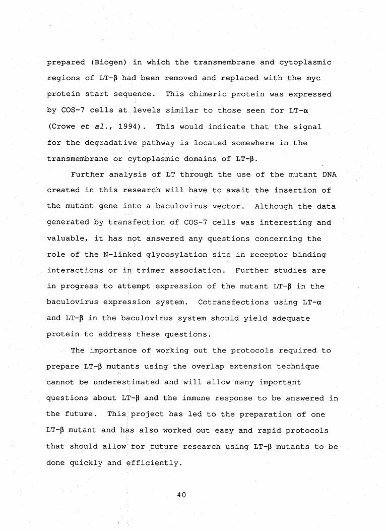
(Growe et al., 1994). This would indicate that the signal
for the degradative pathway is located somewhere in the
transmembrane or cytoplasmic domains of LT-|3.
Further analysis pf liT through the use of the mutant DNA
created in this research will have to await the insertion of
the mutant gene into a baculovirus vector. Although the data
generated by transfection of COS-7 cells was interesting and
valuable/ it has not answered any questions concerning the
role of the N-linked glycosylation site in receptor binding
interactions or in trimer association. Further studies are
in progress to attempt expression of the mutant LT-p in the
baculovirus expression system. Cotransfections using LT-a
and LT-p in the baculovirus system should yield adequate
protein to address these questions.
The importance of working out the protocols required to
prepare LT-p mutants using the overlap extension technique
cannot be underestimated and will allow many important
questions about LT-p and the immune response to be answered in
the future. This; project has led to the preparation of one
LT-p mutant and has also worked out easy and rapid protocols
that should allow'for future research using LT-p mutants to be
done quickly and efficiently.
40
References
Abbas, A.K., Lichtmann, A.H., and Pober, J,S., "Cellular and Molecular Immunology", W.B. Sanders Co., Philadelphia, Pa., 1991.
Andrews, J.S., Berger, A.E., and Ware, C.F., Characterization of the receptor for tumor necrosis factor (TNF) and lymphotoxin (LT) on human T lymphocytes: TNF and LT differ in their receptor binding properties and the induction of. MHC class I proteins on a human CD4+ T cell hybridoma. J. Iimvnol. 144:2582-2591, 1990.
Androlewicz, M.J., Browning, J.L., and Ware, C.F., Lymphotoxin is expressed in a heterotrimeric complex with a distinct 33 kDa glycoprotein on the surface of an activated human T cell hybridoma. J. Biol. Chem. 276:2542-2547, 1992.
Banner, D.W,, D'Arcy, A., James, W., Gentz, R., Schoenfeld, H-J., Brogeir, C., Loetscher, H., and Lesslauer, W., Crystal structure of the soluble human 55 kd TNF receptor—human TNF-p complex: implications for TNF receptor activation; Cell 71:765-776, 1993.
Browning, J.L., and Ribolini, A., Studies on the differing effects of tumor necrosis factor and lymphotoxin in the growth of several human tumor lines. J. Immunol. 143:1859-1867, 1989.
Browning, J.L., Androlewicz, M.J. and Ware, C.F., Lymphotoxin and an associated 33-kDa glycoprotein are expressed on the surface of an activated human T cell hybridoma. J. Immunol. 147:1230-1237, 1991.
Browning, J.L., Ngam-ek, A., Lawton, P., DeMarinis, J., Tizard, R., Chow, E.P., Hession, C., Greco, B., Foley, S., and Ware, C.F., Lymphotoxin-p: A new member of the TNF family that forms a heteromeric complex with lymphotoxin on the cell surface. Cell 72:847-856, 1993.
Digel, W., Porzolt, F., Schmid, M., Herrmann, F., Lesslauer, W. and Brockiiaus, M., High levels of circulating soluble receptors for tumor necrosis factor in Hairy cell leukemia and|type B chronic lymphocytic leukemia. J. Clin. Jnvlst. 89:1690-1693, 1992.
DiSanto, J.P., Bonnefoy, J,Y,, Gauchat, J.F., Fischer, A., and Saint BaSile, G.de, Ci)40 ligand mutations in X-*-linked immunpdeficiency with hyper-IgM. Nature 361:541-543.
■■■
Engelmann, H., Noyick, D., and Wallach, D., Two tumor necrosis factor binding proteins purified from human urine. <7. B'iol. Chem. 265:1531-1536, 1990.
Goodwin, R.G./ Anderson, D., Jerzy, R., Davis, T., Brannan, C.I., Copelahd, N.G., Jenkins, N.A., and Smith, C.A., Molecular clbning and expression of the type 1 and type 2 murine receptors for tuimor necrosis factor. Mol. Cell. Biol. 11:3020-3026, 1991.
Ho, S.N., Hunt, H.D., Horton, R.M., Pullen, J.K., and Pease, L.R., Site-directed mutagenesis by overlap extension using the polymerase chain reaction. Gene 77:51-59^^ 1989.
Hohmann/ H., Remy> R., Brockhaus, M., and Van Loon, A.P.G.M., Two different cell types have different major receptors for human tuinor necrosis factor (TNF a). J. Biol, Chem. 264:14927-14934, 1989.
Hollenbaugh, D., Grosmaire, L.S., Kullas, O.K., Chalupny, N.J., Braesch-Anderson, S., Noelle, F.J., Stamenkovic, I., Ledbetter, J.A., and Aruffo, A., The human T cell
antigen gp39/ a member of the TNF gene family, is a ligand for the CD40 receptor: Expression of a soluble form of gp39iwith B cell costimulatory activity. EMBO J. 11:4313-4321, 1992.
42
Horiuchi, A., Abe, i.Y,, Miyaki, M., Kimura, K., Hitsumoto, Y., Takeuchi, N.,'anci Kimura, S., Role of membrane-associated lymphotoxin (mLT) in the killing activity of lymphokine-associated killer (LAK) cells towards various tumor cell lines. Clin, Exp. Immunol. 96:152-157, 1994.
Horton, T.M., Ranheim, T.S., Aquino, L., Kusher, D.I., Saha, S.K., Ware, C.F., Wold, W.S.M., and Gooding, L.R., Adenovirus E3 14.7K protein functions in absence of other adenovirus proteins to protect transfected cells from tumor necrosis factor cytolysis. J. Virology 65:2629-2639, 1991.
Jones, E.Y., Stuart, D.I., and Walker, N.P.C., Structure of tumor necrosis factor. Nature 338:225-228, 1989.
Korthauer, U., Graf, D., Mages, H.W., Briere, F., Padayachee, M., Malcolm; S., Ugazio/ A.G., Notarangelo, L.D., Levinsky, R.J., and Kroczek, R.A., Defective expression of T-cell Cb40 causes X-linked immunodeficiency with hyper-IgM. Nature 3261:539-541, 1993.
Kriegler, M., Perez, C., DeFay, K., Albert, I., and Lu, S.U., A novel form;,of TNF/Cachectin is a cell surface
Cytotoxic transmembrane protein: Ramifications for the complex physiology of TNF. Cell 53:45-53, 1988.
Nedwin, G.E., Naylor, S.L., Sakaguchi, A.Y., Smith, D., Jarrett-Nedwin, J., Pennica, D., Goeddel, D.V., and Gray, P.W., Human lymphotoxin and tumor necrosis factor genes: Structure, horiiology and chromosomal location. Nucl. Acids Res. 13:6361-6373, 1985.
Paul, N.L,, and Ruddle, N.H., Lymphotoxin. Ann. Rev. Immunol. 6:407-438, 1988.
Pennica, D., Kohr, W.J., Fendly, B.M,, Shire, S.J., Raab, H.E., Borchardt, P.E., Lewis, M., and Goeddel, D.V., Characterization of a recombinant extracellular domain of the type i tumor necrosis factor receptor: evidence for tumor nedroSis factor-a induced aggregation. Biochemistry 31:1134-1141, 1992.
43
Ruddle, N.H., Bergman, C.M,, McGrath, K.M., Ligenheld, E.G., Grunnet, M.Li, Padula, S.J., and Clark, R.B., An antibody to lymphotoxin and tumor necrosis factor prevents transfer of experimental allergic encephalomyelitis. J. Exp. Med. 172:1193-1200, 1990.
Scharf, S.J., Horn, G.T., and Erlich, H.A., Direct cloning and sequence analysis of enzymatically amplified genomic sequences. Science, 233:1076-1078, 1986.
Schultze, S., Potthof, K., Machleidt, T., Berkovic, D., Wiegmann, K., and Kronke, M., TNF activates NF-kB by phosphatidylcholine-specific phospholipase C-induced "acidic" sphingomyelin breakdown. Cell 71:765-776, 1992.
Smith, C.A., Davis, T., Wignall, J.M., Din, W.S., Farrah, T., Upton, C., McFadden, G., and Goodwin, R.G., T2 open reading frame from the shope fibroma virus encodes a soluble form of the TNF receptor. Biochem. Biophys. Res. commun.'176:35-342, 1991.
Smith, C.A., Williams, D., Armitage, R.J., Gliniak, B., Grabstein, K;, Fanslow, W., Farrah, T., Falk, B., Din, W.S., Davis, T., and Goodwin, R.G., The TNF/NGF superfamily of cytokines and receptors. J. Cell. Biochem. 17B:51. Keystone Symposium Feb. 1-8, 1993.
Smith, R.A., and Baglioni, C., The active form of tumor necrosis factor is a trimer. J. Bid. Chem. 262:6951
6954, 1987. ;
Spies, T., Morton,' C.C., Nedospasov, S.A., Fiers, W., Pious, D., and Strominger, J.L., Genes for tumor necrosis factor a and P are linked to the major histocompatability complex. Proc. Natl. Acad. Sci., USA, 83:8699-8702, 1986.
Togni, P.D., Goeliner, J., Ruddle, N.H., Streeter, P.R., Fick, A,, Mariathasan, S., Smith, S.C., Carlson, R., Shornick, L.P., Strauss-Schoenberger, J., Russell, J.H., Karr, R. and\Chapin, D.D., Abnormal development of peripheral lymphoid organs in mice deficient in lymphotoxin. Science 264:703-707, 1994.
Van Ostade, X., Vandenabeele, P., Everaerdt, B., Loetscher, H., Gentz, R,, Brockhaus, M., Lesslauer, W., Travernier, J., Brouckaert, P., and Fiers, W., Human TNF mutants with selective activity on the p55 receptor. Nature 361:266-269, 1993.
44
Vandenabeele, P., Declercq, W., Vercammen, D., Van de Craen, M,, Grooten, J., Loetscher, H., Brockhaus, M., Lasslauer, W., and Fiers, W., Functional characterization of the human tumor necrosis factor receptor p75 in transfected rat/mouse T cell hybridoma. J. Exp. Med. 176:1015-1024, 1992.
Vilcek, J., and .Lee, T.H,, Tumor necrosis factor: New insights into the molecular mechanisms of its multiple actions. J". Biol. Chem. 266:7313-7316, 1991.
Ware, C.F., Andrews, J.S., Shamansky, L.M., and Grayson, M.H.j Regulation of the CTL lytic pathway by tumor necrosis factor. in /Cellular Immunity and the Immunotherapy of Cancer". Lotze, M.T. and Finn, O.J., eds. Wiley—Liss, New York, New York. pp 121—128, 1990.
Ware, C.F., Crowe, p.D., Grayson, M.H., Androlewicz, M.J., and Browning, J.L., Expression of surface lymphotoxin and tumor ndcrosis factor on activated T, B and NK cells. J. Immunol, 149:3881-3888, 1992.
Zhang, X-M., Webber, I., and Chen, M-J., Site directed mutational analysis of human necrosis factor-a receptor binding site and structural-functional relationship. J. Biol. Chem. 267:24069-24074, 1992.