Preparation and Characterization of 80 S Ribosomes of AscitesTumor Cells Active in Polypeptide Synthesis
Karl Letnansky
Institute for Cancer Research, University of Vienna, Vienna, Austria
SUMMARY
The preparation of tumor ribosomes that are essentially freeof polyribosomes and active in polypeptide synthesis isdescribed. Most active preparations were obtained byincubating ribosomal suspensions 20 min under conditions ofprotein synthesis. After this time, all polysomes had beenconverted to 80 S particles which could be stimulated topolypeptide synthesis by polyuridylic acid or liver mRNA.High-speed supernatant from rat or pig liver was used for theincubation procedure and in the cell-free system, rather thantumor supernatant, which caused almost complete inactivationof the ribosomes. The most active high-energy system was 10mM 3-phosphoglycerate; optimum ATP and Mg2+
concentrations were 1 and 7 mM, respectively.Deoxycholate was used to solubilize membranous fractions
and to liberate bound polysomes. Ribosomes, having beenprepared by deoxycholate treatment and successiveincubation, were free of polyribosomes. However, a smallamount of ribosomal subunits was always observed underthese conditions. Large-scale preparation of structurally intactsubunits by Mg2+ deficiency or mild ethylenediaminetetra-
acetic acid treatment is complicated by the high affinitybetween 40 and 60 S particles. In this respect, ribosomes ofthe Ehrlich carcinoma are comparable to those of rapidlygrowing hepatomas. However, they are strikingly differentfrom ribosomes of slowly growing hepatomas and normaltissues.
INTRODUCTION
Cytoplasmic ribosomes are distributed in eukaryotic cells invariable proportions as polyribosomes, single ribosomes, andsubribosomal particles (6, 14, 16, 22, 23, 50, 51). Some ofthe polysomes are bound to membranes of the endoplasmicreticulum, and these fulfill special requirements.
Among the different tumors, the relative proportion ofpolyribosomes to single ribosomes may be strikingly different.This is demonstrated by Morris Hepatoma 5123D, which ischaracterized by a high content of polyribosomes (50), ascompared with the Krebs 2 tumor (6) and the McCoy MDAB,Dunning, and Yoshida hepatomas (14, 50), in which there is ahigh proportion of single ribosomes and dimers. An exampleof rather equal distribution within the ribosomal profile wouldbe the Landschützascites tumor, which has been reported toshow 45% single ribosomes and 38% polyribosomes (16).Great differences also have been reported with respect to thecellular content of membranes and bound polyribosomes (14,42).
Received January 20, 1972; accepted August 1, 1972.
A basic requirement for studies of specific protein synthesisin cell-free systems is a ribosomal preparation lackingendogenous mRNA and polyribosomes. Most of theinvestigations into cell-free protein synthesis of tumor systems,however, have paid very little attention to this fact. This paperreports on some distribution studies of Ehrlich ascitescarcinoma ribosomes and on the influence of some methodsfor polysomal breakdown and removal of endogenous mRNAon the ribosomal profile.
These investigations were undertaken to obtain ribosomalpreparations with maximum contents of single ribosomes orsubunits, stimulated maximally by synthetic and naturalmessengers.
MATERIALS AND METHODS
Methods of Polysomal Disaggregation
EAC1 were harvested 7 to 10 days after their peritoneal
inoculation into Swiss mice and were washed twice with 0.9%NaCl solution. After suspension in Buffer A (50 mM Tris-HCl,pH 7.6; 25 mM KC1; 10 mM MgCl2 ; and 250 mM sucrose), thecells were homogenized according to the method ofMerkenschlager et al. (28). Nuclei and cell debris weresedimented by 10 min of centrifugation at l,500Xg;mitochondria were separated by 20 min centrifugation at16,000 X g. One part of the postmitochondrial supernatantwas centrifuged for 90 min at 120,000 X^, yieldingsupernatant fractions required for incubation of the ribosomesand in the cell-free system. The other part was similarlycentrifuged after DOC (final concentration, 0.25%, dissolvedin 0.2 M glycyl glycine, pH 8.0) had been added (in mostcases) to the supernatant. The ribosomal pellet was treatedfurther by either EDTA, dialysis, or incubation.
EDTA Treatment. Ribosomes that had been prepared in theabsence of DOC were suspended in Buffer Ci (10 mMTris-HCl, pH 7.4; 100 mM KC1; and 0.8 mM EDTA) and freedof aggregated material by centrifugation at 1,500 X g for 5min. Sedimentation profiles were then analyzed at aconcentration of 4 mg/ml (referred to as ribonucleoprotein) ina Beckman Model E analytical ultracentrifuge. The timeinterval between resuspension and the 1st photograph, takenafter attainment of 18,000 rpm, was 60 min. The temperaturewas kept constant at 4°.At the same time, a similarly treatedpreparation was tested for incorporation of phenylalanine-14 Cin the cell-free system after the addition of polyuridylic acidand optimum amounts of Mg2+.
'The abbreviations used are: EAC, Ehrlich-Lettre ascites carcinomacells; DOC, deoxycholate; PGA, 3-phosphoglycerate.
Dialysis. The ribosomal pellet, obtained after DOCtreatment and centrifugation of the postmitochondrialsupernatant, was resuspended in Buffer A0 (50 mM Tris-HCl,pH 7.0; 25 mM KC1; and 10 mM MgCl2) and dialyzed for 16hr against Buffer B (10 mM Tris-HCl, pH 7.4; and 0.05 mMMgCl2)at4°.
Incubation. Ribosomes, with or without DOC treatment,were resuspended in Buffer A and incubated at 37°for 20 min
in a medium consisting of 12 ml ribosomal suspension (about10 mg/ml), 12 ml (120,000 X g) supernatant from rat or pigliver or Ehrlich tumor, 8 mg ATP, and 30 mg3-phosphogly cerate. Following the incubation, anyprecipitated material was removed by centrifugation at9,OOOX g for 15 min, and the ribosomes were sedimentad at120,000 X g for 90 min. The pellet was resuspended in BufferA at a concentration of 4 mg/ml, and the suspension wasstored at -28°.
Preparation of 120,000 X g Supernatant from Liver
Rat or pig liver was minced and homogenized with 2volumes of Buffer A in a Potter-Elvehjem homogenizer at 0°.
Further processes used were similar to those described fortumor supernatant.
Cell-free System
Incubation mixtures consisted of the following: ATP, 0.25to 2.0mM;GTP,0.15 mM;MgCl2, 5 to 13mM;KCl, lOOmM;Tris-HCl, pH 7.4 60 mM; 120,000 X g supernatant, 100/¿I/mlincubation mixture; creatine phosphate, 5 mM, plus creatinephosphokinase, 10 Mg/ml (or PGA, 3 to 10 mM, orphosphoenolpyruvate, 5 mM, plus pyruvate kinase, 40 Mg/ml);and ribosomes, 400 Mg/ml. When polyphenylalanine synthesiswas investigated, phenylalanine-14C (specific activity, 450
mCi/mmole), 0.6 juCi/ml, and polyuridylic acid, 60
were added to the incubation mixture. Protein synthesis wasfollowed in the presence of leucine-14C (specific activity, 300
mCi/mmole), 0.6 /nCi/ml; amino acid mixture (containing 20amino acids, leucine excluded, 50 juM each); and messengerfrom pig liver, 100 Mg/ml. The total volume was 0.5 ml;incubation temperature was 37°.Samples were taken 20, 40,
and 60 min after the start and were prepared for PackardTri-Carb liquid scintillation counting according to the methodof Mans and Novelli (25).
The phenol method, as described by Georgiev et al. (12,13), was used to extract mRNA from pig liver. Determinationsof ribosomal protein and RNA were made by use of themethods of Lowry et al. (24) and of Mejbaum (27),respectively. In addition, concentrations of ribosomalsuspensions were determined spectrophotometrically, with therelation Al'¿0RNA= 250 (40). Except where otherwise stated,
concentrations are given as mg rRNA per ml.RNase activity was determined according to the method of
Roth (37), with 1% yeast RNA (C. F. Boehringer, Mannheim,Germany) or pig liver mRNA as a substrate. Each assaycontained 100 M! of the material to be tested in a total volumeof 3.0 ml. RNase activity is given as absorbance at 260 nm ofthe acid-ethanol supernatant fraction. All chemicals, purchasedfrom Merck, Darmstadt, Germany, were of analytical gradepurity. Radiochemicals were obtained from the RadiochemicalCentre, Amersham, England, and biochemicals were obtainedfrom Boehringer.
RESULTS
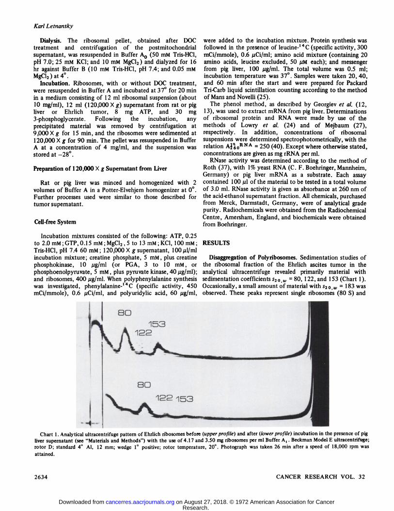
Disaggregation of Polyribosomes. Sedimentation studies ofthe ribosomal fraction of the Ehrlich ascites tumor in theanalytical ultracentrifuge revealed primarily material withsedimentation coefficients 52o,HP=80, 122, and 153 (Chart 1).Occasionally, a small amount of material with s20 w = 183 wasobserved. These peaks represent single ribosomes (80 S) and
Chart 1. Analytical ultracentrifuge pattern of Ehrlich ribosomes before (upper profile) and after (lower profile) incubation in the presence of pigliver supernatant (see "Materials and Methods") with the use of 4.17 and 3.50 mg ribosomes per ml Buffer A,. Beckman Model E ultracentrifuge;rotor D; standard 4°Al, 12 mm; wedge 1°positive; rotor temperature, 20°.Photograph was taken 26 min after a speed of 18,000 rpm was
polysomes with 2 to 4 ribosomes per unit. The sedimentationcoefficients are in good agreement with those reported forother tumors and normal tissues (3, 15, 38, 42, 45). Theproportion of dimers in the ribosomal preparation is about 6%,and that of trimers is 31% (Table 1). This pattern is verysimilar to that of the Landschütz ascites tumor, whichcontains 38% polyribosomes (16), although that tumor ischaracterized by a higher content of ribosomal subunits (17%).
Table 1Effect of DOC treatment and incubation on the composition of
ribosomal preparations from Ehrlich ascites tumor cells
0 Values were obtained from analytical ultracentrifuge patterns andrepresent percentages of the total ribosomal content. Sedimentationconditions were as described in Chart 1.
80 S Ribosomes of Ascites Tumor Cells
The addition of 0.8 mM EDTA resulted in markeddisaggregation of polyribosomal structures, but no significantamount of subunits was found in these preparations (Chart 2),even after prolonged centrifugation. The bulk of the materialwas still present as 80 S particles. Despite the very low EDTAconcentration, corresponding to just the amount of Mg2+
bound in the ribosomes (3), decreased activity of thesepreparations in the cell-free system was already observed
(Table 2).Optimum polypeptide-synthesizing capacity is shown by
preparations of single ribosomes, obtained by incubationunder the conditions of protein synthesis (Table 2). Suchpreparations are characterized by a low content ofpolyribosomes and some material with a sedimentationcoefficient lower than 80 S, presumably ribosomal subunits(Chart 1). Membrane-bound ribosomes, which have beenreported to be of limited significance in the Ehrlich carcinoma(32, 39), could be separated in good yield frompostmitochondrial supernatants by sucrose gradientcentrifugation. Most of the membranous material can besolubilized by treatment with DOC. This treatment results inan increase of the peak in the sedimentation profile,representing free ribosomes (Chart 3). It was observed,
I
Chart 2. Analytical ultracentrifuge pattern of EDTA-treated ribosomes.
Table 2Incorporation of phenylalanine- ' 4C by variously treated Ehrlich ascites tumor ribosomes in a cell-free system in the
presence and absence of polyuridylic acidThe treatment of ribosomes and the composition of the cell-free system were as described in "Materials and Methods."
Incor- Incorporation of phenylalanine-14C (cpm/mg rRNA) following
Chart 3. Effect of DOC on the composition of the postmitochondrialfraction of Ehrlich ascites carcinoma homogenates. The homogenatewas freed of nuclei and cell debris, and DOC was added to 0.25%. Afterremoval of the mitochondria by centrifugation for 20 min at11,500 X g, ribosomes were sedimented at 120,000 X g (90 min). Thepellet was resuspended in Buffer A, (about 4 mg rRNA per ml), and470 Mgribosomes were separated in a sucrose density gradient (25 ml, 5to 20% sucrose in Buffer A0, layered upon 3 ml 60% sucrose, SW 25.1rotor, 160 min, 25,000 rpm, 5°).Sedimentation is from right to
left, , profile after DOC treatment; —¿�,profile of ribosomalpreparations, not treated with DOC.
however, that a fraction of the membranous materialapparently cannot be solubilized by DOC. The sedimentationbehavior of this material seems unaltered after the DOCtreatment. A similar observation has been reported byRobinson (35), who used rat liver microsomes.
Although the liberation of bound polysomes resultsprimarily in a decrease in protein-synthesizing activity, thismay be overcome in part by subsequent incubation (Table 2).Diminished activity as a result of DOC treatment was alsoreported with ribosomes from mouse or rat spleen, in contrastto the activation of ribosomes from rat liver or plasma cells(44). Ribosomes prepared by DOC treatment and subsequentincubation are characterized by a very low proportion of
polyribosomes and an elevated content of subribosomalparticles (Chart 4). A 16-hr dialysis against Buffer B alsoresulted in an activation of DOC-treated ribosomes (Table 2).This activation may be further enhanced by additionalincubation. An incubation time of 20 min has proven mostsatisfactory (Chart 5).
The activity of the ribosomal preparation is highlydependent on the nature of the 120,000 X g supernatant usedduring the incubation and in the cell-free system. The
15 20 25
fraction number
Chart 4. Sucrose density gradient profile of DOC-treated andincubated (20 min, pig liver supernatant) ribosomes (115 Mg,5 to 20%sucrose in Buffer A0, SW 25K rotor, 25,000 rpm, 215 min, 5°).
Sedimentation is from right to left. The main peak consists of singleribosomes.
mI
or~X
0)
E
BO BO 1OO
minutes pre - incubation
Chart 5. Effect of preincubation on the phenylalanine-14 Cincorporation by DOC-treated and dialyzed EAC ribosomes. After apreincubation period of 20 to 90 min, 60 Mgpolyuridylic acid per mlwas added, and incubation was continued for an additional 20 min.
exclusive use of tumor supernatant results in a complete lossof ribosomal activity. However, if incubations are performedin the presence of rat or pig liver supernatant and if tumorsupernatant is used only in the test system, some amino acidincorporation is observed. Optimum activity is observed withliver supernatant in both the incubation and the test system,and there is only a small decrease in this activity when tumorsupernatant is substituted for liver supernatant in theincubation (Chart 6). The presence of supernatant fractions inthe incubation system is essential, since ribosomes have tocomplete their cycle to give monomeric units that are free ofmRNA and nascent protein. Another important factor seemsto be the RNase inhibitor, found by Roth (36) in liversupernatant, which prevents the degradation of thepolyribosome into monosomes by the action of RNase's. To
test the significance of these reactions, determinations ofthe RNase activity in the high-speed supernatant of both liverand tumor cells were performed. As demonstrated in Table 3,there is a high RNase activity in rat liver as well as in tumorsupernatant. Normally, more than 90% of the enzyme activityis bound in the liver, whereas all of the activity is free in thetumor. After the addition of liver supernatant to the tumorpreparation, however, a 90% inhibition of the tumor enzymewas also observed.
In an attempt to inhibit endogenous RNase activities duringthe preparation of the ribosomes, we added rat liversupernatant to the homogenization medium of the tumor cells.There was no significant difference, however, between theultracentrifuge patterns of ribosomes prepared in the presenceand in the absence of rat liver supernatant, indicating that thehigh proportion of 80 S monomères is not an artifact causedby nuclease digestion of polysomes. This is further substantiated by dissociation experiments in media of high ionicstrength. According to current concepts (10, 20, 54), adissociation of 80 S particles into smaller particles in 0.5 M
KC1 or NaCl is achieved only with single ribosomes and notwith monosomes, which carry mRNA fragments. As demonstrated in Chart 7, suspending DOC-treated and preincubatedribosomes in 0.5 M NaCl-0.01 M Tris, pH 7.4, results in adegradation into particles with sedimentation constants ofabout 35 and 65 S. Thus, the pattern demonstrated in thisstudy with tumor cells resembles to a high degree the patternobtained by Yang et al. (53) with the use of salt treatment ofreticulocyte ribosomes. The observation that the peak,consisting of small subparticles, exceeds that of the largesubparticles may be explained by some further degradation ofthe 65 S particle.
For ribosomes prepared by DOC treatment and incubation,the ratio of absorbances at 260 and 280 nm is 1.79. The samefigure is reported by Bielka et al. (3) for purified rat liverribosomes, composed of 50.9% RNA and 49.1% protein.
Table 3RNase activity of high-speed supernatant and ribosomes
from Ehrlich osciles tumor and rat liverActivity is given as absorbance at 260 nm of the acid-ethanol
" Direct assay.b Assay with p-chloromercuribenzoate, 1 mM.
0)
IC
Ë
.o L 10
o T
SO BO
Chart 6. Effect of 120,000 X g supernatants from EAC and pig liver, used during the incubation of the ribosomes and in the cell-free system, onthe synthesis of polyphenylalanine. A, ribosomes, preincubated with tumor supernatant; B, ribosomes, preincubated with pig liver supernatant;
, absence of polyuridylic acid; —¿�,presence of polyuridylic acid; T, EAC supernatant in the cell-free system; L, liver supernatant in thecell-free system. Conditions were as described in "Materials and Methods."
Ribosomes from other tissues are characterized by very similarcompositions and absorbance ratios (8, 33). The determinationof the RNA and protein content of the tumor ribosomalpreparations by the methods of Mejbaum (27) and Lowry etal. (24), respectively, are in good agreement with these values,resulting in a RNA content of 53.2% and a protein content of46.8%.
Activity of the Ribosomes in the Cell-free System. Theinfluence of the high-energy phosphate compounds on thepolyphenylalanine synthesis in the cell-free system of theEhrlich ascites carcinoma is demonstrated in Chart 8. Inaddition to sufficient amounts of ATP and GTP, anenergy-generating system is required. The widely usedphosphoenolpyruvate-pyruvic kinase is not very active in oursystem. However, phosphocreatine-creatine kinase and,particularly, PGA, proved to be very effective. On the otherhand, in the pig liver cell-free system, highest activities havebeen observed with phosphocreatine. Possibly, the proportionof microsomes, polyribosomes, and single ribosomes in theribosomal preparation is reflected to some extent by theeffectivity of these phosphate compounds. This would besuggested by the results of Todd and Campbell (48) and ofFalvey and Staehelin (10), who found high activity ofphosphoenolpyruvate in a liver microsomal system, in contrastto the low activity of this compound that was found insystems with polysomes or single ribosomes and polyuridylicacid. Moreover, both phosphoenolpyruvate and ATP mayinfluence the Mg2+ concentration by forming chelates (41).
Under our conditions, optimum concentrations for ATP andMg¿fwere 1 and 7 mM, respectively (Charts 9 and 10), and
PGA stimulated maximally at concentrations > lOmM (ChartHi-
Only limited stimulation of the leucine-'4C incorporation
into natural proteins was observed after mRNA extracted frompig liver was added to a cell-free system with untreated tumor
30
0)
10
C 1O
0Ü
¿JO BO
minutes
Chart 8. The effect of different high-energy phosphate compoundson the incorporation rate of the cell-free system. Each ml contained400 ¿igDOC-treated ribosomes (prepared in the presence of EACsupernatant); lOO MgP'g liver supernatant; 0.6 /¿Ciphenylalanine-14C;100 M!high-energy phosphates; and, where required, 60 Mgpolyuridylicacid. The concentration of the high-energy phosphates per ml in thecell-free system was as follows: 1 /¿moleATP, 0.15 Minole GTP, and 5/¿molesPGA [or 5 Amólesphosphocreatine (PC) + 10 jugcreatine kinase,or 5 /¿molesphosphoenolpyruvate (PEP)+ 40 jug pyruvate kinase].Total Mg2* concentration was 9 /imoles/ml. All otherconcentrations were as described in "Materials and Methods." Final
volume was 0.5 ml. , absence of polyuridylic acid; —¿�,presence ofpolyuridylic acid.
Chart 7. Analytical ultracentrifuge pattern of salt-treated ribosomes. Upper pattern, ribosomes that weredissociated by treatment with 0.5 M NaCl-0.01 M Tris,pH 7.4; lower pattern, untreated control (sedimentation coefficient of the main peak, 81 S).
Chart 9. Effect of the ATP concentration on the incorporation ofphenylalanine-14 C into polyphenylalanine. Each ml of the cell-free
system contained 0.25, 0.5, 1.0, 1.5, or 2.0 AmólesATP, 5 Amólesphosphocreatine, 10 jig creatine kinase, and 9 AmólesMg'*. The other
conditions were as described for Chart 7.
ribosomes, although these preparations contain a considerableproportion of 80 S ribosomes (Chart 1). Our preliminaryexperiments were not indicative of the existence of a majorproportion of metabolic inactive monosomes originating frompolysome degradation by RNase's within this 80 S fraction.
Rather, they suggested that in vivo 80 S ribosomes differfunctionally from polysome-derived 80 S particles, as is alsosupposed for rat liver ribosomes (9). Incubation and/or DOCtreatment of the crude ribosomal preparation results in amarked decrease of the incorporation activity in the absenceof the messenger, as a consequence of polysomal disaggrega-tion. However, these ribosomes are stimulated to a high degreeby the addition of mRNA, resulting in incorporation rates asobserved with the untreated ribosomes (Chart 12). Thestimulation is of the same magnitude as that found in otherheterologous, cell-free systems of eukaryotic origin, as wasdemonstrated by Armentrout and Weisberger (1) on areticulocyte system with lymph node polysomal messengerribonucleoprotein, and by Ishikawa et al. (21) on anEscherichia coli system with rat liver nuclear ribonucleoprotein.
DISCUSSION
According to current concepts of the ribosome cycle, theribosome is released from the polysome after termination assubunits. These subunits are in equilibrium with 80 S singleribosomes. Preceding the initiation of a new cycle, a
dissociation of single ribosomes into subunits is required, ifprotein synthesis is directed by natural messengers. Only in thesimplified system, with the use of polyuridylic acid as artificialmessenger, may 80 S units be bound directly (9).
The low proportion of subunits within ribosomalpreparations of certain tumor cells possibly reflects aparticular significance of single ribosomes for protein synthesisin these systems. In fact, Hogan and Korner (17) suppose thesingle ribosomes of the Landschützascites tumor to be somesort of a pool for ribosomal subunits. In the tumor describedin this paper, single ribosomes were also found in a highconcentration. This differentiates the Ehrlich tumor clearlyfrom the Yoshida ascites hepatoma, in which there is a highproportion of very stable dimers (14), and from MorrisHepatoma 5123D, which is very rich in polyribosomes (50).
Under certain conditions, ribosomes of the Ehrlichcarcinoma seem to be rather sensitive to the influence ofRNase's of the supernatant fraction. Similar observations have
been reported for a series of other ribosomal systems.Primarily, the larger subunit has been observed to be attackedby RNase's and interrelations between RNase activity and
incorporation activity of different ribosomal preparations havebeen found (4, 8, 11,31). In our system, incubation in media
60
Chart 10. Effect of the Mg2* concentration on the incorporation ofphenylalanine-14 C into polyphenylalanine. Each ml of the cell-free
system contained 5, 7, 9, 11, or 13 AmólesMgCl,, 5 AmólesPGA, 1Mmole ATP, and 60 Mg polyuridylic acid (poly U). Other conditionswere as described for Chart 7.
Chart 11. Effect of the PGA concentration on the incorporation ofphenylalanine-1 4C into polyphenylalanine. One ml of the test systemcontained 3, 5, or 10 jumólesPGA, 1 jumóleATP, 9 jumólesMg2*and 60
/jg polyuridylic acid. Other additions were as described for Chart 7.
2OOO0)
160O
'8 BOO-1
4OO-
20 ¿tominutes
60
Chart 12. Stimulation of the incorporation of leucine-'*C intotrichloroacetic acid-insoluble material by a natural mRNA, isolatedfrom pig liver. Tumor ribosomes were prepared by incubation inpresence of pig liver supernatant and/or DOC treatment. The cell-freesystem was as described in "Materials and Methods," and it contained
liver supernatant and PGA. , absence of exogenous messenger; —¿�-,presence of exogenous messenger; Curve 1, untreated ribosomes; Curve2, DOC-treated ribosomes; Curve 3, DOC-treated, incubated ribosomes.
containing 120,000 X g supernatant from tumor cells results ina complete inactivation of the ribosomes. The fact thatincubations with liver supernatant yield very activepreparations is possibly explained by the existence of RNaseinhibitors in the liver. This would also account for the stabilityof the assay system; amino acid-incorporating activity isobserved for at least 60 min at an almost constant ratewhereas, in most cell-free systems described up to now,protein synthesis is impaired after much shorter incubationperiods due to degradation of the messenger (2, 8,19).
A series of methods that are capable of releasing 80 Sparticles or subunits from polyribosomes have been described,including treatment with fluoride, puromycin, EDTA, PPj,RNase, and high-ionic-strength salt solutions, as well as dialysisand incubation under conditions of protein synthesis (26, 30,33, 34, 49). Only a limited number of methods yieldpreparations also active in protein synthesis. It seemed mostpromising to us to investigate the usefulness of EDTAtreatment and incubation for the preparation of tumorribosomes that were free of endogenous messengers and thatwere stimulated by the addition of exogenous messengers. Thechanges in ribosomal distribution were followedat the same time by sucrose gradient centrifugation or in theanalytical ultracentrifuge.
Tumor ribosomes differ from ribosomes of normal tissues orbacteria in their behavior against EDTA. With bacterialribosomes, low Mg2+ concentration of the suspension medium
is sufficient to cause dissociation into subunits (47), whereaswith liver ribosomes all of the bound Mg2+ must be removed
Incubation under the conditions of protein synthesis hasbeen found to be one of the most satisfactory means ofobtaining single ribosomes, free of mRNA and active inpolypeptide synthesis after the addition of exogenousmessengers (10, 52). In our experience, this method yields themost active preparations also from tumor cells, if certainrequirements are met. Additional treatment with DOC resultsin the liberation of membrane-bound ribosomes. In contrast toliver ribosomes, tumor ribosomes show a decrease inincorporation activity after DOC treatment, which could bepartially counteracted, however, by a subsequent incubationstep (Table 2). This may possibly be explained by the capacityof DOC to act not only as a detergent but also as a chelatingagent for bivalent metallic ions (7). The different responses of
tumor and liver ribosomes to Mg2+deficiency and the resultant
structural alterations or nuclease attack may account for thedifferent reaction of these particles.
REFERENCES
1. Armentrout, S. A., and Weisberger, A. S. Polypeptide SynthesisDirected by Lymph Node Ribonucleoprotein in a ReticulocyteCell-free System. Arch. Biochem. Biophys., ¡44:322-329, 1971.
2. Baiondes, S. H., and Nirenberg, M. W. Fate of a SyntheticPolynucleotide Directing Cell Free Protein Synthesis. I.Characteristics of Degradation. Science, ¡38:810-813, 1962.
3. Bielka, H., Welfle, H., Böttger, M., and Förster, W. StructuralChanges and Dissociation of Liver Ribosomes as a Function of theMg2*Concentration. European J. Biochem., 5: 183-190, 1968.
4. Bont, W. S., Rezelman, G., Meisner, 1., and Bloemendal, H. Studieson Cytoplasmic RNA from Rat Liver. VIII. Stability ofPolyribosomes from Normal and Regenerating Rat Liver. Arch.Biochem. Biophys., 119: 36-40, 1967.
6. Dalgarno, L., Cox, R. A., and Martin, E. M. Polyribosomes inNormal Krebs 2 Asches Tumor Cells and in Cells Infected withEncephalomyocarditis Virus. Biochim. Biophys. Acta, 138:316-328, 1967.
7. Dickman, S. R., Madison, J. T., and Holtzer, R. L. Preparation andProperties of Beef Pancreas Microsomal Fractions. Biochemistry, 1:568-574, 1962.
8. Earl, D. C. N., and Morgan, H. E. An Improved Preparation ofRibosomes and Polysomes from Cardiac Muscle. Arch. Biochem.Biophys., 128: 460-469, 1968.
9. Falvey, A. K., and Staehelin, T. Structure and Function ofMammalian Ribosomes. II. Exchange of Ribosomal Subunits atVarious Stages of in Vitro Polypeptide Synthesis. J. Mol. Biol., 53:21-34,1970.
10. Falvey, A. K., and Staehelin, T. Structure and Function ofMammalian Ribosomes. I. The Isolation and Characterization ofActive Liver Ribosomal Subunits. J. Mol. Biol., 53: 1-19, 1970.
11. Fenwick, M. L. The Effect of Ribonuclease on Polysomes andRibosomes of Bacteria and Animal Cells. Biochem. J., 707:481-489,1968.
12. Georgiev, G. P. Ribonucleic Acid of the Chromosomal-NucleolarApparatus. Biokhimija, 26: 1095-1107, 1961.
13. Georgiev, G. P., Samarina, O. P., Lerman, M. I., Smirnov, N. N.,and Severtzov, A. N. Biosynthesis of Messenger and RibosomalRibonucleic Acids in the Nucleochromosomal Apparatus of AnimalCells. Nature, 200: 1291-1294, 1963.
14. Grávela, E., Caratterizzazione dell'Apparato Ribosomiale delle
Cellule di Epatoma Ascile di Yoshida. Tumori, 54: 398-400,1968.
15. Hogan, B. L. M., and Korner, A. Ribosomal Subunits ofLandschützAscites Tumor Cells. Biochem. J., 100: 74P, 1966.
16. Hogan, B. L. M., and Korner, A. Ribosomal Subunits ofLandschützAscites Cells during Changes in Polysome Distribution.Biochim. Biophys. Acta, 169: 129-138, 1968.
17. Hogan, B. L. M., and Korner, A. The Role of Ribosomal Subunitsand 80 S Monomèresin Polysome Formation in an Ascites TumorCell. Biochim. Biophys. Acta, 769: 139-149, 1968.
18. Huez, G., Burny, A., Marbaix, G., and Lebleu, B. Release of mRNAfrom Rabbit Reticulocyte Polyribosomes at Low Concentrations ofDivalent Cations. Biochim. Biophys. Acta, 145: 629-636, 1967.
19. Hunter, A. R., and Korner, A. The Response of Rat LiverPolysomes to Added Homopolynucleotides: The Removal of
20. Infante, A. A., and Graves, P. N. StabUity of Free Ribosomes,Derived Ribosomes and Polysomes of the Sea Urchin. Biochim.Biophys. Acta, 246: 100-110, 1971.
21. Ishikawa, K., Kuroda, C., Ueki, M., and Ogata, K. MessengerRibonucleoprotein Complexes Released from Rat Liver Nuclei byATP. I. Characterization of the RNA Moiety of MessengerRibonucleoprotein Complexes. Biochim. Biophys. Acta, 213:495-504, 1970.
22. Kuff, E. L., and Roberts, N. E. In Vivo Labeling Patterns of FreePolyribosomes: Relationship to Tape Theory of MessengerRibonucleic Acid Function. J. Mol. Biol., 26: 211-225, 1967.
23. Laga, E. M., Baliga, B. S., and Munro, H. N. Isolation andProperties of Polysomes from Human Placenta. Biochim. Biophys.Acta, 213: 391-400, 1970.
24. Lowry, O. H., Rosebrough, N. J., Farr, A. L., and Randall, R. J.Protein Measurement with the Folin Phenol Reagent. J. Biol.Chem., 795:265-275, 1951.
25. Mans, R. J., and Novelli, G. D. Measurement of the Incorporationof Radioactive Amino Acids into Protein by a Filter-Paper DiscMethod. Arch. Biochem. Biophys., 94: 48-53, 1961.
26. Means, A. R., and Baker, C. A. Isolation of Polyribosomes andCharacteristics of an Amino Acid-Incorporation System from theLiver of a Primate (Papio papio). Biochim. Biophys. Acta, 182:461-468,1969.
27. Mejbaum, W. Über die Bestimmung kleiner Pentosemengen,insbesondere in Derivaten der Adenylsäure.Z. Physiol. Chem.,258: 117-120,1939.
28. Merkenschlager, M. M., Schlossmann, K., and Kurz, W. Einmechanischer Zellhomogenisator und seine Anwendbarkeit aufbiologische Probleme. Biochem. Z., 329: 332-340, 1957.
29. Miall, S. H., and Walker, I. O. Structural Studies on Ribosomes. II.Denaturation and Sedimentation of Ribosomal Subunits Unfoldedin EDTA. Biochim. Biophys. Acta, 174: 551-560, 1969.
30. Näslund,P. H., and Hultin, T. Effects of Potassium Deficiency onMammalian Ribosomes. Biochim. Biophys. Acta, 204: 237-247,1970.
31. Nicholls, D. M., Ryan, M. P., Miall, S. H., Westall, C. G., andCappon, I. D. The Regulation of Protein Synthesis in NephroticRat Kidney Ribosome Preparations. Can. J. Biochem., 48:308-315, 1970.
32. Palade, G. E. A Small Particulate Component of the Cytoplasm. J.Biophys. Biochem. Cytol., 7: 59-68, 1955.
33. Pollack, M. S., Schapira, G., and Dreyfus, J. C. The Inhibition ofRibosomes and Ribosomal Subunits in Mammalian Tissue withSpecial Reference to Skeletal Muscle. Bull. Soc. Chim. Biol., 52:891-903,1970.
34. Pragnell, I. B., and Arnstein, H. R. V. Interaction of HemoglobinMessenger RNP with the Small Subparticle of ReticulocyteRibosomes. Federation European Biochem. Soc. Letters, 9:331-334, 1970.
35. Robinson, G. B. The Contamination of Rat-liver PolyribosomalPreparations by Non-ribosomal Proteins. Federation EuropeanBiochem. Soc. Letters, 4: 190-192, 1969.
36. Roth, J. S. Ribonuclease. V. Studies on the Properties andDistribution of Ribonuclease Inhibitor in the Rat. Biochim.Biophys. Acta, 21: 34-43, 1956.
37. Roth, J. S. In: H. Busch (ed.), Methods in Cancer Research, Vol. 3,pp. 153-242. New York: Academic Press, Inc., 1967.
38. Schlossberg, A. H., and Hollenberg, C. H. Amino AcidIncorporation by a Cell-free Preparation from Rat Adipose Tissue.Effect of Fasting. Can. J. Biochem., 48: 113-121, 1970.
39. Selby, C. C., Biesele, J. J., and Grey, C. E. Electron Microscope
Studies of Ascites Tumor Cells. Ann. N. Y. Acad. Sci., 63:748-773, 1956.
40. Sheard, B., Miall, S. H., Peacocke, A. R., Walker, I. O., andRichards, R. E. Proton Magnetic Relaxation Studies of the Bindingof Manganese ions to Escherichia coli Ribosomes. J. Mol. Biol., 28:389-402, 1967.
41. Staehelin, T. Polyphenylalanine Synthesis by Rat Liver RibosomesDerived from Polysomes by Ethionine Treatment in Vivo or byPreincubation in Vitro. Biochim. Biophys. Acta, 174: 713-721,1969.
42. Stahl, J., and Bielka, H. On the Effects of RNase on the MembraneBound Ribosomes of Liver and Hepatoma Tissue. Acta Biol. Med.Ger.,/7.-K5-K8.1966.
43. Stahl, J., Lawford, G. R., Williams, B., and Campbell, P. N. ARequirement for the Presence of Cell Sap in the ReversibleDissociation of Rat Liver Polysomes to Ribosomal Subunits.Biochem. i.,109: 155-157,1968.
44. Talal, N., and Kaltreider, H. B. Functional and Structural Studiesof Membrane-bound and l-'ree Ribosomes from Rat Spleen. J. Biol.
Chcm., 243: 6504-6510, 1968.45. Tashiro, Y., and Siekevitz, P. Ultracentrifugai Studies on the
Dissociation of Hepatic Ribosomes. J. Mol. Biol., //: 149-165,
1965.46. Temmerman, J., and Lebleu, B. Evidence for the Detachment of a
47. Tissières,A., Watson, J. D., Schlessinger, D., and Hollingworth, B.R. Ribonucleoprotein Particles from Escherichia coli J. Mol. Biol.,/. 221-223,1959.
48. Todd, P. S., and Campbell, P. N. The Use of High EnergyPhosphate Compounds in in Vitro studies on Protein Synthesis: In:P. N. Compbell and J. R. Sargent (eds.), Techniques in ProteinSynthesis, pp. 251-256. London: Academic Press, Inc., 1969.
49. Ts'o, P. O. P., and Vinograd, J. Studies of Ribosomes fromReticulocytes. Biochim. Biophys. Acta, 49: 113-129, 1961.
50. Utsunomiya, T., and Roth, J. S. Studies on the Function ofIntracellular Ribonucleases. IV. Some Observations on theProperties of Ribosomes and Polysomes from Rat Liver andHepatomas. J. Cell. Biol., 29: 387-393, 1966.
51. Webb, T. E., Blobel, G., and Potter, V. R. Polyribosomes in RatTissues. I. A Study of in Vivo Patterns in Liver and Hepatomas.Cancer Res., 24: 1229-1237, 1964.
52. Weksler, M. E., and Gelboin, H. V. Studies on the Removal ofEndogenous Messenger Ribonucleic Activity from Rat LiverMicrosomes. J. Biol. Chem., 242: 727-735, 1967.
53. Yang, P. C., Hamada, K., and Schweet, R. Studies on Salt-treatedReticulocyte Ribosomes. Arch. Biochem. Biophys., 725: 506-513,1958.
54. Zylber, E. A., and Penman, S. The Effect of High Ionic Strength onMonomères,Polyribosomes, and Puromycin-treated Polyribosomes.Biochim. Biophys. Acta, 204: 221-229, 1970.