+ All Categories
Home > Education > Presentation 229 b pamela kittrell safety of peg tube insertion in patients wirth als using propofol...

Presentation 229 b pamela kittrell safety of peg tube insertion in patients wirth als using propofol...

Date post: 13-Jul-2015
Category:
Upload: the-als-association
View: 285 times
Download: 0 times
Share this document with a friend
Popular Tags:
25
Safety of Peg Tube Insertion in Patients with ALS Using Propofol Sedation in an Outpatient Surgical Center Pamela Kittrell MSN RN CCRC Clinical Research Nurse Senior The University of Texas Health Science Center at San Antonio
Transcript

Safety of Peg Tube Insertion

in Patients with ALSUsing Propofol Sedation in an Outpatient Surgical Center

Pamela Kittrell MSN RN CCRC

Clinical Research Nurse Senior

The University of Texas Health Science Center at San Antonio

Objectives

• Review the nutritional needs of the ALS

patient

• Explain the different methods used in tube

placement

• Discuss the results of the PEG tube

outpatient placement study and apply

them in a clinical setting

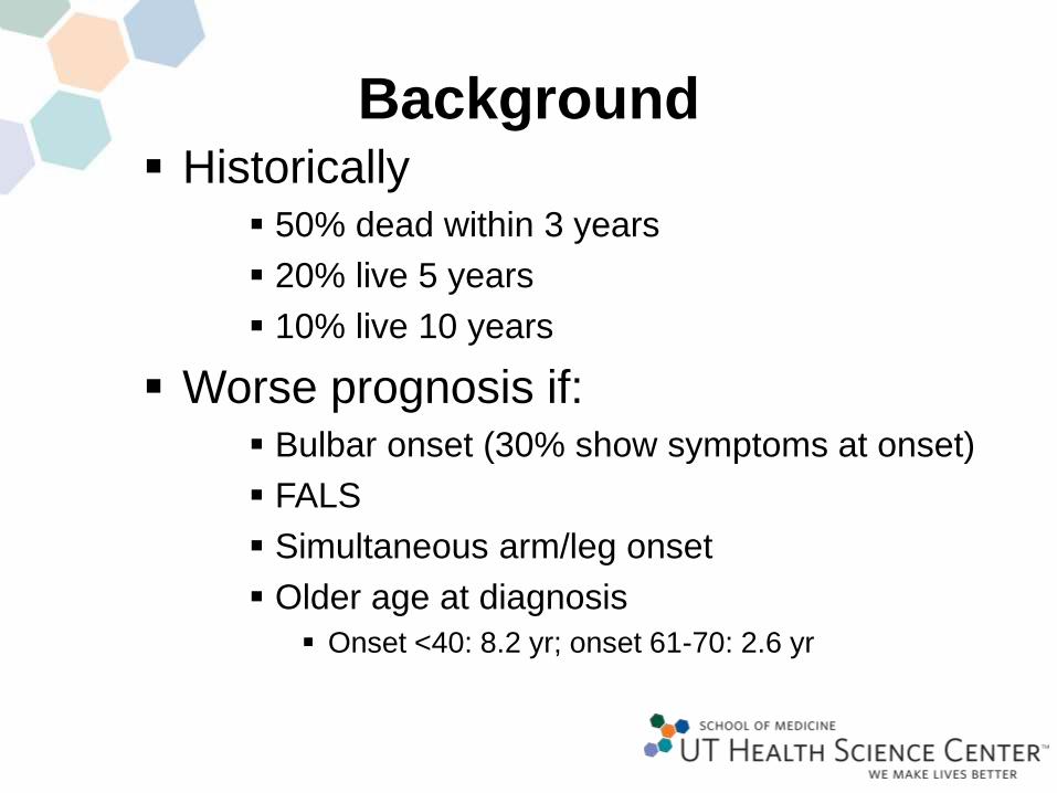
Background Historically

50% dead within 3 years

20% live 5 years

10% live 10 years

Worse prognosis if: Bulbar onset (30% show symptoms at onset)

FALS

Simultaneous arm/leg onset

Older age at diagnosis

Onset <40: 8.2 yr; onset 61-70: 2.6 yr

AAN Guidelines

• In patients with ALS, impaired oral food intake, enteral

nutrition via PEG should be considered to stabilize body

weight

• Insufficient data to support or refute specific timing of

PEG insertion in ALS patients

• Peg should be considered for prolonging survival in

patients with ALS

Procedure Options

Percutaneous Endoscopic Gastrostomy (PEG)

Percutaneous Endoscopic Jejunostomy (PEJ)

Radiologically Inserted Gastrostomy (RIG)

Complications

• Aspiration

• Bleeding

• Bowel

perforation

• Peritonitis

• Death

• Local infection

• Buried bumper

• Tube

blockage/breakdown

• Stoma leakage

• Inadvertent peg tube

removal

Our Study

Primary

• To review the authors’ experience of PEG

tube placement in patients with

amyotrophic lateral sclerosis with different

degrees of impaired respiratory function

• Secondary• To demonstrate the safety profile, peri-procedural and

post-interventional complications of PEG tube insertion

performed in an outpatient surgical center.

What we did

Retrospective medical records review

of patients treated at the University of

Texas Health Science Center of San

Antonio ALS clinic who were referred

for PEG tube placement using propofol

sedation at an outpatient surgical

center between 2011-2014

Methodology

• Three year period:

– Total referrals to Gastroenterology 145

• Less those that did not meet criteria 27

• Total number of charts reviewed 118

• Less those who refused peg 28

76% of all referrals resulted in peg placement• 24% of all referrals were refused
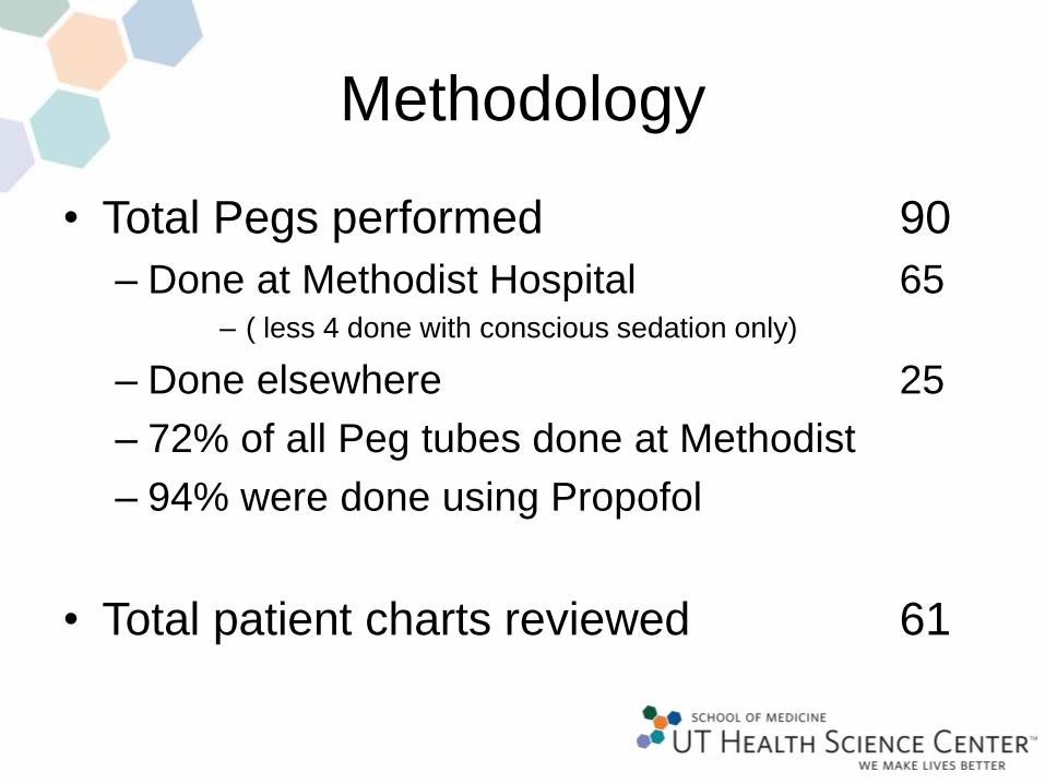
Methodology

• Total Pegs performed 90

– Done at Methodist Hospital 65– ( less 4 done with conscious sedation only)

– Done elsewhere 25

– 72% of all Peg tubes done at Methodist

– 94% were done using Propofol

• Total patient charts reviewed 61

Age

0

5

10

15

20

25

30

35

40

>75 (7) 55-75 (37) <55 (17)

Oldest

84 Y 7M

Youngest

31 Y 6 M

Median 64

Mean 62

Mode 64

Forced Vital Capacity

0

5

10

15

20

25

30

<30% (9) 30-50% (26) >50% (24)

Lowest FVC 13%

Highest FVC 102%

BMI

0

5

10

15

20

25

30

35

40

High-38 Median-24 Mean-25 Mode-24 Low-13

ALSFRS-R

0

5

10

15

20

25

30

35

40

45

50

High (45) Median (22) Mean (24) Mode (17) Low (8)

Observations

• Procedure duration (anesthesia time/surgery time) averaged 29/11

minutes (range 19-45/4-27).

• Total propofol dose ranged from 60 to 500mg.

• Patients currently on Bipap brought their own devices and used

them in the intra and post-operative period as needed.

• No patients required invasive ventilatory support in the immediate

post op period

• No symptomatic cardiac arrhythmias or hypotension.

• Very low use of post operative pain medication.

• All patients that developed complications also had significant

comorbidities.

• There was no correlation between complications and FVC, BMI,

disease duration or ALSFRS-R scale.

Complications

0 50 100

C-1 at 1 day

C-2 at 2 days

C-3 at 10 days

C-4 at 2 weeks

C-5 at 30 days

Age

FVC

BMI

ALSFRS-R

Mean Survival

• FVC <30% 7 months (4-22 M)

• FVC 30-50% 11 months (2 D-29 M)

• FVC >50% 10 months (1D-30M)

GI Findings

• Gastric Polyps (5)

• Gastritis (4)

• Barrett’s Esophagus (3)

• Hiatal Hernia (3)

• Esophagitis (1)

• Esophageal Thrush (1)

• Duodenitis (1)

• GERD (1)

• Schatzki’s Ring (1)

• Diverticulum (1)

What does this mean?

PEG tube placement with propofol sedation and Bipap support

performed at a specialized outpatient surgical center can be achieved at

a reasonable risk even in patients with very low FVC (less than 30%).

Relevant findings

• Wide range of FVC (including several with very low)

• Short procedure time

• No need for invasive respiratory support at time of procedure

• No intraoperative complications

• Overall low incidence of complications

• Low need for other medication besides propofol

• No inpatient admissions post procedure

Future Research

• Quality of life for patients/caregivers

– Does this really make a difference

• Replicate this finding in other centers

– Controlled environment/providers/staff

Resources• Amornyotin S, et al. Propofol-Based Sedation Does not Increase Rate of Complication during

Percutaneous Endoscopic Gastrostomy Procedure. Gastroenterology Research and Practice Vol

2011, Article 1D 134819.

• Chio A, et al. Percutaneous radiological gastrostomy: a safe and effective method of nutritional

tube placement in advance ALS. Journal of Neurology Neurosurgery Psychiatry 2004: 74:645-647

• Czell D, et al. Outcomes of percutaneous endoscopic gastrostomy tube insertion in respiratory

impaired amyotrophic lateral sclerosis patients under noninvasive ventilation. Respiratory Care

May 2013; 58(5): 838-44.

• Duzak R, et al. Percutaneous Gastrostomy and Jejunostomy.

http://emedicine.medscape.com/article/182157. Updated 1/14/2014; 1-15.

• Gregory S, et al. Gastrostomy insertion in ALS patients with low vital capacity: respiratory support

and survival. Neurology 2002; 58(3):485-487.

• http://www.alsa.org/als-care/resources/publications-videos/factsheets/feeding-tubes.html

• Jawaid A, et al A decrease in body mass index is associated with faster progression of motor

symptoms and shorter survival in ALS. Amyotrophic Lateral Sclerosis December 2010; 11(6):

542-548.

• Laasch, HU et al. Gastrostomy insertion: comparing the options—PEG, RIG or PIG? Clinical

Radiology. May 2003: 58(5):398-405.

• Lynch CR, et al. Prevention and Management of Complications of Percutaneous Endoscopic

Gastrostomy Tubes. Practical Gastroenterology November 2004: 66-76.

Resources

• Miller RG, et al. Practice parameter: the care of the patient with amyotrophic lateral sclerosis (an

evidence-based review): report of the Quality Standards Subcommittee of the American Academy

of Neurology: ALS Practice Parameters Task Force. Neurology 1999;52:1311-1323.

• Mitsumoto H, et al. Percutaneous endoscopic gastrostomy (PEG) in patients with ALS and bulbar

dysfunction. Amyotrophic Lateral Sclerosis Other Motor Neuron Disorders. 2003; 4(3): 177-85.

• Potack JZ, et al. Complications of and Controversies Associated with Percutaneous Endoscopic

Gastrostomy: Report of a Case and Literature Review.

http://www.medscape.com/viewarticle/573059 6/17/2008.

• Procaccini NJ, et al. Percutaneous Endoscopic Gastrostomy in the patient with ALS Risk vs

Benefit? Practical Gastroenterology March 2008: 24-34.

• Stavroulaskis T, et al. A Prospective Multi-centre evaluation of Gastrostomy in Patients with MND.

24th International Symposium on ALS/MND December 2013: Abstract CW20 page 39.

• Thornton FJ, et al. Amyotrophic Lateral Sclerosis: enteral nutrition provision—endoscopic or

radiologic gastrostomy. Radiology. 2002 Sept; 224(3):713-717.

Acknowledgments

Alejandro Tobon MD

Christopher Fincke MD

Tina Motazedi MS3

Carlayne Jackson MD

The University of Texas Health Science Center at San

Antonio, Department of Neurology,

Neuromuscular Division

Methodist Healthcare System, Methodist Hospital

[email protected]


Recommended