2
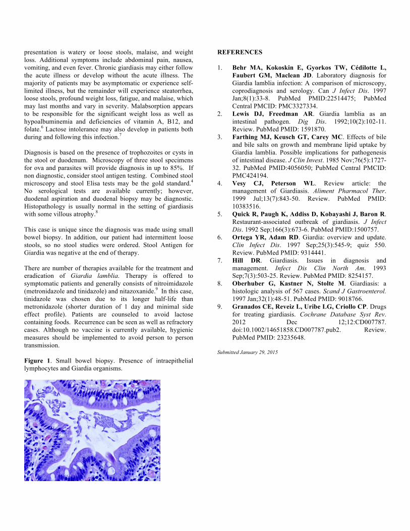
Proceedings of UCLA Healthcare -VOLUME 19 (2015)- CLINICAL VIGNETTE Presentation of Giardia lamblia Found on a Small Bowel Biopsy Rimma Shaposhnikov, M.D., and Roman Leibzon, M.D. Abstract Giardia lamblia is a ubiquitous intestinal protozoan with fecal-oral transmission. Patients may present with acute symptoms such diarrhea, malaise, or abdominal pain. However, they may also present with subacute nausea, anorexia, or weight loss. Chronic symptoms may develop in up to half of individuals and include steatorrhea, malabsorption, and weight loss. Enzyme immunoassay and direct fluorescent-antibody assay for antigen detection in stool sample; small intestine biopsy provide the best diagnostic sensitivity. The most effective drugs in the treatment of this infection are metronidazole and tinidazole. We report a case of G. lamblia infection in a patient with interstitial lung disease, rheumatoid arthritis on prednisone, and pulmonary arterial hypertension. The patient presented with a 3-month history of malaise, weight loss, and loose stools. Patient denied any recent travel. The diagnosis was established after duodenal biopsy. The patient was successfully treated with one dose of tinidazole. Case report A 61-year-old Hispanic male with underlying interstitial lung disease, rheumatoid arthritis, scleroderma, and pulmonary hypertension presented with a new onset of early satiety and weight loss. He reported a 10 lb weight loss over the past three months and no loss of appetite. He noted fatigue, looser stools, and increased flatulence, but he denied diarrhea, foul smelling, or fatty stools. He denied fever, chills, vomiting, and nausea. Patient denied sick contacts or recent travel. He has small grandchildren in good health. His medications included prednisone 10 mg, azathioprine 50 mg, bosentan 125 mg, leflunomide 20 mg, tadalafil 40 mg, Lisinopril, and furosemide. His physical appearance and exam were unremarkable. He had long standing macrocytic anemia with Hgb 12.4 and MCV 105, attributed to azathioprine. Other than prednisone taper, all other medications were unchanged. Prior to GI consultation, patient was evaluated by his pulmonary specialist and cardiologist, and no changes in the status of his chronic problems were noted. Endoscopy was performed due to persistent weight loss and intermittent symptoms of looser stools. Small bowel appeared normal, but the biopsy returned positive for multiple organisms consistent with Giardia lamblia, as well as an increase in intraepithelial lymphocytes. (Figure 1) His prior colonoscopy was in 2013, at which time two polyps were noted and removed, and small bowel was biopsied showing normal ileal mucosa with benign lymphoid aggregates, consistent with Peyer's patches. There was no evidence of microscopic or macroscopic colitis. Patient was treated with one dose of tinidazole and noted immediate improvement in his stools, weight, and energy level. Patient was counseled to avoid lactose containing foods. Discussion Giardia lamblia is the most common protozoal infection found in the GI tract with prevalence from 7-30% worldwide, especially in areas with poor sanitation and poor water treatment. 1 It has been reported to cause gastroenteritis, traveler’s diarrhea, and outbreaks of diarrhea in children at daycare centers. The infection results from infestation with parasite Giardia intestinalis, also known as G. duodenalis or G. lamblia. There are two stages in its life cycle: an actively replicating trophozoite and a resistant cyst. Cysts are formed as the parasite passes from the small bowel of the host. 2 Prior studies have identified the need for host bile acid for the growth of organisms. This may explain the preferred location of the duodenum and proximal jejunum. 3 Infection can be caused by ingestion of as low as 10 cysts. After excystation, replicative trophozoites may attach to the small bowel wall, but do not invade it. Trophozoites may encyst and be shed in feces for potential ingestion by a different host. Although trophozoites may cause an infection, they have limited ability to survive outside the bowel. 4 Giardia cysts may be transmitted via water, raw and undercooked food, or fecal-oral transmission. There is significant controversy regarding the zoonotic nature of giardiasis since no human outbreak can be linked to a specific animal, and yet there are significant genetic similarities between humans and animal isolates of Giardia. Severity is variable since up to 15% of individuals shed cysts asymptomatically. Symptomatic infection, seen in up to 50% of patients, depends on the virulence of the isolate, the parasite load, as well as the host’s immune system. 2 The incubation period ranges from 7 to 14 days but may be as long as 45 days with symptoms lasting up to four weeks. 5 The most common