Presented at the 2011 COMSOL Conference in Boston Deformation of Biconcave Red Blood Cell in the Dual-Beam Optical Tweezers Lingyao Yu 1 , Yi He 2 , Arthur Chiou 2 , and Yunlong Sheng 1 1 Center of Optics, Photonics and Lasers, Dept. of Physics, University Laval, Quebec, Canada 2 Institute of Biophotonics Engineering, National Yang-Ming University, Taipei, Taiwan
Transcript
Presented at the 2011 COMSOL Conference in Boston
Deformation of Biconcave Red Blood Cell in the Dual-Beam
Optical Tweezers
Lingyao Yu1, Yi He2, Arthur Chiou2, and Yunlong Sheng1
1 Center of Optics, Photonics and Lasers,
Dept. of Physics, University Laval, Quebec, Canada
2 Institute of Biophotonics Engineering,
National Yang-Ming University, Taipei, Taiwan
Content
1. Manipulating RBC with optical tweezers
2. Steps of calculation
3. Models of COMSOL Multiphysics
4. Computation and Experiment results
5. Conclusions and Prospects
Manipulating the human red blood cell (RBC) with optical tweezers
Transportability RBC
(erythrocyte)
Mechanical force
of cell Deformability
Optical traps
Cited papers about manipulating the RBC with optical tweezers
Statistics from the web of science database: http://apps.webofknowledge.com/CitationReport.do?product=WOS&search_mode=CitationReport&SID=4Ea9dB6o@LaEK7LG6nJ&page=1&cr_pqid=7&viewType=summary
Acc
ount
s of
Pap
ers
Year
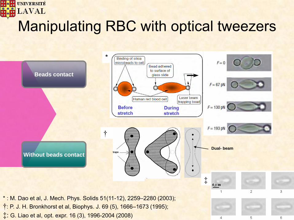
Manipulating RBC with optical tweezers
*
Beads contact
Without beads contact
* : M. Dao et al, J. Mech. Phys. Solids 51(11-12), 2259–2280 (2003); †: P. J. H. Bronkhorst et al, Biophys. J. 69 (5), 1666–1673 (1995); ‡: G. Liao et al, opt. expr. 16 (3), 1996-2004 (2008)
†
Dual- beam
‡
Dual-beam optical tweezers
†: G. B. Liao et al, Opt. Express 16(3), 1996–2004 (2008); ‡: Y. Sheng at el, COMSOL Conference Boston (2010).
‡
†
Jumping beam
Advantages of the dual-beam optical tweezers
No physical contact to the specimen
Probing the characteristics of the cellular membrane and cytoskeleton by Manipulating living biological cells
Photonics’ shear force is in the same order of magnitude (pN) as the mechanical force for deforming the cell
Steps of Simulation
1. Geometric construction of the biconcave
human RBC
2. The background electromagnetic fields
of dual-beam optical tweezers;
3. Compute stress distribution with Maxwell
Stress tensor in RF ModuleTM
4. Compute Deformation of RBC with solid mechanicsTM module
Geometry of a biconcave RBC
4
0
222
220
22
100
22
041
D
zxc
D
zxcc
D
zxDy
* : E. Evans, and Y. Fung, Microvascular research, 4 (1972) 335-347
D0= 7.8 μm,
c0= 0.207161,
c1= 2.002558,
c2= -1.122762.
*
RBC model
PML Buffer
RBC membrane
cytosol
RBC solid geometry
n1=1.335
n2=1.378
d=1.5%D0
Introduction of background field
Spherical wave
Linear/radial polarized
Gaussian intensity
Highly focused (1.25NA)
Introduction of background field
212222
220
11 22jexp)2(exp zySxzW
ySx
zW
WAEb
2/12
00 1)(
z
zWzW
n
NAW
10
sintan
20
0W
z
212222
220
22 22jexp)2(exp zySxzW
ySx
zW
WAEb
Spherical wave Gaussian intensity Relative to
beam power
Maxwell stress tensor
zzyyxxn nEnEnEnEE
nEEn
nnn tn
22
22
212
221
0
2
zyx
zyxt
nnn
EEE
zyx
nEE
ˆˆˆ
IBEBBEET 2
0
2
0
1211
tangent
normal
Interface of our model
Two electromagnetic waves modules as dual-beam optical tweezers,
respectively
Stress calculated from the RF
modules will be loaded in Solid
mechanics module
Constraints of prescribed displacement have also been set
Initial Stress on cell surface
The normalized stress distribution in different beam separations S=3.1 (a), 3.8 (b), 4.5 (c), 5.2 (d), 5.9 (e), 6.6 (f), 7.0 (g), and 7.3 (h) µm with COMSOL multiphysics.
Redistribution of stress on the deformed cells
1 2 3
4 5 6
7 8 9
Final deformations
Fitting to Experimental Results
E= 650 Pa; Eh=24μN/m; Gh=17μN/m
Conclusion
RF module is used to compute the scattered EM field instead of geometrical optics;
RF module and Structural mechanics module are combined with ComsolTM strongly coupled solver;
Natural biconcave shape of RBC is calculated instead of the swollen spherical RBC;
Computed deformations are fit to experimental data to determine the elasticity of the RBC .
Prospects
• The deformation of the arbitrary shape of the cell can be simulated with the same method as well as the organelle and biomolecules (like the cell membranes, proteins, and DNAs).
• A variety of mechanical characteristics of human cells can be explored