Pressure Sores The complex etiology of pressure sores requires a multidisciplinary approach to treatment. The surgeon cannot simply schedule the patient for surgery and expect a healed wound that will not reoccur. Nursing home caregivers and primary care physicians should regularly evaluate their elderly and wheelchair-bound patients for the first signs of tissue necrosis. Internists can manage metabolic and physiologic diseases such as providing tight glucose control in diabetics. Dietitians can determine energy requirements and suggest nutritional supplements to meet the patient’s caloric goals. Social workers should investigate the patient’s home for environmental hazards and help eliminate financial barriers to therapeutic intervention. The plastic surgeon examines the patient for severity of the wound and determines the best surgical course of action, if indicated. The plastic surgeon can coordinate and mobilize the many team members. Case managers schedule appointments, coordinate trans- portation for routine checkups, and help obtain needed equipment. If a surgical intervention is pursued, specially trained nurses can assist with postsurgical flap checks, dressing changes, and other nursing functions; they are an extension of the plastic surgeon throughout the acute setting. Finally, physical and occupational therapists evaluate patient mobility and recommend custom equipment to help with healing and prevention of future pressure sore formation. These therapists can assist with range of motion exercises to help treat and prevent flexion contractures as well as activities of daily living. With a combined multidisciplinary team, patients receive the best prognosis for long-term pressure sore treatment. Terminology The immobilized patient is at risk for developing breakdown of his or her skin and soft tissue. This breakdown is the necrosis of skin, subcutaneous tissue, and muscle, collectively referred to as a pressure sore by the medical community. The term pressure sore has several anecdotal synonyms rooted in history, including bedsore and decubitus ulcer. The term decubitus comes from the Latin word decumbere meaning Curr Probl Surg 2007;44:101-143. 0011-3840/2007/$30.00 0 doi:10.1067/j.cpsurg.2006.12.007 Curr Probl Surg, February 2007 101
Transcript
TasceIpephtsitpsptFrfmap
T
hssau
C0d
C
Pressure Sores
he complex etiology of pressure sores requires a multidisciplinarypproach to treatment. The surgeon cannot simply schedule the patient forurgery and expect a healed wound that will not reoccur. Nursing homearegivers and primary care physicians should regularly evaluate theirlderly and wheelchair-bound patients for the first signs of tissue necrosis.nternists can manage metabolic and physiologic diseases such asroviding tight glucose control in diabetics. Dietitians can determinenergy requirements and suggest nutritional supplements to meet theatient’s caloric goals. Social workers should investigate the patient’some for environmental hazards and help eliminate financial barriers toherapeutic intervention. The plastic surgeon examines the patient foreverity of the wound and determines the best surgical course of action,f indicated. The plastic surgeon can coordinate and mobilize the manyeam members. Case managers schedule appointments, coordinate trans-ortation for routine checkups, and help obtain needed equipment. If aurgical intervention is pursued, specially trained nurses can assist withostsurgical flap checks, dressing changes, and other nursing functions;hey are an extension of the plastic surgeon throughout the acute setting.inally, physical and occupational therapists evaluate patient mobility andecommend custom equipment to help with healing and prevention ofuture pressure sore formation. These therapists can assist with range ofotion exercises to help treat and prevent flexion contractures as well as
ctivities of daily living. With a combined multidisciplinary team,atients receive the best prognosis for long-term pressure sore treatment.
erminologyThe immobilized patient is at risk for developing breakdown of his orer skin and soft tissue. This breakdown is the necrosis of skin,ubcutaneous tissue, and muscle, collectively referred to as a pressureore by the medical community. The term pressure sore has severalnecdotal synonyms rooted in history, including bedsore and decubitus
lcer. The term decubitus comes from the Latin word decumbere meaning
to lie down.” Each of these words attempts to identify the underlyingathophysiology of wounds due to physical stress. However, these termsail to address the existence of pressure sores not due to lying in a beduch as in wheelchair-bound patients and in nondependent locations.1
urrently, the term pressure sore best describes these complex woundsecause they are multifactorial in nature and can occur anywhere on theody.
tiologyPressure sores are complex wounds that result from 1 or more
ontributing factors. Stress, time, spasticity, infection, edema, denerva-ion, moisture, and poor nutrition are considered fundamental issues thatesult in or contribute to pressure sore development. More than 60% ofressure sores develop within the hospital wards.2 One probable source ishe increasing geriatric population requiring hospitalization in whom onehird to one half experience a functional decline.3 Debilitation andevelopment of a pressure sore type of injury poses a legal liability risko healthcare practitioners and hospital risk managers.4,5 This nosocomialondition requires vigilant preventative measures.
tressPosition and contact with the environment is a critical component to theressure sore injury. The gravitational pull on the human body results inontact with the bed, wheelchair, or other surface. This contact is enhancedt surfaces of the body that overly bony prominences6 (Fig 1). The softissues become compressed between hard external objects and thenderlying bone, leading to localized ischemia and eventual necrosis.his force exerted on tissues is considered the sine qua non of pressureore formation.The famous Robert Pershing Wadlow provides a lesson in the pathologyf how position and contact with objects can lead to detrimental effects.r. Wadlow holds the Guinness Book of World Records title for theorld’s tallest man at 8 feet 11 inches (272 cm) and weighing approxi-ately 200 kg.7 He developed a blister on his ankle that became infected
fter being poorly fitted for leg braces. One week later he died from sepsist the age of 22.
imeProlonged contact between the human body and hard objects results in
schemic necrosis and pressure sore formation of the intervening tissues.
osiak and colleagues, demonstrated the inverse parabolic relationship
02 Curr Probl Surg, February 2007
bmapi
FuP
F
C
etween time and pressure in pressure sore formation using a canineodel8 (Fig 2). A high level of pressure induces tissue ulceration withinshort time interval, and conversely, low levels of pressure require a
rolonged timeframe to cause tissue damage. The 1 caveat in this study
IG 2. Time versus pressure curve for tissue breakdown in a canine model.
s the use of dogs, a loose-skinned animal with less subcutaneous tissue
urr Probl Surg, February 2007 103
tctimsdtto
saieCasrc
S
ppo(
F
1
han in humans. A porcine model, whose subcutaneous tissue morelosely models humans, was used by Dinsdale to establish irreversibleissue damage after 2 hours of constant pressure of 70 mm Hg.9 However,f the pressure was relieved for at least 5 minutes, tissue damage wasinimized even at elevated levels of stress. Husain used a rat pressure
ore model to conclude that sustained low pressure induces more tissueamage than brief periods of high pressure.10 These basic science investiga-ions form the core body of knowledge describing how the element ofime and its relationship to soft tissue necrosis is critical to the formationf pressure sores.Prolonged contact with objects is associated with patient immobility for
everal reasons, including broken bones, paralysis, psychiatric diagnoses,nd loss of consciousness. Paralysis may be due to trauma, neoplasm,nfection, iatrogenic injury, and radiation.11-15 Suspicion of an infectioustiology requires careful delineation between a bacterial source such asampylobacter jejuni and viral subtypes that include the West Nile virusnd the rare poliomyelitis.16 Psychiatric diseases such as catatonicchizophrenia should be treated promptly.17 All of these patients are atisk for the development of pressure sores as the time of immobilityontinues.
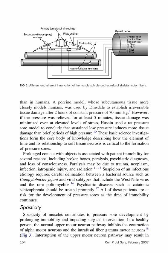
pasticitySpasticity of muscles contributes to pressure sore development byrolonging immobility and impeding surgical intervention. In a healthyerson, the normal upper motor neuron pathway inhibits the contractionf alpha motor neurons and the intrafusal fiber gamma motor neurons18
IG 3. Afferent and efferent innervation of the muscle spindle and extrafusal skeletal motor fibers.
Fig 3). Interruption of the upper motor neuron pathway may result in
04 Curr Probl Surg, February 2007
hTatst
OrbsGsbpsastpmmc
I
imadt
T
(1
C
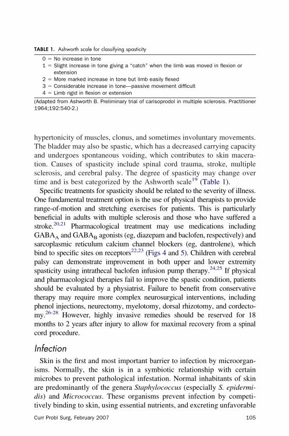
ypertonicity of muscles, clonus, and sometimes involuntary movements.he bladder may also be spastic, which has a decreased carrying capacitynd undergoes spontaneous voiding, which contributes to skin macera-ion. Causes of spasticity include spinal cord trauma, stroke, multipleclerosis, and cerebral palsy. The degree of spasticity may change overime and is best categorized by the Ashworth scale19 (Table 1).Specific treatments for spasticity should be related to the severity of illness.ne fundamental treatment option is the use of physical therapists to provide
ange-of-motion and stretching exercises for patients. This is particularlyeneficial in adults with multiple sclerosis and those who have suffered atroke.20,21 Pharmacological treatment may use medications includingABAA and GABAB agonists (eg, diazepam and baclofen, respectively) and
arcoplasmic reticulum calcium channel blockers (eg, dantrolene), whichind to specific sites on receptors22,23 (Figs 4 and 5). Children with cerebralalsy can demonstrate improvement in both upper and lower extremitypasticity using intrathecal baclofen infusion pump therapy.24,25 If physicalnd pharmacological therapies fail to improve the spastic condition, patientshould be evaluated by a physiatrist. Failure to benefit from conservativeherapy may require more complex neurosurgical interventions, includinghenol injections, neurectomy, myelotomy, dorsal rhizotomy, and cordecto-y.26-28 However, highly invasive remedies should be reserved for 18onths to 2 years after injury to allow for maximal recovery from a spinal
ord procedure.
nfectionSkin is the first and most important barrier to infection by microorgan-
sms. Normally, the skin is in a symbiotic relationship with certainicrobes to prevent pathological infestation. Normal inhabitants of skin
re predominantly of the genera Staphylococcus (especially S. epidermi-is) and Micrococcus. These organisms prevent infection by competi-
ABLE 1. Ashworth scale for classifying spasticity
0 � No increase in tone1 � Slight increase in tone giving a “catch” when the limb was moved in flexion or
extension2 � More marked increase in tone but limb easily flexed3 � Considerable increase in tone—passive movement difficult4 � Limb rigid in flexion or extension
Adapted from Ashworth B. Preliminary trial of carisoprodol in multiple sclerosis. Practitioner964;192:540-2.)
ively binding to skin, using essential nutrients, and excreting unfavorable
urr Probl Surg, February 2007 105
F
Fmr
1
IG 4. The GABAA receptor.
IG 5. The ryanodine receptor is responsible for mediating intracellular calcium mobilization inuscle cells. Dantrolene prevents calcium release by the ryanodine receptor in the sarcoplasmic
eticulum.
06 Curr Probl Surg, February 2007
cwb
cccitTpipdpd
Sbabcwifpgr
E
aosiphco
c
C
ompounds.29 An example of the latter is Propionibacterium acnes,hich secretes oleic acid, an inhibitor of fungi and Gram-negativeacteria.By maintaining proper hygiene, the opportunity for pathologic bacteria to
ause infection is reduced.30 Similarly, nosocomial sources of infection areombated by frequent hand washing and the use of alcohol-based skinleansers.31 Soiling of the skin—such as from urine and feces—canntroduce microorganisms to the skin surface. This is especially of concern inhe paraplegic, wheelchair-bound patient with a developing pressure sore.he use of Foley catheters or intermittent catheterization is a must-userevention technique for patients with neurogenic bladders. The presence ofnfectious microbes, which flourish in the media-like cellular breakdownroducts due to tissue necrosis, at a minimum prevents healing of theamaged tissues. Soft tissue infections can spread superficially along tissuelanes and deeper into subcutaneous structures, requiring advanced surgicalebridement, which complicates reconstructive efforts.Several studies have examined colonization of traumatized tissue.urgical sites can become infected from distant, endogenous sources ofacteria, and a pressure sore can become a nidus for infection.32 Afterttaching metallic spheres to rabbit gluteal muscles, Groth injected aacterial solution into their veins.33 The bacteria localized to sites ofompression and resulted in necrosis at lower pressures when comparedith control animals not receiving the bacterial injection. Similarly,
njection of bacteria and then applying pressure serves as a homing deviceor microbes.10 In fact, experimental surgical wounds created at sites ofressure that are infected with a known bacterial load have bacterialrowth that is 102 higher than controls.34 Thus, compressed skin has lessesistance to bacterial invasion.35
dema, Denervation, and MoistureThe fluid environment of pressure sores contributes to both infection
nd necrosis. Fluid accumulates either as edema in the interstitial spacesr as moisture surrounding the outside of the wound area. Compression ofoft tissues causes edema by occluding venous outflow and raising thenterstitial pressure in the affected tissue. Edema can be secondary toatient disease states. Malnutrition and cirrhosis of the liver causingypoalbuminemia, a low cardiac ejection fraction, and renal diseases canause edema. The resulting edema accumulates in the dependent regionsf the body, which is a factor of positioning in the bed or chair.The investigation into how edema affects tissue perfusion has been
ritical to our understanding why tissue necrosis occurs. Landis used a
urr Probl Surg, February 2007 107
mcopFcrid
ittDn
tmAflSaedcttda
mTuip(r
N
p
1
icroinjection technique to discover the blood pressure of individualapillaries.36 An external pressure of greater than 12 mm Hg will result incclusion at the venous end of the capillary. End capillary venousressure then rises and causes plasma extravasation and ensuing edema.urther increasing the pressure to 32 mm Hg occludes blood flow into theapillary, preventing tissue perfusion with oxygen and nutrients andemoval of toxic metabolic waste products. With the passage of time,schemia and necrosis result, and this promotes pressure sore formation asiscussed previously.A contributing factor to edema is denervation of blood vessels. The
nability of the capillary’s afferent supply to regulate its diameter defaultso a state of dilation.37 Such denervation edema has been documented inhe ipsilateral one half of the tongue after reconstructive surgery.38
ocumentation of denervation edema is best accomplished using mag-etic resonance imaging.39
Several studies on denervation edema demonstrate the serious nature ofhis problem. Acutely denervated rat cremaster muscles experience aore severe ischemia-reperfusion injury after 3 hours of ischemia.40
lison and colleagues studied the effects of denervation on island pedicleaps in sheep buttocks that receive an intradermal inoculation with 107
taphylococcus aureus.41 The prolonged denervation group demonstrated25-fold increase in bacterial counts and a significant increase in tissue
dema. A small study comparing 6 patients with chronic sympatheticenervation to 5 healthy controls found that nondenervated limbs de-reased blood flow by 50% when venous pressure was elevated to morehan 30 mm Hg.42 At 40 mm Hg, nondenervated limbs have one third lesshe capillary filtration of chronically denervated limbs. These studiesemonstrate the importance of intact innervation to regulate blood flownd prevent edema in tissues.Moisture is the external accumulation of fluid on skin. The source ofoisture can be from perspiration, incontinence, and wound exudate.hese liquids cause maceration—the wrinkling of skin—which contrib-tes to the setup for infection after prolonged exposure. Thus, macerations a contributing factor to the prevention of wound healing. Moisture onressure sores should be prevented by (1) frequent dry dressing changes,2) the use of urinary catheters for incontinent patients, and (3) bowelegimens that include examination for and cleansing of stool.
utritionNutritional intervention for pressure sore patients must be part of the
lastic surgical evaluation. Poor nutrition is a risk factor for the formation
08 Curr Probl Surg, February 2007
opescmaenbo
EtAemvhbf
cspnidapish
pfi
T
C
f pressure sores, their exacerbation, and prevent healing from surgicalrocedures. Certain basic metabolic components are recognized asssential for survival and the repairing of surgical wounds.43 The patienthould be evaluated carefully by a nutritional expert to determine thealoric needs and whether these demands are being met. The causes ofalnutrition are manifold: poor intake (eg, alcoholism), chronic nausea
nd vomiting (eg, pancreatitis), hypermetabolic states (eg, burns), socio-conomic forces (eg, natural disasters), and internal organ diseases (eg,ephrotic syndrome). Supplementing nutritional intake is best performedy encouraging an appropriate oral diet. However, duodenal feeding tubesr parenteral feeds should be used to prevent a catabolic state.Proper caloric intake is necessary for the human body to function.nergy requirements are a product of the body’s basal energy expendi-
ure. This can be calculated using the Harris Benedict formula (Table 2).lthough debatable, a patient’s daily caloric need is the basal energy
xpenditure multiplied by a stress factor: 1.3 for mild stress, 1.4 foroderate stress, and 1.5 for severe stress. In comparison, burn patients are
ariably hypermetabolic and should be examined clinically at the time ofospitalization.44 Protein requirement also ranges from 1 to 2 g/kg ofody weight per day.45 The end goal is to meet nutritional requirementsor normal wound healing without overfeeding.Evaluation of the patient’s current nutritional status often includes
linical laboratory tests such as albumin or prealbumin. Fifty percent oferum protein synthesized by the liver is albumin, the most abundantlasma protein. Yet, serum albumin levels are not valuable indicators ofutritional status in the marasmic and critically ill patient.46 An exclusions made for individuals with kwashiorkor whereby the high carbohydrateiet results in a state of low serum albumin and tissue edema.47 Studieslso fail to link prealbumin as a predictor of patient nutritional status andrognosis in critically ill patients.48 Clinical judgment should be appliedn patients receiving nutritional therapy; the patient should be gainingtrength and the wound site should be inspected regularly for signs ofealing.Many nutrients, vitamins, and minerals contribute to the wound healingrocess. One well-demonstrated example is the synthesis of collagen by
broblasts (Fig 6). Amino acids, particularly lysine and proline, must be
urr Probl Surg, February 2007 109
avhtihp
ottvct
dnhhgshgao
F
1
vailable to synthesize the collagen protein. Additionally, oxygen, iron,itamin C, and alpha-ketoglutarate are necessary cofactors for theydroxylation of lysine and proline to allow intermolecular bonding ofhe collagen polypeptides into the triple helix procollagen. Failure tongest adequate amounts of vitamin C results in decreased strength ofealing scars, a prolonged healing process, and even dehiscence ofreviously healed scars.49
Other supplements should be provided to the pressure sore patient forptimal wound healing. Calcium is used for the conversion of procollageno fibrillar collagen. Iron, folate, and vitamin B12 supplementation helpso optimize erythropoiesis in the anemic patient. Adequate levels ofitamin K are essential for blood clotting. Additionally, patients takingorticosteroids should receive vitamin A supplementation to counteracthe lysosomal stabilization that retards the wound healing process.50
In addition to nutritional deficiencies, metabolic disturbances such asiabetes should be monitored carefully and treated to prevent delaying theormal healing process.51-53 Diabetes mellitus is a chronic disorder ofyperglycemia requiring daily monitoring for euglycemia and quarterlyemoglobin A1C levels. Several problems are associated with the hyper-lycemic state. In a diabetic rat model, Komesu and colleagues demon-trated a prolongation in the inflammatory response phase of woundealing.54 Fahey and colleagues established that diabetic mice fail toenerate proper interleukin-6 levels after 7 days of healing.55 Histologicalnalysis of these wounds found decreased neovascularization and less
IG 6. The pathway for collagen synthesis.
rganization of granulation tissue in the wounds of diabetic mice.
10 Curr Probl Surg, February 2007
irgoIidmi
L
pbflt
FJC
C
Robson and Heggers56 performed in vitro and in vivo studies onnfection and varying glycemic levels. They demonstrated an increasedisk of Gram-positive septicemia in patients with elevated levels of serumlucose of at least 130 mg/100 mL.56 Conversely, Gram-negative sepsisccurred in patients with blood glucose levels of less than 110 mg/100 mL.t is not surprising that a diabetic patient with an ulcer is at greater risk ofnfection and complications above and beyond nondiabetic patients,espite tight glucose control and meticulous wound care.53 Thus, theetabolic status of diabetic patients affects their ability to resist bacterial
nfection, prevent skin breakdown, and promote wound healing.
ocation of Pressure SoresThe vast majority of pressure sores result from contact between theatient and the posterior or lateral surface of the pelvis. The surgeon muste cognizant that other locations of the body can undergo stress necrosisrom prolonged contact with objects (Figs 7 and 8). This includes armseaning over bed rails, nasogastric and feeding tubes taped upward against
IG 7. Bed rail-induced pressure sore. (With permission from Colen SR. Pressure sores. In: McCarthyG, editor. Plastic Surgery: The Trunk and Lower Extremity. Philadelphia: WB Saunders; 1990;hapter 77, pp 3797-838.)
he nasal ala, and the forehead or mental area in prone patients.
urr Probl Surg, February 2007 111
ppiesdgtapbasroa
F
1
As noted previously, the immobilized person is most susceptible toressure sore formation. From the Yeoman and Hardy study of 240araplegics we know that more than 50% of pressure sores form at theschium and sacrum57 (Table 3). The study also documented the heel,xternal malleoli, tibial crest, and costal margin as nonpelvic sitesusceptible to pressure sore formation. Such information was virtuallyuplicated by Dansereau and Conway in their study of 2000 paraple-ics with pressure sores, adding only the high incidence of trochan-eric pressure sores58 (Table 4). In a study from Denmark by Petersennd Bittmann, almost two thirds of pressure sores occurred whileatients are in the hospital.2 Fifty-three percent of patients wereedridden, one third were wheelchair bound, and only 10% werembulatory patients. Again, sacral, ischial, and trochanteric pressureores were the most common sites (Fig 9). Preventive measuresequire early ambulation, frequent turning of their patients, and the usef adequate support surfaces to disperse the patient’s weight evenly
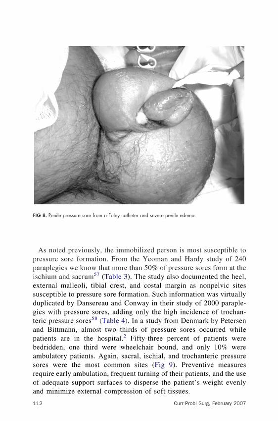
IG 8. Penile pressure sore from a Foley catheter and severe penile edema.
nd minimize external compression of soft tissues.
12 Curr Probl Surg, February 2007
C
(pAhatp
bdn
T
(T
T
(T3
C
one of NecrosisThe configuration of the pressure sore is that of a 3-dimensional cone61
Fig 10). Similar to the proverbial iceberg, the majority of injury due toressure necrosis is deeper and larger below the surface (Fig 11).lthough pressure causes ischemia of the skin, the subcutaneous muscleas a higher metabolic activity and thus is more sensitive to prolongednoxia. Further, objects trap the underlying muscle between the skin andhe deeper bony prominence. Thus, the human endoskeleton acts as aressure point for the external object.Nola and Vistnes performed histological analysis of skin and muscle overony prominences subjected to pressure in animals.62 Bony prominences thateveloped superficial ulceration demonstrated breakdown of the dermis and
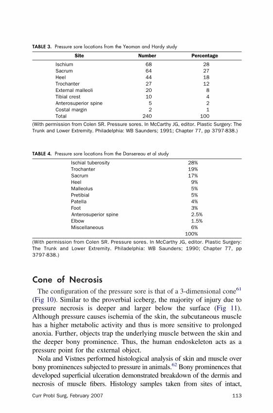
ABLE 3. Pressure sore locations from the Yeoman and Hardy study
With permission from Colen SR. Pressure sores. In McCarthy JG, editor. Plastic Surgery:he Trunk and Lower Extremity. Philadelphia: WB Saunders; 1990; Chapter 77, pp797-838.)
ecrosis of muscle fibers. Histology samples taken from sites of intact,
urr Probl Surg, February 2007 113
nDflsba(
S
igP
FD
1
onulcerated skin showed significant muscle necrosis underneath. Similarly,aniel and Faibisoff found almost total muscle atrophy in myocutaneousaps used to cover pressure sores.63 These studies suggest that plasticurgeons should avoid transposing myocutaneous flaps to cover pressure-earing bony prominences. However, muscle can be used to obliterate annatomical dead space from excision of bone and scar tissue, and innervatedie, sensory) flaps can be used to prevent recurrence.64
tages of Pressure SoresThere are 4 generally accepted stages of pressure sores (Table 5). Stage I
s exemplified by continued erythema of skin that cannot be blanched with aloved finger. The skin is still intact and no ulceration is yet evident.
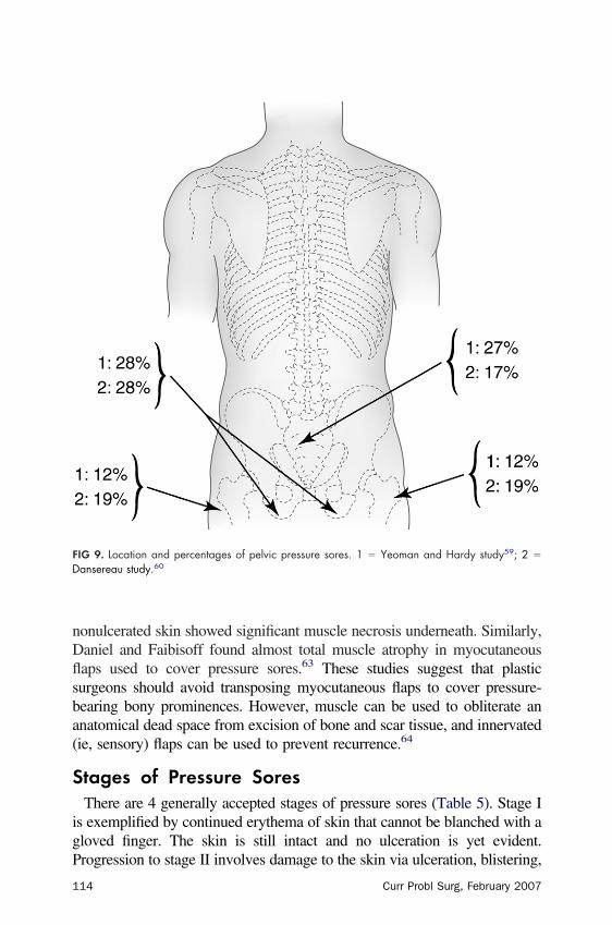
IG 9. Location and percentages of pelvic pressure sores. 1 � Yeoman and Hardy study59; 2 �
ansereau study.60
rogression to stage II involves damage to the skin via ulceration, blistering,
14 Curr Probl Surg, February 2007
omssionbd
FJS
C
r abrasion. Often these 2 stages are indicators of deeper necrosis. They alsoay occur independently as sheer injuries, caused by sliding against sheets or
upport surfaces. Stage III lesions represent full-thickness destruction of thekin. Although the ulcer may not visibly extend into muscle, the pressuretself usually causes necrosis of the underlying muscle as discussed previ-usly. Stage IV lesions include involvement of muscle, tendons, joints,erves, and even bone. Stage III and IV lesions require surgical interventionecause these defects are often large and infected and require sharp
IG 10. The cone of necrosis. (With permission from Phillips LG, Robson MC. Pressure ulcerations. In:urkiewicz MJ, Krizek TJ, Mathes SJ, Ariyan S, editors. Plastic Surgery: Principles and Practice. Vol. 2.t Louis: CV Mosby; 1990; pp 1223-51.)
ebridement.
urr Probl Surg, February 2007 115
P
cpraolap
Ffe
T
SS
S
S
AAR
1
revention StrategiesSince the vast majority of pressure sores occur in the hospital,
aregivers must have a regimen for inspecting the skin of immobilizedatients. In addition, the presence of spasticity in a limb, proximity of bedails, nasogastric tubes, and other solid objects should encourage frequentssessment for skin breakdown. The formation of a pressure sore dependsn the severity of pressure, shape of the object creating the pressure,ocation on the body, and the length of time. Thus, patient repositioningnd the use of padding or special beds (see below) is a crucial
IG 11. External appearance versus the large base of a trochanteric pressure sore. (With permissionrom Phillips LG, Robson MC. Pressure ulcerations. In: Jurkiewicz MJ, Krizek TJ, Mathes SJ, Ariyan S,ditors. Plastic Surgery: Principles and Practice. Vol. 2. St Louis: CV Mosby; 1990; pp 1223-51.)
ABLE 5. Consensus classification/stages of pressure sores
Pressure sore staging system
tage I Nonblanchable erythema of intact skin (heralding lesion of skin ulceration).tage II Partial-thickness skin loss involving epidermis or dermis. The ulcer is superficial
and presents clinically as an abrasion, blister, or shallow crater.tage III Full-thickness skin loss involving damage to or necrosis of subcutaneous tissue
that may extend down to, but not through, underlying fascia. The sorepresents clinically as a deep crater with or without undermining of adjacenttissue.
tage IV Full-thickness skin loss with extensive destruction, tissue necrosis, or damagein muscle, bone, or supporting structures (for example, tendon or jointcapsule).
dapted from U.S. Department of Health and Human Services (HHS): Pressure Ulcers indults: Prediction and Prevention. Clinical Practice Guideline No. 3, Publication 97-0047. HHS,ockville, MD, 1992.
rophylactic mechanism for tissue breakdown.
16 Curr Probl Surg, February 2007
apacfaFoap
spwtatf
dcftsb
T
S
M
A
M
N
F
AC
C
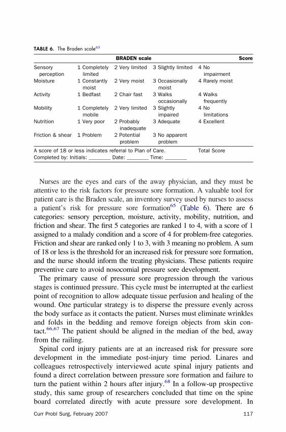
Nurses are the eyes and ears of the away physician, and they must bettentive to the risk factors for pressure sore formation. A valuable tool foratient care is the Braden scale, an inventory survey used by nurses to assess
patient’s risk for pressure sore formation65 (Table 6). There are 6ategories: sensory perception, moisture, activity, mobility, nutrition, andriction and shear. The first 5 categories are ranked 1 to 4, with a score of 1ssigned to a malady condition and a score of 4 for problem-free categories.riction and shear are ranked only 1 to 3, with 3 meaning no problem. A sumf 18 or less is the threshold for an increased risk for pressure sore formation,nd the nurse should inform the treating physicians. These patients requirereventive care to avoid nosocomial pressure sore development.The primary cause of pressure sore progression through the various
tages is continued pressure. This cycle must be interrupted at the earliestoint of recognition to allow adequate tissue perfusion and healing of theound. One particular strategy is to disperse the pressure evenly across
he body surface as it contacts the patient. Nurses must eliminate wrinklesnd folds in the bedding and remove foreign objects from skin con-act.66,67 The patient should be aligned in the median of the bed, awayrom the railing.Spinal cord injury patients are at an increased risk for pressure soreevelopment in the immediate post-injury time period. Linares andolleagues retrospectively interviewed acute spinal injury patients andound a direct correlation between pressure sore formation and failure tourn the patient within 2 hours after injury.68 In a follow-up prospectivetudy, this same group of researchers concluded that time on the spine
ABLE 6. The Braden scale65
BRADEN scale Score
ensoryperception
1 Completelylimited
2 Very limited 3 Slightly limited 4 Noimpairment
oisture 1 Constantlymoist
2 Very moist 3 Occasionallymoist
4 Rarely moist
ctivity 1 Bedfast 2 Chair fast 3 Walksoccasionally
4 Walksfrequently
obility 1 Completelymobile
2 Very limited 3 Slightlyimpaired
4 Nolimitations
utrition 1 Very poor 2 Probablyinadequate
3 Adequate 4 Excellent
riction & shear 1 Problem 2 Potentialproblem
3 No apparentproblem
score of 18 or less indicates referral to Plan of Care. Total Scoreompleted by: Initials: ________ Date: ________ Time: ________
oard correlated directly with acute pressure sore development. In
urr Probl Surg, February 2007 117
aaph
hhiaTsaiKpmtpt
otmaitcblcppsfe
cbmsm
1
ddition, there is an inverse relationship between systolic blood pressurend early pressure sore formation.69 Thus, regular turning of immobilizedersons, especially spinal cord injury patients, should begin within 2ours of injury, even during assessment and transport.Hospital staffing can be a challenge, especially when the wards haveigh numbers of immobilized patients or those with impaired mobility. Toelp alleviate this nursing burden, several bed technologies exist to assistn patient turning, also known as kinetic therapy. These include the Fosternd Stryker frames, the CircOlectric bed, and the Keane RotoRest bed.he Foster and Stryker frames alternate the patient from a prone to aupine position via hand turning of the frames. The CircOlectric bed is anutomatic turning device allowing for vertical elevation of the head, buts rather large, expensive, and time-consuming to monitor (Fig 12). Theeane RotoRest bed is the least burdensome of the group (Fig 13). Nurseslace patients on this 4-inch thick mattress with numerous side attach-ents and forward braces. The bed then automatically and gently rocks
he patient from side to side. The benefit of kinetic therapy is to shift theressure points and to enhance lung perfusion, which minimizes atelec-asis and nosocomial pneumonia.70,71
Regular turning of patients should be supplemented with minimizationf point pressure. Three types of bed technology are available to addresshis issue: the air mattress, waterbeds, and air-fluidized beds. Airattresses are categorized by 2 types: the static air mattress and the
lternating pressure mattress. The static air mattress is lightweight andnexpensive but not favored by nurses who must wrestle with the deviceo move patients. The alternating pressure mattresses utilize internal airells and an electric air pump to inflate and deflate the air pockets. Theseeds require correct patient positioning, monitoring of the cells foreakage, and an electrical source. Leakage in the tubing or mattress cellsan burn up the electric pump, which creates noise pollution to those inroximity of the pump’s normal operation. The efficacy of alternatingressure mattresses is questionable with few well-controlled studies toupport the device.72 Water mattresses are heavy, bulky, and cannot beashioned into a seated position. Leakage of their water volumes canndanger the patient and flood the room.All of the previously mentioned mattresses have polyvinyl outer
overings. Patients often complain of the additional heat retention createdy the material. Also, the occlusive nature to the mattress shell preventsoisture wicking and promotes skin maceration, infection, and pressure
ore formation. To combat the numerous tribulations associated with
attresses, the air-fluidized bed uses both warmed air and optical glass
18 Curr Probl Surg, February 2007
sb1nf
felMc
FJS
C
pheres to literally float patients. The best known of these is the Clinitroned (Fig 14). The maximal pressure exerted on the patient skin surface is0 mm Hg, which effectively eliminates the pressure element of tissueecrosis. Additionally, the polyester sheet containing the spheres allowsor absorption and drying of the patient contact surface.The sparse popularity and limitation of the air-fluidized bed use comes
rom its disadvantages. Dehydration may occur particularly in thextensively burned patient who cannot effectively limit evaporative waterosses.73 The bulky largesse of a bed weighs approximately 1 metric ton.
oving the air-fluidized bed requires extreme caution because the device
an gain momentum and injure the patient or transporting team. Bolsters,
urr Probl Surg, February 2007 119
ddp
C
pstpappnfm
F
1
ressings, or pillows effectively negate the bed’s ability to evenlyistribute pressure throughout the patient’s contact surface area. Elderlyatients may develop disorientation in these beds.
linical EvaluationAll patients admitted to the hospital should be evaluated continually forossible pressure sore formation. This begins with a clinical index ofuspicion based on admitting diagnosis, current state of mobility, type ofreatment being rendered to the patient, and the iatrogenic effect on theatient’s life. Those patients with a history of a previous pressure sore(s)re of particular concern. Patients with diabetes, paraplegia, and quadri-legia may have some component of insensate body parts over bonyrominences. The inability to recognize numbness or pain due to pressureecrosis increases a person’s susceptibility to ulceration and pressure soreormation. Further, physically incapable or deconditioned persons cannot
IG 13. A Keane RotoRest bed.
ove despite their desire to readjust and relieve the pressure. All these
20 Curr Probl Surg, February 2007
pp
cPpnat
r
FJS
C
atients need a caregiver to be mindful of the condition and help move theatient every 2 hours or sooner.The initial discovery of skin breakdown can come from the patient or
aregiver. Drainage from the wound soils the patient’s clothes or linens.atient discomfort during the recovery from their medical illness mayrecede external evidence of pressure sore development in the subcuta-eous tissues. The usual method of pressure sore discovery comes fromthorough physical examination despite the absence of signs or symp-
oms.Inspection of the wound itself demonstrates the classic hallmark signs:
ubor (erythema), tumor (edema), calor (warmth), and dolor (pain) in the
urr Probl Surg, February 2007 121
sronatPr
sa“fiwtocdw
awartUp
iactimbmcd
w
1
ensate patient. Gentle pushing on the wound with a gloved hand mayeveal underlying fluctuance or loculated fluid from liquefaction necrosisf subcutaneous tissues. This may be covered by an eschar composed ofecrotic skin that has desiccated. Such wounds slough their eschar capnd reveal a deep, cone-shaped ulcer cavity.74 If the wound is suspectedo connect with surrounding structures, a sinogram may be desired (Fig 15).ressure sores can involve surrounding bone, joint spaces, and sometimesectal or urethral fistulas with infection and exacerbation of injury.Stage IV pressure sores have abundant necrotic tissue and often
urrounding cellulitis. Blood cultures are in order if the patient has signsnd symptoms of sepsis: fever, elevated white blood cell count with aleft” shift, rigors, sweating, or change in mental status. Contributingactors must be addressed. This includes biopsies of the pressure soretself to be sent for quantitative and qualitative wound cultures. Theound probably has a bacterial count greater than 105 bacteria/g of tissue,
hus negating the effect of systemic antibiotics.75 Further, the poorxygen tension of the wound is a function of distant blood vessels, whichontributes to poor circulation of antibiotics. Once the wound is surgicallyebrided, targeted antibiotic therapy and local wound care promoteound healing.Urine incontinence is defined as involuntary loss of urine and must be
ddressed to prevent maceration and soiling of the wound. Urine cultureith request for bacterial sensitivities should be sent to the laboratory, andFoley catheter can prevent further urine contact with the wound. A
aison de=être must be investigated because some patients may havereatable conditions such as a urinary tract infection or bladder spasticity.rology consultation can provide a diagnosis and treatment regimen foratients.Stool incontinence warrants frequent dressing or clothing changes. This
s particularly difficult for patients suffering from diarrhea. Diarrhea thatccompanies recent administration of antibiotics mandates sending stoolultures for possible Clostridium difficile infection despite the absence ofhe cytotoxin.76 If no pathological or bacterial source of the diarrhea isdentified, the use of codeine or Lomotil can assist the bowel to createore formed stools. Spinal cord injury patients with formed stools may
enefit from routine use of suppositories to induce scheduled bowelovements. Patients with recurrent pressure sores complicated by fecal
ontamination should be referred to a general surgeon for a possibleiverting colostomy, although this practice should be used with caution.77
Preoperative labs can identify other sources of problems that delay
ound healing. The presence of anemia and hypoalbuminemia should
22 Curr Probl Surg, February 2007
tHdt
FPW
C
rigger suspicion of a poor nutritional intake or other chronic disease state.emoglobin A1C and random blood glucose levels are necessary in theiabetic patient. All such issues should be addressed via consultation with
IG 15. A sinogram demonstrating joint capsule involvement. (With permission from Colen SR.ressure sores. In McCarthy JG, editor. Plastic Surgery: The Trunk and Lower Extremity. Philadelphia:B Saunders; 1990; Chapter 77, pp 3797-838.)
he appropriate specialist.
urr Probl Surg, February 2007 123
psppne
nconcsmrfi
dppplmtu
T
h(
cpfcaeaat
1
Auscultation of the lungs is a vital component to the preoperativeatient evaluation. Detection of wheezing, crackles, or coarse breathounds merits further analysis. Chest radiographs are warranted inatients with poor diaphragmatic excursion. This includes quadriplegicatients and those with chronic obstructive pulmonary disease. Pulmo-ary insufficiency can be secondary to a cardiac source, and occasionally,chocardiography is a necessary preoperative measure.The continued popularity of nicotine and the many products containingicotine—including cigarettes, cigars, and smokeless “chew”—should beonsidered in performing flaps. Cigarette smoking decreases subcutane-us tissue oxygen tension lasting up to 50 minutes after smoking due toicotine’s vasoconstrictor effects.78 Reus and colleagues compared out-omes in elective free-tissue transfers between 51 nonsmokers and 93mokers who quit perioperatively.79 There was no difference in anasto-otic patency, but smokers experienced delayed wound healing at the
ecipient site and more often required an additional procedure to achievenal wound closure.Designing a care plan for the pressure sore requires a full and frankiscussion of expected and possible outcomes. The patient needs com-lete understanding of the large investment being made to treat thisotentially lethal condition. The clinician must carefully assess theatient’s ability to understand and provide the acute cooperation and theong-term commitment of preventing recurrence. Family care providersust be included both to educate the patient and to provide an outline for
he ensuing care, which has the greatest chance of compliance andltimate success.
reatment of Pressure SoresTherapeutic surgical intervention follows maximization of patientealth and education. The goals are similar in all pressure sore casesTable 7).The history and physical examination of the pressure sore patient is
ritical to determine which goals may be achieved. Some debilitatedatients cannot tolerate a surgical procedure and should be consideredor nonoperative therapeutic intervention. Comorbid conditions in-lude congestive heart failure, noncardiogenic pulmonary diseases,therosclerosis with carotid or coronary artery disease, and uncoop-rative patients. Terminally ill patients are treated more supportivelynd less aggressively. The specific problem should be evaluated by thenesthesiologist, psychologist, or social worker to determine limita-
ions or causes of a suboptimal outcome, including recurrence. All
24 Curr Probl Surg, February 2007
prgc
N
TottAbt6
macstlPsabl
T
1
2
3
4
5
6
7
C
atients require the same basic care to treat local or distant infection,emove necrotic tissue if indicated, and promote healing. Once theseoals are attained, treatment is directed to surgical or nonsurgicallosure of the wound.
onsurgical TreatmentMyriad information is available on the subject of topical wound therapy.opical therapy for the treatment of a pressure sore includes a spectrumf enzymes for debridement, elemental pastes, antimicrobials, and solu-ions.80 Kucan and colleagues compared the efficacy of silver sulfadiazineo saline and povidone-iodine in preparing pressure ulcers for closure.81
ll wounds treated with silver sulfadiazine (15 patients) reduced theacterial count to less than 105/g of tissue within 3 weeks. Comparatively,he use of saline was more effective than povidone-iodine (79% versus4%, respectively).Topical antimicrobials include compounds such as bacitracin, poly-yxin, neomycin, silvadene, and sulfamylon. One problem with these
ntibiotics is patient sensitization, allergy, and systemic toxicity. Thelinician should look for these complications when treating large woundsubjected to frequent application of the compound in question. Althoughhere is no such concept as the typical pressure sore—only typicalocations—the microbial flora is presumed to be either Staphylococcus,roteus, Pseudomonas, or Bacteroides species.82 The astute clinicianhould send tissue biopsies before starting any broad-spectrum topicalntimicrobial. The wound-specific flora is best treated with an antimicro-ial causing the highest level of growth inhibition as determined by
ABLE 7. Goals of treatment
. Total excisional debridement of the necrotic tissue, ulcer base, scar tissue, bursa,undermined skin (if not extensive), and heterotopic calcification
. Excision of infected bone, bony prominence recontouring to eliminate isolated pressurepoints
. Acquisition of hemostasis after debridement to healthy tissue and placement of lowsuction drainage(s), if appropriate
. Obliteration of the anatomical dead space using muscle, myocutaneous flaps, and/ordeepithelialized skin flaps to provide sufficient bulk to cushion bony prominences
. Coverage of the wound with vascularized flaps placed to create a tension-free repair overthe defect
. Placement of the flap so as to position the stress point away from the suture line, apoint of diminished blood flow
. Restoration of the patient’s activities of daily living before developing the pressure sore
aboratory testing. Systemic antibiotics are then used as an adjunct to the
urr Probl Surg, February 2007 125
ts
etaii
tspctitiGt
ptoadpaOCcdwd
vTldrfiA
1
opical antimicrobials for treatment of cellulitis, osteomyelitis, or joint-pace infections.Spontaneous wound closure in small to medium size pressure sores
nhances as the bacterial counts decline. The finalized product is scarissue with poorly developed skin covering with a poor vascular supplynd requires manual lubrication.83 The stiff and friable skin can readily benjured by pressure or shearing, and recurrence is higher than for surgicalntervention.Topical application of growth factors should be considered in treating
he nonoperative pressure sore. Robson and colleagues demonstrated aubstantial reduction in pressure ulcer volume from the application oflatelet-derived growth factor, a potent activator of wound healingells.84,85 Conversely, a prospectively randomized, blind, placebo-con-rolled trial failed to demonstrate improved pressure ulcer healing withnterleukin-1�.86 For the more challenging diabetic patients, local injec-ion of basic fibroblast growth factor improves wound breaking strengthn a diabetic rat model to a statistically significant level (P � 0.001).87
rowth factor treatment of pressure sores remains a clinical trialreatment, in part due to its significant cost.The main goal in treating pressure sores is to alleviate the pressureoint. Although infection is 1 of the many inhibitors to wound healing,he pressure and shearing forces must be eliminated or closure will notccur. Therefore, the patient should be shifted in position every 2 hoursnd 1 of the bed technologies should be employed to maximize pressureistribution. Additionally, there are health care products designed as localressure distributors. One such product is Allevyn Adhesive, whichbsorbs moisture and attempts to redistribute the wound pressure forces.ther brand name products include Tielle, Mepilex Border, Tegaderm,ombiDERM, Lyofoam, and Biatain. There are very few randomized,ontrolled trials with these products. Each manufacturer has specificesigns and patterns to fit variously shaped pressure ulcers based on theound location. This adds to the complexity in choosing a product andeciding about the cost effectiveness.Another technology-based wound closure device is the subatmosphericacuum-assisted closure technique.88 Vacuum Assisted Closure (V.A.C.)herapy, promoted by Kinetic Concepts, Incorporated, uses controlled,
ocalized negative pressure to promote granulation tissue formation,ecrease tissue edema, and remove infectious materials (Fig 16). In aetrospective review of 25 patient charts, Weed and colleagues, did notnd the V.A.C. to reduce bacterial colonization but rather increased it.89
prospective, randomized trial of 54 patients found vacuum-assisted
26 Curr Probl Surg, February 2007
cib
soosdrtoad
liab
F
C
losure to decrease nonfermentative Gram-negative bacilli (P � 0.05),ncrease S. aureus (P � 0.05), and effect no change in quantitativeacterial load.90
The V.A.C. is used for numerous wound types. A thorough literatureearch identifies numerous case reports describing V.A.C. therapy forpen sternal wounds, fascial reapproximation in trauma patients withpen abdomens, gynecologic oncology wound failures, securingplit-thickness skin grafts, and full-thickness skin graft positioning foregloving injuries.91-96 However, toxic shock syndrome has beeneported from the use of the V.A.C., and 1 meta-analysis sharply noteshe weak evidence for V.A.C. therapy versus conventional meth-ds.97-99 In a porcine model, Miller and colleagues failed to establishhistologically based benefit to the V.A.C. therapy versus wet-to-dry
ressing changes.100
The use of V.A.C. therapy in pressure ulcers is unsubstantiated by theiterature. Again, an operative intervention will provide the ideal outcomen larger pressure sores. The surgeon must decide the most practicalpproach and potential benefit of this emerging technique on a per-patient
IG 16. Vacuum-assisted closure device applied to a wound.
asis.101
urr Probl Surg, February 2007 127
O
rsfscpsscHoa
nppegw
eoot
upncmrmpsadcc
1
perative TechniquePressure sore wound debridement is best performed in the operating
oom under general endotracheal anesthesia, where lighting and hemo-tasis are superior. The anesthesiologist can carefully monitor the patientor signs of pain, excess blood loss, hypotension, and shock while theurgeon focuses on treating the wound. Local therapy is directed to obtainomplete removal of necrotic debris, decreased bacterial load, andreparation for wound closure and healing. Beyond sharp debridement theurgeon can use pulsating jet lavage on the wound. Animal studiesuggest that substantial wound bacterial count reductions can moreonsistently be achieved using pulsating jet lavage versus bulb syringe.102
owever, 2 studies of high pressure jet lavage performed on ex vivovine muscle suggest this therapy may cause damage to healthy tissuesnd bacterial retention.103,104
The surgeon should remain cognizant that there may be extensiveecrosis within the wound that involves muscle, fat, and skin and berepared to debride as needed. The patient’s condition may haverogressed to involve the underlying bone or nearby joint. The surgicalnd result must be well-vascularized tissues with increased tissue oxy-enation and removal of the physical barriers to wound healing byhatever closure technique selected.Appropriate patient position in the operating room gives optimal
xposure of the wound. The wound is cleaned and repaired in a positionf maximal tension, whereby all other physical positions decrease tensionn the suture lines. Thus, any size limitations are addressed intraopera-ively and the possibility of postoperative dehiscence is minimized.Progressing to the operating room for sharp debridement is delayedntil all potential sources of infection have been addressed. Hence, theossibility of respiratory, urinary, or other site infections cross-contami-ating the debrided pressure sore is minimized. The wound itself almostertainly contains a substantial bacterial load and necessitates debride-ent. Studies have demonstrated that freshly incised skin has less
esistance to bacterial incursion, and measures should be taken toinimize seeding of the cleansed wound.105,106 Standard antiseptic skin
rep and aseptic technique block external sources of infection such as theurgeon and operating room equipment. This is further enhanced bydministering perioperative systemic antibiotics to prevent seeding ofistant bacteria into the surgically cleansed wound.107 The wound can beleansed with buffered Dakin’s solution (sodium hypochlorite, 0.025%
oncentration), which is both bactericidal and nonhazardous to neutro-
28 Curr Probl Surg, February 2007
pf
dtioslgfip
cncaotsv
S
tnpldescr
ippwci
C
hils and fibroblasts, although Silvadene, mafenide, and Betadine arerequently used.Ensuring complete pressure ulcer excision necessitates the removal ofead tissue to prevent incomplete excision of skin margins. Such an efforto clean these fastidious wounds would be marred by recurrence ofnfection and wound dehiscence. One technique to ensure completenessf debridement is the use of a dilute 1% solution of methylene blue, whichtains the exposed tissues. This can be followed with a hydrogen peroxideavage to remove excess dye.108 Excision of all stained tissues visuallyuides the surgeon’s blade. This is supplemented with the presence ofresh bleeding of tissues, although hemostasis must be achieved. Follow-ng skin and necrotic tissue excision is bone debridement and contouringerformed with rongeurs and rasps.A debrided wound is now ready for closure. Surgical closure of the
leansed pressure sore is best achieved using local rotation, fasciocuta-eous, or musculocutaneous flaps. Skin grafting is an option for superfi-ial ulceration, but the long-term stability is around 30%.109 Both animalnd clinical studies confirm the superiority of musculocutaneous flapsver skin grafting of infected wounds.110 Paraplegic patients are suscep-ible to flap loss secondary to postoperative hematoma as a function ofympathetic tone loss and physiologic inability to vasoconstrict bloodessels.71,111
acrumThe supine, bedridden patient and the acutely injured spinal cord patient
ypify the victim of skin and tissue breakdown over the sacral promi-ence. Small sacral wounds can, theoretically, be excised and closedrimarily. Such primary repairs place tension on the wound and a sutureine directly over a pressure point. As a result, the wound may not heal,ehisce, or reoccur as the patient returns to the supine position postop-ratively. Skin grafting is another possible option, but the nature of healedkin grafts is to provide skin coverage and not padding. This type oflosure has the best results in ambulatory patients that are sensate andecovering from an acute illness or injury.Sacral wound repair requires adequate site preparation. When perform-
ng the bone ostectomy/contouring, the surgeon should take care not toerforate the anterior sacral cortex over which lies the presacral venouslexus. Various flaps may be designed to cover the debrided sacralound. The local buttock rotation flap is an excellent primary method for
losure of the prepared sacral wound. The design requires an elliptical
ncision extending from the superior pole of the wound then superiorly
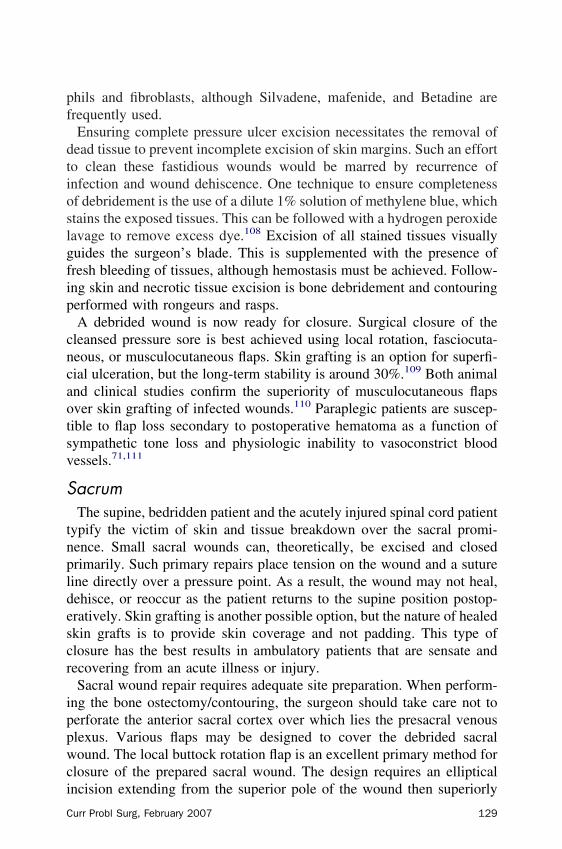
urr Probl Surg, February 2007 129
aBAs
ivfl(h
flwsgso
cyar
FJS
1
nd laterally toward the posterior iliac crest and then inferiorly (Fig 17).oth the superior and inferior gluteal arteries supply this portion of tissue.ny lateral defect can be closed with a skin graft, and recurrence of the
acral pressure sore allows for readvancement of the gluteal flap.The transverse lumbosacral flap is an arterial skin flap. This reliable flap
s supplied by the lumbar perforating vessels and uninterrupted subdermalascular plexus to the terminal portion.112 A large, transverse U-shapedap is elevated so that the inferior portion can cover the wound defectFig 18). The created superior deficit is closed with a skin graft. This flapas less subcutaneous tissue to pad the sacrum than the buttock flap.With both the local buttock rotation flap and the transverse lumbosacralap, a deficit is created so that the primary wound can close the sacralound. Closure of these iatrogenic, secondary wounds is performed with
kin grafting. As discussed above, closure of the primary wound with skinrafting is suboptimal at best. However, these secondary wounds are notubjected to the pressure and stress compared with the sacral area andther pressure points and will heal.Skin rotation flaps receive their blood supplies from deeper musculo-
utaneous blood vessels that perforate and branch upward. The last 30ears of plastic surgery has been devoted to the incorporation of musclend skin along a vascular pedicle to ensure tissue viability, increase the arc of
otation, and gain length. Ger is attributed with early use of muscle flaps
30 Curr Probl Surg, February 2007
tiwcs
ptoinarsa
I
pc1
FuP
C
o close leg ulcers and later pressure sores.113,114 Muscle flaps werenitially used to cover exposed bone, and then the muscle was coveredith a skin graft.115 Skin-bearing portions of muscle, termed musculo-
utaneous flaps, were attempted for pressure sore closure and demon-trated the viability of this closure method.116
Sacral pressure sores can be closed using musculocutanous flaps whenrimary closure or a local rotation flap is not an option. The majority ofhese musculocutaneous flaps are based on some portion of the superiorr inferior gluteal arteries.117 The musculocutaneous flap can be fash-oned for V to Y closure.118 Gaining popularity is the use of fasciocuta-eous flaps to close various wounds, including flaps based on the glutealrtery for sacral pressure sores.119 Borman and Maral demonstrated theeliability of the gluteal fasciocutaneous flap with V to Y closure for 15acral pressure sore patients without wound dehiscence or flap necrosis atminimum follow-up time of 6 weeks.120
schiumIschial pressure sores are common to the wheelchair-bound seatedatient. These wounds befuddled early attempts at nonoperative care andonservative closure with skin grafts. Conway and Griffith documented
00 ischial pressure sore patient care events whereby nonoperative
urr Probl Surg, February 2007 131
tmscmb
pimflrittfs
mbfflgaip
tamtswflid7flt
1
reatment resulted in a recurrence rate of 77% and total success rate oferely 18%.121 Comparatively, split-thickness skin grafting yielded a
imilarly high recurrence rate and a total success rate of 11%. The majoromplicating factor for ischial pressure sore patients is the necessity forobility and eventual return of the patient to the source of tissue
reakdown, usually the wheelchair.The ischium is a critical structure to pelvic stability in the seatedosition, and these pelvic bony prominences are subject to high pressuresn the supine patient. The high recurrence rate with ischial pressure soresandates surgical intervention. Recontouring the underlying bone beforeap closure has been extensively investigated.122 Conway and Griffitheduced their ischial pressure sore recurrence rate from 38% to 3% viaschiectomy. Others have recommended prophylactic bilateral ischiec-omy for unilateral disease.123 The result of bilateral ischiectomy isransferred pressure to the perineum where the patient is at increased riskor developing a urethral fistula, urethral diverticula, or perineal pressureore, and is a practice which should not be followed.124-127
Flap design depends on the size of the defect and previous closureethods for recurrent ischial pressure sores. Local random skin flaps are
est used for small, shallow wounds. Other options include the tensorascial lata flap, a reliable pedicle with a length advantage, although thisap may not provide adequate bulk for some patients128 (Fig 19). Theluteal thigh flap is another option that provides bulk, reliability,nd—interestingly—sensation to the ischial area.129 Other described flapsnclude use of the hamstrings and total thigh flaps for multiple, recurrentressure sores.130-132
In a rather unique maneuver, Jones accomplished coverage and sensa-ion for a T7 paraplegic patient with recurrent ischial pressure sores using
free medial gastrocnemius flap.133 Sensation was restored by anasto-osis between a proximal intercostal nerve, sural nerve graft, and the
ibial motor nerve branch of the gastrocnemius. Hallock has also reporteduccess with a free medial gastrocnemius flap to close a recurrent ischialound.134 If muscle is not desired, an inferior gluteal artery perforatorap can be fashioned.135 Homma and colleagues confirmed excellent
schial pressure sore repair in 10 paraplegic patients using a posterome-ial thigh fasciocutaneous flap with no recurrence in 7 of the patients at7 months postoperatively.136 The authors attribute their success of thisap to meticulous outlining of the musculocutaneous perforator, centering
he flap over the artery, and preservation of the proximal fascia.
32 Curr Probl Surg, February 2007
T
plCsas
FPe
C
rochanterSkin and tissue breakdown over the greater trochanter—the lateralrominence of the proximal femur—is most associated with prolongedateral decubitus positioning and large patients in narrow wheelchairs.onway and Griffith studied 82 patients with 122 trochanteric pressure
ores.121 Conservative, nonsurgical therapy in this cohort worked inpproximately 41% of cases, and skin grafting these wounds demon-
IG 19. Tensor fascia lata musculocutaneous flap for ischial pressure sores. (With permission fromhillips LG, Robson MC. Pressure ulcerations. In: Jurkiewicz MJ, Krizek TJ, Mathes SJ, Ariyan S,ditors. Plastic Surgery: Principles and Practice. Vol. 2. St Louis: CV Mosby; 1990; pp 1223-51.)
trated a 33% long-term success rate. These tenacious wounds are like
urr Probl Surg, February 2007 133
iph
wttcup
Ffe
1
schial and sacral pressure sores. They require the typical approach forroper healing: excision of dead and necrotic tissue, debridement down toealthy and viable tissue, and proper flap coverage.The primary flap used to cover debrided trochanter pressure soreounds is the tensor fascia lata musculocutaneous flap (Fig 20). The
ensor fascia lata is a workhorse flap applicable to both ischial, trochan-eric, and sometimes sacral defects.128 In a small series by Josvay andolleagues, 8 of 9 attempts resulted in closure of trochanteric woundssing the tensor fascia lata flap.137 All of these wounds were closed
IG 20. Tensor fascia lata musculocutaneous flap for trochanteric pressure sores. (With permissionrom Phillips LG, Robson MC. Pressure ulcerations. In: Jurkiewicz MJ, Krizek TJ, Mathes SJ, Ariyan S,ditors. Plastic Surgery: Principles and Practice. Vol. 2. St Louis: CV Mosby; 1990; pp 1223-51.)
rimarily with the V to Y advancement technique. If closure of the
34 Curr Probl Surg, February 2007
hoam
wtpRpa
lp
P
oagmDnsfl
ppipwwoTpanamat
C
arvest site cannot be accomplished primarily, a skin graft is used. Otherptions include the use of muscle flaps without skin, such as the gluteusnd vastus lateralis; coverage is then achieved with a skin graft over theuscle belly.The main cause of pressure recurrence is the insensate nature of theound site. The tensor fascia lata flap is the ideal method to close
rochanteric pressure sores because of the lumbar innervation thatrovides sensation in those patients with low spinal cord injuries.estoring sensation is a powerful preventative measure for pressure soreatients as they can determine themselves when to shift position andvoid tissue breakdown.Dibbell and colleagues have used the tensor fascia lata flap with its
ateral femoral cutaneous nerve for pressure sores in meningomyeloceleatients.138
ostoperative CareMuch of the care for the postoperative patient is the same for the closuref pressure sore wounds as other flaps. The suture edges are covered withnonadherent mesh such as Adaptic. This is covered with absorptive
auze to remove serosanguineous drainage from the skin and preventaceration. The gauze is changed as necessary to keep the skin clean.rains are useful monitoring devices for excessive bleeding from under-eath the flap; drains also help prevent flap loss from hematoma oreroma formation by controlled, low-pressure evacuation and help theap adhere to the underlying wound bed.The operating physician should monitor the patient’s postoperativerogress at regular intervals. However, the nursing staff is the crucialhysician extender to keep an eye on the patient and report problemsmmediately. The patient should be moved every 2 hours to preventressure sore development at other sites, pressure necrosis of the flap, oround dehiscence. Another option is the use of an air-fluidized bed,hich extends the time between turning patients. Whatever the method-logy, the wound should avoid excess moisture and fecal contamination.he majority of focus will be on the flap tissue, but other classicostoperative issues should not be ignored. Pain is controlled withppropriate analgesic medications such as morphine, fentanyl, oronsteroidal anti-inflammatory drugs. For patients with neuropathicnd refractory pain, Neurontin should be considered.139 The patientay have nausea, ileus, and poor ventilatory effort from the general
nesthetic. Feeding should begin as tolerated by the patient to enhance
issue healing. Scheduled nebulizer therapy and deep breathing exer-
urr Probl Surg, February 2007 135
cptce
olorBs3ebc
usirmaEtpm
1
ises are useful for atelectasis. Muscle spasms not adequately treatedreoperatively require immediate pharmacological therapy becausehis condition can compromise the flap and certainly challenges patientomfort. Valium and baclofen are reliable options to manage postop-rative muscle spasms.140
The last goal of surgical intervention is to restore the patient’s levelf independence before the pressure sore developed. This requires ateast 2 months of postoperative attention by the surgeon and a lifetimef care by the patient. After 1 to 2 weeks, the drains can be carefullyemoved if the serous fluid is clear and less than 30 mL per day.eginning at 2 weeks, the wound edges are sufficiently healed to
upport the patient’s sitting or lying on the flap for up to 15 minutestimes per day, gradually increasing by small increments in time. The
xception to this is for ischial pressure sore repairs, which require arief period of pressure relief every 10 minutes, even after healing isomplete.Several unresolved problems may prevent achievement of a healed,ncomplicated wound. Pain can be related to distress and depressiveymptoms.141 The patients must be compliant and motivated to heal theirnjury despite their altered body image. This requires good nutrition,ehabilitation, and strengthening of their deconditioned status, and continenceaintenance if necessary. Comatose, spinal cord injured, and elderly patients
re the most challenging to heal and require the highest level of nursing care.ach of these patients may not be able to sense, determine, or comply with
he need to shift their body. Should recurrence of the wound develop, theatient, similar to other victims of chronic disease states, needs encourage-ent, support, and commitment from their caregivers.
REFERENCES1. Bergstrom N, Bennett A, Carlson CE, et al. Pressure ulcer treatment: a critical
practice guideline. In: AHCPR Pub No 95-0653. Rockville, MD: U.S. Departmentof Health and Human Services, Public Health Service, Agency for Health CarePolicy and Research; 1994.
2. Petersen NC, Bittmann S. The epidemiology of pressure sores. Scand J PlastReconstr Surg 1971;5:62-6.
3. Capezuti EA, Braun JA. Medico-legal aspects of hospital siderail use. In: KappMB, editor. Ethics, Law, and Aging Review. New York: Springer Publishing;2001; pp 25-57.
4. Bass MJ. Risk management, health care, and the geriatric patient population. In:Kapp MB, editor. Ethics, Law, and Aging Review. New York: Springer Publishing;2001; pp 11-24.
5. Leape LL, Brennan TA, Laird N, Lawthers AG, Localio AR, Barnes BA, et al. The
36 Curr Probl Surg, February 2007
C
nature of adverse events in hospitalized patients. Results of the Harvard MedicalPractice Study II. N Engl J Med 1991;324:377-84.
6. Lindan O, Greenway RM, Piazza JM. Pressure distribution on the surface of thehuman body. I. Evaluation in lying and sitting positions using a “bed of springs andnails”. Arch Phys Med Rehabil 1965;46:378-85.
7. http://www.guinnessworldrecords.com/. Accessed December 13, 2005.8. Kosiak M, Kubicek WG, Olson M, Danz JN, Kottke FJ. Evaluation of pressure as
a factor in the production of ischial ulcers. Arch Phys Med Rehabil 1958;39:623-9.9. Dinsdale SM. Decubitus ulcers: role of pressure and friction in causation. Arch
Phys Med Rehabil 1974;55:147-52.10. Husain T. An experimental study of some pressure effects on tissues, with reference
to the bed-sore problem. J Pathol Bacteriol 1953;66:347-58.11. Abu-Rustum NR, Rajbhandari D, Glusman S, Massad LS. Acute lower extremity
paralysis following radiation therapy for cervical cancer. Gynecol Oncol1999;75:152-4.
12. Buchan AC, Fulford GE, Harris P, Jellinek E, Kerr WG, Kirkland I, et al. Apreliminary survey of the incidence and aetiology of spinal paralysis. Paraplegia1972;10:23-8.
13. Johnson KL, Cheung RB, Johnson SB, Roberts M, Niblett J, Manson D.Therapeutic paralysis of critically ill trauma patients: perceptions of patients andtheir family members. Am J Crit Care 1999;8:490-8.
14. Rotondo MF, McGonigal MD, Schwab CW, Kauder DR, Hanson CW. Urgentparalysis and intubation of trauma patients: is it safe? J Trauma 1993;34:242-6.
16. Solomon T, Willison H. Infectious causes of acute flaccid paralysis. Curr OpinInfect Dis 2003;16:375-81.
17. Rohland BM, Carroll BT, Jacoby RG. ECT in the treatment of the catatonicsyndrome. J Affect Disord 1993;29:255-61.
18. Greenberg MS. Handbook of Neurosurgery. New York: Thieme MedicalPublishers; 2001.
19. Ashworth B. Preliminary trial of carisoprodol in multiple sclerosis. Practitioner1964;192:540-2.
20. Brown TR, Kraft GH. Exercise and rehabilitation for individuals with multiplesclerosis. Phys Med Rehabil Clin N Am 2005;16:513-55.
21. Wang RY, Chen HI, Chen CY, Yang YR. Efficacy of Bobath versus orthopaedicapproach on impairment and function at different motor recovery stages afterstroke: a randomized controlled study. Clin Rehabil 2005;19:155-64.
22. Maruyama T, Behrends JC, Akaike N. Inhibition of the voltage-dependent calciumcurrents in isolated frog sensory neurons by GABA-related agonistic compounds.Neurosci Res 1988;6:97-105.
23. Zhao F, Li P, Chen SR, Louis CF, Fruen BR. Dantrolene inhibition of ryanodinereceptor Ca2� release channels. Molecular mechanism and isoform selectivity.J Biol Chem 2001;276:13810-6.
24. Fares Y, Khazim RM, del Barrio ER, Burzaco JA. Dosage of intrathecal baclofenmaintenance therapy in the spastic syndromes. J Med Liban 2004;52:13-8.
25. Albright AL, Awaad Y, Muhonen M, Boydston WR, Gilmartin R, Krach LE, et al.
Performance and complications associated with the synchromed 10-ml infusion
urr Probl Surg, February 2007 137
1
pump for intrathecal baclofen administration in children. J Neurosurg 2004;101:64-8.
26. Garland DE, Lucie RS, Waters RL. Current uses of open phenol nerve block foradult acquired spasticity. Clin Orthop Relat Res 1982;217-22.
27. Herz DA, Looman JE, Tiberio A, Ketterling K, Kreitsch RK, Colwill JC, et al. Themanagement of paralytic spasticity. Neurosurgery 1990;26:300-6.
28. Sindou M, Millet MF, Mortamais J, Eyssette M. Results of selective posteriorrhizotomy in the treatment of painful and spastic paraplegia secondary to multiplesclerosis. Appl Neurophysiol 1982;45:335-40.
29. Jani P, Trice J, Umejiego C. http://www.arches.uga.edu/�pjani/Chioma.html.Accessed June 16, 2005.
30. Larson E. Skin hygiene and infection prevention: more of the same or differentapproaches? Clin Infect Dis 1999;29:1287-94.
31. Boyce JM, Pittet D. Guideline for Hand Hygiene in Health-Care Settings:Recommendations of the Healthcare Infection Control Practices Advisory Com-mittee and the HICPAC/SHEA/APIC/IDSA Hand Hygiene Task Force. InfectControl Hosp Epidemiol 2002;23:S3-40.
32. Howard JM, Barker WF, Culbertson WR, et al. Post-operative wound infections:the influence of ultra-violet radiation of the operating rooms and various otherfactors. Ann Surg 1964;160:32-81.
33. Groth K. Klinische Beobachtungen und experimentelle Studien uber die Enstehungdes Dekubitis. Acta Chir Scand 1942;87:1-209.
34. Robson MC, Krizek TJ. The role of infection in chronic pressure ulcerations.Symposium on Neurologic Aspects of Plastic Surgery. St Louis: CV Mosby; 1978.
36. Landis EM. Micro-injection studies of capillary blood pressure in human skin.Heart 1930;15:209-28.
37. Exton-Smith AN, Crockett DJ. Nature of oedema in paralysed limbs of hemiplegicpatients. BMJ 1957;3:1280-83.
38. Ho TL, Lee KW, Lee HJ. Subacute mandibular and hypoglossal nerve denervationcausing oedema of the masticator space and tongue. Neuroradiology 2003;45:262-6.
39. Bredella MA, Tirman PF, Fritz RC, Wischer TK, Stork A, Genant HK. Denervationsyndromes of the shoulder girdle: MR imaging with electrophysiologic correlation.Skeletal Radiol 1999;28:567-72.
40. Chen LE, Seaber AV, Urbaniak JR. Combined effect of acute denervation andischemia on the microcirculation of skeletal muscle. J Orthop Res 1992;10:112-20.
41. Alison WE Jr, Phillips LG, Linares HA, Hui PS, Hayward PG, Broemeling LD,et al. The effect of denervation on soft-tissue infection pathophysiology. PlastReconstr Surg 1992;90:1031-5.
42. Henriksen O, Sejrsen P, Paaske WP, Eickhoff JH. Effect of chronic sympatheticdenervation upon the transcapillary filtration rate induced by venous stasis. ActaPhysiol Scand 1983;117:171-6.
44. Dickerson RN, Gervasio JM, Riley ML, Murrell JE, Hickerson WL, Kudsk KA,
38 Curr Probl Surg, February 2007
C
et al. Accuracy of predictive methods to estimate resting energy expenditure ofthermally-injured patients. JPEN J Parenter Enteral Nutr 2002;26:17-29.
45. Mandt J, Teasley-Strausburg K, Shronts E. Nutritional requirements. In: Teasley-Strausburg K, editor. Nutritional Support Handbook. Cincinnati: Harvey WhitneyBooks; 1992.
46. Fuhrman MP. The albumin-nutrition connection: separating myth from fact.Nutrition 2002;18:199-200.
47. Golden MH, Golden BE. Severe malnutrition. In: Garrow JS, James WPT, RalphA, editors. Human nutrition and dietetics. Edinburgh: Churchill Livingstone; 2000;p 515.
48. Lim SH, Lee JS, Chae SH, Ahn BS, Chang DJ, Shin CS. Prealbumin is not sensitiveindicator of nutrition and prognosis in critical ill patients. Yonsei Med J2005;46:21-6.
54. Komesu MC, Tanga MB, Buttros KR, Nakao C. Effects of acute diabetes on ratcutaneous wound healing. Pathophysiology 2004;11:63-7.
55. Fahey TJ 3rd, Sadaty A, Jones WG 2nd, Barber A, Smoller B, Shires GT. Diabetesimpairs the late inflammatory response to wound healing. J Surg Res 1991;50:308-13.
56. Robson MC, Heggers JP. Effect of hyperglycemia on survival of bacteria. SurgForum 1969;20:56.
57. Yeoman MP, Hardy AG. The pathology and treatment of pressure sores inparaplegics. Br J Plast Surg 1954;7:179-92.
58. Dansereau JG, Conway H. Closure of decubiti in paraplegics. Report of 2000 cases.Plast Reconstr Surg 1964;33:474-80.
59. Yeoman MP, Hardy AG. The pathology and treatment of pressure sores inparaplegics. Br J Plast Surg 1954;7:179-92.
60. Dansereau JG, Conway H. Closure of decubiti in paraplegics. Report of 2000 cases.Plast Reconstr Surg 1964;33:474-80.
61. Daniel RK, Priest DL, Wheatley DC. Etiologic factors in pressure sores: anexperimental model. Arch Phys Med Rehabil 1981;62:492-8.
62. Nola GT, Vistnes LM. Differential response of skin and muscle in the experimentalproduction of pressure sores. Plast Reconstr Surg 1980;66:728-33.
63. Daniel RK, Faibisoff B. Muscle coverage of pressure points—the role of myocu-taneous flaps. Ann Plast Surg 1982;8:446-52.
64. Krupp S, Kuhn W, Zaech GA. The use of innervated flaps for the closure of ischialpressure sores. Paraplegia 1983;21:119-26.
65. Braden BJ, Bergstrom N. Clinical utility of the Braden scale for predicting pressure
sore risk. Decubitus 1989;2:44-51
urr Probl Surg, February 2007 139
1
66. Stauffer ES. Rehabilitation of post-traumatic cervical spinal cord-quadriplegia andparaplegia. In: Bailey R, editor. The cervical spine. Philadelphia: JB Lippincott;1983.
67. Ungar GH. The care of the skin in paraplegia. Practitioner 1971;206:507-12.68. Linares HA, Mawson AR, Suarez E, Biundo JJ. Association between pressure sores
and immobilization in the immediate post-injury period. Orthopedics 1987;10:571-3.69. Mawson AR, Biundo JJ Jr., Neville P, Linares HA, Winchester Y, Lopez A. Risk
factors for early occurring pressure ulcers following spinal cord injury. Am J PhysMed Rehabil 1988;67:123-7.
70. Mullins CD, Philbeck TE Jr, Schroeder WJ, Thomas SK. Cost effectiveness ofkinetic therapy in preventing nosocomial lower respiratory tract infections inpatients suffering from trauma. Managed Care Interface 2002;15:35-40.
71. Raoof S, Chowdhrey N, Raoof S, Feuerman M, King A, Sriraman R, et al. Effectof combined kinetic therapy and percussion therapy on the resolution of atelectasisin critically ill patients. Chest 1999;115:1658-66.
72. Vasconez LO, Schneider WJ, Jurkiewicz MJ. Pressure sores. In: Current Problemsin Surgery. Chicago: Year Book Medical Publishers; 1977;14:1-62.
73. Berecek KH. Treatment of decubitus ulcers. Nurs Clin North Am 1975;10:171-210.74. Robson MC. Difficult wounds: pressure ulcerations and leg ulcers. Clin Plast Surg
1979;6:537-40.75. Robson MC, Edstrom LE, Krizek TJ, Groskin MG. The efficacy of systemic
antibiotics in the treatment of granulating wounds. J Surg Res 1974;16:299-306.76. Lashner BA, Todorczuk J, Sahm DF, Hanauer SB. Clostridium difficile culture-
positive toxin-negative diarrhea. Am J Gastroenterol 1986;81:940-3.77. Deshmukh GR, Barkel DC, Sevo D, Hergenroeder P. Use or misuse of colostomy
oxygen. Arch Surg 1991;126:1131-4.79. Reus WF 3rd, Colen LB, Straker DJ. Tobacco smoking and complications in
elective microsurgery. Plast Reconstr Surg 1992;89:490-4.80. Morgan JE. Topical therapy of pressure ulcers. Surg Gynecol Obstet 1975;141:945-7.81. Kucan JO, Robson MC, Heggers JP, Ko F. Comparison of silver sulfadiazine,
povidone-iodine and physiologic saline in the treatment of chronic pressure ulcers.J Am Geriatr Soc 1981;29:232-5.
82. Daltrey DC, Rhodes B, Chattwood JG. Investigation into the microbial flora ofhealing and non-healing decubitus ulcers. J Clin Pathol 1981;34:701-5.
84. Robson MC, Phillips LG, Thomason A, Robson LE, Pierce GF. Platelet-derivedgrowth factor BB for the treatment of chronic pressure ulcers. Lancet1992;339:23-5.
85. Robson MC, Phillips LG, Thomason A, Altrock BW, Pence PC, Heggers JP, et al.Recombinant human platelet-derived growth factor-BB for the treatment of chronicpressure ulcers. Ann Plast Surg 1992;29:193-201.
86. Robson MC, Abdullah A, Burns BF, et al. Safety and effect of topical recombinanthuman interleukin-1B in the management of pressure sores. Wound Repair Regen1994;2:177-81.
87. Phillips LG, Abdullah KM, Geldner PD, Dobbins S, Ko F, Linares HA, et al.
40 Curr Probl Surg, February 2007
1
1
1
1
1
1
1
C
Application of basic fibroblast growth factor may reverse diabetic wound healingimpairment. Ann Plast Surg 1993;31:331-4.
88. Argenta LC, Morykwas MJ. Vacuum-assisted closure: a new method for woundcontrol and treatment: clinical experience. Ann Plast Surg 1997;38:563-76.
89. Weed T, Ratliff C, Drake DB. Quantifying bacterial bioburden during negativepressure wound therapy: does the wound VAC enhance bacterial clearance? AnnPlast Surg 2004;52:276-9.
90. Moues CM, Vos MC, van den Bemd GJ, Stijnen T, Hovius SE. Bacterial load inrelation to vacuum-assisted closure wound therapy: a prospective randomized trial.Wound Repair Regen 2004;12:11-7.
91. Harlan JW. Treatment of open sternal wounds with the vacuum-assisted closuresystem: a safe, reliable method. Plast Reconstr Surg 2002;109:710-2.
92. Garner GB, Ware DN, Cocanour CS, Duke JH, McKinley BA, Kozar RA, et al.Vacuum-assisted wound closure provides early fascial reapproximation in traumapatients with open abdomens. Am J Surg 2001;182:630-8.
93. Schimp VL, Worley C, Brunello S, Levenback CC, Wolf JK, Sun CC, et al.Vacuum-assisted closure in the treatment of gynecologic oncology wound failures.Gynecol Oncol 2004;92:586-91.
94. Scherer LA, Shiver S, Chang M, Meredith JW, Owings JT. The vacuum assistedclosure device: a method of securing skin grafts and improving graft survival. ArchSurg 2002;137:930-3.
95. DeFranzo AJ, Marks MW, Argenta LC, Genecov DG. Vacuum-assisted closure forthe treatment of degloving injuries. Plast Reconstr Surg 1999;104:2145-8.
96. Josty IC, Ramaswamy R, Laing JH. Vacuum assisted closure: an alternativestrategy in the management of degloving injuries of the foot. Br J Plast Surg2001;54:363-5.
97. Gwan-Nulla DN, Casal RS. Toxic shock syndrome associated with the use of thevacuum-assisted closure device. Ann Plast Surg 2001;47:552-4.
98. Fisher A, Brady B. Vacuum assisted wound closure therapy. Issues Emerg HealthTechnol 2003;44:1-6.
99. Sibbald RG, Mahoney J, Group VACTCC. A consensus report on the use ofvacuum-assisted closure in chronic, difficult-to-heal wounds. Ostomy WoundManage 2003;49:52-66.
00. Miller Q, Bird E, Bird K, Meschter C, Moulton MJ. Effect of subatmosphericpressure on the acute healing wound. Curr Surg 2004;61:205-8.
01. Dieu T, Leung M, Leong J, Morrison W, Cleland H, Archer B, et al. Too muchvacuum-assisted closure. ANZ J Surg 2003;73:1057-60.
02. Tabor OB Jr, Bosse MJ, Hudson MC, Greene KG, Nousiainen MT, Meyer RA Jr,et al. Does bacteremia occur during high pressure lavage of contaminated wounds?Clin Orthop 1998;117-21.
05. Burke JF. Preventive antibiotic management in surgery. Annu Rev Med1973;24:289-94.
06. Miles AA, Miles EM, Burke J. The value and duration of defence reactions of the
skin to the primary lodgement of bacteria. Br J Exp Pathol 1957;38:79-96.
urr Probl Surg, February 2007 141
1
1
1
1
11
1
1
1
1
1
1
1
1
1
1
1
1
1
1
1
1
07. Jennings SA, Robson MC, Jennings MM. Preventing wound infection from distantendogenous sources in the rat. J Surg Res 1977;22:16-22.
08. Jones NF, Wexler MR. Delineation of the pressure sore bursa using methylene blueand hydrogen peroxide. Plast Reconstr Surg 1981;68:798-9.
09. Conway H. The plastic surgical closure of decubitus ulcers in patients withparaplegia. Surg Gynecol Obstet 1947;85:321-2.
10. Bruck JC, Buttemeyer R, Grabosch A, et al. More arguments in favor ofmyocutaneous flaps for the treatment of pelvic pressure sores. Ann Plast Surg1991;26:85.
11. Findlay GM. Histamine and infection. J Pathol Bacteriol 1928;31:633.12. Hill HL, Brown RG, Jurkiewicz MJ. The transverse lumbosacral back flap. Plast
Reconstr Surg 1978;62:177-84.13. Ger R. The surgical management of ulcerative lesions of the leg. In: Ravitch MM,
editor. Current Problems in Surgery. Chicago: Year Book Medical Publishers;1972.
14. Ger R, Levine SA. The management of decubitus ulcer by muscle transposition: an8-year review. Plast Reconstr Surg 1976;58:419-28.
15. Vasconez LO, Bostwick J 3rd, McCraw J. Coverage of exposed bone by muscletransposition and skin grafting Plast Reconstr Surg 1974;53:526-30.
16. Vasconez LO, McCraw JB. The use of muscle in plastic and reconstructive surgery.In: Krizek TJ, Hoopes J, editors. Symposium on Applications of Basic Science toPlastic Surgery. St Louis: CV Mosby; 1976.
17. Coskunfirat OK, Ozgentas HE. Gluteal perforator flaps for coverage of pressuresors at various locations. Plast Reconstr Surg 2004;113:2012-7.
18. Lee HB, Kim SW, Lew DH, Shin KS. Unilateral multilayered musculocutaneousV-Y advancement flap for the treatment of pressure sore. Plast Reconstr Surg1997;100:340-9.
19. Leow M, Lim J, Lim TC. The superior gluteal artery perforator flap for the closureof sacral sores. Singapore Med J 2004;45:37-9.
20. Borman H, Maral T. The gluteal fasciocutaneous rotation-advancement flap withV-Y closure in the management of sacral pressure sores. Plast Reconstr Surg2002;109:2325-9.
21. Conway H, Griffith BH. Plastic surgery for closure of decubitus ulcers in patientswith paraplegia; based on experience with 1,000 cases. Am J Surg 1956;91:946-75.
22. Blocksma R, Kostrubala JG, Greeley PW. The surgical repair of decubitis ulcers.Plast Reconstr Surg 1947;2:403.
23. Arregui J, Cannon B, Murray JE, O=Leary JJ Jr. Long-term evaluation ofischiectomy in the treatment of pressure ulcers. Plast Reconstr Surg 1965;36:583-90.
24. Karaca AR, Binns JH, Blumenthal FS. Complications of total ischiectomy for thetreatment of ischial pressure sores. Plast Reconstr Surg 1978;62:96-9.
25. Eltorai I, Khonsari F, Montroy R, Comarr AE. Urinary fistulae after radicalischiectomies in surgery of ischial pressure sores. Paraplegia 1985;23:379-85.
26. Hackler RH, Zampieri TA. Urethral complications following ischiectomy in spinalcord injury patients: a urethral pressure study. J Urol 1987;137:253-5.
28. Nahai F, Silverton JS, Hill HL, Vasconez LO. The tensor fascia lata musculocu-taneous flap. Ann Plast Surg 1978;1:372-9.
29. Hurwitz DJ, Swartz WM, Mathes SJ. The gluteal thigh flap: a reliable, sensate flapfor the closure of buttock and perineal wounds. Plast Reconstr Surg 1981;68:521-32.
30. Tavakoli K, Rutkowski S, Cope C, Hassall M, Barnett R, Richards M, et al.Recurrence rates of ischial sores in para- and tetraplegics treated with hamstringflaps: an 8-year study. Br J Plast Surg 1999;52:476-9.
31. Josvay J, Donath A. Modified hamstring musculocutaneous flap for the coverage ofischial pressure sores. Plast Reconstr Surg 1999;103:1715-8.
32. Berger SR, Rubayi S, Griffin AC. Closure of multiple pressure sores with split totalthigh flap. Ann Plast Surg 1994;33:548-51.
33. Jones JW. Reinnervated medial gastrocnemius free flap for closure of a recurrentischial pressure sore: case report. J Reconstr Microsurg 2002;18:397-400.
34. Hallock GG. Closure of an ischial pressure sore using a free gastrocnemiusmusculocutaneous flap with a long venous pedicle. Br J Plast Surg 1995;48:504-6.
36. Homma K, Murakami G, Fujioka H, Fujita T, Imai A, Ezoe K. Treatment of ischialpressure ulcers with a posteromedial thigh fasciocutaneous flap. Plast ReconstrSurg 2001;108:1990-7.
37. Josvay J, Sashegyi M, Kelemen P, Donath A. Modified tensor fascia latamusculofasciocutaneous flap for the coverage of trochanteric pressure sores. Br JPlast Surg 2005 (PMID: 16309648).
38. Dibbell DG, McCraw JB, Edstrom LE. Providing useful and protective sensibilityto the sitting area in patients with meningomyelocele. Plast Reconstr Surg 1979;64:796-9.
39. Gabapentin (Neurontin) for chronic pain. Med Lett Drugs Ther 2004;46:29-31.40. Gay S, Egon G. [Spasticity. Physical therapy, preventive measures and treatment].
Ann Fr Anesth Reanim 2005;24:663-6.41. Roth RS, Lowery JC, Hamill JB. Assessing persistent pain and its relation to
affective distress, depressive symptoms, and pain catastrophizing in patients withchronic wounds: a pilot study. Am J Phys Med Rehabil 2004;83:827-34.