THE JOURNAL OF BIOLOGICAL CHEMISTRY Vol. 258, No. 22, Issue of November 25, pp. 13833-13840.1983 Prrnted in U. S.A. Presteady StateKinetic Analysis of Vanadate-induced Inhibition of the Dynein ATPase* (Received for publication, April 1, 1983) Takashi ShimizuS and Kenneth A. Johnsons From the Department of Biochemistry, Microbiolo.m, Molecular and Cell Biology, The Pennsylvania State University, University Park, Pennsylvania 16802 ~. The effects of vanadate on the kinetics of ATP bind- ing and hydrolysis by Tetrahymena 30 S dynein were examined by presteady state kinetic analysis. Up to a concentration of 400 PM, vanadate did not inhibit the rate or amplitude of the ATP binding-induced dissocia- tion of the microtubule-dynein complex measured by stopped flow light-scattering methods. Chemical quench flow experiments showed that vanadate (80 PM) did not alter the rate or amplitude of the presteady state ATP binding or ATP hydrolysis transients, but the steady state hydrolysis of ATP was blocked imme- diately after a single turnover of ATP. Preincubation of the enzyme with ADP and vanadate inhibited both presteady state and steady state hydrolysis. These data suggest that vanadate acts as a phosphate analog to form an enzyme-ADP-vanadate complex, analogous to the transition state during catalysis, by the following pathway: D + ATP + D-ATP e D-ADP-Pi + D-ADP 8 D + ADP TL k”[VI D-ADP-V where V represents vanadate and D represents a dy- nein active site. ADP and vanadate, added together, induced dissocia- tion of the microtubule-dynein complex at a maximum rate of 0.6 s-l. These observations imply that a micro- tubule-dynein-ADP-vanadate complex was formed which subsequently dissociated as shown below: M-D + ADP + V e M-D-ADP- V + M + D-ADP- V where M denotes a microtubule. The ADP plus vana- date-induced dissociation may represent the reverse of the normal forward pathway involving the binding of a dynein-ADP-phosphate complex to a microtubule. Vanadate has been shown to inhibit a variety of phosphate- related enzymes including some ATPases (Cantley et al., 1977; Goodno, 1979; Shimizu, 1981). While most of them are inhib- ited in the micromolar range, the sensitivities of two ATPases to vanadate, the ciliary 30 S dynein ATPase and the (Na/K)- * The costs of publication of this article were defrayed in part by the payment of page charges. This article must therefore be hereby marked “advertisement” in accordance with 18 U.S.C. Section 1734 solely to indicate this fact. $ Current address, Research Institute for Polymers and Textiles, 1-1-4 Yatabe-Higashi, Tsukuba, Ibaraki Pref. 305, Japan. 3 Recipient of National Institutes of Health Grant GM-26726. To whom correspondence should be addressed. ATPase, are very high with half-maximal inhibition observed at 10 to 50 nM (Shimizu, 1981; Cantley et al., 1977). The mechanism of inhibition of the myosin ATPase (Goodno, 1979; Goody et al., 1981; Goodno and Taylor, 1982) and the (Na/K)-ATPase (Cantley et al., 1978)havebeenshown to occur by the binding of vanadate at their phosphate sites. For example, myosin forms a very stable dead end enzyme-ADP- vanadate complex analogous to the normal enzyme-ADP-Pi intermediate (Goodno, 1979; Goodno and Taylor, 1982). On the other hand, the mechanism of the vanadate-induced inhibition of the dynein ATPase is less understood. Since the dynein ATPase plays an important role in ciliary or flagellar motility, vanadate is a potent inhibitor of the motility itself (Kobayashi et al., 1978; Gibbons et al., 1978). Sale and Gib- bons (1979) reported that the active sliding of the doublet microtubules of trypsin-digested flagellar axonemes was in- hibited by vanadate but that the banana-peel disintegration was not. They also observed that the rigor wave relaxation occurred even in the presence of vanadate. Therefore, they suggested that vanadate must have inhibited a step subse- quent to ATP binding, such as ATP hydrolysis or reattach- ment of the dynein, although their data could not establish the mode of inhibition. Okuno (1980) investigated the effects of vanadate on the beat frequency of the reactivated sperm and on the relaxation of the rigor sperm model. He concluded that vanadate bound to some form of the enzyme-nucleotide complex to stop the reaction. Satir et al. (1981) also suggested that vanadate stops the ATPase reaction of the dynein in the midst of the cycle. Vanadate fixed the cilia or flagella in a relaxed state only if the substrate was provided. Theyassumedthatvanadate inhibited ATP hydrolysis, but not ATP binding, and used that assumption in an attempt to establish whether ATP hydrolysis was required for dynein cross-bridge release. Recently, Shimizu (1981) showed by steady state analysis of the ciliary dynein ATPase from Tetrahymena that the inhibition by vanadate was apparently noncompetitive versus ATP. The inhibitioncould be a noncompetitive type if vana- date binds to the dynein-ADP complex at the phosphate site to form a dynein-ADP-vanadate complex provided that ADP release from the dynein-ADP intermediate is readily reversi- ble. If ADP release is irreversible (i.e. due to a low ADP concentration), the apparent noncompetitive type inhibition could be explained by the binding of vanadate to the active site of the free dynein molecule as well as to the dynein-ADP complex. A third but less likely suggestion was that vanadate was chelated by ATP to form an inhibitory complex (Ander- son and Purich, 1982). All of these alternatives can be readily distinguished by presteady state kinetic analysis. The dynein ATPase has been suggested to exhibit an initial burst of phosphate liberation as indicated by extrapolation of 13833 by guest on February 15, 2018 http://www.jbc.org/ Downloaded from
Transcript
THE JOURNAL OF BIOLOGICAL CHEMISTRY Vol. 258, No. 22, Issue of November 25, pp. 13833-13840.1983 Prrnted in U. S.A.
Presteady State Kinetic Analysis of Vanadate-induced Inhibition of the Dynein ATPase*
(Received for publication, April 1, 1983)
Takashi ShimizuS and Kenneth A. Johnsons From the Department of Biochemistry, Microbiolo.m, Molecular and Cell Biology, The Pennsylvania State University, University Park, Pennsylvania 16802
~.
The effects of vanadate on the kinetics of ATP bind- ing and hydrolysis by Tetrahymena 30 S dynein were examined by presteady state kinetic analysis. Up to a concentration of 400 PM, vanadate did not inhibit the rate or amplitude of the ATP binding-induced dissocia- tion of the microtubule-dynein complex measured by stopped flow light-scattering methods. Chemical quench flow experiments showed that vanadate (80 PM) did not alter the rate or amplitude of the presteady state ATP binding or ATP hydrolysis transients, but the steady state hydrolysis of ATP was blocked imme- diately after a single turnover of ATP. Preincubation of the enzyme with ADP and vanadate inhibited both presteady state and steady state hydrolysis. These data suggest that vanadate acts as a phosphate analog to form an enzyme-ADP-vanadate complex, analogous to the transition state during catalysis, by the following pathway:
D + ATP + D-ATP e D-ADP-Pi + D-ADP 8 D + ADP TL k”[VI
D-ADP-V
where V represents vanadate and D represents a dy- nein active site.
ADP and vanadate, added together, induced dissocia- tion of the microtubule-dynein complex at a maximum rate of 0.6 s-l. These observations imply that a micro- tubule-dynein-ADP-vanadate complex was formed which subsequently dissociated as shown below:
M-D + ADP + V e M-D-ADP- V + M + D-ADP- V
where M denotes a microtubule. The ADP plus vana- date-induced dissociation may represent the reverse of the normal forward pathway involving the binding of a dynein-ADP-phosphate complex to a microtubule.
Vanadate has been shown to inhibit a variety of phosphate- related enzymes including some ATPases (Cantley et al., 1977; Goodno, 1979; Shimizu, 1981). While most of them are inhib- ited in the micromolar range, the sensitivities of two ATPases to vanadate, the ciliary 30 S dynein ATPase and the (Na/K)-
* The costs of publication of this article were defrayed in part by the payment of page charges. This article must therefore be hereby marked “advertisement” in accordance with 18 U.S.C. Section 1734 solely to indicate this fact.
$ Current address, Research Institute for Polymers and Textiles, 1-1-4 Yatabe-Higashi, Tsukuba, Ibaraki Pref. 305, Japan.
3 Recipient of National Institutes of Health Grant GM-26726. To whom correspondence should be addressed.
ATPase, are very high with half-maximal inhibition observed at 10 to 50 nM (Shimizu, 1981; Cantley et al., 1977). The mechanism of inhibition of the myosin ATPase (Goodno, 1979; Goody et al., 1981; Goodno and Taylor, 1982) and the (Na/K)-ATPase (Cantley et al., 1978) have been shown to occur by the binding of vanadate a t their phosphate sites. For example, myosin forms a very stable dead end enzyme-ADP- vanadate complex analogous to the normal enzyme-ADP-Pi intermediate (Goodno, 1979; Goodno and Taylor, 1982).
On the other hand, the mechanism of the vanadate-induced inhibition of the dynein ATPase is less understood. Since the dynein ATPase plays an important role in ciliary or flagellar motility, vanadate is a potent inhibitor of the motility itself (Kobayashi et al., 1978; Gibbons et al., 1978). Sale and Gib- bons (1979) reported that the active sliding of the doublet microtubules of trypsin-digested flagellar axonemes was in- hibited by vanadate but that the banana-peel disintegration was not. They also observed that the rigor wave relaxation occurred even in the presence of vanadate. Therefore, they suggested that vanadate must have inhibited a step subse- quent to ATP binding, such as ATP hydrolysis or reattach- ment of the dynein, although their data could not establish the mode of inhibition.
Okuno (1980) investigated the effects of vanadate on the beat frequency of the reactivated sperm and on the relaxation of the rigor sperm model. He concluded that vanadate bound to some form of the enzyme-nucleotide complex to stop the reaction. Satir et al. (1981) also suggested that vanadate stops the ATPase reaction of the dynein in the midst of the cycle. Vanadate fixed the cilia or flagella in a relaxed state only if the substrate was provided. They assumed that vanadate inhibited ATP hydrolysis, but not ATP binding, and used that assumption in an attempt to establish whether ATP hydrolysis was required for dynein cross-bridge release.
Recently, Shimizu (1981) showed by steady state analysis of the ciliary dynein ATPase from Tetrahymena that the inhibition by vanadate was apparently noncompetitive versus ATP. The inhibition could be a noncompetitive type if vana- date binds to the dynein-ADP complex at the phosphate site to form a dynein-ADP-vanadate complex provided that ADP release from the dynein-ADP intermediate is readily reversi- ble. If ADP release is irreversible (i.e. due to a low ADP concentration), the apparent noncompetitive type inhibition could be explained by the binding of vanadate to the active site of the free dynein molecule as well as to the dynein-ADP complex. A third but less likely suggestion was that vanadate was chelated by ATP to form an inhibitory complex (Ander- son and Purich, 1982). All of these alternatives can be readily distinguished by presteady state kinetic analysis.
The dynein ATPase has been suggested to exhibit an initial burst of phosphate liberation as indicated by extrapolation of
steady state ATPase measurements (Takahashi and Tono- mura, 1979; Shimizu et al., 1979; Evans, 1982) and vanadate appeared to have little effect on the amplitude of the burst (Shimizu et al., 1979; Evans, 1982). More recently, the burst has been established by using a quench flow apparatus and the rate constants and amplitudes of the ATP-binding and the ATP-hydrolysis transients have been determined (John- son, 1983; Johnson and Porter, 1982). In this report, we describe a presteady state kinetic analysis of vanadate action and establish the mechanism of the vanadate-induced inhi- bition of the dynein ATPase. A preliminary account of this data has been presented (Shimizu and Johnson, 1982).
MATERIALS AND METHODS
Dynein was prepared from cilia of Tetrahymena therrnophila strain B-255 as described by Porter and Johnson (1983a, 198315). Unless noted otherwise, the dynein purified by DEAE-Sephacel (Pharmacia) chromatography was used. The 30 S dynein, purified further by sucrose density gradient centrifugation, was used where noted. Bovine brain tubulin was prepared and used as described (Porter and John- son, 1983a). PIPES,' phenylmethylsulfonyl fluoride, HEPES, vana- dium-free ATP, ADP, and dithiothreitol were obtained from Sigma Chemical Co. ADP was further purified by DEAE-Sephadex chro- matography, eluting with a gradient of triethylammonium bicarbon- ate (Goody and Eckstein, 1971). [y-"PIATP was synthesized by the method of Schendel and Wells (1973) and was purified by DEAE- Sephadex chromatography. Sodium orthovanadate (Fisher Co.) was used throughout the present study. Due to the unknown number of waters of hydration in commercial preparations of sodium orthovan- adate, the concentration of the vanadate solution was determined by the colorimetric method using H2S04 and Hz02, standardized with vanadium pentoxide (Vogel, 1961). All other chemicals were of the highest grade. Water was distilled and deionized by MilliQ (Millipore CO.).
The concentrations of 30 S dynein and tubulin were determined by the method of Lowry with a bovine serum albumin standard. The dynein concentration determined by the Lowry method is within 3% of that obtained by measurements of absorbance at 280 nm using an extinction coefficient of 0.97 cm2/mg, based upon calibration using dry weight.'
Chemical quench flow experiments were performed as already described (Johnson, 1983). Dynein was mixed with [Y-~'P]ATP and after 5-100 ms, the reaction was quenched by the addition of either 1 N perchloric acid (acid quench) or a 200-fold excess of unlabeled ATP (ATP quench). The ATP quench was followed in 1-2 s by the addition of acid to stop further reaction; this time period represents 10-20 half-lives of the enzyme turnover and was thus sufficient to allow hydrolysis and release of all tightly hound ATP. The rate of steady state turnover was determined independently by manually mixing to obtain reaction times from 5-30 s (data not shown). Mixing with 1 N perchloric acid (final concentration) gave the time course of the formation of acid-labile phosphate, which we will refer to as hydrolysis. Mixing with 3 mM nonradioactive ATP (final concentra- tion) followed by manually mixing with 1 N perchloric acid after 1-2 s (ATP quench) provided a measurement of the kinetics of ATP binding. In each case, the presteady state transient was followed by linear steady state turnover of ATP (Johnson, 1983).
Porter and Johnson (1983h). Apparent ADPase assays were carried The stopped flow experiments were carried out as described by
out in a solution consisting of 50 mM HEPES (pH 7.4), 4 mM MgC12, 0.02-0.5 mM ADP, and the indicated concentration of the protein. The reaction was terminated by adding final 1 N perchloric acid and the phosphate released was determined by the method of Fiske and SubbaRow (1925). Inclusion of 5-50 PM P,,P6-di(adenosine-5-)pen- taphosphate did not affect the results.
All of the stopped flow and chemical quench flow measurements were performed at 28 "C in a buffer consisting of 50 mM PIPES, 4 mM MgCl,, pH 7.0. The concentrations given are the final concentra- tions during the reaction, after mixing, unless noted otherwise.
' The abbreviations used are: PIPES, piperazine-N,N'-bis(Z-eth- anesulfonic acid); HEPES, N-2-hydroxyethylpiperazine-N'-2-eth- anesulfonic acid.
D. B. Clutter and K. A. Johnson, manuscript in preparation.
RESULTS
Kinetics of A T P Binding and Hydrolysis by Dynein in the Presence of Vanadate
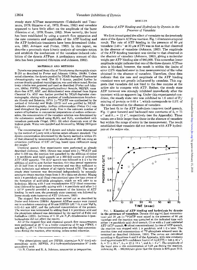
We first investigated the effect of vanadate on the presteady state of the dynein ATPase reaction. Fig. 1 illustrates a typical result. The rate of ATP binding in the presence of 80 p~ vanadate (140 s" at 30 FM ATP) was as fast as that observed in the absence of vanadate (Johnson, 1983). The amplitude of the ATP-binding transient was similar to that observed in the absence of vanadate (Johnson, 1983), giving a molecular weight per ATP binding site of 980,000. This somewhat lower amplitude might indicate that one of the three dynein ATPase sites is blocked however, the result is within the limits of error (12% standard error in four measurements) of the value obtained in the absence of vanadate. Therefore, these data indicate that the rate and amplitude of the ATP binding transient were not greatly influenced by vanadate. This sug- gests that vanadate did not bind to the free enzyme at the active site to compete with ATP. Rather, the steady state ATP turnover was strongly inhibited immediately after the transient with no apparent lag. Under this experimental con- dition, the steady state rate was inhibited to 1.6 nmol of Pi/ min/mg of protein or 0.03 s-', which corresponds to 0.5% of the rate observed in the absence of vanadate.
The best fit to the ATP hydrolysis transient (acid quench, Fig. 1) gave forward and backward rate constants of kz = 75 s" and k".L = 12 s-', respectively (see the Appendix). These values are a little larger than those in the absence of vanadate but within the range of error in the measurement. The result established that vanadate did not interfere with ATP hydrol- ysis at the active site.
I .2
I .o
w
v) \ w 2 0.6 I v) n
x 0.4 0 a
0.8
0.2
0
I I I 1 1 I
0 TIME (msec)
FIG. 1. Kinetics of ATP binding and hydrolysis by dynein in the presence of vanadate. Dynein (0.6 mg/ml final concentra- tion) and 30 PM [y-32P]ATP were mixed in the presence of 80 gM vanadate and the reaction was quenched at the indicated time with either 1 N perchloric acid (Acid quench, 0) or with a 3 mM unlabeled ATP (ATP quench, 0) (final concentrations). In case of ATP quench, the reaction was stopped with 1 N perchloric acid 1-2 s later. The reaction time and concentration of 3ZP-phosphate released were de- termined as described (Johnson, 1983). The curves are calculated according to Equations 8 and 9 of the Appendix, with rate constants (and approximate 90% confidence limits) of: k,(ATP] = 140 k 40 s", kf = 75 k 30 s-l, k--a = 12 -C 10 s-', and k3 = 8 s-'. The amplitude of the burst gave a site concentration of 0.49 WM during the reaction, indicating M , = 980,00O/site given that the dynein is 80% pure 30 S.
It was thus shown that the very first cycle of the ATPase reaction occurred in a normal manner even in the presence of a high concentration of vanadate, but the ATPase was inhib- ited just after a single turnover. This result ruled out the possibility of vanadate binding to the free enzyme at the catalytic site or to ATP. Moreover, it indicates that vanadate binds to an intermediate formed following ATP hydrolysis. This suggests that phosphate release precedes ADP release and vanadate binds to the dynein-ADP intermediate to form a dead end dynein-ADP-vanadate complex (see “Discussion”).
Effect of Vanadate on the ATP-induced Dissociation of the Microtubule-Dynein Complex
ATP induces dissociation of the microtubule-dynein com- plex, which can be monitored by means of light scattering. The rate of dissociation of dynein from the microtubule was previously shown to provide a direct measurement of the rate of ATP binding to dynein (Porter and Johnson, 1983a, 1983b; Johnson, 1983). We investigated the effect of vanadate on the ATP-induced dissociation of the microtubule-dynein com- plex, since the stopped flow light-scattering experiment pro- vides a much more accurate measurement of the kinetics of ATP binding and can be performed in the presence and absence of vanadate using the same sample. As shown in Fig. 2, vanadate did not affect the rate or amplitude of the light- scattering transient at 20 PM ATP. This result demonstrates unequivocally that vanadate did not affect the rate of ATP binding, in agreement with the result of the chemical quench flow experiment described above.
Dissociation of the Microtubule-Dynein Complex by ADP Plus Vanadate
The results described above showed that vanadate did not affect the binding or the hydrolysis of ATP at the active site of dynein. Therefore, inhibition must occur at a subsequent
- R E 2.25 ’ ..
- “ 2.10 q E 1.95
“ t 1 t.65
1.50 0 10 20 30 40
TIME ( msec) FIG. 2. Effect of vanadate on kinetics of 20 JIM ATP-induced
dissociation of the microtubule-dynein complex. The microtu- bule-dynein complex was mixed with ATP in the stopped flow appa- ratus and the light scattering a t 420 nm (arbitrary intensity units) was recorded. The figure shows two sets of data: one with vanadate and the other without. Final concentrations were: tubulin, 0.12 mg/ ml; dynein, 0.15 mg/ml; ATP, 20 pM; and vanadate, 400 pM. The data were stored in a computer and fitted to a single exponential (smooth line). The rate constants were 68 s-l in the absence and 72 s-’ in the presence of vanadate.
TABLE I Apparent ADPase activity of dynein and the microtubule proteins In order to assess the adenylate kinase activity, apparent ADPase
assays were performed as described under “Materials and Methods.” Sample Concentration of ADP Activity
step and it is likely that the formation of a ternary dynein- ADP-vanadate complex accounts for the inhibition. Accord- ingly, we might expect to observe the formation of the ternary complex by incubation of dynein with ADP plus vanadate. This complex might mimic the conformation of the normal dynein-ADP-Pi intermediate (Johnson, 1983) and induce the dissociation of the dynein from the microtubule. A similar situation has been shown to be the case for actomyosin (Goodno and Taylor, 1982).
Effect of A D P Alone-First, we investigated the effect of ADP alone on the light-scattering intensity of the dynein- microtubule complex. Although commercial ADP induced a light-scattering decrease, it seemed to be due to some ATP contamination. ADP purified by DEAE-Sephadex column chromatography induced only a very slow decrease in light scattering, at a rate of approximately 0.01 s-’ at 0.2 mM ADP (data not shown). Furthermore, the dissociation was incom- plete with an amplitude of only one-third relative to the decrease of the light scattering observed with ATP.
There may be a possibility that the observed light-scattering decrease was due to ATP formed by the action of contami- nating adenylate kinase (Kaji, 1973). Therefore, the adenylate kinase activity of the dynein fraction and of the microtubule fraction was estimated by apparent ADPase measurements coupled with dynein ATPase activity (Table I). If the ATP- generating activity of the adenylate kinase is less than the ATP-hydrolyzing activity of the dynein ATPase, this meas- urement provides a reasonable value of the adenylate kinase activity by measuring apparent ADPase activity. Only a neg- ligible adenylate kinase activity was detectable in our prepa- rations of DEAE-purified dynein (specific activity of approx- imately 0.05 nmol/mg/min) which is 4000-fold lower than the ATPase activity. A slightly higher activity was present in the microtubule protein preparation (Table I) but still, under our conditions for the light-scattering measurements, the rate of ATP formation should be less than 1 nmol/min/ml. In addi- tion, since the dynein ATPase activity is so high, the actual concentration of ATP must be maintained at much less than 1 PM. At this concentration, no dissociation of the microtu- bule-dynein complex is observed in the absence of vanadate (Shimizu and Johnson, 1983). Since the apparent ADP-in- duced dissociation of the microtubule-dynein complex was relatively slow, we could effectively examine the dissociation induced by ADP plus vanadate on a shorter time scale (less than 20 s) without any detectable contribution from ADP alone.
Effect o f ADP Plus Vanadate-The combination of ADP and vanadate induced a fairly rapid light-scattering decrease (Fig. 3A). Increasing the vanadate concentration up to 0.1 mM in the presence of 0.5 mM ADP enhanced the rate of the light-scattering change and a plateau in rate was attained above that concentration (Fig. 3B). The light-scattering
A I I I I biphasic light-scattering transients and the somewhat sig- moidal concentration dependence of the rate are indicative of a multistep dissociation pathway and further work is under
2 2.40 - -
0 paper describes the biphasic kinetics observed at low concen- way to explore the kinetics in more detail. The following
c ::
terms of a three-headed dynein molecule (Shimizu and John- ,*.;-y:y.. . .. .. . a Y +> ..... * ...... ~ _ , - t 2.25
trations of ATP and provides a reasonable interpretation in
son, 1983). In the present case, the observed biphasic trace ... E
, '. -.. ...... could be a function of a multiple-step dissociation pathway 2.10 - ':i
. . .... due to the three dynein heads or due to binding of ADP
. -. .. c We next confirmed that the light-scattering decrease, in- .,...
1 I 1 I . .-. .. - ..-. - UQyx.
duced by ADP plus vanadate, reflected the genuine dissocia-
....... . . '2 "* .,._ .. (3
..T." "... ,-~ -.--'L,^. - -, .-,. . . . .
'C. -.. A.,
- - . . ... k +
5: 1.95 +
-
- 3 1.80
I I...
. . \._ '"
% , .. . . . . followed by vanadate. The observation of a lag at saturating . -.
4 ADP would tend to argue against the latter explanation. .:. >_ '\ .. ............... b ... .'< ._ > .'-%
CONCENTRATION Of VANADATE (*) FIG. 3. ADP plus vanadate-induced dissociation of the mi- Z
crotubule-dynein complex. A, stopped flow measurements of the L 0.4 light scattering. The microtubule-dynein complex was mixed with f
- -
change upon vanadate concentration. As described in text, the slower 40 p ~ , or ( e ) 400 p ~ . B, dependence of the rate of the light-scattering concentrations of vanadate were: (a ) 0 pM, (b ) 8 pM, ( c ) 20 pM, ( d ) 4 0.2 - - tubulin, 0.12 mg/ml; dynein, 0.13 mg/ml; and ADP, 0.5 mM. The final was monitored as described in Fig. 2. Final concentrations were: ADP and varying concentrations of vanadate and the light scattering - 4/
effect observed a t 28 p~ vanadate. 0 0. I 0.2 0.3 0.4 0.5 calculated hyperbola with a maximum rate of 0.7 s-l and half-maximal 0 I I I 1 I was plotted against the concentration of vanadate. The curue is a phase of the data was fitted with a single exponential and the rate
/ P
CONCENTRATION OF Mg-ADP ( m u ) curves were biphasic and rate measurements were obtained FIG. 4. ADP plus vanadate-induced dissociation of the mi- by fitting to the slow phase. A similar relationship was dem- CrotUbule-dYnein complex- A, stopped flow measurements of the onstrated with a fixed vanadate concentration and varying light scattering. The experiments were done in the same way as
ADP concentration (Fig- 4). The effects were varied. The final concentrations were: tubulin, 0.10 mg/ml; dynein, described in Fig. 3A except that the Mg-ADP concentration was
observed at 0.028 mM vanadate Or at 0.1 mM ADP when the 0.12 mg/ml; and vanadate, 400 p ~ . In order to keep the MgZ+ other ligand was at saturation. The maximal rate of the light- concentration approximately constant, Mg2+ was added at the same scattering change was 0.5 to 0.7 s" depending upon the concentration as ADP, in addition to the 4 mM MgCb already present. preparation of dynein. Vanadate alone up to 0.4 mM did not The final concentrations of Mg-ADP were: (a ) 0 mM, (b ) 0.05 mM,
any significant light-scattering decrease of the micro- ( c ) o.l mM, and ( d ) l.o m'. B, dependence of the rate of the light- tubule-dynein complex within 20 s. It should be noted that mated and plotted in the Same way as in F ~ ~ . 3B. The solid line is a
scattering change upon Mg-ADP concentration. The rate was esti-
the plateau in rate established unequivocally that the observed calculated hyperbola with a maximum rate of0.6 s-1 and half-maximal dissociation was not due to ATP contamination. Also, the effect observed a t 0.1 mM Mg-ADP.
FIG. 5. Dissociation of the microtubule-dynein complex by ADP plus vanadate as demonstrated by electron microscopy. The microtubule-dynein complex was formed as described under "Materials and Methods" a t final concentration of 0.2 mg/ml tubulin and 0.26 mg/ml dynein. The samples were applied to carbon and Formvar-coated grids, negatively stained with uranyl acetate, and viewed with a Philips EM300 operated at 60 kV. A, microtubule- dynein complex. R, microtubule-dynein complex 30 s after treatment with 50 p~ Mg-ADP and 40 p~ vanadate. X 88,000.
I I I I I 0 I 2 3 4 5
TIME (eec) FIG. 6. Dissociation of the microtubule-30 S dynein com-
plex by ADP plus vanadate. The microtubule-30 S dynein complex was made in the same way as the microtubule-DEAE dynein complex. Final concentrations were: tubulin, 0.09 mg/ml; 30 S dynein, 0.11 mg/ml. The concentration of the ligands were: ( a ) 400 p~ vanadate, ( b ) 0.5 mM ADP, and (e) 0.5 mM ADP plus 400 pM vanadate.
tion of dynein from the microtubule. Electron microscopic observation showed that even in the presence of 0.5 mM MgADP or 0.2 mM vanadate alone, decoration of the micro- tubule walls by dynein was apparent. In the presence of ADP plus vanadate, however, only naked microtubules were ob- served (Fig. 5). Therefore, it could be concluded that ADP plus vanadate induced essentially complete dissociation of the dynein from the microtubule.
The dynein fraction used above was purified by DEAE- Sephacel column chromatography and was a mixture of the 30 S dynein (80%) and 14 S dynein (Porter and Johnson,
1983a). Porter and Johnson (1983a, 198313) showed that only the 30 S dynein was responsible for the light-scattering change. As shown in Fig. 6, the 30 S dynein could substitute for DEAE-dynein in terms of ADP plus vanadate-induced light-scattering change indicating that the 30 S dynein was the major, if not exclusive, contributor. In addition, the rate of the light-scattering decrease at 0.5 mM ADP plus 0.2 mM vanadate was about 0.6 s-' which was almost the same as that obtained with the column purified dynein.
Inhibition of the Binding of ATP by Preincubating the Dynein with ADP plus Vanadate
As described above, the data argue that a dynein-ADP- vanadate ternary complex was formed when the dynein was incubated with ADP plus vanadate. In turn, we expect that preincubation of the dynein with ADP plus vanadate should block the binding of ATP to the enzyme site, although this step was not inhibited by incubation with vanadate alone.
Since ADP alone might inhibit the binding of ATP, a low concentration of ADP (5 p ~ ) was chosen. At this concentra- tion, ADP alone had little effect on the binding of ATP to the dynein ATPase as measured by chemical quench flow methods (Fig. 7, filled circles). In contrast, preincubation of the dynein with 5 p~ ADP plus 80 p~ vanqdate for 20 min a t 28 "C nearly eliminated the ATP-binding transient (Fig. 7, open circles). A small amplitude of the ATP-binding transient remained but this may be due to the incomplete binding of ADP and vanadate to the dynein since experiments performed at a lower ADP concentration gave an increased amplitude (data not shown). Nonetheless, ADP plus vanadate but not
TIME (msec) FIG. 7. Inhibition of ATP binding of the dynein by prein-
cubation with ADP plus vanadate. ATP quench experiments were performed as described in Fig. 1. The first syringe contained 0.75 mg/ ml dynein with 5 p~ ADP (0) or with 5 p~ ADP plus 80 pM vanadate (0). The mixture in the first syringe was allowed to stand for 20 min a t 28 "C before use. The second syringe contained 60 p~ [y-'*P]ATP (initial concentration before mixing). Equal volumes from the both syringes were mixed and, after a delay of the time indicated, allowed to enter the second mixing chamber where 3 mM unlabeled ATP was added. The enzyme reaction was stopped 1-2 s later by adding 1 N perchloric acid manually. The curue is calculated according to the method described previously (Johnson, 1983) with the following rate constants: kl[ATP] = 140 s-I, k, = 70 s-I, k-2 = 10 s-I, k, = 8 s-I. The amplitude of the ATP-binding transient (0) gave a site concentration of 0.402 p ~ , suggesting a molecular weight per ATP binding site equal to 750,000.
ADP or vanadate alone inhibited the ATP-binding. These results strongly suggest the formation of a dynein-ADP-van- adate complex at the active ATPase site with binding that is tighter than that for ADP or ATP or vanadate alone.
Effect of ADP Pllls Phosphate on the Light-scattering of the Microtubule-Dynein Complex
In combination with ADP, phosphate had some effect on the intensity of light scattering by the microtubule-dynein complex although more than 10 mM phosphate was necessary to exert a detectable change (data not shown). This high concentration of phosphate induced the reduction of light- scattering intensity even in the absence of ADP and in the presence of the microtubule stabilizing drug taxol. We could not exclude the possibility that the dissociation of dynein molecules was due to the high ionic strength, so the accurate determination of the rate constant was not warranted. How- ever, the effectiveness of phosphate was estimated to be at least lo4 times less than that of vanadate. At 50 mM phosphate in combination with ADP, the observed rate constant was about 0.05 s" which corresponds to the rate observed at 4 PM vanadate (Fig. 3B).
DISCUSSION
Porter and Johnson (1983a, 1983b; Johnson, 1983) have investigated the reaction mechanism of the dynein ATPase and established the following scheme:
M-D + ATP 2 M-D-ATP .1
D + ATP 1 D-ADP a D-ADP-P, 4 D + ADP + P, 2
SCHEME 1
where M and D represent microtubule and dynein, respec- tively. In the presence of a saturating concentration of ATP (e.g. more than 10 phi) step 3 is rate-limiting in the steady state (Johnson, 1983). With this background, we investigated the effect of vanadate on each reaction step.
Vanadate did not affect the binding of ATP (steps 1 and 1') as shown by the chemical quench flow (Fig. 1, ATP quench) and the stopped flow experiments (Fig. 2). Our pre- liminary results3 also showed that vanadate did not inhibit the slow dissociation of the microtubule-dynein complex in- duced by the nonhydrolyzable analog (&y-imido)ATP. The lack of any observable effect of vanadate on the kinetics of dissociation of the microtubule-dynein complex induced by (P,y-imido)ATP or 20 phi ATP indicates that vanadate did not exert its effect by binding to a site other than the catalytic site. Moreover, ATP hydrolysis (step 2) was unaffected by vanadate as shown by the acid quench experiments (Fig. 1).
These observations establish that the inhibition occurs subsequent to ATP hydrolysis. It should be emphasized that, in the presence of a sufficient concentration of vanadate, the inhibition takes place immediately after one turnover. This implies that the first catalytic cycle must produce an inter- mediate that is required for inhibition to occur and that the binding of vanadate to this intermediate must be relatively rapid. The observed dissociation of the microtubule-dynein complex by ADP plus vanadate and the inhibition of the ATP binding/hydrolysis transient by preincubating the enzyme with ADP plus vanadate argue very strongly that the inhibited state is an enzyme-ADP-vanadate ternary complex. Thus, vanadate bound tightly to the catalytic center as an analog of the y-phosphate of ATP only when the ADP site was occu-
D. B. Clutter, M. E. Porter, T. Shimizu, and K. A. Johnson, unpublished results.
pied. An earlier report of a phosphate burst for dynein, occurring over a period of minutes and having an apparent amplitude of " 5 0 mol ATP/500,000 g of dynein (Nakamura and Masuyama, 1977) is probably due to the slow formation of the dynein-ADP-vanadate complex with a low concentra- tion of contaminating vanadate in the ATP solution (Cantley et al., 1977).
We present the following scheme for the vanadate-induced inhibition of the dynein ATPase:
D + ATP + D-ATP + D-ADP-Pi -+ D-ADP + Pi D + ADP 1 2 3 4
TJ 5 D-ADP-V
SCHEME 2
This scheme implies the existence of a D-ADP intermediate during the cycle of the ATPase reaction, with phosphate release occurring first and ADP second.
The overall scheme parallels the vanadate inhibition of the myosin ATPase although the individual rate constants are different (Goodno, 1979; Goody et al., 1981; Goodno and Taylor, 1982). The rates of association and dissociation of vanadate are a factor of approximately lo6 faster for dynein than for the corresponding reactions with myosin, although the equilibrium constants for the formation of the enzyme- ADP-vanadate comlex may be similar for dynein and myosin. The observed difference in rates is sufficient to account for the selective inhibition of dynein at low vanadate concentra- tions over a period of time where myosin is not inhibited.
Is it possible to explain the noncompetitive type inhibition by this reaction scheme? Shimizu (1981) previously reported steady state ATPase measurements in the presence of an ATP-regenerating system (pyruvate kinase + phosphoenol- pyruvate) that showed that the inhibition was noncompeti- tive. This analysis was based upon the assumption that the ATP concentration during the measurement was the same as that added, but this assumption may not be correct. There should have been a steady state between ATP and ADP and some of the nucleotide should have bound to the active sites of the enzymes. Therefore, the actual concentration of ATP must have been lower, especially when the concentration of the added ATP was less than 5 ph i . In fact, we have prelimi- nary evidence suggesting that the free ATP concentration is considerably lower than the total one in such a system (Shim- izu and Johnson, 1983).
With this consideration, we must reevaluate the inhibition pattern obtained previously. Qualitatively, it could be deduced that the inhibition may not be purely noncompetitive but a mixed type of noncompetitive and uncompetitive inhibition. As a matter of fact, such type of inhibition was observed with the dATPase of dynein (Shimizu, 1982) and with t h e 1 T P a ~ e . ~
Let us consider the kinetics of Scheme 2. If we assume that the phosphate binding ( k 3 ) is negligible, the enzyme activity ( u ) is given by the following equation:
where V,,, = k3k4EO/(k3 + k4), Eo is the total concentration of the dynein, K , = kz(k-, + k3)/(k,(k3 + k4)), and KADp and KI are dissociation constants for ADP and vanadate, respec- tively, and I denotes vanadate. Qualitatively, the equation has both an ordinate intercept effect and a slope effect if the ratio of [ADP]/KADP # 0, and so the inhibition would be a noncom- petitive type. Quantitatively, we could account for the pre- vious mixed noncompetitive inhibition pattern (Shimizu, 1981) by assuming the following constants: kg = 8 s-', k , =
10-20 s” (Porter and Johnson, 1983b; Johnson, 1983), and k-4 = 2-5 X lo6 M-’, with an ADP concentration of 0.3 pM during the steady state measurements. Although these rates are reasonable, further analysis will be required to establish these parameters by direct measurement. One should also note that the form of the equation would be altered due to the tight binding of vanadate, if one wants to more precisely relate the free concentration of inhibitor, [ I ] , to the total concentration.
Anderson and Purich (1982) reported results, in their stud- ies using a crude extract of sea urchin flagellar dynein, indi- cating that the addition of diadenosine pentaphosphate led to an increase in ATPase activity and gave a linear noncom- petitive inhibition pattern of vanadate versus ATP. On the basis of this sole observation, they concluded that vanadate must form a vanadate-ATP chelate complex to inhibit the enzyme. This postulate cannot explain the present data, es- pecially the demonstration that the binding and hydrolysis of 30 p~ ATP was unaffected by 80 ~ L M vanadate. There are problems with the data presented by Anderson and Purich. 1) The K , they reported for their dynein preparation uersus ATP was 20 p ~ , which is approximately 1 order of magnitude higher than that normally observed; this suggests that their dynein preparation was fully activated (Gibbons and Fronk, 1979) and not in a native state. 2) There is no reasonable explanation, and the authors do not offer any, for the rather strange effect of diadenosine pentaphosphate that they ob- served in accelerating the ATPase; and 3) the adenylate kinase activity was equal to one-third of the ATPase activity in their preparations. Our purification of dynein effectively removes contaminating adenylate kinase activity and the addition of diadenosine pentaphosphate during the past sev- eral years of experiments has had no effect on any of the results.
The following scheme provides the minimum pathway con- sistent with the dissociation of the microtubule-dynein com- plex by ADP plus vanadate:
M - D + ADP & M-D-ADP + V a M-D-ADP-V b
1 c M + D-ADP-V
SCHEME 3
where V represents vanadate. The forward reaction of step c has a rate constant of 0.5-0.7 s-’. The dissociation of the microtubule-dynein complex may occur as the reverse of the normal forward pathway involving the recombination of the dynein-ADP-P, intermediate with the microtubule, with re- lease of products accompanying cross-bridge movement (Johnson, 1983).
Phosphate in combination with ADP had the same sort of effect as vanadate on the light-scattering property of the microtubule-dynein suspension, although the effect of phos- phate was much weaker. Since the phosphate-binding at the y-phosphate site of the catalytic center is very likely, the result supports the interpretation that vanadate acted as a phosphate analog to form the dynein-ADP-vanadate ternary complex. The much weaker effect of phosphate than that of vanadate may be consistent with the observation that phos- phate is not a strong inhibitor of the dynein ATPase (Shimizu and Kimura, 1977; however, cf. Otokawa, 1973). In fact, van- adate has been shown to be much more effective in inhibiting a variety of enzymes than phosphate (Shimizu, 1981). Since vanadate can form a pentacoordinated species (Cotton and Wilkinson, 1966), the complex of ADP and vanadate may resemble the transition state for ATP hydrolysis with the p- phosphate of ADP contributing one of the five oxygen ligands
on the vanadate. This would explain the very tight binding of vanadate in the presence, but not in the absence of ADP. The weak binding by phosphate suggests that there may be a large free energy decrease accompanying phosphate release similar to that observed for actomyosin (Taylor, 1979) and that this step may normally be coupled to force production in the cilium.
Okuno (1980) reported that ADP plus vanadate or phos- phate induced no relaxation of the rigor wave of Triton models of sea urchin spermatozoa. This result differs from our obser- vations using a simpler system and suggests that relaxation of the rigor wave requires simultaneous detachment of a large fraction of the microtubule-dynein complexes. In the axo- neme, an equilibrium between attached and detached states will be established and the high local microtubule concentra- tion will favor the attached state.
In contrast, ATP plus vanadate caused relaxation of the rigor wave (Okuno, 1980) and the addition of vanadate in- creased the rate of relaxation a t low ATP concentration. This is consistent with our observation that vanadate prevents the multiple turnovers of ATP and should allow relaxation at a level of only one ATP per dynein site. The final equilibrium state after the addition of ADP or ATP with vanadate should be the same, with nearly all of the sites occupied with ADP and vanadate and some equilibrium between attached and detached cross-bridges. The different effects observed with ATP and ADP may suggest a higher level of complexity. There is reason to believe that dynein cross-bridges occur only with the formation of a bend and that other structures in the axoneme normally serve to prevent the simultaneous association of all the cross-bridges. The more rapid dissocia- tion of the dynein in the presence of ATP may allow for complete relaxation of the wave prior to establishment of the final equilibrium state involving the reassociation of the dy- nein-ADP-vanadate complex with the microtubule.
APPENDIX
Vanadate Inhibition of Dynein: Kinetics of a Single Turnover of ATP
In the presence of a high concentration of vanadate, all the sites of the dynein ATPase were occupied with products (i.e. ADP) and the inhibitor, vanadate ( V ) immediately after a single turnover of ATP (Shimizu and Johnson, 1983). There- fore, we could assume the following scheme.
E + A T P ~ E - A T P e E - A D P - P , ~ E - A D P - + E + A D P k k2 k k4
k-z r4 ~ J V I E-ADP- V
Because of the tight binding of ATP, k-* is assumed to be negligible relative to kZ (Johnson, 1983) and due to the low concentration of phosphate, the binding of phosphate to the E - ADP complex (k -J could also be neglected.
Since a high concentration of vanadate inhibits the enzyme after a single turnover, one can model the system by assuming that k,[V] is much larger than k3 and k4. Accordingly, the pathway given above can be simplified to the following:
k kz k3
k-2 E + ATP E-ATP a E-ADP-Pi -+ E-ADP-V ( 2 )
This leads to the following set of differential equations:
d [ E l / d t = -k*[EI[Tl
d [ E T ] / d t = k J E ] [ T ] + k-z[ED] - kz[ET]
where T, Eo, E, ET, ED, and E V represent ATP, total enzyme, free enzyme, E-ATP complex, E-ADP-Pi complex, and E- ADP-V complex, respectively, and [Eo] = [E] + [ET] + [ED] + [EV].
This leads to the following third order differential equation: [ED]"' + ( k , [ T ] + k, + k-, + k3)[ED]"
which has a solution of the form: [ED]/[Eo] = Cle-Al' + Cze-'2l' + C3e-A3t (4)
The rate constants, X,, are given by:
A1 = MTI (5) X2, A3 = ( ( k , + k-z + k3) f [ ( k z + k-2 + k3)' - 4kzk3]"}/2
C1, C2, and CB are constants:
C1 = kzXl/((Az - A d ( A 3 - AI))
c2 = k2Al/((X2 - Xl) (AZ - A311 (6)
c3 = kZAl/((A3 - A l ) ( A 3 - X Z ) )
to be 8 s-l from our measurement in the absence of vanadate (Johnson, 1983). This is reasonable since vanadate should not alter the rate of product release, but will only bind the site left empty following dissociation of phosphate. Moreover, changes in the value of k3 had little effect on the fit to the experimental data. The remaining constants, k2 and k-?, were obtained as fits to the rate and amplitude of the Acid quench experiment. The fitting process was refined and approximate 90% confidence limits were calculated based upon the sum square error as described previously (Johnson, 1983).
REFERENCES Anderson, S. A,, and Purich, D. L. (1982) J. B i d . Chem. 2 5 7 ,
6656-6658 Cantley, L. C., Jr., Josephson, L., Warner, R., Yanagisawa, M.,
Lechene, C., and Guidotti, G. (1977) J . Biol. Chem. 252,7421-7423 Cantley, L. C., Jr., Cantley, L. G., and Josephson, L. (1978) J. Bid.
Chem. 253,7361-7368 Cotton, A. F., and Wilkinson, G. (1966) Advanced Inorganic Chem-
istry, 2nd ed., pp. 811-813, John Wiley and Sons, New York Evans, J . A. (1982) Ph.D. thesis, University of Hawaii Fiske, C. H., and Subbarow, Y. (1925) J. Biol. Chem. 66,375-400 Gibbons, I. R., and Fronk, E. (1979) J. Bid. Chen. 254,187-196 Gibbons. 1. R.. Cosson. M-P.. Evans. J. A., Gibbons, B. H., Houck,
The time dependence of E V can be obtained by integration N& Acad, sei, U, s, A , 75, 2220-2224 of the equation: Goodno. C. C. (1979) Proc. Natl. Acad. Sci. U. S. A . 76 , 2620-2624
B. Martinson, K. H.', Sale,'W. S., and Tang, W-J..Y. (1978) Proc.
which can be solved to yield
[ E V ] / [ E o ] = - (1 - C A I t ) k3CI
A1
Goodno; C. C., and Taylor, E. W. (1982) Proc. Natl. Acad. Sci. U. S. A .
Goodv. R. S.. and Eckstein, F. (1971) J . Am. Chem. Soc. 9 3 , 79,21-25
The final solution can be calculated in parts using a micro- computer with the appropriate substitutions of Equations 4-7 into Equations 8 and 9 for the following sums measured by the ATP and Acid quench experiments:
ATP quench = ( [ E T ] + [ E D ] + [ E V ] ] / [ E o ] (8)
= 1 - [ E ] / [ E o ] = 1 - e+lf
Acid quench = [ED]/[Eo] + [EV]/ [Eo] (9)
The solution for the time dependence of the Acid quench and the ATP quench was used with a computer graphics subroutine to fit the quench flow data by trial and error as described previously (Johnson, 1983). The value of the rate constant for ATP binding was obtained directly from the ATP quench data as a fit to a single exponential (Equation 8). The Acid quench data theoretically should exhibit a triple exponential, the fitting of which is clearly beyond the limits of error in the data. Nonetheless, the fit to a simpler equation, without including all three constants, is incorrect and would lead to an underestimate of the rate of hydrolysis. TO obtain a solution, the rate constant for product release was assumed
6252-6257 Goody, R. S., Hofman, W., and Konrad, M. (1981) FEBS Lett. 129 ,
Johnson, K. A. (1983) J . Biol. Chem. 2 5 8 , 13825-13832 168-172
173 Johnson. K. A,, and Porter, M. E. (1982) Cell Motil. Suppl. 1,101-106 Kaji, K. (1973) Ph.D. thesis, University of Tokyo Kobayashi, T., Martensen, T., Nath, J., and Flavin, M. (1978)
Nakamura, K., and Masuyama, E. (1977) Biochim. Biophys. Acta
Okuno, M. (1980) J. Cell Bid. 85 , 712-725 Otokawa, M. (1973) Biochim. Biophys. Acta 292,834-836 Porter, M. E., and Johnson, K. A. (1983a) J. Biol. Chem. 2 5 8 ,
Porter, M. E., and Johnson, K. A. (1983b) J. Bid. Chem. 2 5 8 ,
Sale, W. S., and Gibbons, I. R. (1979) J . Cell Biol. 8 2 , 291-298 Satir, P., Wais-Steider, J., Lebduska, S., Nasr, A., and Avolio, J.
Schendel, P. F., and Wells, R. D. (1973) J. Biol. Chem. 2 4 8 ,
Shimizu, T. (1981) Biochemistry 2 0 , 4347-4354 Shimizu, T. (1982) Cell Motil. Suppl. 1 , 107-112 Shimizu, T., and Johnson, K. A. (1982) Biophys. J. 37,346a Shimizu, T., and Johnson, K. A. (1983) J. Biol. Chem. 248,8319-8321 Shimizu, T., and Kimura, I. (1977) J. Biol. Chem. 258 , 13841-13848 Shimizu, T., Murofushi, H., Kimura, I., and Sakai, H. (1979) FEBS
Takahashi, M., and Tonomura, Y. (1979) J . Biochem. (Tokyo) 86 ,
Taylor, E. W. (1979) Crit. Reu. Biochem. 6 , 103-164 Vogel, A. 1. (1961) A Textbook of Quantitative Inorganic Analysis, 3rd
Biochem. Biophys. Res. Commun. 81, 1313-1318
48 1,660-666
6575-6581
6582-6587
(1981) Cell Motil. 1, 303-327
8319-8321
Lett. 108,215-218
413-423
ed., pp. 790-791, John Wiley and Sons, Inc., New York


![3D-AFM Nano-structural Features and Magnetic Properties of Indium-Doped Vanadate ... · 2020. 9. 27. · thermodynamic stability of bismuth vanadate-based compositions [9]. Based](https://static.documents.pub/doc/80x56/6081c316e3d07c0e3b322aa6/3d-afm-nano-structural-features-and-magnetic-properties-of-indium-doped-vanadate.jpg)