+ All Categories
Home > Documents > Prevalence and treatment of painful diabetic neuropathyclok.uclan.ac.uk/11313/2/Aslam Final e-Thesis...

Prevalence and treatment of painful diabetic neuropathyclok.uclan.ac.uk/11313/2/Aslam Final e-Thesis...

Date post: 15-Mar-2018
Category:
Upload: hathuan
View: 213 times
Download: 0 times
Share this document with a friend
195
1 Prevalence and treatment of painful diabetic neuropathy By Amir Aslam (MB BS, MRCP (UK), MRCGP) A thesis submitted in partial fulfilment of the requirements for the degree of MSc (by Research) at the University of Central Lancashire August 2014
Transcript

1

Prevalence and treatment of painful diabetic

neuropathy

By

Amir Aslam (MB BS, MRCP (UK), MRCGP)

A thesis submitted in partial fulfilment of the requirements for the degree

of MSc (by Research) at the University of Central Lancashire

August 2014

2

University of Central Lancashire

STUDENT DECLARATION FORM

Concurrent registration for two or more academic awards

*I declare that while registered as a candidate for the research degree, I have not been a

registered candidate or enrolled student for another award of the University or other

academic or professional institution

Material submitted for another award

*I declare that no material contained in the thesis has been used in any other

submission for an academic award and is solely my own work

Collaboration

Where a candidate’s research programme is part of a collaborative project, the thesis

must indicate in addition clearly the candidate’s individual contribution and the extent

of the collaboration. Please state below:

Signature of Candidate

______________________________________________________

Amir Aslam

Type of Award ………………. MSc by Research…………………………

School …………..Pharmacy and Biomedical Sciences…………….

3

Acknowledgement

I give my sincere thanks to Professor Jaipaul Singh for his continuous support, valuable

advice and help in preparation of this thesis. I am also thankful for Professor Rajbhandari

for his dedication, support and practical advice in designing the research, ethical

clearance, and continuous supervision throughout this Masters project. Due to the

immense support and guidance of Professor Rajbhandari and Professor Singh, I was able

to publish papers in leading journals. I also appreciate the expertise and motivation given

to me by both supervisors regularly in meetings held to discuss my progress. My special

appreciation goes to the University of Central Lancashire and Lancashire Hospitals NHS

Trust for providing the facilities and great research environment for carrying out this

Masters project.

Finally, I am forever indebted to my parents, my wife Dania, my sisters, and brother for

their never-ending encouragement, love, and unconditional support all the way. I am

extremely proud to dedicate this thesis to my Family with Love.

4

Declaration

I declare that this thesis has been composed by myself and that, whilst registered as a

candidate for the degree of Master of Science by Research, I have not been registered as

a candidate for any other awarding body.

Amir Aslam

5

Abstract

The prevalence of diabetes is rising globally and, as a result, its associated complications

are also rising. Painful diabetic neuropathy (PDN) is a well-known complication of

diabetes and the most common cause of all neuropathic pain. About one-third of all

diabetes patients suffer from PDN. The reported prevalence of PDN varies from 11% in

Rochester, Minnesota, USA to 53.7% in the Middle East. One UK study, published in

2011, reported that the prevalence of PDN was 21.5% in type 2 diabetes patients and

13.4% in type 1 diabetes patients, resulting in an overall prevalence of 21%. Numerous

studies have found cardiovascular risk factors—including increased age, longer duration

of diabetes, higher weight, smoking, poor glycaemic control, renal impairment and high

cholesterol—to be associated with PDN. This disorder has a huge effect on people’s daily

lives both physically and mentally. Despite huge advances in medicine, the treatment of

PDN is both challenging for physicians and distressing for patients. In this thesis, three

studies were carried out on the following topics: prevalence and characteristics of painful

diabetic neuropathy, PDN patients’ quality of life, and treatment employing lignocaine.

This first study assessed the prevalence of painful diabetic neuropathy (PDN) and its

relationship with various cardiovascular characteristics in diabetes subjects. This was

done through an observational study of diabetes subjects in Northwest England, UK (n

=204). The self-completed Leeds Assessment of Neuropathic Symptoms and Signs

questionnaire was sent by post to the subjects and used to diagnose PDN. Consent for

participation and access to blood results was given by the study participants. Ethical

approval for the study was also granted by National Research Ethics Committee UK. The

results of the study showed that the crude prevalence of PDN among subjects was 30.3%.

The prevalence of type 2 diabetes subjects was higher (33.1 %) than that of type 1 diabetes

6

subjects (14.1%). There was a significant association of obesity, smoking and height in

males with PDN compared to the non-PDN group (P <0.05). The results also showed a

significant trend of increasing PDN prevalence with duration of diabetes, increasing

HbA1c and increasing BMI (P<0.05). There was a trend of increasing prevalence with

age as well (P>0.05); however, due to the small sample size, the data was not statistically

significant. There was no relationship of PDN with systolic or diastolic blood pressure,

nephropathy, alcohol intake or blood cholesterol (P>0.05). These results highlight the

importance of better control of modifiable factors, including smoking, glycaemic control

(HbA1c) and obesity.

The second study assessed the impact of painful diabetic neuropathy on quality of life

(QoL), mood and anxiety by comparing patients suffering from painful diabetic

neuropathy (PDN group) with diabetes patients not known to have PDN (control group,

C). The study used short form (SF) 36 and Hospital Anxiety and Depression Scale (HADS

Scale) questionnaires. For the PDN group, 25 adult subjects (mean age 56, standard

deviation (SD) +/- 11 years, male 15, female 10) were randomly selected from patients

attending the painful diabetes neuropathy clinic at Chorley Hospital. For the control

group, 25 adult diabetic subjects (mean age 56, SD +/- 14 years, male 14, female 9) were

randomly selected from patients undergoing General Practitioner Surgery. Both groups

completed the HADS and SF36 questionnaires. Subjects in the PDN group had

significantly lower SF36 summary scores in both the physical health (P ˂0.0001) and

mental health domains (P= 0.026) compared with the C group. HADS data showed that

56% subjects in the PDN group could be diagnosed anxiety compared to only 20% in the

C group (P=0.018); and 60% of the PDN group received the diagnosis of depression

compare to 44% in the C group (P=0.396). The results also show that PDN was

significantly associated with impaired QoL, both physically (p<0.0001) and mentally

7

(p<0.026). Anxiety was significantly associated with the PDN group compared to control

(p<0.018), and depression was 16% more prevalent in PDN group than in the control

group.

The final study assessed the efficacy of lignocaine infusion as a treatment for PDN in

challenging cases where conventional treatment had not helped. A total 11 patients

participated; 7 patients were referred from the pain clinic (non-PDN group), and 4 were

referred from the foot clinic (PDN group). All were given lignocaine infusion as a

treatment for chronic pain. Participants from both groups were on multiple pain

medications with minimal results. All participants gave consent for participation and

filled out a McGill short form (SF) questionnaire before and after lignocaine infusion.

The results showed a 33% reduction in the visual analogue pain score after lignocaine

infusion in PDN group compared to an 11% reduction in the non-PDN group. The data

were statistically significant (P<0.05). Similarly, there was significant (p<0.05)

reduction of affective pain score: 41% after lignocaine infusion in PDN group, compared

to 21% in non-PDN group. In contrast, no significant difference was seen between groups

for the sensory pain score reduction after lignocaine infusion: 23% in PDN group

compared to 17% in non-PDN group (P>0.05). None of the 11 patients reported adverse

effects from the treatment and their observations were within normal limits throughout

the lignocaine infusion. Overall, the study showed that lignocaine infusion is effective

and safe in reducing the chronic intractable pain when conventional treatments are

intolerable or unhelpful. The treatment is also more effective for painful diabetic

neuropathy than for other forms of chronic pain.

8

Contents

Acknowledgement & Declaration……………………………………………….3

Abstract…………………………………………………………………………...5

Contents………………………………………………………………...................8

List of Figures ……………………………………………………………………12

List of Tables …………………………………………………………………….13

Abbreviations……………………………………………………………………..14

Chapter 1- ………………………………………………………………….......15

Introduction: Diabetes Mellitus and Painful diabetic neuropathy

1.1 Diabetes Mellitus…………………………………………………………........16

1.1.1 Type 1 and type 2 diabetes…….…………………………………………..17

1.1.2 Sign and symptoms of diabetes……………………………………………18

1.1.3 Diagnosis of diabetes…………………………………………………........18

1.1.4 Macrovascular complications of diabetes………………………………….19

1.1.5 Chronic microvascular complications of diabetes……….………………...19

1.1.6 Management of diabetes……………………………………………………21

1.2 Painful diabetic neuropathy………………………………………………….....26

1.2.1 Physiology of pain………………………………………………………….28

1.2.2 Neuropathic pain generation pathogenesis…………………………………30

1.2.2.1 Ectopic electrical impulses…………………………………………………30

1.2.2.2 Change in glucose flux and pain……………………………………………30

1.2.2.3 Role of dorsal root ganglion in neuropathic pain……………………….......33

1.2.2.4 Methyglyoxal and pain………………………………………………...........34

1.2.2.5 Sympathetic modulation of pain……………………………………………34

1.2.2.6 Gate control theory………………………………………………………….35

1.2.2.7 Central sensitization………………………………………………………...37

1.2.2.8 Central inhibition & Central facilitation …………………………………...38

1.2.2.9 Thalamic abnormalities…………………………………………………….39

1.2.2.10 Chronic neuropathic pain and plasticity of brain…………………...39

9

1.2.3 Diagnosis of painful diabetic neuropathy……………………………………40

1.2.3.1 Scales available to aid the diagnosis of neuropathic pain………………….41

1.2.4 Management of painful diabetic neuropathy……………………………….42

1.2.4.1 Pharmacological therapies………………………………………………….45

1.2.4.1.1 Antidepressants…………………………………………………………45

1.2.4.1.2 Anticonvulsants…………………………………………………………46

1.2.4.1.3 Opioid Agonists…………………………………………………………49

1.2.4.1.4 Topical agents…………………………………………………………...49

1.2.4.1.5 Other pharmacological therapies……………………………………….....50

1.2.4.2 Non Pharmacological therapies……………………………………………..52

1.2.4.2.1 Transcutaneous electrical nerve stimulation (TENS)…………………...52

1.2.4.2.2 Acupuncture……………………………………………………………..52

1.2.4.2.3 Electrical spinal cord stimulation……………………………………….53

1.2.4.2.4 Psychological therapies…………………………………………………53

1.2.4.3 Combination treatment and national guidance……………………………...54

1.2.5 Prognosis………………………………………………………………………57

Chapter 2- ……………………………………………………………..60

Prevalence and characteristics of painful diabetic neuropathy in the diabetic

population of Northwest England.

2.1 Abstract…………………………………………………………………………61

2.2 Introduction……………………………………………………………………..62

2.3 Subjects and Methods…………………………………………………………...64

2.3.1 Statistical analysis…………………………………………………………….65

2.4 Results…………………………………………………………………………..65

2.5 Discussions……………………………………………………………………...73

2.5.1 Comparison with existing data………………………………………………..73

2.5.2 Strength and limitation of study………………………………………………77

2.6 Conclusion………………………………………………………………………78

10

Chapter 3-…………………………………………………………….79

The impact of painful diabetic neuropathy on quality of life

3.1 Abstract ………………………………………………………………………..80

3.2 Introduction…………………………………….…………………………….81

3.3 Methods………………………………………….…………………………...83

3.3.1 Participants…………………………….……………………………….83

3.3.2 Study design……………………………….…………………………...83

3.3.3 Assessment of QoL, anxiety and mood………………………………..84

3.3.4 Statistical analysis………………………………………………….......85

3.4 Results………………………………………......................................................85

3.5 Discussions…………………………………………...…………………………90

3.5.1 Comparison of existing data……………………………………..……..91

3.5.2 Strength and limitation of study…………………..……………………92

3.5.3 Conclusion…………………………………………………………………..93

Chapter 4…………………………………………………………………………94

Treatment of Painful diabetic neuropathy Vs chronic pain with intravenous

lignocaine infusion

4.1 Abstract …………………………………………………………………………95

4.2 Introduction……………………………………………………………………...97

4.3 Subjects and Methods……………………………...............................................99

4.3.1 Statistical analysis…………………………………………………………….100

4.4 Results…………………………………………………………………………..100

4.5 Discussion……………………………………………………………………....109

4.5.1 Comparison with existing data………………………………………………..110

4.5.2 Strength and limitation of study………………………………………………112

4.6 Conclusion………………………………………………………………………112

11

Chapter 5- …………………………………………………………………….114

General discussions, conclusion and future scope of this thesis

5.1 General discussion…………………………………………………………...115

5.2 Conclusion…………………………………………………………………...118

5.3 Scope for future studies...…………………………………............................118

References……………………………………………………………………….120

Appendix………………………………………………………………………...147

Appendix 1 S-LANSS questionnaire…………………………………………….148

Appendix 2 SF – 36 questonnaire………………………………………………..151

Appendix 3 HADS questionnaire………………………………………………...160

Appendix 4 McGill SF pain score………………………………………………..161

Publications & Presentation..…………………………………………………..162

Aslam A, Rajbhandari S, Singh J. Diagnosis and treatment of atypical painful neuropathy

due to “Insulin neuritis” in patients with diabetes. International Journal of Diabetes and

Metabolism 2014, XX-XX (In press)

Aslam A, Singh J, Rajbhandari S (2014). The impact of painful diabetic neuropathy on

quality of life. Diabetes & Primary Care; 16: XX-X (In press)

Amir Aslam, Jaipaul Singh, and Satyan Rajbhandari, “Pathogenesis of Painful Diabetic

Neuropathy,” Pain Research and Treatment, vol. 2014, Article ID 412041, 7 pages,

2014. doi:10.1155/2014/412041

A Aslam, J Byrne, SM Rajbhandari. Abdominal Pain and Weight Loss in New-Onset

Type 1 Diabetes. Clinical Diabetes: 2014, 32(1); 26-27

Aslam A, Singh J, Rajbhandari S (2013). Poster Presentation: Depression is more

common among General Practice attendees. National Primary Care Diabetes

Conference Birmingham (Nov 2013)

A Aslam, SM Rajbhandari. Deprivation of liberty to safeguard against recurrent

ketoacidosis. Practical Diabetes International: 2013, 30(2); 60-62

12

List of Figures

Figure 1.1: Arteriolar attenuation (A), tortuosity (B), aterio-venous shunting (C) and

proliferation of newly formed vessels (D) of the vasa nervosum seen in the sural nerve of

a patient with insulin neuritis (photo courtesy of Tesfaye and Boulton)…………….......32

Figure 1.2 Visual description of Gate control theory …………………………………..36

Figure 1.3: Schematic pathway of pain and sites of action of pain-relieving drugs. AMPA,

alpha-amino-3-hydroxy-5-methyl-4-isoxazolepropionic acid; DRG, dorsal root ganglion;

GABA, γ-amino butyric acid; 5-HT, serotonin; mGlur, metabotropic glutamate receptor;

NMDA, N-methyly-D-aspartate; TCA, tricyclic antidepressant……………………….44

Figure 2.1: Prevalence of PDN in relation to duration of DM. ……………………….70

Figure 2.2: Prevalence of PDN in relation to duration of diabetes in type 1 DM……...70

Figure 2.3: Prevalence of PDN in relation to duration of diabetes in type 2 DM………71

Figure 2.4: Prevalence of PDN in relation to HbA1c in mmol/mol. ……………………71

Figure 2.5: Prevalence of PDN in relation to body mass index (BMI) kg/m2………….72

Figure 2.6: Prevalence of PDN in relation to Age in years. ……………………………72

Figure 3.1: The box plot analysis shows the overall physical and mental health domains

aggregate score of SF36 in DPN and C groups. …………….…………………………88

Figure 3.2: The box plot analysis shows the HADS anxiety and depression scores in DPN

and C groups. …………………………………………………………………………...89

Figure 4.1: Box plot showing McGill SF somatic score, affective score and visual

analogue score (VAS) compared before (B) and after (A) lignocaine infusion in chronic

pain subjects. ………………………………………………………………………....104

Figure 4.2: Box plot showing visual analogue score either before (B) or after (A)

lidocaine infusion in PDN compared to non-PDN groups. …………………………..106

Figure 4.3: Box plot showing affective score either before (B) or after (A) lidocaine

infusion in PDN compared to non-PDN groups. …………………………………….107

Figure 4.4: Box plot showing somatic score either before (B) or after (A) lidocaine

infusion in PDN group compared to non-PDN group……………………………….108

13

List of Tables

Table: 1.1: Examples of Neuropathic Pain…………………………………………….29

Table: 1.2: Pharmacological Therapies………………………………………………..43

Table 1.3: Treatment Algorithm of Painful diabetic neuropathy……………………....56

Table 2.1: Prevalence of PDN in the study population. Data expressed as percentages...66

Table 2.2: Prevalence of PDN in Hospital and GP groups for comparison. Data expressed

as percentages. …………………………………………………………………………67

Table 2.3: Demographic and clinical variables and characteristics comparing subjects

between PDN and non- PDN groups. Data are mean +_SD; * p<0.05 statistical

significant………………………………………………………………………………68

Table 3.1: SF 36 eight domains data in DPN and control group. ……………………...86

Table 4.1: Demographics and baseline characteristics of patients participated in the

study…………………………………………………………………………………...102

14

Abbreviations

A: After

ACE: Angiotensin converting enzyme inhibitor

ADA: American diabetes association

B: Before

C: Control

CVD: Cardiovascular disease

DM: Diabetes Mellitus

DN: Diabetic Neuropathy

HADS: Hospital anxiety and depression score

IV: Intravenous

McGill SF pain score: McGill Short form questionnaire

N= Total

NICE: National Institute for Health and Care Excellence

PDN : Painful diabetic neuropathy

QoL: Quality of life

S- LANSS questionnaire: Self report -Leeds assessment of neuropathic symptoms and

signs questionnaire

SD: Standard deviation

SF 36: Short Form 36 questionnaire

SIGN: Scottish Intercollegiate Guidelines Network

VAS: Visual analogue score

15

Chapter 1

Introduction: Diabetes Mellitus and Painful Diabetic Neuropathy

16

1.1 Diabetes Mellitus

Diabetes mellitus (DM) is a chronic metabolic disorder characterized by hyperglycaemia

due to either a lack of insulin or the presence of factors opposing insulin’s actions (Harris

& Zimmet, 1997). DM is a common health condition worldwide, and there are currently

about 2.9 million people diagnosed with diabetes in the UK. Its prevalence is also rising;

in the UK in 2006, prevalence of DM was 3.54% and currently the figure is at 4.6%. It

has been estimated that by the end of 2025, about 4 million people in the UK will be

suffering from diabetes (Diabetes.uk.org, 2013). DM has huge impact on conferring

increased risk for macrovascular complications such as cardiovascular disease

(myocardial infarction, peripheral vascular disease & stroke) and microvascular

complications such as neuropathy, nephropathy, retinopathy and erectile dysfunction

(Turner & Wass, 2009)

Diabetes was first described by Indian physicians in 1500 BC as “honey urine,”

after they noted that ants were attract by the urine of these patients. The name Diabetes

Mellitus was given by Greek physician Apollonius of Memphis, with diabetes meaning

‘siphon’ (movement of fluid due to change in pressure) and mellitus meaning ‘sugar’.

Together, these describe the hallmark symptoms of uncontrolled diabetes, including

hyperglycaemia with osmotic symptoms of polyuria and polydipsia. Type 1 and type 2

diabetes were first identified as a separate conditions in 400-500 CE by Indian physicians

who noted the association of type 1 with young individuals and type 2 with middle aged

obese (Poretsky, 2009). In the 18th century, Cawley linked diabetes with the pancreas

(Cawley, 1788). In 1921, Banting and Best discovered insulin (Banting, 1942). After the

discovery of insulin, the life expectancy of diabetes patients dramatically improved. Due

to a better understanding of disease and advances in pharmacological treatments, diabetes

17

is now much better controlled. As a result people, are living longer with the long-term

complications of diabetes, which include cardiovascular disease, nephropathy,

retinopathy, erectile dysfunction and neuropathy.

1.1.1 Type 1 and Type 2 Diabetes

Type I, or Insulin-Dependent Diabetes Mellitus (IDDM), is caused by the deficiency of

insulin. The onset of type 1 DM is typically during childhood and its pathogenesis

involves environmental triggers that may activate autoimmune mechanisms in genetically

susceptible individuals, leading to progressive loss of pancreatic islet cells (Harrison et

al., 1999). Islet cell antibodies are present in most patients and are a diagnostic criterion

of type 1 DM; however, these disappear over time. Other antibodies to specific proteins

have recently been identified: these include antibodies to glutamic acid decarboxylase

and tyrosine phosphatase. The presence of these antibodies in a non-diabetic individual

indicates an 88% chance of developing diabetes within 10 years (Zimmet et al., 2001).

Type 2, or Non-Insulin-Dependent Diabetes Mellitus (NIDDM), is associated

with insulin resistance and obesity, in which target tissues fail to respond appropriately

to insulin. Typically, the onset of this disease occurs in adulthood. In some patients, the

insulin receptor is abnormal, while in others, one or more aspects of insulin signalling are

defective. And in another group of DM patients, no defect has been identified. For most

patients, insulin release is not usually impaired (at least initially) and insulin injections

are therefore not useful for therapy. Rather, the disease is controlled through dietary

therapy and hypoglycaemic agents (Harris & Zimmet, 1997; Moller, 2001; Zimmet et al.,

2001 ; Kumar & Clark, 2002).

18

1.1.2 Sign and Symptoms

The symptoms of diabetes mellitus are similar in both types of diabetes, including non-

specific symptoms such as tiredness, fatigue, and as well as more specific osmotic

symptoms such as polyuria, polydipsia, and blurred vision. Because of the total lack of

insulin in type 1 DM, symptoms progress rapidly and more severely with the presence of

diabetic ketoacidosis (DKA) (Alterman, 1997; Kumar & Clark, 2002). Longstanding

undiagnosed diabetes sometime present with the complications of DM, such as a

cardiovascular event (ischaemic heart disease, stroke), renal failure (chronic kidney

disease), visual impairment (retinopathy), erectile dysfunction, foot ulcers & pain in legs

(neuropathy) (Kumar & Clark, 2002 ; Bracken et al., 2003 ; Fallow& Singh, 2004).

1.1.3 Diagnosis of diabetes

Traditionally, a fasting blood glucose (FBG) level above 7 mmol/litre, random blood

glucose (RBG) above 11 mmol/litre, or a two-hour oral glucose tolerance test (OGTT)

above 11mmol/litre have been used to diagnose diabetes (NICE, 2009 & SIGN 2010). In

2011, the World Health Organization introduced HbA1c for the detection of DM, with a

cut-off 48 mmol/mol. To confirm the diagnoses of diabetes, the physician needs any two

abnormal readings of FBG, RBG or HbA1c 2 weeks apart, or any one abnormal reading

with osmotic symptoms of polyuria, polydipsia and visual disturbance.

19

1.1.4 Macrovascular complications of diabetes

Diabetes mellitus is a major risk factor for the formation of atherosclerosis, which causes

the narrowing and hardening of blood vessels and leads to the development of

cardiovascular disease (CVD) including myocardial infarction, stroke and peripheral

vascular disease. As a result, people with diabetes have an increased risk of cardiovascular

disease compared to the general population. CVD is a major cause of death and disability

in people with diabetes, accounting for 44% of fatalities in people with type 1 diabetes

and 52% of deaths in people with type 2 diabetes (Diabetes.uk.org, 2013). Stroke is twice

as likely to occur if a person has diabetes, and myocardial infarction is 3–5 times as likely.

Peripheral vascular disease can lead to gangrene and amputation, and is 50 times more

likely in a person with diabetes (Kumar & Clark, 2002).

1.1.5 Chronic microvascular complications of diabetes

Diabetes mellitus with chronic uncontrolled hyperglycaemia has a direct effect on small

blood vessels. As a result, it causes microvascular complications with neuropathy,

nephropathy, retinopathy and erectile dysfunction (Turner & Wass, 2009).

Diabetes nephropathy is a well-known microvascular complication of diabetes

and is the most common cause of end-stage renal disease requiring dialysis (Satirapoj,

2012). The diagnosis of diabetic nephropathy relies on proteinuria. A urine spot albumin

& creatinine ratio (ACR) above 2.5 mg/mmol in males and 3.5 mg/mmol in females

classifies micro-albuminuria—the earliest sign of diabetic nephropathy. Urine proteinuria

above 300 mg/day or urine spot ACR above 30 suggests a clear diagnosis of diabetic

nephropathy (SIGN, 2010; CKS nephropathy, 2013). Research has shown a strong

20

correlation between micro-albuminuria and cardiovascular events (Viana et al., 2012). A

Cochrane review by Strippoli et al. (2006) showed that the angiotensin converting

enzyme (ACE) inhibitor are the drugs of choice for preventing the progression of diabetic

kidney disease. These drugs are also recommended by Scottish Intercollegiate Guidelines

Network (SIGN) and National Institute for Health and Care Excellence (NICE) even with

normal creatinine levels and eGFR. If there is evidence of micro or macro albuminuria,

the patient needs to commence treatment with an ACE inhibitor as soon as possible. Also,

as there is a strong relationship between micro-albuminuria and cardiovascular events.

Blood pressure needs to be optimized at target levels of 130/80 mm of Hg.

Diabetic retinopathy is another well-known microvascular complication of

diabetes. It is estimated that, in England, there are 1,280 new cases of blindness every

year, with 4,200 people are at risk for blindness caused by diabetic retinopathy (Diabetic

eye screening UK, 2012). The United Kingdom Prospective Diabetes study (UKPDS)

emphasized the importance of controlling both blood glucose and blood pressure in order

to minimise the risk of developing sight-threatening retinopathy (Kohner, 2008).

Diabetic neuropathy affects 8.3% to 60% (Shaw & Hodge 1998, Boru et al., 2004)

of all diabetic patients. It presents as a feeling of numbness in symmetrical stocking-glove

pattern, with the involvement of distal peripheral nerves. Because of the lack of sensation,

subjects are not aware of stepping on sharp objects, having a cut or blister, or touching

something too hot or cold. Complications of diabetic neuropathy include pain, ulcers,

infections and amputation (Tesfaye and Boulton, 2009). NICE (2009) and SIGN (2010)

recommend feet examination upon diagnosis of diabetes and at least annually, including

the 10-gram monofilament test for sensation, and searching for ulcers, calluses,

deformities and pulses.

21

Erectile dysfunction is one of the microvascular complications that result from the

neurovascular and autonomic neuropathy caused by diabetes. One study found that ED

was three times more common in patients with diabetes mellitus. Erectile dysfunction in

diabetes is strongly linked with macro-vascular diabetes complications (Watkins, 2003).

This thesis focuses mainly on painful diabetic neuropathy (PDN).

1.1.6 Management of diabetes

The management of diabetes from initial assessment to further review should include the

following components:

1. Structured diabetes education

2. Diet and lifestyle modification

3. Glucose control

4. Blood pressure control

5. Assessment of need for lipid modification therapy

6. Consideration of whether the person should be taking antithrombotic therapy

Structured diabetes education

Structured diabetes education has been shown to lead to significant reduction in HbA1c

and weight. The UK has in place a dedicated, well-structured programme called Diabetes

Education and Self-Management for On-going and Newly-diagnosed Diabetes

(DESMOND). Davies et al. (2008) demonstrated the DESMOND programme’s

effectiveness in a cluster randomised controlled trial of 824 adults with a diagnosis of

diabetes. The structured six-hour education programme delivered by two trained health

22

professionals was compared with usual care. At the end of a 12-month follow-up period,

HbA1c had decreased by 1.49% in the intervention group compared with 1.21% in the

control group. The programme group also showed a weight reduction of 2.98 Kg,

compared with 1.86 Kg for controls (P=0.027). A positive association was also found

between weight loss and a change in perceived personal responsibility at 12 months

(P=0.008). In sum, the DESMOND programme led to greater improvements in weight

loss, beliefs about the illness, and reduction in glycated haemoglobin (HbA1c) levels in

newly diagnosed type 2 diabetes patients up to 12 months after diagnosis.

Another structured educational programme for diabetes called “X-pert diabetes”

consists of 6 sessions delivered weekly. The programme focuses on diabetes education, a

patient-centred approach and self-empowerment. A randomized controlled trial showed

significant improvement in clinical parameters, lifestyle and psychosocial well-being for

programme participants with recent onset and long-term diabetes (Deakin et al., 2005).

Another programme, Dose Adjustment For Normal Eating (DAFNE) is a

structured diabetes type 1 education programme focused mainly on carbohydrate intake

control and injected insulin dosage, along with hypoglycaemia awareness and general

diabetes education. A follow-up study showed significant reduction of HbA1c in program

participants, as well as improved quality of life at 12-month follow-up (Speight et al.,

2010).

Diet and lifestyle modification

Diet and lifestyle changes have been recommended by NICE (2009) and SIGN (2010) as

the major element of diabetes management. Dyson et al. (2011) explained that lifestyle

interventions are effective for weight loss, improving glycaemic control and reducing

23

cardiovascular risk in people with type 2 diabetes. Outside of pharmacological and

surgical interventions, a combination of diet and physical activity is the standard and most

successful route to achieving weight loss. NICE (2009) and SIGN (2010) recommend that

people with type 2 DM should aim for 30 minutes of physical activity at least five days a

week and be provided is the structured dietary advice that may help in the reduction of

weight and better glycaemic control. Dietary options include simple caloric restriction,

reducing fat intake, consumption of carbohydrates with low rather than high glycaemic

index, and restricting the total amount of dietary carbohydrate (a maximum of 50 g per

day appears safe for up to six months).

Glucose control

Glucose control is paramount in the management of diabetes. In type 1 diabetes, the main

form of glucose control is the commencement of insulin treatment upon diagnosis. In type

2 DM, the main treatment is oral medication or a combination of oral medications and

insulin. When to initiate the treatment has been a controversial topic among national

guidance organizations. NICE (2009) recommends diet and lifestyle modifications for the

first 3 months, and if a target HbA1c of < 48 mmol/mol is not achieved then the oral

medication, metformin, would be started. However, SIGN (2010) suggests offering

pharmacological treatment from the time of diagnosis, along with diet and lifestyle

changes. A 10-year follow-up UKPDS looked at 5,102 type 2 DM patients who were

randomly assigned to either conventional treatment (dietary restriction only) or intensive

treatment (metformin or sulfonylurea, plus insulin). The HbA1c differences initially seen

were lost after 1 year of follow-up. In the sulfonylurea-insulin group, relative reductions

in risk for any diabetes-related end point (9%, P=0.04) and microvascular disease (24%,

P=0.001) persisted at 10 years. Furthermore reductions in risk for myocardial infarction

24

(15%, P=0.01) and death from any cause (13%, P=0.007) emerged over time. In the

metformin group, significant risk reductions persisted for all diabetes-related end points

(21%, P=0.01), myocardial infarction (33%, P=0.005), and death from any cause (27%,

P=0.002). This study concluded that, despite an early loss of glycaemic differences, a

continued reduction in microvascular risk and emergent risk reductions for myocardial

infarction and death from any cause were observed during 10 years of post-trial follow-

up. A continued benefit of metformin therapy was evident among patients (Holman et al.,

2008). Therefore, better early control of glycaemia has a long-term effect in the

prevention of micro- and macro-vascular disease.

Blood pressure control

Diabetes is itself a risk factor for cardiovascular events and the UKPDS risk calculator

shows a direct relationship between hypertension and cardiovascular risks (Stevens et al.,

2001; Kothari et al., 2002). The UKPD study of long term follow-up after tight control of

blood pressure in type 2 diabetes showed significant relative risk reductions during the

trial for all diabetes-related end points, diabetes-related death, microvascular disease, and

stroke in the group receiving tighter blood-pressure control. However, the benefit of

previous blood pressure control was lost when blood pressure improvements in both

groups were not sustained during the post-trial follow-up. Thus, the study demonstrated

the significance of good control of blood pressure in the long term for prevention

cardiovascular events (Holman et al., 2008). There are several antihypertensive

medications available to control BP. A Cochrane review showed the effectiveness of ACE

inhibitors and angiotensin II receptor antagonists for the prevention and the progression

of diabetic kidney disease alongside the control of blood pressure (Strippoli, 2006).

Hence, ACE inhibitors are the drugs of choice for treating hypertension in diabetes.

25

Lipid lowering medication

Hypercholesterolemia is one of the known risk factors for cardiovascular disease (Stevens

et al., 2001; Kothari et al., 2002). Hence, lowering the cholesterol levels should lower the

cardiovascular risk. Both NICE (2009) and SIGN (2010) advise the anti-lipid treatment

simvastatin 40 mg for pre-existing cardiovascular disease. If the patient is on anti-lipid

treatment, the target is a total serum cholesterol of <4.0 mmol/litre and LDL of <2.0

mmol/litre. According to SIGN (2010), for primary prevention, all diabetes patients above

the age of 40 should be on statins.

Antiplatelet therapy in diabetes

Anti-platelet therapy has been shown to have clear benefits in reducing cardiovascular

risk. Traditionally, it has been used both for primary and secondary prevention in

diabetes. However, recent randomized controlled trials showed benefits only in secondary

prevention; for primary prevention, the trials did not show a reduction of cardiovascular

death. At the same time, there is increasing evidence of GI bleeding caused by aspirin. It

has thus been concluded that aspirin is not effective in the primary prevention of

cardiovascular disease (De Berardis et al., 2009; Sacco et al., 2003). SIGN 2010 also

suggests not to use Aspirin for primary prevention in diabetes.

As this thesis focuses mainly on PDN, the rest of this chapter will discuss the

details of the pathogenesis and treatment of PDN.

26

1.2 Painful diabetic neuropathy (PDN)

Diabetic Neuropathy (DN) is a well-known long-term complication of diabetes that can

cause significant morbidity and mortality (Tapp and Shaw, 2009), and may affect up to

50% of diabetic population (Vinik et al., 1994). DN encompasses variety of clinical and

sub clinical presentations depending on the involvement of sensory, motor or autonomic

nerve fibres of the peripheral nerves. Thomas (1997) proposed the classification of DN

into generalized, focal, and multifocal. Generalized DN includes diabetic peripheral

neuropathy, painful neuropathy and autonomic neuropathy. Focal and multifocal

neuropathies include mononeuritis multiplex, amyotrphy radiculopathy and entrapment

of the median nerve causing carpal tunnel syndrome (Fonseca, 2006). This chapter

focuses mainly on PDN.

Diabetes peripheral neuropathy is length dependent and manifests as a loss of

sensation in a stocking pattern. Patients may present with the adverse consequences to the

loss of sensation, such as plantar ulcers and arthropathy, mainly due to large fibre disease.

One study on diabetic patients attending the diabetes clinic showed that 25% of patients

exhibited symptoms of neuropathy and 50% were given a diagnosis of neuropathy after

simple clinical tests such as the vibration perception test or ankle jerk (Larsen and

Kronenberg, 2002).

PDN is a common presentation of diabetic neuropathy and the most common

cause of neuropathic pain in Europe (Chong and Hester, 2007). The reported prevalence

of PDN varied from 11% in Rochester, Minnesota, USA, (Dyck et al, 1993) to 53.7 % in

the Middle East (Jambart et al, 2011). One UK study published in 2011 reported that the

27

prevalence of PDN was 21.5% in type 2 diabetes patients and 13.4% in type 1 diabetes

patients, resulting in an overall prevalence of 21%. (Abbott et al, 2011). In the large,

prospective EURODIAB study in 16 European countries, almost one-quarter of type 1

DM patients developed new onset PDN over a seven year period (Tesfaye et al. 1996). A

prospective study in Finland followed newly diagnosed diabetes patients between the

ages of 45 and 64 years for 10 years. It found a 6% prevalence at the time of diagnosis of

diabetes and a 26.4% prevalence at the 10-year follow (Partanen et al, 1995). In a large

UK-based community diabetic population, Abbot et al. (Abbott et al, 2011) observed that

increasing age was directly related to painful symptoms of neuropathy. Most studies

found no significant difference in genders, however, Abbot et al. (Abbott et al, 2011)

reported a slightly higher prevalence of painful symptoms of neuropathy in females (38%)

than males (31%). The same study also found a higher prevalence of painful symptoms

in South Asians (38%) compared to Europeans (32%).

PDN symptoms exhibit a symmetrical “stocking and glove” distribution and are

often associated with nocturnal exacerbation. It can present from a mild “pins and

needles” sensation to stabbing, burning, unremitting or even unpleasant electric shock

sensation. There can be allodynia in the form of cutaneous hypersensitivity leading to

acute distress on contact with an external stimulus, such as clothing (Larsen and

Kronenberg, 2002). The pain is often worse at night and disturbs sleep, causing tiredness

during the day. Some patients present with distressing allodynia and severe pain in the

legs. This may be so painful that it prevents them from performing their daily activities,

thereby impacting their employment and social life. The constant, unremitting pain and

withdrawal from social life often results in depression (Quattrini and Tesfaye, 1996). In

extreme cases, patients lose their appetite and experience significant weight loss, which

28

is reported in the literature as “diabetic neuropathic cachexia” (Larsen and Kronenberg,

2002).

1.2.1 Physiology of pain

Pain is the body’s perception of actual or potential damage to the nerve or tissue by

noxious stimuli. The sensory afferent nerves carry sensations from the skin, joints and

viscera via large and small fibres. Large fibres, such as A-alpha, are responsible for limb

proprioception and A-beta fibres carry sensations of limb proprioception, pressure and

vibration. Large A-delta myelinated fibres and small C unmyelinated fibres are mainly

responsible for carrying nociceptive sensations. Superficial pain is often a sharp or

pricking sensation and is transmitted by A-delta fibres. A deep seated, burning, itching,

aching type of pain is often accompanied with hyper-algesia and allodynia and is

transmitted via slow, unmyelinated C fibres. Tissue damage results in the release of

inflammatory chemicals, such as prostaglandins, bradykinins and histamines, at the site

of inflammation, which triggers the depolarization of nociceptors, thereby generating an

action potential. The action potential transmits the nociceptive sensation, via the dorsal

root ganglion (DRG), to the dorsal horn of the spinal cord. The release of glutamate and

substance P results in the relay of nociceptive sensations to the spinothalamic tract,

thalamus and, subsequently, the cortex, where pain is interpreted and perceived (Willis

and Westlund, 1997).

Nociceptive pain is the normal response to noxious insult or injury of tissues such as skin,

muscles, visceral organs, joints, etc. Nociceptive pain usually subsides upon the healing

of the tissue injury. On the other hand, neuropathic pain arises as a direct consequence of

29

a lesion or disease affecting the somatosensory system without any noxious stimuli.

Neuropathic pain is caused by damage or pathological change and is characterised by the

activation of abnormal pathways of pain at the peripheral nerves and posterior roots

(peripheral neuropathic pain) or spinal cord and brain (central pain) (Treed et al, 2008).

Neuropathic pain manifestation can be focal, multifocal or generalized depending on the

involvement of peripheral or central origin and cause of the disease. A few examples of

neuropathic pain are listed in Table 1.1.

Table: 1.1: Examples of Neuropathic Pain

Origin of Pain

Structures Examples

Peripheral Nervous

System

Nerve

Dorsal Root

Diabetic painful neuropathy

Neuroma

Phantom limb pain

Trigeminal Neuralgia

Lumbosacral plexopathy

Post-herpetic neuralgia

Brachial plexus avulsion

Central Nervous System

Spinal Cord

Thalamus

Spinal cord injury

Spinal cord infarction

Multiple sclerosis

Infarct

Tumour

Parkinson disease

30

1.2.2 Neuropathic pain generation pathogenesis

The origin of pain in PDN is not fully understood. The abnormalities in the peripheral or

central nervous system could be related to hyperglycaemia, as this is the key metabolic

abnormality of diabetes. There are many other conditions that produce pain similar to that

of PDN and they may also aid our understanding of the pathophysiology of PDN.

1.2.2.1 Ectopic electrical impulses

Chronic hyperglycaemic (HG) damage to the nerves can cause regeneration of nerve

sprouts, called neuromas, at the stump. The sprouting of the new nerves in all directions

cause collateral damage of otherwise undamaged nerves and expands the sensitized area

(Devor et al, 1994). Hyper-excitability in the neuroma generates ectopic impulses that

affect neighbouring intact afferents and the cell bodies of the DRG. It leads to

spontaneous, exaggerated, abnormal hyper-excited responses, along with increased

sensitivity to a given stimulus (Study and Kral, 1996). This phenomenon is called

peripheral sensitization. Electrical impulses from the axons of small fibres at the dorsal

horn of the spinal cord are increased and, hence, it alters the “gate” (described below) and

causes the release of substance P and glutamate. This causes a relay of the impulses to

the ascending track, which is perceived as pain (Campbell et al, 1988).

1.2.2.2 Change in Glucose flux and pain

Treatment induced acute painful neuropathy due to rapid glycaemic control in the first

month of the initiation of insulin or oral hypoglycaemic agents has been reported in the

literature as ‘insulin neuritis’. In 1933, Caravati first described the observation that acute

31

painful neuropathy might follow a sudden change in glycaemia control, suggesting that

blood glucose flux could precipitate pain. This observation was experimentally tested in

rats by Kihara et al, in 1994. In their study, they infused insulin under non-hypoglycaemic

conditions and evaluated its effect on endoneurial oxygen tension, nerve blood flow, and

the oxy-haemoglobin dissociation curve of peripheral nerves in normal and diabetic rats.

Their results showed that insulin administration caused a reduction in nerve nutritive

blood flow and an increase in arterio-venous shunt flow. When the arterio-venous shunts

were obliterated by the infusion of 5-hydroxytryptamine, endo-neurial oxygen reverted

to normal. Sudden changes in glycaemia may induce relative hypoxia in nerve fibres,

which contributes to the generation of impulses, thereby indicating that it is the

combination of structural and functional changes in peripheral nerves that cause the pain.

In 1996, Tesfaye et al observed neurovascular changes in vivo in five human

diabetic patients with insulin neuritis. These patients presented with severe sensory

symptoms but clinical examination and electrophysiological tests were normal, except in

one subject who had severe autonomic neuropathy. On sural nerve exposure in vivo,

epineural blood vessels showed severe structural abnormalities resembling the

retinopathy changes normally seen in the retina, including arteriolar attenuation,

tortuosity and arterio-venous shunting and the proliferation of newly formed vessels.

They hypothesized that the structural abnormalities in epineural blood vessels, together

with the formation of new vessels, caused a steal effect and, hence, resulted in hypoxia

and neuropathic pain. It can now be postulated that a sudden change in glycaemic control

can cause flux effects that result in structural and functional changes in the epineural

blood vessels of nerves, which, in turn, can lead to neuropathic pain or “insulin neuritis”

(Boulton, 1992; Tesfaye et al, 1996) (see Figure: 1.1). Symptoms can be mild and often

go unreported, but may present with severe, excruciating neuropathic pain. Symptoms

32

usually last up to six months and respond to treatment that is usually needed for up to six

months (Larsen and Kronenberg, 2002).

Figure 1.1: An image showing arteriolar attenuation (A), tortuosity (B), aterio-venous

shunting (C) and proliferation of newly formed vessels (D) of the vasa nervosum seen in

the sural nerve of a patient with insulin neuritis (Photo courtesy of Tesfaye and Boulton,

1996).

33

1.2.2.3 Role of the dorsal root ganglion (DRG) in neuropathic pain

The expression of voltage-gated sodium and calcium channels and voltage-independent

potassium channels in the DRG has a significant role in the generation of nociceptive

sensation and peripheral sensitization that leads to central sensitization. Hyper-excited

ectopic impulses are generated by the expression of various voltage-gated sodium

channels, such as Nav 1.3, Nav1.7 and Nav1.8 (Black et al, 2008). The voltage-gated

sodium channel Nav1.3 probably plays a key role in the development of neuropathic pain

(Cummins et al, 2001). Amir et al described after nerve injury, in the DRG, there is a

sustained phasic discharge that results in repeated firing (Amir et al 1999). The voltage-

dependent sodium channel alternates with a voltage-independent potassium leak to

oscillate membrane potentials. When these oscillations reach the threshold amplitude,

they result in the generation of ectopic impulses and, hence, lead to sustained peripheral

sensitization (Amir et al 2002). In addition to the voltage-gated sodium channels, the

expression of voltage-gated calcium channels were also found in neuropathic pain

(Mathews et al, 2001), specifically subtype Cav 3.2 is highly expressed in DRG neurons

and showed strong correlation with allodynia (Bourinet et al, 2005). Calcium entry

through voltage-gated calcium channels causes the release of substance P and glutamate,

which results in the modulation of pain at the dorsal horn (White and Zimmermann,

1988). The up-regulation of transient receptor potential expression is also found to be

associated with neuropathic pain. Studies found a direct relationship between TRPV1

(transient receptor potential vanilloid 1) and neuropathic pain. A few animal studies

suggest that hyper-algesia does not develop in TRPV1-deficient mice and TRPV1

antagonists reduce pain behaviour in mice (Caterina et al, 2000; Hudson et al, 2001).

34

1.2.2.4 Methylglyoxal (MGO) and pain

Methylglyoxal (MGO) is a reactive intracellular by-product of several metabolic

pathways. However, the most important source of MGO is glycolysis and hyperglycaemia

(Inoue and Kimura, 1995). Studies found that PDN patients had significantly higher

concentration of plasma MGO (> 600 nM) compared to healthy controls or diabetes

patients without pain (Bierhaus et al, 2012; Han et al, 2007). MGO depolarizes the

sensory neuron by activating TRPV1 in the DRG (Andersson et al, 2013) and also induces

post-translational modification of the voltage-gated sodium channel Nav 1.8 (Bierhaus et

al, 2012). These changes are associated with increased electrical excitability and facilitate

firing of nociceptive neurons.

1.2.2.5 Sympathetic modulation of pain

Nociceptive A-delta and C fibres are normally not directly connected to sympathetic

nervous system. Several experiments using α-adrenoreceptor agonists found that it did

not activate sympathetic neurons at nociceptor fibres under normal conditions (Elam et

al, 2004; Zahn et al, 2004). It is widely accepted that the sympathetic nervous system does

not activate the sensory nervous system under normal conditions.

Neuropathy causes hypersensitivity in nerves as a result of an abnormal

epinephrine-mediated transmission from one axon to another, this unusual connection is

called ephaptic transmission or cross-talk (Janig et al, 1996). It was also noted that

damaged nerves in the periphery also cause basket formation, called sympathetic

sprouting in the DRG, which results in the release of noradrenaline (Kanno et al, 2010).

Both sympathetic sprouting and ephaptic transmission release adrenaline and cause

35

sympathetic-sensory coupling. This leads to an increase in ectopic and spontaneous firing.

This unusual connection is called sympathetically maintained pain.

Several studies proved this hypothesis and showed dramatic improvement in pain

relief after sympathetic blockage (Yoo et al, 2011), sympathetectomy (Sekiguchi et al,

2008) or temporary blockage with α-adrenergic antagonists with intravenous

phentolamine (Raja et al, 1991)

1.2.2.6 Gate control theory

In 1965, Melzak and Wall described, for the first time, that nervous connections from the

peripheral to central nervous system and to the brain is not a seamless transmission of

information. They described the gate mechanism at the dorsal horn of the spinal cord,

which inhibits or facilitates the flow of afferent impulses from peripheral nerves to the

spinal cord before it evokes pain perception. The activity at the gate is primarily

dependent on the transmission of impulses along small or large nerve fibres. Small nerve

fibres, unmyelinated C fibres, and myelinated A-delta fibres tend to open the gate and

large A-beta fibres tend to close the gate. Opening and closing of the gate depends on the

number of input impulses. Thus, if nociceptive input from C- and A-delta fibres exceeds

A-beta fibre input, then the gate is open and nociceptive impulses ascend to the spinal

cord. On the other hand, if A-beta fibre input (touch, vibration and pressure) exceeds that

of C- and A-delta fibre input (pain), then gate is closed; nociceptive impulses only pass

through when the gate is open (see figure 1.2). The classic example of this phenomenon

is the rubbing of an injured site immediately after suffering from trauma, which results in

gate closure (Melzack and Wall, 1965).

36

Figure 1.2: Visual description of Gate control theory Freudenrich C. "How Pain

Works" (2007). HowStuffWorks.com. http://science.howstuffworks.com/life/inside-the-

mind/human-brain/pain4.htm. (Accessed on 06 August 2014)

37

1.2.2.7 Central sensitization

Central sensitization was first described by Woolf in 1983. Non-noxious stimuli

transmitted from the periphery with A-beta fibres (touch) was perceived as painful by

patients with allodynia (Woolf, 1983). A-delta fibres and C fibres are innervated in

laminae I-II and A-delta fibres also innervated in lamina V of the dorsal horn. The

majority of spinal cord neurons that express the substance P receptor are located in lamina

I, or have their cell bodies in laminae III-IV, but extend their dendrites to lamina I. The

pain mediation of noxious stimuli occurs by releasing substance P, mainly in lamina I of

the dorsal horn. A-beta fibres are innervated deep in laminae III to V and are responsible

for touch mediation (Woolf et al, 1992; Koerber et al, 1994; Bouhassira, 1999). Peripheral

sensitization and sustained hyper-excited impulses at the dorsal horn cause an increase in

responsiveness to noxious and non-noxious stimulation. This was believed to be due to

the structural plasticity of sprouting of A-beta fibres, which leads to “rewiring” of the

dorsal horn laminae in the central nervous system (CNS) (Bouhassira, 1999). As a result,

the CNS pathway, which is responsible for transmitting only non-noxious stimuli (touch),

was replaced by sprouting A-beta fibres that transmit non-noxious impulses and release

substance P in the dorsal horn, thereby mediating allodynia (Harris, 1999). This

hypothesis was mainly based on experiments that showed that the uptake of the cholera

toxin B (CTB) subunit, which is a selective tracer for large myelinated A-fibres,

terminated in lamina II (Lekan et al, 1996). The selectivity of this toxin after peripheral

nerve injury is somewhat controversial. Experiments demonstrated that uptake of the

CTB tracer was not selective and that CTB was found in axons of all types including, A-

delta fibres and C fibres, and that the CTB tracer incorporated in C fibres that terminated

in lamina II (Hughes et al, 2003). This contradicts the hypothesis of structural plasticity

and A-beta fibres sprouting in lamina II. However, studies with immune-staining and

38

electrophysiological recordings have clearly established that peripheral nerve injury

causes large myelinated fibres to begin to drive nociceptive neurons in superficial lamina

(Bester et al, 2000; Woodbury et al 2008). The persistent incoming nerve impulses lead

to activation of N-methyl-D-aspartate (NMDA) receptors on post-synaptic membranes in

the dorsal horn of the spinal cord. This leads to the release and binding of glutamate (an

excitatory neurotransmitter), which causes an influx of sodium and calcium and an efflux

of potassium. This generates a larger post-synaptic action potential and augments the

perception of normal stimuli, thereby resulting in allodynia (Chen and Huang, 1992).

1.2.2.8 Central inhibition & Central facilitation

Impulses from the brainstem nuclei descend to the spinal cord and influence the

transmission of pain signals at the dorsal horn. The periaqueductal grey matter (PAG),

locus coeruleus, the nucleus raphe magnus and several bulbar nuclei of reticular formation

give rise to descending modulatory pathways. These pathways dampen or enhance the

pain signal. Increased descending facilitation has been demonstrated in chronic pain

models. The injection of lidocaine in to the rostral ventromedial medulla of rats with

peripheral nerve injury abolished the enhance abnormal pain (Pertovaara et al, 1996). The

projections from the nucleus raphe magnus to the spinal cord are the major source of

serotonin in the spinal cord. Exogenous opioids imitate the endogenous opioids and

induce analgesia by acting upon the PAG, reticular formation and the spinal dorsal horn

(Willis and Westlund, 1997). The antidepressant serotonin and norepinephrine reuptake

inhibitors (SNRIs) (Goldstein et al, 2005) and opioids (Harati, et al, 1998) have been

found to be beneficial in the treatment of neuropathic pain as these medications increase

the availability of these neurotransmitters and, hence, increase inhibition at the spinal

39

cord. Psychological factors, such as fear and anxiety, can influence the inhibitory

mechanism through the modulatory system. Cognitive behavioural therapies are thought

to be beneficial in modulating the pain by reducing the fear and anxiety (Otis et al, 2013).

1.2.2.9 Thalamic abnormalities

The nociceptive hyper-excited impulse generated within primary afferent nerves is not

only modulated and amplified at the DRG-spinal cord level, but also at the thalamic

ventral posterolateral (VPL) level, before being relayed to the cerebral cortex. This was

experimentally proved in streptozotosin rat model with PDN. The experiment

demonstrated hyper-excitability in thalamic VPL neurons, with increased responses to

phasic brush, press, and pinch stimuli applied to peripheral receptive fields. VPL

neurones from diabetic rats also displayed enhanced spontaneous activity, independent of

ascending afferent impulses, and enlarged receptive fields (Fischer et al, 2009).

Salverajah et al investigated this further in humans using a magnetic resonance (MR)

perfusion scan in patients with PDN. This study demonstrated increased thalamic

vascularity and sluggish blood flow (Salverajah et al, 2011). Similar vascular perfusion

findings were also observed at the sural nerve in patients with PDN (Eaton et al, 2003).

It was suggested that increased perfusion at thalamus VPL neurons in PDN patients

causes an increase in neuronal activity and, hence, further modulates pain and central

sensitization.

1.2.2.10 Chronic neuropathic pain and plasticity of brain

Neuroplasticity or plasticity of the brain is the term used to describe the adaptive change

in structure, chemical balance and function of the brain in response to changes within the

40

body or in the external environment. In response to chronic neuropathic pain,

neuroplasticity is associated with somatosensory cortex remodelling, reorganization and

hyperexcitability in the absence of external stimuli. A study of patients with chronic

neuropathic and non-neuropathic pain using functional and anatomical magnetic

resonance imaging found cortical reorganization and changes in somatosensory cortex

activity only in the neuropathic pain group (Gustin et al, 2012). Provoked pain and

spontaneous stimuli may reverse the remodelling and reorganization at the somatosensory

cortex. Other studies have also shown a beneficial effect of pain relief with transcranial

magnetic stimulation (TMS) and transcranial direct current stimulation (tDCS), which

suggests a reversal of plasticity (Knotkova and Cruciani, 2010; Treister et al, 2013).

1.2.3 Diagnosis of painful diabetic neuropathy

Diagnosis of painful diabetic neuropathy is mainly based on a clinical history of pain.

The classical description of PDN pain is that it usually begins distally from the feet

bilaterally, or in both feet and hands in the “gloves and stockings” distribution, with

progressive or spontaneous burning sensations, shooting pains similar to electric shock,

stabbing, pins and needles, tingling, and hot or cold feelings with or without contact

hypersensitivity (allodynia). In rare cases, it can focally affect the dermatome region.

(Fonseca A, 2006)

Sensory assessment using nerve conduction studies, vibration perception

threshold tests, or the 10-gram monofilament test could be normal, as these tests assess

only large A Beta fibres. As discussed earlier, pain is generated and mediated solely via

small C fibres and large A delta fibres (Larsen and Kronenberg, 2002). Quantitative

sensory testing (QST) is the means of testing to assess the thermal pain thresholds to hot

41

(C fibre) and cold (A delta fibre) (Sorensen et al., 2006; Kelly et al., 2005). However,

these assessments are known to be highly subjective and also not widely available;

therefore, they are not commonly used in clinical practice. Direct examination of nerve

fibres by punch biopsy found a loss of intra-epidermal nerve fibres (IENF) in the small

fibres of patients with painful neuropathy. However, loss of IENF cannot explain pain

in all cases, suggesting that different pain mechanisms trigger pain in neuropathy

(Sorensen et al., 2006). So far there is no consensus supporting the use punch biopsy in

clinical practice, and it would be difficult to do this invasive procedure in all patients.

There have, however, been advancements in the detection of pain using functional

Magnetic Resonance Imaging (fMRI), which measures the changes in brain in the form

of a pain matrix after painful stimulus. This method can help in quantifying the intensity

and location of pain (Melzac, 1999). However, further studies are needed before this

mode of imaging can be fully utilized in clinical practice.

1.2.3.1 Scales available to aid the diagnosis of neuropathic pain

There are several validated neuropathic pain scales available to aid the diagnosis of

neuropathic pain, such as the Neuropathic Symptom Score (NSS), the neuropathic pain

scale (NPS), the Douleur Neruopathicque en 4 Questions (DN4), the Leeds Assessment

of Neuropathic Symptoms and Signs (LANSS) scale, and the Self completed LANSS

(S-LANSS). These scales have been used in clinical practice to diagnose painful

diabetic neuropathy (Jambart et al., 2011; Abbott et al., 2011; Erbas et al., 2011,

Liberman et al., 2014; Gu et al., 2012; Yunus and Rajbhandari, 2011). The visual

analogue score is widely used in monitoring the pain (Athanasakis et al., 2013). The

McGill pain questionnaire has been used to assess the severity of the symptoms of

neuropathic pain (Melzack, 1975); however, it is not widely used in clinical practice.

42

The brief pain inventory (BPI) has been used to assess the severity of pain, response to

medications, and the physical and psychological impact of pain. The BPI has been

shown to be effective in evaluating painful diabetic neuropathy (Zelman et al., 2005).

1.2.4 Management of painful diabetic neuropathy

There are several pharmacological and non-pharmacological therapies that have been

proven to alleviate neuropathic pain, but not a single therapy restores nerve function. The

main aim of treatment is symptomatic relief. Table 1.2 display the various

pharmacological treatments and adverse reactions. Figure 1.3 displays the various

pharmacological treatments with modes of action (see Figure 1.3 and Table 1.2)

43

Table: 1.2: Pharmacological therapies for the treatment of PDN

Drug Classes Examples Adverse reactions

Tricyclic antidepressants

Serotonin norepinephrine

reuptake inhibitor (SNRI)

antidepressants

Anticonvulsants

Topical Anaesthetic

Opioids

Amitriptyline

Nortriptyline

Duloxetine

Pregabalin

Gabapentin

Carbamazepine

Lidocaine patch 5%

Tramadol

Morphine

Oxycodone

Agitation, anxiety, ataxia,

confusion, dry mouth,

arrhythmia

Nausea

Somnolence, headache,

dizziness, insomnia, diarrhoea,

constipation, decreased appetite

Oedema, somnolence,

dizziness, ataxia, fatigue

Decrease appetite, weight loss,

somnolence, dizziness, fatigue

Burn

Nausea, vomiting, drowsiness,

somnolence, constipation

44

Figure 1.3: Schematic pathway of pain and sites of action of pain-relieving drugs.

AMPA, alpha-amino-3-hydroxy-5-methyl-4-isoxazolepropionic acid; DRG, dorsal root

ganglion; GABA, γ-amino butyric acid; 5-HT, serotonin; mGluR, metabotropic

glutamate receptor; NMDA, N-methyly-D-aspartate; TCA, tricyclic antidepressant.

(Modified from Vinik & Mehrabyan, 2004)

45

1.2.4.1 Pharmacological Therapies

1.2.4.1.1 Antidepressants

Tricyclic Antidepressant (TCA)

The TCA amitriptyline has been the drug of first choice for neuropathic pain since 1970

(Collins et al., 2000). Several studies have reported the significant relief of symptoms of

neuropathic pain in diabetes patients using this drug (Nash, 1999; McQuay et al., 1996).

TCAs relieve pain by inhibiting the reuptake of 5-HT and noradrenaline and blocking the

sodium and calcium channels (Jensen et al., 2006). Side effects such as dry mouth,

sweating, sedation and dizziness are mainly due to anti-cholinergic actions. The starting

dose of Amitriptyline is 10 mg, and can gradually be titrated up to a maximum of 75 mg

at night (NICE guidance on PDN, 2013)

Serotonin noradrenaline reuptake inhibitors (SNRI)

The efficacy of the SNRI duloxetine in PDN has been investigated in several studies and

found to be effective pain in relief at the doses of 60 and 120 mg/day (Jensen et al., 2006;

Goldstein et al., 2005). SNRIs relieve the pain by increasing the availability of serotonin

and noradrenaline in the descending pathways, which are inhibitory to pain impulses. The

most frequently reported side effects are nausea, somnolence, dizziness and constipation

(Goldstein et al., 2005; Raskin et al., 2005). Duloxetine is licensed for the treatment of

painful diabetic neuropathy in UK (NICE guidance on PDN, 2013)

Other studies have found that the SNRI venlafaxine is also effective for pain relief in

painful diabetic neuropathy (Kadiroglu et al., 2008). Side effects of this drug included

somnolence, nausea, hypertension and in some cases, ECG changes. Because arrhythmia

46

is a major concern, especially when diabetes patients have coexisting cardiovascular

disease, venlafaxine is not licensed for PDN (Jensen et al., 2006).

1.2.4.1.2 Anticonvulsants

There are several old-fashioned and newer generation anticonvulsants that have been

found to have beneficial effects in painful diabetic neuropathy. These include

carbamazepine, phenytoin, sodium valproate, pregabalin, gabapentin, lamotrigine and

topiramate. Anti-convulsants inhibit pain by either blocking sodium channels or binding

to calcium ion channels, reducing the flux of sodium or calcium and thus reducing the

release of neurotransmitter in hyperexcited neurones (Tesfaye, 2009). The common side

effects from the use of anticonvulsants are somnolence, dizziness and in rare cases, liver

derangements (Wong et al., 2007).

Carbamzepine

Carbamzepine stabilizes membranes by inhibiting sodium channels. Several double-blind

placebo-controlled studies have demonstrated carbamazepine’s effectiveness in the

management of painful diabetic neuropathy, finding that it is particularly useful for

lightning-like or shooting pain (Vinik et al., 1992). Carbamazepine is known to be

associated with bone marrow suppression and osteoporosis. Due to its toxic side effects,

and the development of newer anticonvulsants, its use is limited in PDN (Chong and

Hester, 2007).

Phenytoin

Phenytoin was one of the first sodium channel blockers and it has long been used in PDN.

Two crossover studies with phenytoin conducted in 1970 showed some benefit at 5 weeks

47

of treatment compared to placebo, but no benefit at 20 weeks (Chadda and Mathur, 1978).

The long-term use of phenytoin is also known to be associated with osteoporosis,

peripheral neuropathy and cerebellar ataxia. It is known to be toxic to the liver and thus

requires monitoring of liver function. For these reasons, phenytoin is not generally used

in painful diabetic neuropathy (Chong and Hester, 2007).

Sodium Valproate

Sodium valproate potentiates the inhibitory neurotransmitter γ-Aminobutyric acid

(GABA) in the brain. Its mechanism of action in neuropathic pain is still not fully

understood. Double blind studies have shown modest benefits from sodium valproate

treatment compared to placebo in PDN (Kochar et al., 2002; Sindrup et al., 2003).

However, the long-term use of sodium valproate is associated with hair loss, weight gain,

and in rare cases, liver toxicity. Because of the adverse effects and modest evidence of

efficacy, sodium valproate is not widely used for PDN (Tesfaye, 2009; Chong and Hester,

2007).

Lamotrigine

Lamotrigine is a new anticonvulsant sodium channel and presynaptic glutamate therapy,

which may possess beneficial properties for pain relief. Studies have shown the possible

benefits of lamotrigine in the treatment of PDN. However, these studies were small in

sample size (n=10) and (n=59) (Eisenburge et al., 2001a; Eisenburge et al., 2001b).

Lamotrigine is also known to be associated with Steven Johnson syndrome and

bradycardia; therefore, careful titration of dose is needed (Fonseca, 2006). Due to limited

evidence at present, lamotrigine is not widely used in painful diabetic neuropathy.

48

Gabapentin

Gabapentin has been used since 1994 as an effective anticonvulsant that also has an

analgesic effect in neuropathic pain (Gorson et al., 1999). Gabapentin inhibits voltage-

gated sodium and calcium channels and has an analgesic effect at spinal cord (Backonja,

1999). Several studies have reported significant pain relief in PDN along with positives

effect on mood and quality of life (Backonja, 1999; Vinik et al., 1998). A dosage of 1800

mg to 3600 mg per day may be required (American Diabetes Association (ADA) guidance

on PDN, 2013). Doses that high, however, may have untoward side effects, the most

disconcerting being weight gain (Fonseca, 2006). Gabapentin is licensed in the US for

the treatment of PDN (ADA guidance on PDN, 2013).

Pregabalin

Pregabalin is structurally related to gabapentin and has the same mode of action. Several

studies have shown significant alleviation of pain in painful diabetic neuropathy

(Rosenstock et al., 2004; Lesser et al., 2004; Richter et al., 2005; Freyhagen et al., 2005).

While the doses used in these studies range from 150 mg/day to 600 mg/day, the drug

was found to be significantly more effective at 300 mg/day to 600 mg/day. Rapid dose

titration increases the risk of sedation and dizziness. High doses of pregabalin were

reported to cause ankle edema and weight gain, and abrupt discontinuation could lead to

cerebral edema (Oaklander and Buchbinder, 2005). In US and UK, pregabalin is licensed

to treat PDN (NICE guidance on PDN, 2013; ADA guidance on PDN, 2013).

49

1.2.4.1.3 Opioids agonists

Opioid agonists modulate pain by acting on peripheral nociceptors, presynaptic receptors,

postsynaptic receptors, and on the descending system (Tesfaye, 2009). Tramadol has been

found to show significant pain relief in PDN. In a randomised controlled study of 130

patients, tramadol at an average dose of 200 mg/day for 6 weeks showed statistically

significant pain relief compared to placebo (Harati et al., 1998). However, higher doses

(300 to 400 mg/day) are associated with high incidence of adverse effects, such as

drowsiness, headache, nausea and constipation. The other opioid reported to be beneficial

in PDN is oxycodone. Studies on oxycodone have shown alleviation of pain compared

with placebo (Gimbel et al., 2003; Watson et al., 2003). Physicians are generally reluctant

to use opioids for the long term in PDN due to serious adverse effects, including opioid

dependency, constipation and impaired cognitive function.

1.2.4.1.4 Topical agents

Capsaicin 0.075 % cream

Capsaicin, a natural colloid extracted from red chilli peppers, works by depleting

substance P from nerve terminals and has been found to be effective in neuropathic pain

(Donofrio and Walker, 1991). Several studies have reported significant pain relief with

topical application of capsaicin 0.075% in patients with PDN (Scheffler et al., 1991; Chad

et al., 1990; Low et al., 1995). Sometimes, within the first 2 to 4 weeks of application, the

treatment may actually cause worsening of neuropathic pain symptoms, including

burning, tingling, stinging and erythema at application site. However, in general, when

used sparingly 3 to 4 times a day on affected areas, it can provide effective relief of pain.

50

Lidocaine 5% Patch

The lidocaine 5% patch acts as a local anaesthetic by blocking sodium channels and

studies have reported significant improvements in the treatment of PDN (Devers and

Galer, 2000; Barbano et al., 2004). One systematic review in 2010 compared lidocaine

5% plaster with various other medications in PDN and that found it to be comparable to

amitriptyline, capsaicin, gabapentin and pregabalin, with no significant adverse effects

reported with topical application (Wolff et al., 2010).

Topical nitrate

The impairment of nitric oxide synthesis contributes to the pathogenesis of diabetic

neuropathy. Topical nitrate acts by producing nitric oxide and working locally at the

nerve site. Several studies on patients with PDN have demonstrated significant

improvement in pain relief upon topical application of isosorbide dinitrate spray or

glyceryl trinitrate patches (Yuen et al., 2002; Rayman et al., 2003).

1.2.4.1.5 Other Pharmacological Treatments

Dextromethorphan

Dextromethorphan is an NMDA receptor antagonist found to be effective in painful

diabetic neuropathy. A randomized control trial comparing the drug to placebo reported

significant pain relief in diabetic painful neuropathy using dextromethorphan (Nelson et

al., 1970). However, the sample size was too small (n=13) to provide convincing evidence

of efficacy. Further large studies are needed.

51

Lignocaine infusion

Lignocaine is a sodium channel blocker first synthesized by Swedish chemist Nils

Lofgren in 1943 (Lofgren et al., 1946). Lignocaine is widely used as a local anaesthetic

and peripheral nerve blocker. It has been used intravenously for the treatment of

arrhythmias and has also been found effective in chronic neuropathic pain (Tremont-

Lukats et al., 2006) and chronic pain disorders (Cahana et al., 1998; Wallace et al., 2000).

Furthermore, it is not associated with any significant side effects (Challapalli et al., 2005).

The potential use of lignocaine infusion as a treatment for PDN was first evaluated by

Kastrup in 1986 (Kastrup et al., 1987). Since then, several studies have reported pain

relief in PDN. The duration of pain relief post lignocaine transfusion was variable among

studies, from 3 days to 28 days (Bach et al., 1990; Kastrup et al., 1987; Viola et al., 2006).

Lignocaine infusion is often reserved only for patients with persistent excruciating pain

and for whom other medications are not beneficial. Due to practicalities of lignocaine

infusion, including intravenous mode of administration the need for cardiac monitoring,

its use is very limited.

Mexiletine

Mexiletine, the structurally-similar, oral analogue of lidocaine, has the same mechanism

of action, the blockade of sodium channels. The evidence thus far has shown variable

pain relief in PDN (Jarvis and Couked, 1998). Two studies have reported significant pain

reduction compared to placebo (Dejgard et al., 1988; Oskarsson et al., 1997), while two

others reported no pain reduction compared to placebo (Stracke et al., 1992; Wright et al.,

1997). Since it is an analogue of lidocaine, it could therefore be a drug of choice for those

52

people who respond well to IV lignocaine. However, further studies are needed to assess

the efficacy of mexiletine.

1.2.4.2 Non- Pharmacological Therapies

1.2.4.2.1 Transcutaneous electrical nerve stimulation (TENS)

TENS has been used in variety of pain syndromes and has been found to be beneficial in

PDN (Meyler et al., 1994). Its mode of action is thought to be the stimulation of nerves

causing release of endogenous opioids and induction of the gate principle to prevent pain

mediation (Shafter and Kitay, 1988). Several studies on TENS treatment have reported

amelioration of pain perception in painful diabetic neuropathy (Alvero et al., 1999;

Kumar and Marshall, 1997; Kumar et al., 1998; Julka et al., 1998). The advantage of

TENS treatment is that it is portable and can be done by the patient, the current is low

frequency, and it is safe to use (apart from someone who has pacemaker, in which case it

is contraindicated).

1.2.4.2.2 Acupuncture

Acupuncture works on the same principle of TENS. It is a well-known non-

pharmacological method of treatment for a variety of pain syndromes. Several studies

have shown that acupuncture significantly reduces pain perception in patients with PDN

(Ahn et al., 2007; Kasuya, 2012; Abuaisha et al., 1998; Ewins et al., 1995). There are,

however, limitations to the use of acupuncture, as it requires specialized skills and there

is a lack of trained specialists in this field. Further research is needed to determine

whether it is cost effective to provide acupuncture as a treatment under NHS.

53

1.2.4.2.3 Electrical spinal cord stimulation

Electrical spinal cord stimulation is an invasive treatment for PDN. An electrode is fitted

into the spinal cord epidural space in the thoracic or lumbar region, and on stimulation it

causes the release of endogenous opioids. Thus, it works on the same principle as TENS

or acupuncture. Tesfaye et al. (1996) used electrical spinal cord stimulation for the first

time in 1996 on patients with PDN and found promising results: 8 out of 10 patients

showed significant pain reduction. In another study performed on 9 patients with whom

conventional treatment was ineffective, 8 out of 9 patients reported to have significant

pain relief for up to 6 months. For 6 of these patients, it was their only pain treatment (de

Vos et al., 2008). There was another large multicentre prospective study on 36 patients

with PDN resistant to conventional treatment used spinal cord stimulator and found 59%

had adequate response till 6 months (Slangen et al, 2014). Electrical spinal cord

stimulation has also been found to be safe overall, with the only side effects being peeling

of the skin at the site of stimulator, and accidental damage of electrodes causing the need

for replacement (Daousi et al., 2005).

1.2.4.2.4 Psychological therapy

Painful diabetic neuropathy has a huge psychological impact on patients, causing both

physical and mental distress. The unremitting pain with contact hypersensitivity

(allodynia) causes disturbances in sleep and withdrawal from social activity. Some

patients may enter into a depressive phase. Psychological treatment mainly involves

learning how to tackle the thoughts, emotions and distress that come with chronic pain.

The aim is to train the patients cognitively in order to influence their thoughts and

perceptions of the pain response, thus leading to diminution of distress and improvement

54

in activity and performance (Pither and Nicholas, 1991). A randomized controlled study

of patients with PDN compared cognitive behavioural therapy (CBT) to treatment as

usual (TAU). Out of the 20 patients who participated, 12 received CBT and 8 received

TAU. Participants receiving CBT showed a significant decrease in pain severity and

interference compared to the TAU group (Otis et al., 2013). There exist several challenges

to the success of psychological therapy, including patient commitment, compliance with

therapy, and availability of resources, such as the ability to provide such a service within

the diabetes neuropathy clinic.

1.2.4.3 Combination treatment and National Guidance

In March 2013, NICE (UK) issued their guidance regarding pharmacological treatment

of neuropathic pain including PDN (NICE guidance on PDN, 2013). The first line

treatment recommended by NICE is either duloxetine, amitriptyline, pregabalin or

gabapentin. The choice of drug should be patient centred and consider tolerance of side

effects and comorbidities. If one drug is not effective or not tolerated then switch over to

one of the remaining 4 drugs. Tramadol to considered as a rescue drug for pain relief. The

recommended starting duloxetine dose is 30 mg/day, with upward titration to a maximum

of 120 mg/day. Amitriptyline starting dose at 10 mg/day with upward titration to a

maximum of 75 mg/day. Pregabalin starting dose is 150 mg/day in divided dose to a

maximum of 600 mg/day in divided dose and gabapentin starting dose is 300mg/day in

divided dose to maximum 1800mg/day with upward titration. If first line treatment is

unable to achieve satisfactory pain relief at the maximum tolerated dose, then NICE

recommended, as a second line, to switch over to, or employ combinations of, other first-

line medications. For example, if the first-line medication was with duloxetine, then the

patient would switch to amitriptyline or pregabalin or combine duloxetine with

55

pregabalin. If first-line treatment was amitriptyline, then the patient would switch to

pregabalin or combine amitriptyline with pregabalin. The third-line treatment

recommended is to switch over to or add tramadol from 50 to 100 mg, 4 hourly, to a

maximum of 400 mg/day. If pain control is still not satisfactory, this will require referral

to a specialist in painful neuropathy.

For the US, the American Diabetes Association’s (ADA) 2013 clinical practice

recommendation for the treatment of PDN (ADA guidance on PDN, 2013) recommends

first-line treatment with Amitriptyline, at a dose of 25 mg to 150 mg at bed time. The

second line of treatment is to add on gabapentin gradually titrated up to 1.8 gram /day in

three divided doses. The third line is to add on tramadol or oxycodone, and if pain control

is not achieved, then consider referral to the pain clinic.

A large multinational double-blind study on 804 PDN patients evaluated

duloxetine or pregabalin as a monotherapy at higher doses (duloxetine 120 mg/day and

pregabalin 600mg/day) vs. combination therapy with standard doses of duloxetine (60

mg/day) and pregabalin (300mg/day). The study found no difference between the

higher-dose monotherapy and the standard-dose combination therapy. However, at

standard doses duloxetine was found to be superior in monotherapy compared to

pregabalin (Tesfaye et al., 2013). The treatment algorithm flow chart, expressed as per

National guidance NICE/ADA, is presented in Table 1.3 below.

.

56

Table 1.3: Treatment Algorithm of Painful diabetic neuropathy

TREATMENT ALGORITHM

SNRI- Duloxetine (60mg daily to Max 120mg/day)

OR

TCA- Amitriptyline (10-25mg at night up to 75mg)

OR

Antiepileptics

Pregabalin ( 75mg twice a day to maximum 150mg twice a day)

OR

Gabapentin (300 mg/day to maximum 1800 mg/day in three divided doses)

Combination of SNRI-Duloxetine with Antiepileptics-Pregabalin OR Gabapentin

OR

Combination of TCA-Amitriptyline with Pregabalin OR Gabapentin

Tramadol, Morphine Sulphate or Oxycodone

AND/OR

Topical Lidocaine

TENS

Psychological therapy

Acupuncture

Spinal cord stimulation

Step 2 Combination Therapy

Step 1 Monotherapy

Step 3 Add on or Switch over

Step 4 Add on or Switch over to Non pharmacological Therapies

57

1.2.5 Prognosis

Patients with PDN usually suffer from constant and unremitting neuropathic pain, causing

disturbances to sleep and having a huge impact on daily life. Social withdrawal and

constant pain causes lowered mood and depression (Archer et al., 1983).

Acute painful neuropathy is usually observed in newly diagnosed diabetes or in

patients with poor control of diabetes after starting insulin or other hypoglycaemic agents,

termed in the literature as insulin neuritis (Larsen and Kronenberg, 2002; Caravati, 1933).

Several case studies have reported the acute symptoms of painful neuropathy, including

weight loss termed diabetic cachexia (Ellenberg, 1974; Larsen and Kronenberg, 2002;

Archer et al.,1983; Caravati, 1933; Dabbya et al., 2009; Wilson et al., 2003). In all the

cases studied, symptoms completely resolved and patients regained weight under

neuropathic treatment within 6 months.

Despite advances in treatment, the chronic symptoms of PDN are challenging for

clinicians and distressing to patients. Boulton et al. (1983) followed 39 patients with PDN

over a period of 4 years after treatment and found no significant difference in the intensity

of pain. Another 5-year follow-up study on PDN with conventional treatment reported

that symptoms were resolved in only 23% of patients (Daousi et al., 2006). There have

been limited studies on the natural course and prognosis of painful neuropathy; thus,

further studies are needed. However, clinicians should be aware of the negative

symptoms. If pain has resolved, the feet need to be examined. Sensory neuropathy may

have gotten worse, which would cause disappearance of pain.

58

(a) Working Hypothesis:

Different aetiological factors are associated with painful diabetic neuropathy, including

longer duration of diabetes, poor glycaemic control, increasing age, smoking, renal

impairment and increased prevalence in Northwest England—all of which have

significant impact on the quality of life for patients. In most cases, patients can be

symptomatically treated with medications such as duloxetine, amitriptyline, and/or

pregabalin, with lignocaine infusion used in challenging cases.

(b) Main aim:

The main aim of the project was to investigate the prevalence of PDN in the Chorley &

Whiston towns of England, to identify the risk factors associated with the disorder, and

to investigate the treatment and psychological impact of PDN.

(c) Specific Aims of the Research:

1. To identify prevalence of PDN in the Chorley and Whiston towns of England and

identify the association of gender, age, duration of diabetes, smoking, alcohol, HbA1c,

lipid profile and eGFR as risk factors for painful diabetic neuropathy as compared to

diabetic neuropathy without pain.

2. To evaluate the psychological and physical impact of PDN on patients’ lives.

3. To evaluate the effectiveness of lignocaine infusion treatment for PDN in challenging

cases.

59

Format of the thesis

This MSc by Research thesis contains one review article (part of the Introduction in

Chapter 1) and three original research papers (each including an abstract, introduction,

materials and methods, results and discussion) that are presented in Chapters 2, 3 and 4.

All references are provided at the end of the paper. In addition, a general discussion is

presented in Chapter 5, with concluding remarks and suggestions for future study.

60

Chapter 2

Prevalence and characteristics of painful diabetic neuropathy in

the diabetic population of Northwest England.

61

Submitted to the Canadian Journal of Diabetes (under review)

2.1 Abstract

Objective: This study was conducted to assess the prevalence of painful diabetic

neuropathy (PDN) and its relationship to various cardiovascular characteristics in

diabetes subjects.

Methods: This was an observational study conducted in Chorley & Whiston towns of

England, UK (n =204). The Self-completed Leeds Assessment of Neuropathic Symptoms

and Signs (S-LANSS) questionnaire was used by post to diagnose PDN. Consent for

participation and access to blood results were provided by the diabetes subjects and

ethical approval was granted National Research Ethics Committee UK.

Results: In this study, the crude prevalence of PDN was 30.3%. The prevalence of type

2 DM in the subjects was higher (33.1%) than type 1 (14.1%). We found a significant

association of obesity, smoking and height in males to PDN, compared with the non-PDN

group (P <0.05). We also saw a significant trend of increasing prevalence of PDN with

duration of diabetes, increased HbA1c and increased BMI (P<0.05). A trend of increasing

prevalence with age was also found (P>0.05); however, due to the small sample, the data

was not statistically significant. There was no relationship between PDN and systolic or

diastolic blood pressure, nephropathy, alcohol intake or blood cholesterol (P>0.05).

Conclusion: In this study, about 1/3 of all diabetic subjects suffered from PDN diabetic

neuropathy. PDN was twice as prevalent in type 2 DM than in type 1, and a significant

correlation with smoking, weight and height were seen. Prevalence of PDN increased

with age, duration of diabetes, poor glycaemic control and obesity. These results highlight

the importance of achieving better control of modifiable factors such as smoking,

glycaemic control (HbA1c) and obesity.

62

2.2 INTRODUCTION

Diabetes mellitus (DM) affects about 382 million people worldwide and its prevalence is

expected to increase to 592 million by the year 2035 (International Diabetes Federation

(IDF), 2013). Diabetic neuropathy (DN), a well-known, long-term complication of DM,

may affect almost half of the diabetic population (Tapp and Shaw, 2009) and is associated

with higher morbidity and mortality (Vinik et al., 1994). DN encompasses a variety of

clinical and sub-clinical presentations. Painful diabetic neuropathy (PDN) is a common

type of diabetic neuropathy and the most common cause of neuropathic pain (Chong and

Hester, 2007). The reported prevalence of PDN has varied from 11% in Rochester,

Minnesota, USA, (Dyck et al., 1993) to 53.7 % in the Middle East (Jambart et al., 2011).

One UK study published in 2011 reported the prevalence of PDN to be 21.5% in type 2

(T2) DM patients and 13.4% in type 1 (T1) DM patients, resulting in an overall prevalence

of 21% (Abbott et al., 2011). Several studies have observed that that duration of DM and

increased age are directly related to PDN (Jambart et al., 2011; Abbott et al., 2011;

Tesfaye et al., 1996, Partanen et al., 1995). In a large, prospective EURODIAB study

conducted in 16 European countries, almost one-quarter of type 1 DM patients developed

new-onset PDN over a seven-year period (Tesfaye et al., 1996). A prospective study in

Finland followed newly diagnosed diabetes patients between the ages of 45 and 64 years

for 10 years and found a 6% prevalence of PDN at the time of DM diagnosis and a 26.4%

prevalence at the 10-year follow-up (Partanen et al., 1995). Most studies found no

significant difference in genders; However. Abbot et al. (2001), reported a slightly higher

prevalence of painful symptoms of neuropathy in females (38%) than in males (31%).

The same study also found a higher prevalence of painful symptoms in South Asians

(38%) compared to Europeans (32%).

63

Several validated diagnostic questionnaires are available to aid in the diagnoses

of neuropathic pain, including the Neuropathic Symptom Score (NSS), the Douleur

Neruopathicque en 4 Questions (DN4), the Leeds Assessment of Neuropathic Symptoms

and Signs (LANSS) scale and the Self completed LANSS (S-LANSS). These

questionnaires have all been used in various prevalence studies of PDN. Jambart et al.

(2011) used the DN4 questionnaire in the Middle East and reported highest prevalence of

PDN found to date, at 53.7%. Erbas et al. (2011) used the LANSS questionnaire and

reported a PDN prevalence of 16% in the Turkish diabetic population. Abbot et al. (2011)

used the NSS questionnaire and observed a 21% prevalence in United Kingdom diabetic

population. S-LANSS is a self-completed version of LANSS. Bennett et al. (2005)

compared the S-LANSS postal survey with the interview format and found that the S-

LANSS scale correctly identified 75% of pain types when self-completed and 80% when

used in interview format. These findings support the use of the S-LANSS scale as a valid

and reliable self-report instrument for identifying neuropathic pain that is also acceptable

for use in postal survey research. As such, several studies have used the S-LANSS

questionnaire to diagnose neuropathic pain including PDN (Yunus and Rajbhandari,

2011; Cho et al., 2014, Torrance et al., 2013). Liberman et al. (2014), for example, used

the S-LANSS questionnaire and observed a 46.5% prevalence of PDN in the Israel

diabetic population, while Younis and Rajbhandari (2011) used the S-LANSS to confirm

the presence of neuropathic discomfort in diabetic foot ulcers.

The S-LANSS questionnaire is based on self-assessment performed by the patient

and thus does not require healthcare professional input or examination to complete. It is

a validated tool, is easy to use, and data can be collected easily from the targeted

population via postal service. It is also routinely used in some diabetes neuropathy clinics.

In contrast, the NSS questionnaire used by Abbot et al. (2011) in a PDN prevalence study

64

on 10,000 diabetic patients required assessment and examination by the healthcare

professionals in order to complete the questionnaire. Although the S-LANSS

questionnaire is comparatively easier to use, its completion depends on patient

understanding of the questions. We chose the S-LANSS questionnaire for this study due

to its ease of use and the fact that it is a validated tool that can be administered via postal

survey (Bennett et al., 2005).

The main aim of this study was to assess the prevalence of PDN using the postal

self-administered S-LANSS questionnaire and to identify the associations of gender, age,

duration of diabetes, smoking, alcohol, HbA1c, lipid profile and eGFR as risk factors for

PDN in comparison to diabetic neuropathy without pain.

2.3 SUBJECTS & METHODS

Primary care subjects were identified from patients with the diagnosis of DM in the

General Practice database of Aston Healthcare, Whiston, Merseyside. Secondary care

subjects were identified from the Diabetes Alliance for Research in England (DARE)

database at Lancashire Teaching Hospitals NHS Trust. Patients under 16 and over 80

years of age were excluded. All patients were mailed a Modified S-LANSS questionnaire

through the postal service. An information leaflet, consent form for participation and

access to blood results, and a self-addressed return envelope were included in the mailing.

In the modified S-LANSS questionnaire, a score of 12 or more and a bilateral stockings,

or stockings and gloves distribution of pain were the criteria for the diagnosis of PDN in

this study. The laboratory data and medical records held at Lancashire hospitals NHS

Trust and Aston Health Care, Whiston were used for this study. Ethical approval was

granted by the National Research Ethics Service, UK, and institutional approvals were

65

obtained from Lancashire Hospitals NHS Trust, Aston Health Care, Whiston, and the

University of Central Lancashire.

2.3.1 Statistical Analysis

Data were analysed using Graph Pad software (Graphpad Inc USA, 2013). The

continuous variable were normally distributed and expressed as means, +/- standard

deviation (SD), median, 95% confidence interval and P value. The means were analysed

by unpaired Student’s t-test. Categorical data were expressed as frequency distribution

and percentage of subjects groups along with p value. The categorical data were also

analysed by 2x2 table using Fischer’s exact test. The continuous variable descriptive

statistics and trend in groups were calculated with the chi-square test using Minitab

statistical software (Minitab statistical software, 2013).

2.4 RESULTS

A total of 205 patients with diabetes were identified from primary care and 266 from

secondary care and sent the pre-paid postal questionnaire. The total of 204 (43.3%)

returned the postal modified S-LANSS questionnaire with the signed consent form. The

number of secondary care subjects that responded was 48.7% (n=130) compared to 36%

(n=74) from the primary care group. The mean age (+/-SD) of subject was 64.1 +/- 12.11

years. There was a total of 125 males (61.2%) with mean age (+/-SD) of 64.5 +/- 11.5

years, and 79 females (38.7%) with mean age (+/-SD) of 63.7 +/- 13.2 years. The ages in

both genders were similarly distributed (P > 0.05). A total of 123 (60.2%) subjects with

diabetes reported pain in the questionnaire. Further S-LANSS questionnaire assessment

was done on all 123 of the subjects who reported pain. A total of 62 (50.4%) of the

subjects who had complained of pain fulfilled the criteria of PDN with the mean (+/-SD)

66

S-LANSS score of 18.1 (+/- 4.0). The overall prevalence of PDN in the population studied

was 30.3% (n = 62, confidence interval (CI) = 24.4 – 37.0), and fairly comparable for

males (30.4%, n=38, CI 23.0 – 38.9) and females (30.3 %, n= 24, CI 21.3 – 41.2) (P =

1.0). The prevalence of PDN among T2DM patients was 33.1% (n= 57, CI 26.5 – 40.4),

which was significantly (P < 0.05) higher than in T1DM (14.2% (n= 4, CI 5.0 – 32.1) (P

= 0.048) (see Table 2.1).

Table 2.1: Prevalence of PDN in the study population. Data expressed as percentages.

Groups Subjects with PDN Prevalence of PDN (n) (n) (Age) (Duration of DM) (%) (95% CI) Yrs +/- SD Yrs +/-SD

Total study group (n=204) 62 62+/- 10 12.9+/-8.8 30.3 24.4 – 37.0

Males (n=125) 38 63.7+/- 13.2 30.4 23.0 – 38.9

Females (n=79) 24 60+/- 12.1 30.3 21.3 – 41.2

TIDM (n=28) 4 55.5+/- 9.11 31.75+/-13.2 14.2 5.0 – 32.1

T2DM (n=172) 57 62.93+/- 10.94 11.79+/-6.82 33.1 26.5 – 40.4

Unknown type (n=4) 1 25 3.4 – 71

67

The overall prevalence of PDN in the secondary care group was 33% (n=43). This

was statistically no different from the primary care group (25.6%; n=19) (P= 0.34).

Details are provided in Table 2.2.

Table 2.2: Prevalence of PDN in Hospital and GP groups. Data expressed as percentages.

Prevalence of PDN

Group (n) Hospital Group (n) GP Group (n) P value

(n = Total ) (n = subjects with PDN)

Male (125) 34.5% (28) 22.7% (10) 0.222

Female (79) 30.6% (15) 30.0% (9) 1.000

Type 1 Diabetes (28) 11.5% (3) 50% (1) 0.269

Type 2 Diabetes (172) 39.0% (39) 25.0% (18) 0.070

The clinical and biochemical characteristics of the study groups, either with or

without PDN, are shown in Table 2.3. Taller height in males, increasing body weight and

BMI, and smoking history, were associated with the presence of PDN (p<0.05).

68

Table 2.3: Demographic and clinical variables and characteristics comparing subjects

between PDN and non- PDN groups. Data are mean +_SD; * p<0.05 statistical significant

(Non PDN group) (PDN group)

Variables (n = 142) (n = 62) P value

Mean +/- SD Mean +/- SD

Age (years) 64 +/- 12 62 +/- 10 0.179

Males (n=125) 64.5 +/- 11.5 63.7 +/- 13.2 0.634

Females (n=79) 65.0 +/- 13.5 60+/- 12.1 0.177

Height (cm) 168.96+/- 8.94 170.36+/-11.07 0.423

Male 173.3 +/- 6.4 176 +/- 6.7 0.023*

Female 160.2 +/- 6.3 159.1 +/- 7.61 0.591

Weight (Kg) 90.1 +/- 23 106.6 +/- 27 0.0001*

˃ 80 Kg 99.9 +/- 20.8 110.4 +/- 26.3 0.015*

˂ 80 Kg 67.4 +/- 8.3 68.0 +/- 4.24 0.894

Body mass index (Kg/m2) 31.8 +/- 8.1 37.1 +/- 9.0 0.0005*

Systolic BP (mm Hg) 135.8 +/- 19.1 138 +/- 16.8 0.473

Diastolic BP (mm Hg) 75.5 +/- 12.0 76.8 +/- 9.1 0.453

Duration of diabetes (years) 13.5 +/- 9.8 12.9 +/- 8.8 0.725

HbA1c (mmol/mol) 59.2 +/- 15.4 60.5 +/- 14.8 0.588

Urine ACR (mg/mmol) 4.8 +/- 21.6 9.6 +/- 32.6 0.233

eGFR (mls/min/1.732) 70.8 +/- 19.3 71.2 +/- 18.3 0.889

Creatinine (umol/L) 92.4 +/- 69.5 90.3 +/- 39 0.826

Total Cholesterol (mmol/L) 4.0 +/- 0.9 4.1 +/- 1.2 0.672

Smoking % (n) 47.1 (67) 74.1 (46) 0.0004*

Alcohol n (%) 85 (59.8) 36 (58.0) 0.877

Management n (%)

Diet only 11 (7.7) 5 (8.0) 1.000

OGLA 68 (47.8) 33 (53.2) 0.540

Insulin 31 (21.8) 9 (14.5) 0.25

OGLA + Insulin 32 (22.5) 15 (24.1) 0.857

69

The results also show a significant (P < 0.0001) linear trend in the prevalence of

PDN with the duration of DM in years overall (<5years: 8%; 5-9: 24.1%; ≥ 10: 40.3%,

trend X2: 99.38, P<0.0001) (Figure 2.1), in type 1 DM (<5years:<1%; 5-9: <1%; ≥ 10:

100%, trend X2: 23.58, P<0.0001) (Figure 2.2), in type 2 DM (<5years:10%; 5-9: 26%; ≥

10: 64%, trend X2: 60.49, P<0.0001) (Figure 2.3), increasing HbA1c (HbA1c < 6.5% (48

mmol/mol): 20%; 6.6% – 7.4% (49-57 mmol/mol): 34%; ≥ 7.5% (58 mmol/mol): 53.1%;

trend X2: 107.83, P<0001) (Figure 2.4) and increasing BMI (BMI <28: 12.7%; 28-34:

34%; ≥ 35: 53.1%; trend X2: 16.27, P<0.023 (Figure 2.5). There was also a linear trend

in the prevalence of PDN observed with increasing age (age < 40: 3.2%; 40-49: 6.4%;

50-59: 17.7%; 60-69: 32.3%; > 65: 40.3%; trend X2: 14.38, P=0.109) (Figure 2.6).

However, these data were not statistically significant.

70

Figure 2.1: Prevalence of PDN in relation to duration of DM in years. Trend X2: 99.38; *P<0.0001.

Figure 2.2: prevalence of PDN in relation to duration of diabetes in type 1 DM in years. Trend X2: 23.58;

*P<0.0001

8

24.1

40.3

0

5

10

15

20

25

30

35

40

45

< 5 5 to 9 ≥ 10

Pre

vale

nce

of

PD

N (

%)

Duration of DM ( years)

Prevalence of PDN in relation to duration of diabetes

0 0

100

0

20

40

60

80

100

120

< 5 5 to 9 ≥ 10

Pre

vale

nce

of

PD

N (

%)

Duration of type 1 DM ( years)

Prevalence of PDN in relation to duration of diabetes in type 1 DM

71

Figure 2.3: prevalence of PDN in relation to duration of diabetes in type 2 DM in years. Trend X2: 60.49;

*P<0.0001

Figure 2.4: Prevalence of PDN in relation to HbA1c in mmol/mol. Trend X2: 107.83;* P<0.0001.

10

26

64

0

10

20

30

40

50

60

70

< 5 5 to 9 ≥ 10

Pre

vale

nce

of

PD

N (

%)

Duration of type 2 DM ( years)

Prevalence of PDN in relation to duration of diabetes in type 2 DM

20

33.3

46.6

0

5

10

15

20

25

30

35

40

45

50

< 48 49 - 57 ≥ 58

Pre

vale

nce

of

PD

N (

%)

HbA1c in mmol/mol

Prevalence of PDN in relation to HbA1c

72

Figure 2.5: Prevalence of PDN in relation to body mass index (BMI) kg/m2. Trend X2: 16.27;* P = 0.023

Figure 2.6: Prevalence of PDN in relation to age in years. Trend X2: 14.38, P > 0.109

12.7

34

53.1

0

10

20

30

40

50

60

< 28 28 - 34 ≥ 35

Pre

vale

nce

of

PD

N (

%)

Body mass index (kg/m²)

Prevalence of PDN in relation to BMI

3.26.4

17.7

32.3

40.3

0

10

20

30

40

50

60

70

80

90

100

Age < 40 Age 40 - 49 Age 50 - 59 Age 60 - 69 Age > 65

Pre

vale

nce

of

PD

N (

%)

Age group (years)

Age wise prevalence of PDN

73

2.5 Discussion:

Painful diabetic neuropathy (PDN) is a common type of diabetic neuropathy and the most

common cause of neuropathic pain (Chong and Hester, 2007). It has a huge impact on

people’s quality of life, both physically and mentally. In this investigation, the crude

prevalence of PDN in the study population in Chorley & Whiston towns of England, UK

was 30.3%. The prevalence of T2DM among subjects was higher (33.1 %) compared to

T1DM subjects (14.1%). There was a significant association of obesity (increasing weight

and BMI), smoking and height in males to PDN, compared with the non-PDN group.

There was also a significant trend of increasing prevalence of PDN with duration of DM,

increasing HbA1c and increasing BMI. There was also a trend of increasing prevalence

with age; however, due to the small sample size, the data were not statically significant.

2.5.1 Comparison with existing data

Numerous studies have reported the prevalence of PDN in diabetes with varied results,

reporting levels from 11% in Rochester, USA (Dyck et al., 1993) to 53% in the Middle

East (Jambart et al., 2011). The variation in PDN prevalence numbers is likely due to the

different diagnostic criteria used in the studies. Likewise, there are some actual

geographical and population contributions to these findings. Jambart et al. (2011)

reported the highest prevalence of PDN seen to date, in the Middle East (53%) using the

DN4 score. Similarly, using the S-LANSS questionnaire, Liberman et al. (2014) observed

46.5% prevalence of PDN in the Israel diabetic population. Erbas et al. (2011) used the

LANSS questionnaire in Turkey and reported 16%. The present study used the S-LANSS

questionnaire in Northwest England and found a crude prevalence of 30.3%, which is

comparable to a study by Abbot et al. (2011) in the Northwest England that used the NSS

questionnaire and reported a prevalence of painful symptoms of 34%. Similarly, a study

74

by Davies et al. (2006) in the Wales population in the UK used neurological history and

examination with the Toronto clinical scoring system and reported a 26.4% prevalence of

PDN in diabetes sufferers.

Type 1 vs. type 2 diabetes

In the present study, the prevalence of PDN in T1DM was 14.1% and 33.1% in T2DM.

These results are more or less similar to the study of Abbot et al. (2011), who reported a

prevalence of 22.7% and 35% for painful symptoms in T1DM and T2DM, respectively.

Gender

In present study, there was no difference in prevalence by gender (males: 30.4% and

females 30.3%). This is similar to other studies, but differs from Abbot et al. (2011) who

reported higher prevalence of in females (38%) compared to males (31%).

Height

The present study also found that increasing height among males was significantly

associated with PDN. This is similar to the results of a EURODIAB study (Tesfaye et al,

1996) in which the authors found an association between increasing height and PDN.

Obesity

Jambart et al. (2011) reported that obesity with BMI greater than 30 was significantly

associated with PDN. Similarly, the present study found a strong association of obesity,

with weight above 80 Kg (P<0.0001) and increasing BMI (P<0.0005) showing a linear

trend of increasing prevalence of PDN (BMI <28: 12.7%; 28-34: 34%; and ≥ 35: 53.1%;

trend X2: 16.27, P <0.05) (Figure 3).

75

Smoking

In the EURODIAB study, Tesfaye et al. (1996) reported smoking to be significantly

associated with PDN, similar to the results from this study where PDN was significantly

associated with smoking (P<0.0004). In contrast, Abbott et al. (2011) found no

correlation between PDN and smoking.

Alcohol

The present study shows no significant association between PDN and alcohol

consumption. These findings are similarly to the studies by Abbot et al. (2011) and

Tesfaye et al. (1996), who reported no significant correlation between PDN and alcohol

consumption.

Cholesterol

The present study demonstrated no correlation between PDN and increasing levels of

cholesterol. This is similar to Tsuji et al.’s (2013) study, in which the authors found no

correlation between PDN and cholesterol. In contrast, the EURODIAB study by Tesfaye

et al. (1996) found a significant correlation between PDN and increasing levels of

cholesterol.

Blood pressure

The present study found no correlation between systolic or diastolic blood pressure and

PDN. These data are similar to those obtained by Tsuji et al. (2013). In contrast, Tesfaye

et al. (1996) found significant correlation between diastolic blood pressure and PDN in

their EURODIAB study.

76

Nephropathy

The present study demonstrated no correlation between renal function and PDN. These

data are similar to those obtained by Tsuji et al. (2013) but in contrast to the findings of

Jambart et al. (2011) of significant correlation between nephropathy with PDN. In the

present study, urine ACR was higher in the PDN group (mean 9.6 +/- 32.6) compared to

the non-PDN group (mean 4.8 +/- 21.6). However, due to the small sample size, the data

were not statistically significant.

Duration of diabetes

The present study also found a statistically significant linear trend of increasing

prevalence with duration of DM—8% in those with DM for less than 5 years, 24.1% with

up to 9 years and almost double, and 40.3% with 10 years. The present data are in close

agreement with those obtained by Jambart et al. (2011) and Tesfaye et al. (1996).

Poor Glycaemic control

Similarly, the present study found a linear trend of increasing prevalence with poor

glycaemic control (HbA1c < 6.5% (48 mmol/mol): 20%; 6.6% - 7.4% (49-57 mmol/mol):

33.3%; and HbA1c ≥ 7.5% (58 mmol/mol): 46.6%; trend X2: 107.83, P<0.0001). In their

study, Tesfaye et al. (1996) found significant correlation between PDN and poor

metabolic control.

Age

The present study also found a linear trend of increasing prevalence of PDN with

increasing age (age < 40: 3.2%; 40-49: 6.4%; 50-59: 17.7%; 60-69: 32.3%; > 65: 40.3%;

77

trend X2: 14.38, P=0.109. This was similar to the findings of Abbot et al. (2011), who

demonstrated a significant increase in PDN prevalence with increasing age. The age-wise

prevalence data in this study, however, were not statistically significant due to a small

sample.

2.5.2 Strengths and limitations of the study

The study population was well defined for both the Hospital and General Practice groups.

All subjects with diabetes between the ages of 16 to 80 registered at GP practice and all

subjects with diabetes in the DARE database had previously agreed to participate in future

research at Lancashire Hospitals NHS Trust, and were invited to participate in our study

by post. Because we aimed to minimize the Berkson selection bias, participants were

recruited from both hospital/secondary care and general practice/primary care. Regarding

responses, both groups of participants responded to the study, but with less than 50% of

the total invited. Of this percentage, 48.7% came from hospital group and 36% from GP

practice group. Both groups completed the S-LANSS questionnaire and provided

demographic data. Both groups were also similar in age and had a similar ratio of males

to females.

A major limitation of the study was related to the selection bias of both Hospital group

and GP group patients. Hospital group patients were selected from the DARE database

where patients were already volunteered for future diabetes research. Secondary care

enrolment suggests severity of the disease with multiple comorbidities. Furthermore

primary care group lies in the low socioeconomic community status area. Poor

socioeconomic areas are known to have higher cardiovascular risks and comorbidities.

Cardiovascular risks and comorbidities are known to have direct association with PDN.

These discrepancies and lack of randomisation in the study, could have led to selection

78

bias, which could have an impact on outcome. Recall bias could exist during completion

of the questionnaire. Questions on the S-LANSS questionnaire were based on current or

recent characteristics of pain; hence, recall bias in the best scenario is expected to be

minimal. However it requires ability to read, understanding of the questions and

physically able to write the response and post it to the researcher. The outcome was based

only on those patients who responded with their understanding of the questions hence

recall bias could not be ruled out.

2.6 Conclusion

The study found that about 1/3 of all diabetic subjects in the study suffered from painful

diabetic neuropathy. It was twice as prevalent in type 2 diabetes than in type 1. There was

a significant correlation of PDN with smoking & height. Prevalence of PDN also

increased with age, duration of diabetes, poor glycaemic control and obesity. As painful

diabetic neuropathy has a huge impact on quality of life (Quattrini and Tesfaye, 1996),

this study highlights the importance of better control of modifiable factors such as

smoking, glycaemic control (HbA1c) and obesity. Controlling these factors may not only

prevent cardiovascular disease but also prevent the occurrence of painful diabetic

neuropathy.

79

Chapter 3

The impact of painful diabetic neuropathy on quality of life

80

Currently in press in the journal Diabetes and Primary Care

3.1 Abstract

Diabetes is a common disorder affecting over 380 million people worldwide. It is

associated with several long-term complications, one of which is painful diabetic

neuropathy (PDN). About a third of diabetes subjects experience PDN, a distressing

condition that affects patients both physically and emotionally. This aim of this study was

to assess the quality of life (QoL), mood and anxiety in diabetic patients with PDN using

the Short form (SF36) and Hospital Anxiety and Depression Scale (HADS Scale)

questionnaires. When PDN patients were compared with diabetic patients without PDN

(Control group), the results revealed that PDN was significantly associated with impaired

QoL, both physically (p<0.0001) and mentally (p<0.026). Anxiety also was significantly

associated with the PDN group compared to control (p<0.018), and depression was 16%

more prevalent in the PDN group than in control.

81

3.2 Introduction

There are currently about 382 million people worldwide living with diabetes mellitus

(DM) and it is estimated that this figure will rise to 592 million by the year 2035

(International Diabetes Federation, 2013). The prevalence of DM-related complications

are also rising. PDN is a common complication of DM, affecting about 1/3rd of all patients

with diabetes (Tesfaye and Boulton, 2009). PDN is characterized by bilateral symmetrical

distal neuropathic pain in the lower extremities with varied symptoms from mild pins and

needles, tingling sensations, shooting pain similar to electric shock, constant burning

sensation with nocturnal exacerbation, and contact hyper-sensitivity-allodynia (Larsen

and Kronenberg, 2002). Relentless pain and allodynia affect patients both physically and

mentally, causing disturbances in sleep, lowered mood, sexual impotence and social

withdrawal. In extreme cases, the patient is unable to walk (Galer et al., 2000; Quattrini

and Tesfaye, 1996; Gardner and Shoback, 2007). PDN can thus significantly alter a

patient’s quality of life. Currently, there are only few studies that have specifically

measured the physical and mental impacts of PDN on patients’ quality of life. This study

was designed to assess the quality of life (QoL), mood and anxiety in patients with PDN

(PDN group) compared to patients with diabetes not known to have PDN (Control group).

There are several health related questionnaires available to assess QoL, and

physical and mental wellbeing (Healthmeasurement.org, 2014). Most researchers

typically use the short form health survey (SF36) for the assessment of QoL and hospital

anxiety and depression scale (HADS) for the assessment of mood and anxiety. Ware and

Sherbourne (1992) introduced the 36-item short form health survey (SF36) in 1992. It

was designed for use in clinical practice and research, health policy evaluations, and

general population surveys.

82

The SF36 includes 36 subjective questions that assess eight health concepts of

QoL from the patient’s point of view. These include:

1) Limitations on physical activities due to health problems.

2) Limitations on social activities due to physical or emotional problems

3) Limitations on usual roles and associated activities due to physical health problems

4) Bodily pain

5) General mental health (psychological distress or well-being)

6) Limitations on usual roles and associated activities due to emotional problems

7) Vitality (energy or fatigue)

8) General health perceptions

SF36 is practical, reliable and valid measure of physical and mental health and

has been used in a variety of chronic health conditions, including diabetic neuropathic

pain (Ware et al., 1994; Garratt, 1993; Vinik et al. 2013; Rosenstock, 2004) and published

in more than 4,000 documents (Turner-Bowker et al., 2002).

The HADS questionnaire was originally developed by Zigmond and Snaith (1983)

for use in psychometric evaluation. Since then, it has been widely used worldwide by

health professionals, both in the community and hospital settings and has been found to

be both a reliable and a valid measure of anxiety and depression (El-Rufaie and Absood,

1987; Nortvedt and Riise, 2006). The HADS questionnaire contains 14 questions, seven

for the assessment of anxiety assessment and seven for depression. HADS provides clear

cut-off scores for the severity of anxiety and depression. Since HADS is believed to be

83

an ideal tool for screening and an index measuring clinical change, it was decided to

employ this questionnaire to measure the QoL in diabetic patients along with the SF-36.

3.3 Methods

3.3.1 Participants

This was an observational study conducted to assess quality of life, mood and anxiety in

patients with PDN attending the Diabetic Neuropathic Pain Clinic at Chorley District

General Hospital (CDGH). The PDN group was compared to diabetic patients not

known to have neuropathic pain (Control group), who attended the Aston Healthcare

General Practice (GP) Surgery for diabetes review at Whiston in Merseyside, UK.

Institutional approvals were obtained at both centres for the study. A total of 25

consecutive patients with PDN were selected at Chorley DGH during their follow-up

visit at the diabetic neuropathy pain clinic. The mean age (+/- SD) of subject was 56.4

+/- 11.4 years. There was a total of 15 males (60%) with mean age (+/- SD) of 55 +/-

10.2 years, and 10 females (40%) with mean age (+/- SD) of 58.6 +/- 13.4 years. The

ages in both genders were similarly distributed (P >0.05). Another 25 consecutive

patients with diabetes but without PDN were selected on their routine visits at GP

surgery. There was a total of 14 males (56%) with mean age (+/- SD) of 57.7 +/- 14.5

years, and 9 females (44%) with mean age (+/- SD) of 55 +/- 15.8 years. The ages in

both genders were similarly distributed (P >0.05). Patients under 16 and over 80 years

of age were excluded from participation. All patients gave consent for participation.

3.3.2 Study Design

The SF36 and HADS (Hospital Anxiety and Depression Score) questionnaires were used

for data collection, based on the rationale described above. The SF36 requires about 15

84

minutes to complete, and HADS 5 minutes, which meant that participants were able to

complete the questionnaires while waiting for their appointments. Alternatively, they

were given the choice to send it through post after completing it at home.

3.3.3 Assessment of quality of life, anxiety and mood.

SF36 used for QoL assessment

The SF36 questionnaire consisted of 36 questions that were scored from 0 (worse possible

functioning) to 100 (highest level of function). Aggregate scores were compiled as a

percentage of the total points possible using the RAND scoring system (Rand.org, 2013).

The average scores from those questions that addressed a specific functional health

domain were the final score of the domain. There were eight domains: four for physical

health (physical function, role limitation due to physical health, pain and general health)

and four for mental health (role limitations due to emotional problems, low

energy/fatigue, emotional well-being and social functioning). The scores for each

individual domain and the average aggregate scores for the physical and mental health

domains were expressed as a percentage, with 0 representing the worse possible health

state and 100 representing highest level of functioning and health.

HADS questionnaire used for the assessment of anxiety and mood

The HADS questionnaire contained a total of fourteen questions, seven questions for

anxiety and seven for depression. Each question was scored from 0 (excellent mental

health) to 3 (poor mental health). The sum of all seven questions score was the final score

for either anxiety or depression, which ranged from 0 to a maximum of 21 (worst possible

mental health). Scores between 0 to 7 were normal HADS scores for both anxiety and

85

depression assessment. Scores 8 and above were considered to be significant for the

diagnosis of both anxiety and depression (El-Rufaie and Absood, 1987; Nortvedt and

Riise, 2006).

3.3.4 Statistical Analysis

Data were analysed using Graph Pad software (Graph pad software Inc. USA, 2013). The

continuous variable of SF36 and HADS were normally distributed and expressed as

means, +/- standard deviation (SD), median, 95% confidence interval and P value. The

means were analysed using an unpaired Student’s t-test. Categorical data were expressed

as frequency distribution and percentage of subjects groups and p value. The categorical

data from the HADS were also analysed by 2x2 table using Fischer’s exact test. Boxplots

were created with descriptive statistics using Minitab statistical software (2013). The

plots display the median (horizontal band) along with minimum and maximum, and the

boxes represent the lower (Q1=25%) and upper (Q3=75%) quartile range.

3.4 Results

Both groups were similarly distributed (P > 0.05) in age and gender. Subjects in the PDN

group had significantly (p<0.05) lower scores in seven out of eight domains of SF36

compared to the control group (Table 3.1). These included physical functioning

(p<0.0001), physical health limitations (p<0.0002), pain (p<0.0005), general health

(p<0.0034), emotional problem limitations (p<0.0188), fatigue (p<0.0073) and social

functioning (p<0.0292). The only exception was emotional well-being, in which the PDN

group was not significantly different from control (p>0.05). Both physical health

(p<0.0001) and mental health (p< 0.026) summary scores were significantly lower in the

86

PDN group compared to the control group. The summary of physical health and mental

health aggregate scores from the SF36 is given in Figure 3.1.

Table 3.1: SF36 eight domains data in PDN and control group.

SF36 Domains PDN Mean Control Mean 95% Confidence interval P value

Physical Functioning

28.38 65.2 18.91 to 54.71 <0.0001*

Physical Health

Limitation

17.0 61.0 21.94 to 66.06 <0.0002*

Pain

29.3 59.9 14.21 to 46.98 <0.0005*

General Health

31.06 52.0 7.27 to 34.59 <0.0034*

Social Functioning

48.8 68.0 2.03 to 36.36 < 0.0292*

Emotional well-being

61.44 69.28 -6.96 to 22.64 0.292

Fatigue

25.36 42.4 4.8 to 29.26 <0.0073*

Emotional Limitation

41.33 71.99 5.30 to 56.02 <0.0188*

87

Table 3.1 gives the SF36 eight domain score means, 95% confidence interval and

P values for the PDN and control groups. The subjects in PDN group had significantly

lower scores compared to control group in physical functioning domain (p < 0.0001),

physical health limitation domain (p < 0.0002), pain domain (p <0.0005), general health

domain (p<0.0034), social functioning domain (P=0.0292), fatigue domain (p<0.0073)

and emotional problem limitation domain (p<0.0188). The data for emotional wellbeing

domain was not statistically significant (P<0.292; not significant).

Subjects in PDN group had significantly (p<0.001) higher HADS anxiety scores

in comparison to the C group. However, HADS depression scores were not statistically

significant (Figure 3.2).

88

Figure 3.1: The box plot analysis shows the overall physical and mental health domain

aggregate scores from the SF36 in the PDN and C groups. Data are mean +/_ SD; n=25.

In the physical health aggregate domain, the PDN group’s mean score was 27.26 (SD

23.15, median 17.5) and the C group’s mean score was 59.52 (SD 29.71, median 60.62)

(P<0.0001). In the mental health aggregate domain, the PDN group’s mean score was

44.43 (SD 27.52, median 35.16) and the C group’s mean score was 62.31 (SD 27.59,

median 72.25) (P<0.0262). The plot shows the median score (horizontal band) along with

the minimum and maximum score. The box represents the lower (Q1=25%) and upper

(Q3=75%) quartile range of the score.

Mental HealthPhysical Health

CPDNCPDN

100

80

60

40

20

0

Ag

gre

ga

te p

erc

en

tag

e s

co

re

Boxplot of Overall Physical Health & Mental Health

89

Figure 3.2: The box plot analysis shows the HADS anxiety and depression scores for the

PDN and C groups. Data are mean +/- SD; n=25. For the HADS anxiety score, the PDN

group’s mean score was 7.32 (SD +/- 3.42, median 8) and the C group’s mean score was

4.72 (SD +/- 4.34, median 4) (P= 0.023). For the HADS depression score, the PDN

group’s mean score was 8.36 (SD +/- 4.05, median10) and the C group’s mean score was

6.6 (SD +/- 4.16, median 7) (p= 0.136). The plot shows the median score (horizontal

band) along with the minimum and maximum score. The box represents the lower

(Q1=25%) and upper (Q3=75%) quartile range of the score.

Depression scoreAnxiety score

CPDNCPDN

18

16

14

12

10

8

6

4

2

0

HA

DS

Sco

re

HADS Anxiety and Depression Score

90

Fourteen (56%) subjects out of 25 had anxiety in the PDN group, mean score was

7.32 +/- 3.42 SD. In the C group, 5 (20%) had anxiety, mean score 4.72 +/- 4.34 SD. P

value calculated by both continuous data of means by unpaired t test (p< 0.023) and

categorical 2x2 table analysis with frequency of anxiety diagnoses (P=0.018). Fifteen

(60%) out of 25 were diagnosed with depression in the PDN group, mean score 8.36 +/-

4.05 SD. In the C group, 11 (44%) were diagnosed with depression, mean score 6.6 +/-

4.16 SD. P value calculated by both continuous data of comparison of means by unpaired

t test (p= 0.136) and categorical 2x2 table analysis with frequency of depression diagnoses

(P= 0.396).

3.5 Discussion

Painful diabetic neuropathy (PDN) is one of the most common complications of diabetes

mellitus, with about 1/3 of all DM patients suffering from diabetic neuropathic pain

(Tesfaye and Boulton, 2009). Although it has a huge impact on quality of life (QoL)

(Galer et al., 2000; Quattrini and Tesfaye, 1996; Gardner and Shoback, 2007), few studies

have specifically reported the impact of DPN on QoL and or looked specifically at the

psychological well-being of diabetes patients (Galer et al., 2000; Quattrini and Tesfaye,

1996; Van Acker, 2009; Benbow et al., 1998; Gore et al., 2005; Argoff et al. 2006). Our

data shows a significant association of PDN with poor QoL and anxiety symptoms, but

not with depression. This could be because a number of patients with PDN are treated

with antidepressants for their neuropathic pain. Hence, the underlying symptoms of

depression could have been minimized to some extent. Also, the control group data,

which were collected from the GP surgery, belong to a low socioeconomic area of

Northwest England. It is known that low socioeconomic community status is associated

91

with higher prevalence of depression (Murali and Oyebode, 2004). These are possible

reasons for the lack of statistical significance in the depression data.

3.5.1 Comparison with existing data

The data from this study showed significant impairment of QoL with lower SF36 scores

for both physical and mental health in the PDN group compared to control. The results

are consistent with a similar study that used the short version 12-item (SF12)

questionnaire. In that study, Van Acker (2009) found significant impairment in both

physical and mental health components of QoL. Another study by Benbow et al. (2000),

used the Nottingham health profile questionnaire and found significant impairment in

QoL for PDN patients in 5 out of 6 domains, including emotional reaction, energy, pain,

physical mobility, and sleep. The only exception was the social isolation domain.

Similarly, in the present study, the data showed significant impairment in 7 our of 8

domains, including physical functioning, physical health limitation, pain, general health,

emotional problem limitation, fatigue and social functioning. The only exception was

emotional well-being. In cases of severe PDN, patients have reported experiencing

constant unrelenting neuropathic pain, disturbance of sleep, and even inability to walk

due to the severity of the pain (Galer et al., 2000; Quattrini and Tesfaye, 1996; Gardner

and Shoback, 2007). Such an experience, in turn, causes withdrawal from routine

activities of life, including employment, and also affects the emotional well-being of a

patient and causes social isolation. The data for the emotional well-being domain in this

study and social isolation domain of Benbow et al. (2000) study were not significant,

perhaps due to a lower number of severe PDN cases with extreme symptoms in the study

groups. However, both studies showed a significant overall impairment of QoL in both

physical and mental components.

92

The HADS score data in the present study showed that more than half of the patients

(56%) in the PDN had anxiety symptoms (HADS A score > 7), significantly higher than

the control group. The data were consistent with those reported by Gore et al. (2005), who

used the HADS questionnaire and found that 35% of their PDN patients showed anxiety

symptoms. However, they used a threshold HADS score of 11 or above (moderate to

severe symptoms). The data for depression symptoms in this study showed that more than

half of the PDN patients (60%) had symptoms of depression (HADS-D score > 7).

However, the results were not statically significant compared to the control group, which

had 44% with depression classification. In contrast, Gore et al. (2005) showed a

significant association between depression and PDN. In their study, the prevalence of

depression was 28% in painful diabetic neuropathy (HADS score 11 or above). A large

of systematic review and meta analysis reported that the prevalence of depression in the

diabetic population is around 17.5% (Ali et al., 2006). In the current study, the random

control group of non-PDN diabetes patients were found to have unusually high prevalence

of depression (44%), which is inconsistent with baseline prevalence previously reported.

Since the control cohort of patients belongs to a poor socioeconomic area, the higher

prevalence of depression in control group could be a confounding factor. The data,

therefore, were not statically significant.

3.5.2 Strengths and limitations of the study

The study population was well-defined for both groups and was assembled with minimal

selection bias since all participants were selected randomly using snowball sampling.

Moreover, both groups of participants completed 100% of both questionnaires (SF36 and

HADS). Both groups were similar in age had a similar ratio of males to females (PDN

group: male 60%, female 40%; C group; male 56%, female 44%). Hence, selection bias

93

was minimal. Recall bias could exist during completion of the questionnaire. However,

most questions from both the HADS and SF36 questionnaires were based on the current

or recent physical and mental well-being of person; hence, recall bias is expected to be

minimal.

A major limitation of the study relates to the selection of the control group. As mentioned

above, the GP surgery from which the control group data were taken lies in an area of

Northwest England with low socioeconomic status. It is known that low socioeconomic

community status is positively associated with prevalence of depression (Murali and

Oyebode, 2004). Furthermore, the two groups were selected from healthcare settings of

different nature. These discrepancies, and the lack of randomisation in the study, could

have led to selection bias, which in turn could have had an impact on outcomes. Data

other than age and sex were not collected for comparison (duration of diabetes, presence

of other complications, and treatment with antidepressants are among the other potential

confounding factors). As with any non-randomised study, it is not possible to infer a

causal relationship and thus our conclusions are tentative at best.

3.5.3 Conclusion

Overall, this study supports past findings that painful diabetic neuropathy has a huge

impact on quality of life and moreover, has a strong association with symptoms of anxiety

and depression. When encountering patients with PDN, clinicians must thus consider

exploring more about the psychosocial and mental well-being of patients and the overall

impact of the condition on patients’ quality of life.

94

.

Chapter 4

Treatment of painful diabetic neuropathy vs. chronic pain with

intravenous lignocaine infusion.

95

Submitted to the journal, Pain Medicine (under review)

4.1 ABSTRACT

Objective:

This study assessed the efficacy of lignocaine infusion as a treatment for chronic

refractory pain where conventional treatment has proven unsatisfactory. We also assessed

the difference in responses between painful diabetic neuropathy (PDN) and chronic pain

(non-PDN).

Methods:

A total of 11 patients participated in the study, with 7 patients referred from pain clinic

(non-PDN group) and 4 patients referred from the diabetes foot clinic (PDN group) for

lignocaine infusion as a treatment for chronic refractory pain. Both groups of participants

were on a combination of pain medications with inadequate response. All the subjects

filled out a McGill short form (SF) questionnaire prior to and after lignocaine infusion to

evaluate the response.

Results:

The mean duration of chronic pain (+/- SD) was 7.1 +/- 4.4 years. The mean somatic pain

score on the McGill SF questionnaire dropped from 20.1 +/- 7.2 to 16.5 +/- 9.5 after

lignocaine infusion (P<0.05). Similarly, the mean affective score dropped from 5.5 +/-

3.1 to 4.0 +/- 3.1 (P<0.05). The results showed a 33% reduction in visual analogue pain

score after lignocaine infusion in the PDN group compared to an 11% reduction in the

non-PDN group. These data were statistically significant (P<0.05). Similarly, there was

a significant (p<0.05) reduction in affective pain score of 41% after lignocaine infusion

96

in the PDN group compared to 21% in the non-PDN group. In contrast, the somatic pain

score reduction after lignocaine infusion was 23% in the PDN group compared to 17% in

non-PDN group. These data were statistically not significant (P>0.05). All 11 patients

reported no adverse effects and their observations were within the normal limits

throughout the lignocaine infusion.

Conclusion:

Overall, the study showed that lignocaine infusion is both effective and safe in reducing

chronic intractable pain when conventional treatments are intolerable or unhelpful. The

treatment was more effective in PDN patients compared to other causes of chronic pain.

97

4.2 INTRODUCTION

The International Association for the Study of Pain (IASP) defines neuropathic pain as

“pain caused by a lesion or disease of the somatosensory nervous system.” Neuropathic

pain is caused by direct injury or damage to, or pathological changes in, the peripheral or

central nervous system. In contrast, nociceptive pain is caused by direct injury or disease

(Treede et al., 2008). Chronic pain is generally defined as pain that lasts for more than 3

to 6 months (Debono et al., 2013). Chronic neuropathic pain is very common around the

world, with almost 6% to 8% of world’s population estimated to suffer from chronic

neuropathic pain (Torrance et al., 2006; Bouhassira et al., 2008). Diabetic neuropathic

pain (aka painful diabetic neuropathy, PDN) is the most common type of chronic

neuropathic pain. Despite advances in treatment options, chronic symptoms of PDN are

challenging for clinicians and distressing for patients. Boulton et al. (1983) followed 39

patients with PDN over the period of 4 years and found no significant decreases in

intensity of pain over time. Another 5-year follow-up study on PDN with conventional

treatment reported that symptoms resolved in only 23% patients (Daousi et al., 2006).

Despite advanced treatments and multiple drug regimes, up to 50% of chronic

neuropathic pain patients are resistant to conventional treatment. One study of chronic

neuropathic pain patients taking combination conventional neuropathic medications

showed poor response (Tesfaye et al., 2013). Furthermore, some treatment-resistant

patients are in intractable pain. These patients have always been a challenge for

physicians. Lignocaine infusion has been reported to show satisfactory response in some

of these challenging, conventional-treatment-resistant patients (Kastrup et al., 1987;

Viola et al., 2006; Bach et al., 1990).

98

Lignocaine is a sodium channel blocker first synthesized by the Swedish chemist

Nils Lofgren in 1943 (Löfgren and Lundqvist, 1946). Lignocaine is widely used as a

local anaesthetic and peripheral nerve blocker. It has been used intravenously for the

treatment of arrhythmias and is not associated with any significant side effects

(Challapalli et al., 2005). It has also been found to be effective in chronic neuropathic

pain (Tremont et al., 2006) and chronic pain disorders (Cahana et al., 1998; Wallace et

al., 2000). Lignocaine is metabolized in the liver and its elimination half-life following

intravenous bolus injection is typically 1.5 to 2 hours. However, when chronic liver

disease and congestive heart failure is present, its half-life may be prolonged.

The potential use of lignocaine infusion as a treatment for PDN was first

evaluated by Kastrup in 1986 (Kastrup et al., 1987). Since then, several studies have

reported pain relief in PDN with lignocaine infusion (Kastrup et al., 1987; Viola et al.,

2006; Bach et al., 1990). Despite its rapid half-life, the duration of pain relief reported

post lignocaine transfusion was up to 28 days (Kastrup et al., 1987; Viola et al., 2006).

This could be due to the central de-sensitization effect of the lignocaine along with its

peripheral action. The side effects in high doses of intravenous (IV) lignocaine can be

sedation, hypotension and arrhythmia. Severe toxicity is rare, but when it does occur,

requires cardio-pulmonary resuscitation using the standard protocol along with

Intralipid infusion via peripheral vein. It expands the intravascular lipid phase that acts

to absorb the unbound circulatory lipophilic lignocaine (Weinberg, 2012). Overall,

studies have found that IV lignocaine infusion is very well-tolerated and safe (Kastrup

et al., 1987; Viola et al., 2006; Wallace et al., 2000). However, because of its practical

limitations, it is often reserved only for patients who have persistent excruciating pain

and where other medications are not beneficial.

99

There are several pain assessment questionnaires available for the assessment of

pain. This study used the McGill short form (SF) questionnaire, which was developed

by Melzack in 1987 (Melzack, 1987). The McGill SF questionnaire is an easy, quick,

and reliable tool to measure the quality of pain in three different aspects, including

somatic, affective and visual analogue scores. It has been used as a measure of pain in a

variety of pain conditions, including PDN (Viola et al., 2006).

The aim of this study was to assess the efficacy of lignocaine infusion in patients

with chronic refractory pain and compare the responses between painful diabetic

neuropathy patients (PDN group) and chronic pain patients (non-PDN group).

4.3 SUBJECTS AND METHODS

A total of 11 subjects participated and completed the McGill SF questionnaire before and

after lignocaine infusion. The mean age (+/- SD) of subjects was 52 +/- 13.96 years. There

were total of 4 males (36%) in PDN group with mean age (+/- SD) 58.7 +/- 15 years and

7 females (64%) in chronic pain group (non-PDN) with mean age (+/- SD) of 49 +/- 13

years. All 4 patients in PDN group had type 2 DM with mean duration of diabetes (+/-

SD) 6.0 +/- 2.4 years. PDN subjects with chronic refractory pain for (+/- SD) 6.5 +/- 3.42

years, who had not responded to standard oral and topical treatments, were identified from

the Foot Clinic at Chorley District General Hospital (CDGH). Chronic pain subjects (non-

PDN) with chronic refractory pain for (+/- SD) 7.75 +/- 4.77 years, who had not

responded to standard oral and topical treatment, were referred for lignocaine infusion

from the Pain Clinic, Lancashire Hospitals NHS Trust. Both groups of patients had

already tried and were currently taking a combination of pain medications without relief.

All subjects attended the study treatment individually and were admitted to the CDGH

100

coronary care unit (CCU) as day cases for 3 hours and given lignocaine infusion 0.2% (2

mg/ml) at 5 mg/kg body weight over 2 hours with throughout monitoring of

electrocardiogram, blood pressure, pulse and oxygen saturations. Nurses administered the

McGill pain short form (SF) questionnaire before and after the infusion for each subject.

All patients returned to the “pain clinic” after 6 weeks for follow-up.

The McGill SF consisted of 15 representative words from the somatic (n=11) and

affective (n=4) pain, as well as visual analogue score (VAS). Each word descriptor was

ranked by the patient on an intensity scale of 0 – none; 1 – mild; 2 – moderate; and 3 –

severe. Somatic pain score ranged from 0 to 33, affective pain score ranged from 0 to 12,

and VAS range from 0 to maximum 10 (Melzack, 1987).

4.3.1 Statistical Analysis

Data were analysed using Graph Pad software (Graphpad Inc USA, 2013). The

continuous variables were normally distributed and expressed as means, +/- standard

deviation (SD), median and P value. The means of the McGill pain scores were analysed

by paired Student’s t-test comparing the before- and after-lignocaine infusion results.

Categorical data were expressed as frequency distribution, percentage of subjects groups,

and p value. The categorical data were analysed by a 2x2 table using Fischer’s exact test.

The boxplots were created with descriptive statistics using Minitab statistical software

(2013). The plots show the median (horizontal band) along with minimum and maximum.

The box represents the lower (Q1=25%) and upper (Q3=75%) quartile range.

4.4 RESULTS

Table 4.1 shows the demographics and baseline characteristics of patients who

participated in the study. The mean duration of chronic pain (+/- SD) was 7.09 +/- 4.37

101

years. The mean somatic score before lignocaine infusion was (+/-SD) 20.14 +/- 7.16

compared to a mean somatic score after lignocaine infusion of (+/-SD) 16.5 +/- 9.52.

There was significant reduction in somatic pain score after lignocaine infusion (P<0.05).

The mean affective score before lignocaine infusion was (+/-SD) 5.5 +/- 3.09 compared

to a mean affective score after lignocaine infusion of (+/- SD) 4.0 +/- 3.13. This represents

a significant reduction in affective pain score after lignocaine infusion (P<0.05). The

mean visual analogue score (VAS) before lignocaine infusion was (+/- SD) 7.72 +/- 1.75

compared to mean a VAS score after lignocaine infusion of (+/- SD) 6.13 +/- 2.53. This

showed a trend for reduction of VAS pain score after lignocaine infusion (P= 0.053).

(See Figure 4.1)

102

Table 4.1: Demographics and baseline characteristics of patients who participated in the study

Patient

No

Age

Yrs

Gender Diagnosis of

pain

Duration

of pain

Yrs

Medication tried not helped Current pain medications Before Lignocaine infusion After Lignocaine infusion

VAS Somatic Affective VAS Somatic Affective

1 79 Male PDN 11 Amitriptyline , Imipramine,

Carbamazepine, Capsaicin cream,

Tramadol, Pregabalin , Mexiletine, GTN patch, Duloxetine, MST, Acupuncture,

Alphalipoic acid, Lignocaine patch

Gabapentin, Oxycontin

7.5 8 1 3.5 3 3.5

2 60 Male PDN 3 Pregabalin, Amitriptyline Duloxetine,

Clonazepam, Durogesic patch, Oramorph PRN.

Pregabalin, Amitriptyline

Duloxetine, Clonazepam, Durogesic patch,

Oramorph PRN.

9 32 7 9 29 9

3 44 Male PDN 5 Gabapentin, Butrans patch, capsaicin cream, colnazepam

Pregabalin, Amitriptyline,

Topiramate

9 12 6 3 4 3

4 52 Male PDN 7 Pregabalin, Gabapentin, topical Capsaicin, Duloxetine, BuTrans patch, Tramadol,

Oxycontin,

Morphine Sulphate, Amitriptyline, Sodium

Valproate

10 22 3 8 21 8

5 41 Female Back pain 5 Carbamazepine, Duloxetine, Amitriptyline, Pregabalin, Ropinerole.

SI joint injections, Facet joint injections,

Butrans patch, TENS machine

Carbamazepine, Duloxetine, Amitriptyline,

Pregabalin, Ropinerole.

8 30 9 8 29 8

6 66 Female Fibromyalgia

3

TENS, acupuncture, physiotherapy,

Gabapentin, amitriptyline, Naproxen,

Codeine, Butrans patch

Ibuprofen 400mg prn

4 20 5 3 11 3

7 53 Female Back pain

9 Epidural steroid injection, Gabapentin Oxycontin, Pregabalin,

Amitriptyline

7 19 4 3 7 3

8 59 Female Angiolipomata

7 Gabapentin, Cocodamol 30/500, SI joint

injection, Facet joint injections, TENS

Carbamazepine,

Duloxetine, Amitriptyline, Pregabalin, Ropinirole

9 18 4 6 15 6

9 26 Female Fibromyalgia

18 Amitriptyline 50 mg, Pregabalin, Gabapentin, Tramadol, psychotherapy

OxyContin, Ibuprofen, Amitriptyline, Duloxetine.

6 16 3 8 18 8

103

10 45 Female Demyelination

of nerves

6 Gabapentin, Pregabalin, Nabilone,

Ketamine, Butranspatch, codeine, Capsaicin cream, Lidocaine patch,

Fentanyl patch, Duloxetine, Topiramate,

Carbamazepine, TENS

Amitriptyline 50 mg

Codeine 60 mg at night

6.5 21.5 6.5 7 17.5 7

11 54 Female Stump pain 4 Paracetamol , Oramorph prn, Oxycodone MR, Pregabalin, Lidocaine patches,

Acupuncture, TENS, carbamazepine

Paracetamol , Oramorph prn, Oxycodone MR,

Pregabalin, Lidocaine

patches

9 26 12 9 27 9

104

Figure 4.1: Box plot showing the McGill SF somatic score, affective score and visual

analogue score (VAS), before (B) and after (A) lignocaine infusion in chronic pain

subjects. Data are mean +/_ SD; n=11, * p<0.05 for somatic score and affective score. *

p=0.053 for VAS score.

The box plot in figure 4.1 shows the McGill pain scores in all 3 sub-categories, including

somatic score, affective score and visual analogue scores (VAS), before (A) and after

(B) lignocaine infusion in all subjects. Before lignocaine infusion, the mean somatic

score was 20.4 +/- 7.16 SD (median 20) compared to a mean somatic score after

lignocaine infusion of 16.5 +/- 9.52 SD (median 17.5) (P< 0.014). Before lignocaine

infusion, the mean affective score was 5.5 +/- 3.09 SD (median 5) compared to a mean

affective score after lignocaine infusion of 4.0 +/- 3.13 SD (median 3.0) (P< 0.013).

Before lignocaine infusion, the mean VAS score was 7.72 +/- 1.75 SD (median 8)

compared to a mean VAS score after lignocaine infusion was 6.13 +/- 2.53 SD (median

7) (P= 0.053). The plot also shows the median score (dark band) along with minimum

A BABAB

35

30

25

20

15

10

5

0

Pa

in s

co

re

Somatic Score Affective Score VAS Score

*

*

*

McGill SF score before (B) and after (A) lignocaine infusion

105

and maximum score. The box represents the lower (Q1=25%) and upper (Q3=75%)

quartile range of score.

All PDN patients were male and all non-PDN patients were female. The ages in

both genders were similarly distributed (P >0.05). The mean duration of pain (+/- SD) in

the PDN group was 6.5 +/- 3.42 years compared to 7.75 +/- 4.77 years in non-PDN group.

The duration of pain in both groups were similarly distributed (P>0.05). All participants

had tried a combination of medications including antidepressants, antiepileptic

medications, and opioid agonists, and moreover, were currently on a combination of

medications with unsatisfactory response.

The results show a 33% reduction of visual analogue pain score after lignocaine

infusion in the PDN group compared to an 11% reduction in non-PDN group. The data

were statistically significant (P<0.05; see Figure 4.2). Similarly, there was a significant

(p<0.05) reduction in affective pain score (41%) after lignocaine infusion in the PDN

group compared to 21% in the non-PDN group (see Figure 4.3). In contrast, the somatic

pain score reduction after lignocaine infusion was similar between groups, with 23%

reduction in the PDN group and 17% in non-PDN group. These data were not statistically

significant (P>0.05; see Figure 4.4)

All 11 patients reported no adverse effects and their observations, including

electrocardiograms, pulse, blood pressure and oxygen saturation, were within normal

limits throughout the lignocaine infusion.

106

Figure 4.2: Box plot showing visual analogue scores before (B) and after (A) lidocaine

infusion in the PDN and non-PDN groups. Data are mean +/-SD; n=4 for PDN and n=7

for non-PDN. * p<0.05 for PDN group compared to non-PDN group. In this and

subsequent figures, PDN(B) = Painful diabetic neuropathy group score before lignocaine

infusion; PDN(A) = Painful diabetic neuropathy group score after lignocaine infusion;

Non-PDN(B) = Non-PDN group score before lignocaine infusion; Non-PDN(A) = Non-

PDN group score after lignocaine infusion; VAS = Visual analogue score

The box plot analysis in Figure 4.2 shows the visual analogue score (VAS) before

(A) and after (B) the lignocaine infusion in PDN and non-PDN groups., In the PDN

group, the VAS mean score before lignocaine infusion was 8.87 +/- 1.03 SD (median 9.0)

compared to a VAS mean score of 5.87 +/- 3.06 SD (median 5.75) after lignocaine

infusion (33% pain reduction). In the Non-PDN group (n=7), the mean VAS score before

lignocaine infusion was 7.07 +/- 1.7 SD (median 7) compared to a VAS mean score of

6.28 +/- 2.43 SD (median 7) after lignocaine infusion (11% pain reduction). The pain

Non-PDN(A)Non-PDN(B)PDN(A)PDN(B)

10

9

8

7

6

5

4

3

VA

S s

co

re

*

*

Visual Analogue Score before (B) and after (A) lignocaine infusion

107

reduction in the PDN group compared to the non-PDN group was statically significant

(P<0.0015). The plot also shows the median score (horizontal band) along with the

minimum and maximum score. The box represents the lower (Q1=25%) and upper

(Q3=75%) quartile range of the score.

Figure 4.3: Box plot showing affective score before (B) and after (A) lidocaine infusion.

Data are mean +/_ SD; n=4 PDN group and n=7 for non-PDN group. * p<0.05 for PDN

group compared to non-PDN group.

The box plot analysis in Figure 4.3 shows the McGill SF affective score before

(B) and after (A) the lignocaine infusion in the PDN and non-PDN groups. The results

show that in the in PDN group, the affective mean score was 4.25 +/- 2.75 SD (median

4.5) before lignocaine infusion compared to an affective mean score of 2.50 +/- 3.11 SD

(median 1.5) (41% pain reduction) after lignocaine infusion. In the non-PDN group (n=7),

Non-PDN(A)Non-PDN(B)PDN(A)PDN(B)

12

10

8

6

4

2

0

Aff

ecti

ve

sco

re

*

*

Affective score before (B) and after (A) lignocaine infusion

108

before lignocaine infusion, the mean affective score was 6.17 +/- 3.54 SD (median 4.5)

compared to an affective mean score of 4.83 +/- 3.31 SD (median 3.5) (21% pain

reduction) after lignocaine infusion was. The pain reduction in the PDN group compared

to the non-PDN group was statistically significant (P<0.0036). The plot shows the

median score (horizontal band) along with the minimum and maximum score. The box

represents the lower (Q1=25%) and upper (Q3=75%) quartile range of the score.

Figure 4.4: Box plot showing McGill SF somatic score before (B) and after (A) lidocaine

infusion. Data are mean +/_ SD; n=4 for PDN group and n=7 for non-PDN group.* p<0.05

for PDN group compared to non-PDN group.

The box plot analysis in Figure 4.4 shows the McGill SF somatic score before (B)

and after (A) lignocaine infusion in the PDN and non-PDN groups. In PDN group, the

somatic mean score was 18.5 +/- 10.75 SD (median 17.0) before lignocaine infusion

compared to a somatic mean score of 14.25 +/- 12.84 SD (median 12.5) (23% pain

Non-PDN(A)Non-PDN(B)PDN(A)PDN(B)

35

30

25

20

15

10

5

0

So

ma

tic s

co

re

*

*

Somatic score before (B) and after (A) lignocaine infusion

109

reduction) after lignocaine infusion. In the non-PDN group, before lignocaine infusion,

the mean somatic score was 21.50 +/- 5.36 SD (median 19.5) compared to a somatic mean

score of 17.83 +/- 8.73 SD (median 16.5) (17% pain reduction) after lignocaine infusion.

The pain reduction in PDN group compared to the non-PDN group was not statically

significant (P=0.3769). The plot shows the median score (horizontal band) along with the

minimum and maximum score. The box represents the lower (Q1=25%) and upper

(Q3=75%) quartile range of the score.

4.5 Discussion

Painful diabetic neuropathy (PDN) is a common complication of diabetes mellitus (DM),

with about 1/3 of all DM patients suffering from diabetic neuropathic pain (Tesfaye,

2009). Moreover, the condition has a huge impact on the quality of life (QoL) of the

patient (Galer et al., 2000; Gardner and Shoback, 2007; Quattrini and Tesfaye, 1996).

Several trials have reported some improvement of PDN symptoms with various

antidepressants, anticonvulsants, opioids and topical medications (Kaur et al., 2011;

Goldstein et al., 2005; Kadiroglu et al., 2008, Rosenstock et al., 2004; Lesser et al., 2004;

Richter et al., 2005; Freyhagen et al., 2005; Backonja, 1999; Vinik et al., 1998; Badran et

al., 1975; Edwards et al., 2000; Donofrio et al., 2005; Harati et al., 1998; Rudroju et al.,

2013; Vinik et al., 2014; Low et al., 1995; Yuen et al., 2002). However, follow-up studies

have revealed that only 23% of patients show satisfactory improvement of PDN

symptoms after conventional treatment (Boulton et al., 1983; Daousi et al., 2006). Most

patients learn to tolerate the residual pain and live with it; however, severe cases of PDN

can include constant unrelenting neuropathic pain, disturbance of sleep, and even inability

to walk due to the severity of the pain (Galer et al., 2000; Gardner and Shoback, 2007;

Quattrini and Tesfaye, 1996). Lignocaine infusion has been used as a treatment in various

challenging cases of chronic pain, including chronic pain syndrome, (Wallace et al., 2000;

110

Challapalli et al., 2005), chronic neuropathic pain (Tremont-Lukats et al., 2006) and PDN

(Kastrup et al., 1987; Viola et al., 2006; Bach et al., 1990), when conventional treatments

proved ineffective or intolerable.

The present data have shown a reduction in all 3 domains of the McGill SF

questionnaire pain scores for the PDN group, including visual analogue score (33%

reduction), affective score (41% reduction) and somatic scores of (23% reduction) after

lignocaine infusion, compared to 11%, 21% and 17%, respectively, in the non-PDN

group. The differences between groups were statistically significant for the VAS and

affective scores, but not for the somatic scores. This could be due to the statistically

significant response of lignocaine infusion on somatic scores in both groups of patients

(see Figure 4.1).

4.5.1 Comparison with existing data

The data from this study have clearly shown significant reduction of McGill SF affective

pain score and visual analogue score after lignocaine infusion in patients with PDN

compared to patients with non-PDN chronic pain. These results are consistent with the

findings of Viola et al. (2006) and Kastrup et al. (1987), who demonstrated significant

reduction in both affective scores and visual analogue scores after lignocaine infusion.

The present study measured the effectiveness of lignocaine infusion as a treatment in

PDN patients compared to patients with chronic pain from other causes. Viola et al.

(2006) and Kastrup et al. (1987), on the other hand, measured the effectiveness of

lignocaine infusion compared to saline infusion in patients with PDN. In our study, the

reduction of the McGill SF somatic pain score was 23% in PDN group compared to 17%

in the non-PDN group. Despite the higher reduction of somatic pain score in the PDN

group, the data were not statistically significant. In contrast, Viola et al. (2006) and

111

Kastrup et al. (1987) showed a significant reduction of McGill somatic pain score in the

PDN group compared to control. This discrepancy could be due to the fact that the

lignocaine infusion response was nearly the same level in both the PDN and non-PDN

groups, making the difference insignificant. The present study was similar to that of Viola

et al. (2006) in that all participants had intractable pain and failure to respond to or tolerate

conventional treatment. It is particularly noteworthy that in this study and the studies

done by Viola et al. (2006) and Kastrup et al. (1987), no participants reported any adverse

effects with lignocaine infusion of 5 m/kg bodyweight. This observation clearly suggests

that this dosage of lidocaine is safe for the treatment of PDN. However, one investigation

(Raphael et al., 2003) has reported that lignocaine infusion caused marked adverse effects

resulting in hypotension and arrhythmia. The study was performed on fibromyalgia

patients and lignocaine infusion was given consecutively for 6 days. Also, the dose was

increased incrementally every day to maximum of 5 mg/kg bodyweight plus 150 mg or

total maximum 550 mg (Raphael et al., 2003).

There are several studies reporting significant reduction in pain after lignocaine

infusion in PDN as well as in a variety of non-PDN conditions including fibromyalgia

(Raphael et al., 2003), headache (Rosen et al., 2009), back pain (Park et al., 2012),

trigeminal neuralgia (Arai et al., 2013) and chronic pain syndrome (Cahana et al., 1998;

Wallace et al., 2000). As with previous investigations, the present study showed a

beneficial effect of lignocaine infusion in treating both the PDN and non-PDN groups.

However, in patients with PDN, lignocaine infusion was statistically more effective than

for other causes of chronic pain. PDN pathogenesis involves peripheral and central

sensitization with neural plasticity (Aslam et al., 2014). The half-life of lignocaine

infusion is only 2 hours; however, the effect of analgesia is reported for up to 28 days

(Kastrup et al., 1987; Viola et al., 2006). This suggests that lignocaine infusion may affect

112

not only peripheral, but perhaps central neural plasticity as well. The possible central

effect of lignocaine could have caused the increased effectiveness in the PDN group.

4.5.2 Strengths and limitations of the study

The study population was well defined for both groups and assembled with minimal

selection bias as the participants for the PDN and non-PDN groups were referred from

the Foot Clinic or Pain Clinic, respectively. Moreover, both groups of participants

completed the McGill SF pain questionnaires 100%. Both groups were similar in age;

however, all participants in the PDN group were males and all in the non-PDN group

were females. Recall bias could exist when participants completed the questionnaire.

However, most questions from the McGill SF questionnaire were based on current or

recent physical and mental well-being of person; hence, recall bias can be assumed to be

minimal. The results also showed that lignocaine infusion had no significant effect on the

ECG, BP, pulse rate or oxygen saturation in the both groups of patients. This was an

observational study and all patients were well aware that they were having treatment with

lignocaine infusion. Therefore, possible placebo effect cannot be ruled out. Also, the

sample size was very small with only 4 in the PDN group and 7 in the non-PDN group.

A further randomized controlled trial on a large sample is needed in order to verify the

results.

4.6 Conclusion

Overall, the study has shown that lignocaine infusion is both effective and safe in reducing

chronic intractable pain in both PDN and non-PDN patients when conventional

treatments are intolerable or unhelpful. The study found that the treatment is more

effective in PDN patients compared to patients with other causes of chronic pain. There

113

is a need for a multicentre randomized controlled trial to verify the effect of lignocaine

infusion, especially in PDN. Chronic neuropathic pain, including PDN, causes the

modulation of pain signalling at the spinal level and plasticity in the brain. As a result, it

is more difficult to treat the refractory pain (Aslam et al., 2014). Perhaps clinicians need

to consider introducing lignocaine infusion in the early stages when conventional

treatments are not helpful.

114

Chapter 5

General discussion, conclusions and future scope of research

115

5.1 General discussion

It was a wonderful experience to complete research projects as a part of MSc by

Research degree. During my academic tenure at University of Central Lancashire

(UCLAN), I learnt several new generic academic skills including academic writing,

research methods, research designing, process of ethical clearance, statistics analyses

and how to write a paper and get it published. During my MSc tenure at UCLAN, I was

able to publish papers in leading journals. It was only possible with the help and support

provided to me from my supervisors and excellent research environment provided by

UCLAN and Lancashire hospitals NHS trust. Apart from generic skills, I have learnt

enormously in the field of diabetes and in particularly PDN.

During my MSc by research, I contributed knowledge in the academic arena and

completed three pilot research projects. The data was either published or is under review

by leading journals. The prevalence and characteristics of PDN study under review in

Canadian journal of diabetes, impact of PDN on quality of life study published in

diabetes & primary care journal and lignocaine infusion as a treatment of PDN under

review in journal of pain medicine.

The study found that about 1/3 of the diabetic subjects tested in Chorley and

Whiston towns of Northwest England suffer from PDN, and that it was twice as prevalent

in type 2 DM than in type 1. These results are similar to other prevalence studies in UK

(Abbot et al., 2011; Davies et al., 2006). There was a significant correlation of PDN with

various cardiovascular risk factors, including smoking, increasing age, duration of

diabetes, poor glycaemic control and obesity. We used S-LANSS questionnaire in postal

survey to diagnose PDN. A major limitation of the study was related to the selection bias

of both Hospital group and GP group patients. Hospital group patients were selected from

116

the DARE database where patients had already volunteered for future diabetes research.

Secondary care enrolment suggests severity of the disease with multiple comorbidities.

Furthermore primary care group lies in the low socioeconomic community status area.

Poor socioeconomic areas are known to have higher cardiovascular risks and

comorbidities. Cardiovascular risks and comorbidities are known to have direct

association with PDN. These discrepancies and lack of randomisation in the study could

have led to selection bias which could have an impact on outcome. Recall bias could exist

during completion of the questionnaire. Questions on the S-LANSS questionnaire were

based on current or recent characteristics of pain; hence, recall bias in the best scenario is

expected to be minimal. However it requires ability to read, understand the questions and

physically able to write the response and post it to the researcher. The outcome was based

only on those patients who responded with their best understanding of the questions hence

recall bias could not be ruled out. There is a need of a large multicentre randomized

controlled study to verify these results.

The study suggests PDN has a huge impact on quality of life of the patients, and

moreover, it has strong association with symptoms of anxiety and depression. When

encountering patients with PDN, clinicians should consider exploring more about the

psychosocial and mental well-being of the patients and the overall impact of the condition

on the patient’s quality of life. A major limitation of the study relates to the selection of

the control group. As mentioned above, the GP surgery from which the control group data

were taken lies in an area of low socioeconomic status. It is known that low

socioeconomic community status is positively associated with prevalence of depression

(Murali and Oyebode, 2004). Furthermore, the two groups were selected from healthcare

117

settings of different nature. These discrepancies and the lack of randomisation in the study

could have led to selection bias, which in turn could have had an impact on outcomes.

Data other than age and sex were not collected for comparison (duration of diabetes,

presence of other complications, and treatment with antidepressants are among the other

potential confounding factors). As with any non-randomised study, it is not possible to

infer a causal relationship and thus our conclusions are tentative at best.

Treatment of PDN is often challenging for physicians and distressing for patients.

Studies have found up to a 50% response rate with combination of treatment (Tesfaye et

al., 2013; Boulton et al., 1983; Daousi et al., 2006). The results of this study has shown

that lignocaine infusion is both effective and safe in reducing chronic intractable pain in

PDN and non-PDN patients when conventional treatments are intolerable or not helpful.

Lignocaine infusion is more effective in PDN patients than in those with other causes of

chronic pain. This was an observational study and all patients were well aware that they

were having treatment with lignocaine infusion. Therefore, possible placebo effect cannot

be ruled out. Also, the sample size was very small with only 4 in the PDN group and 7 in

the non-PDN group. A further multicentre randomized controlled trial on a large sample

is needed in order to verify the results. Chronic neuropathic pain, including PDN, causes

modulation of pain at the spinal level and plasticity of brain; as a result, it is more difficult

to treat the refractory pain (Aslam et al., 2014). Physicians may thus need to consider

introducing lignocaine infusion in early stages when conventional treatments are not

helpful.

118

5.2 Conclusion

The study found that about 1/3 of all diabetic subjects in the study suffered

from PDN. It was twice as prevalent in type 2 DM as in type 1 DM. There was a

significant correlation of PDN with smoking & height. Prevalence of PDN also increased

with age, duration of diabetes, poor glycaemic control and obesity. The study also

supports past findings that PDN has a huge impact on quality of life and moreover has a

strong association with symptoms of anxiety and depression. The study has shown that

lignocaine infusion is both effective and safe in reducing chronic intractable pain in both

PDN and non-PDN patients when conventional treatments are intolerable or unhelpful.

The study found that the treatment is more effective in PDN patients compared to patients

with other causes of chronic pain. There is a need for a multicentre randomized controlled

study on larger sample to verify these results.

5.3 Scope for future studies

About 1/3 of all diabetes patients suffer from PDN, a distressing condition and has a huge

impact on the patient’s quality of life. Despite the development of newer medications, the

treatment of this distressing condition is frequently challenging for physicians. This may

be because we have a poor understanding of pathogenesis of PDN. In this thesis research,

similar to several others, it was shown that various cardiovascular risk factors are

associated with PDN including smoking, increasing age, increasing duration of diabetes,

obesity and poor glycaemic control. So far there is no direct evidence linking the

pathogenesis of PDN with these risk factors. It is assumed that these individual risk

119

factors alone or collectively damage the nerves but our understanding of the pathogenesis

of PDN remains poor. This is an area worthy of extensive study.

The present study found that PDN patients infused with lignocaine responded

better compared to patients suffering from other forms of chronic pain. Although the

half-life of lignocaine infusion is only 2 hours, studies have reported an analgesic effect

of up to 28 days, suggesting that lignocaine may act centrally as well as peripherally.

The studies undertaken in this thesis were observational studies with small samples and

lack of randomization which could have led to selection bias. This in turn could have

had an impact on the outcome. As with any non-randomized study, it is not possible to

infer a causal relationship accurately and, thus, the present conclusions remain tentative.

There is a need for multicentre randomized study on large sample to verify these results.

Also, there is a space for further research in exploring the pathogenesis of PDN. This

may help us to understand the modes of action of current PDN treatments including

lignocaine infusion and may help in creating newer treatments to help in this

debilitating condition.

120

References

121

Abbott C A, Malik R A, van Ross E R, Kulkarni J, Boulton A J (2011). Prevalence

and characteristics of painful diabetic neuropathy in a large community-based

diabetic population in the UK. Diabetes Care; 34 (10): 2220-2224

Abuaisha B B, Costanzi J B, Boulton A J M (1998). Acupuncture for the treat-

ment of chronic painful peripheral diabetic neuropathy: a long-term study.

Diabetes Res Clin Pract; 39: 115–121.

Ahn AC, Bennani T, Freeman R, et al (2007). Two styles of acupuncture for

treating painful diabetic neuropathy--a pilot randomised control trial. Acupunct

Med. (1-2):11-17.

Ali S, Stone MA, Peters JL, Davies MJ, Khunti K (2006). The prevalence of co-

morbid depression in adults with Type 2 diabetes: a systematic review and meta-

analysis. Diabetes Medicine; 23 (11):1165-1173.

Alterman SC (1997). The Insulin Pump and Oral Drugs for Diabetes: In: How

to control Diabetes. Ballantine Pubs. Group, New York. pp 92 – 101 and

pp121 – 126.

Alvero M, Kumar D, Julka IS (1999). Transcutaneous electrostimulation:

emerging treatment for diabetic neuropathic pain. Diabetes Technol Ther; 1

(1):77-80.

American Diabetes Association guidance on painful diabetic neuropathy

(2013).http://professional.diabetes.org/ResourcesForProfessionals.aspx?cid=841

60 Accessed online on 11/06/2013

Amir R, Liu CN, Kocsis JD, Devor M (2002). Oscillatory mechanism in primary

sensory neurons. Brain; 2125, 421–435

122

Amir R, Michaelis M, Devor M (1999). Membrane potentital oscillations in

dorsal root ganglion neurons: role in normal electrogenesis and neuropathic

pain. J Neurosci; 19 (19): 8589-8596

Andersson DA, Gentry C, Light E, Vastani N, Vallortigara J, Bierhaus

A, Fleming T, Bevan S (2013). Methylgyoxal evokes pain by stimulating

TRPA1. PLoS One; 8 (10):e77986

Arai YC, Hatakeyama N, Nishihara M, Ikeuchi M, Kurisuno M, Ikemoto T

(2013). Intravenous lidocaine and magnesium for management of intractable

trigeminal neuralgia: a case series of nine patients. J Anesth. 27(6):960-962

Archer A, Watkins P J, Thomas P K (1983), The natural history of acute painful

neuropathy in diabetes mellitus. J Neurol Neurosurg Psychiatry; 46:491-499

doi:10.1136/jnnp.46.6.491

Argoff CE, Cole BE, Fishbain DA, Irving GA (2006). Diabetic peripheral

neuropathic pain: clinical and quality-of-life issues. Mayo Clin Proc; 81(4):S3-

11.

Aslam A, Singh J, Rajbhandari S (2014). Pathogenesis of painful diabetic

neuropathy. Pain Res Treat; 412041. doi: 10.1155/2014/412041. Epub 2014

May 6.

Athanasakis K, Petrakis I, Karampli E, et al (2013). "Pregabalin versus

gabapentin in the management of peripheral neuropathic pain associated with

post-herpetic neuralgia and diabetic neuropathy: a cost effectiveness analysis for

the Greek healthcare setting". BMC Neurol; 13 (1):56 doi: 10.1186/1471-2377-

13-56

Bach FW, Jensen TS, Kastrup J, et al (1990). The effect of

intravenous lidocaine on nociceptive processing in diabetic neuropathy. Pain; 40

(1): 29-34.

123

Backonja MM (1999). Gabapentin monotherapy for the symptomatic treatment

of painful neuropathy: a multi-centre, double-blind, placebo-controlled trial in

patients with diabetes mellitus. Epilepsia; 40: 557–559.

Badran AM, Aly MA, Sous ES (1975). A clinical trial of carbamazepine in the

symptomatic treatment of diabetic peripheral neuropathy. J Egypt Med Assoc;

58 (11-12):627-631.

Barbano RL, Hermann DN, Hart-Gouleau S, et al (2004). Effectiveness,

tolerability, and impact of quality of life of the 5% lidocaine patch in diabetic

polyneuropathy. Arch Neurol; 61(6): 914-918

Benbow SJ, Wallymahmed ME, MacFarlane IA (1998). Diabetic peripheral

neuropathy and quality of life. QJM; 91(11): 733-737

Bennett MI, Smith BH, Torrance N, Potter J (2005). The S-LANSS score for

identifying pain of predominantly neuropathic origin: validation for use in clinical

and postal research. J Pain; 6 (3):149-158.

Best, C. H. (1942). "Frederick Grant Banting. 1891-1941". Obituary Notices of

Fellows of the Royal Society; 4 (11): 20. doi:10.1098/rsbm.1942.0003.

Bester H, Beggs S, Woolf CJ (2000). Changes in tactile stimuli-induced behavior

and c-Fos expression in the superficial dorsal horn and in parabrachial nuclei after

sciatic nerve crush. J Comp Neurol; 428:45–61

Bierhaus A, Fleming T, Stoyanov S et al (2012). Methylglyoxal modification of

Nav1.8 facilitates nociceptive neuron firing and causes hyperalgesia in diabetic

neuropathy. Nat Med.; 18 (6):926-933.

Black JA, Nikolajsen L, Kroner K, Jensen TS, Waxman SG (2008). Multiple

sodium channel isoforms and MAP kinases are present in painful human

neuromas. Ann. Neurol; 64 (6), 644–653.

124

Boro UT, Alp R, Sargin H, Koçer A, Sargin M, Lüleci A and Yayla A (2004).

Prevalence of peripheral neuropathy in type 2 diabetic patients attending a

diabetes center in Turkey. Endocrine Journal. 51 (6): 563-567

Bouhassira D, Lanteri-Minet M, Attal N, Laurent B, Touboul C (2008).

Prevalence of chronic pain with neuropathic characteristics in the general

population. Pain; 136:380-387

Bouhassira D. Mechanisms of pain in peripheral neuropathy (1999). Acta

Neurologia Scand; 173: 12–29.

Boulton AJ, Armstrong WD, Scarpello JH, et al (1983). The natural history of

painful diabetic neuropathy--a 4-year study. Postgrad Med J. 59 (695): 556–559.

Boulton AJM. (1992). What causes neuropathic pain? J Diabetes Comp. 6: 63–

68.

Bourinet E, Alloui A, Monteil A, Barrère C, Couette B, Poirot O, Pages

A, McRory J, Snutch TP, Eschalier A, Nargeot J (2005). Silencing of the Cav3.2

T type calcium channel gene in sensory neurons demonstrates its major role

innociception. EMBO J; 24 (2):315-324

Bracken, NK, Singh, J, Winlow, W & Howarth, FC (2003). Mechanism

underlying contractile dysfunction in streptozotocin-induced Type 1 and type 2

diabetic cardiomyopathy. In: Athersclerosis, Hypertension and Diabetes (Eds:

Pierce, G.N., Nagano, M., Zahradka, P. and Dhalla, N.S.). Pub: Kluwer

Academic Publishers, Boston, pp 387-408.

Cahana A, Shvelzon V, Dolberg O, Magora F, Shir Y (1998)..

Intravenous lignocaine for chronic pain: an 18-month experience. Harefuah; 134

(9):692-694, 750-751.

125

Campbell JN, Raja SN, Meyer RA, MacKinnon SE (1988). Myelinated afferents

signal the hyperalgesia associated with nerve injury. Pain; 32:89–94.

Caravati CM (1933), Insulin neuritis: a case report. Va Med Monthly. 59: 745-

746

Caterina MJ, Leffler A, Malmberg AB et al (2000). Impaired nociception and pain

sensation in mice lacking the capsaicin receptor. Science; 288 (5464), 306–313.

Cawley T (1788). A singular case of Diabetes, consisting entirely in the quality

of urine, with an inquiry into the different theories of that disease. London Med

J. 9: 286 – 308

Chad DA, Aronin N, Lundstrom R, Mckeon P, Ross D, Molitel M et al (1990).

Does capsaicin relieve the pain of diabetic neuropathy? Pain; 42: 387–388.

Chadda VS, Mathur MS (1978). Double blind study of the effects of

diphenylhydantoin sodium on diabetic neuropathy. J Assoc Physicians India; 26

(5): 403-406

Challapalli V, Tremont-Lukats IW, McNicol ED, Lau J, Carr DB (2005).

Systemic administration of local anesthetic agents to relieve neuropathic

pain. Cochrane Database Syst Rev; CD003345

Chen L, Huang LY (1992). Protein kinase C reduces Mg2+ block of NMDA

receptor channels as a mechanism of modulation. Nature; 356: 521–523.

Cho SI, Lee CH, Park GH, Park CW, Kim HO (2014). Use of S-LANSS, a tool

for screening neuropathic pain, for predicting pos-therpetic neuralgia in patients

after acute herpes zoster events: a single-center, 12-month, prospective cohort

study. J Pain; 15 (2):149-156

Chong M S, Hester J (2007). Diabetic painful neuropathy: current and future

treatment options. Drugs; 67 (4): 569-585

126

CKS Nephropathy Online. http://cks.nice.org.uk/diabetes-type-

2#!scenariorecommendation:37 (Accessed 01 April 2013)

Collins SL, Moore A, McQuay HJ, Wiffen P (2000). Anti-depressants and

anticonvulsants for diabetic neuropathy and postherpetic neuralgia: a quantitative

systemic review. J Pain Symptom Manage; 20: 449–458.

Cummins TR, Aglieco F, Renganathan M, Herzog RI, Dib-Hajj SD, Waxman SG

(2001). Nav1.3 sodium channels: rapid repriming and slow closed-state

inactivation display quantitative differences after expression in a mammalian cell

line and in spinal sensory neurons. J. Neurosci; 21 (16), 5952–5961.

Dabbya R, Sadeha M, Yair Lampla et al (2009). Acute painful neuropathy induced

by rapid correction of serum glucose levels in diabetes patients. Biomed &

Pharmacother; 63 (10): 707-709.

Daousi C, Benbow SJ, MacFarlane IA (2005). Electrical spinal cord stimulation in

the long-term treatment of chronic painful diabetic neuropathy. Diabet Med;

(4):393-398.

Daousi C, Benbow SJ, Woodward A, MacFarlane IA (2006). The natural history

of chronic painful peripheral neuropathy in a community diabetes

population.Diabet Med; (9):1021-1024.

DARE database: http://www.diabetesgenes.org/content/diabetes-alliance-

research-england-dare-previously-known-exeter-research-alliance-extra-stud

(Access on 16/05/2014)

Davies M, Brophy S, Williams R, Taylor A (2006). The prevalence, severity, and

impact of painful diabetic peripheral neuropathy in type 2 diabetes. Diabetes Care;

29 (7):1518-1522.

127

Davies MJ, Heller S, Skinner TC, Campbell MJ, Carey ME, Cradock S, Dallosso

HM, Daly H, Doherty Y, Eaton S, Fox C, Oliver L, Rantell K, Rayman G and

Khunti K (2008). Effectiveness of the diabetes education and self management for

ongoing and newly diagnosed (DESMOND) programme for people with newly

diagnosed type 2 diabetes: cluster randomised controlled trial. BMJ; 336

(7642):491-5. doi: 10.1136/bmj.39474.922025.BE.

De Berardis G, Sacco M, Strippoli G.F (2009). Asprin for primary prevention of

cardiovascular events in people with diabetes: meta-analysis of randomised

controlled trials. BMJ. 339:b4531. doi: 10.1136/bmj.b4531.

de Vos CC, Rajan V, Steenbergen W, et al (2008). Effect and safety of spinal cord

stimulation for treatment of chronic pain caused by diabetic neuropathy. J

Diabetes Complications; 23 (1):40-45

Deakin TA, McShane CT, Cade JE, Williams RD (2005).Group-based self-

management strategies in people with type 2 diabetes mellitus. Cochrane

Database Syst Rev CD003417. Available at: http://bit.ly/L4aww9 (accessed

06.08.2014)

Debono, DJ; Hoeksema, LJ; Hobbs, RD (2013). "Caring for Patients with Chronic

Pain: Pearls and Pitfalls". J Am Osteopath Assoc; 113 (8): 620–627.

Dejgard A, Peterson P, Kastrup J (1988). Mexiletine for treatment of chronic

painful diabetic neuropathy. Lancet; 2: 9–11.

Devers A, Galer BS (2000). Topical lidocaine patch relieves a variety of

neuropathic pain conditions: an open-label study. Clin J Pain; 16 (3): 205-208

Devor M, Lomazov P, Matzner O (1994). Sodium channel accumulation in

injured axons as a substrate for neuropathic pain. In: Boivie J, Hansson P,

Lindblom U, editors. Touch, temperature and pain in health and disease:

mechanisms and assessments. Seattle: IASP Press; pp. 207-230.

128

Diabetes UK (2013). http://www.diabetes.org.uk/Professionals/Publications-

reports-and-resources/Reports-statistics-and-case-studies/Reports/Diabetes-in-

the-UK-2012 (Accessed 01 April 2013)

Diabetic eye screening UK (2012). http://diabeticeye.screening.nhs.uk/sta

tistics (Accessed on 01 April 2013)

Donofrio P, Walker F (1991). Hunt V for the capsaicin study group. Effect of

treatment with capsaicin on daily activities of patients with painful diabetic

neuropathy. Arch Intern Med; 151: 2225– 2229.

Donofrio PD, Raskin P, Rosenthal NR, et al (2005). Safety and effectiveness of

topiramate for the management of painful diabetic peripheral neuropathy in an

open-label extension study. Clin Ther; 27 (9): 1420-1431

Donofrio PD, Raskin P, Rosenthal NR, et al (2005). Safety and effectiveness of

topiramate for the management of painful diabetic peripheral neuropathy in an

open-label extension study. Clin Ther; 27 (9): 1420-1431

Dyck PJ, Kratz KM, Karnes JL, Litchy WJ, Klein R, Pach JM, Wilson DM,

O'Brien PC, Melton LJ 3rd, Service FJ (1993). The prevalence by staged severity

of various types of diabetic neuropathy, retinopathy, and nephropathy in a

population-based cohort: the Rochester Diabetic Neuropathy Study. Neurology;

43 (4): 817-824

Dyson PA, Kelly T, Deakin T, Duncan A, Frost G, Harrison Z, Khatri D, Kunka

D, McArdle P, Mellor D, Oliver L and Worth J (2011).

Diabetes UK Nutrition Working Group (2011). Diabetes UK evidence-

based nutrition guidelines for the prevention and management of diabetes. Diabet

Med. 28 (11):1282-8. doi: 10.1111/j.1464-5491.2011.03371.x.

Eaton SE, Harris ND, Ibrahim S, Patel KA, Selmi F, Radatz M, Ward JD, Tesfaye

S (2003). Increased sural nerve epineurial blood flow in human subjects with

painful diabetic neuropathy. Diabetologia; 46 (7):934-939.

129

Edwards KR, Glantz MJ, Button J, Norton JA, Whittaker T, Cross N (2000).

Efficacy and safety of topiramate in the treatment of painful diabetic neuropathy.

Neurology; 54 (Suppl 3):A81.

Eisenburg E, Alon N, Ishay A, Daoud D, Yarnitsky D (2001). Lamotrigine in the

treatment of painful diabetic neuropathy. Eur J Neurology; 5: 167–173.

Elam M, Macefield VG (2004). Does sympathetic nerve discharge affect the firing

of myelinated cutaneous afferents in humans? Auton. Neurosci. Basic Clin; 111

(2): 116-126

Ellenberg M (1974). Diabetic neuropathic cachexia. Diabetes; 23 (5):418-23.

el-Rufaie OE, Absood G (1987), Validity study of the Hospital Anxiety and

Depression Scale among a group of Saudi patients. Br J Psychiatry; 151: 687-688

Erbas T, Ertas M, Yucel A, Keskinaslan A, Senocak M (2011). Prevalence of

peripheral neuropathy and painful peripheral neuropathy in Turkish diabetic

patients. J Clin Neurophysiol; 28 (1): 51-55.

Ewins DL, Vileikyte L, Borg-Costanzi JB, Carrington A, Boulton AJM (1995).

Acupuncture: a novel treatment for painful diabetic neuropathy. In: Diabetic

Neuropathy: New Concepts and Insights. Amsterdam: Excerpta Medica; 405–

408.

Fallow, GD & Singh, J (2004). The prevalence, type, and severity in

cardiovascular disease in diabetic and non – diabetic patients: A matched paired

retrospective analysis using coronary angiographyas the diagnostic tool . Molec

Cell Biochem 261 (1/2): 263-269

130

Fischer TZ, Tan AM, Waxman SG (2009). Thalamic neuron hyperexcitability and

enlarged receptive fields in the STZ model of diabetic pain. Brain Res; 1268:154-

161

Fonseca A (2006): Clinical Diabetes: Translation in to practice. 1st ed.

Philadelphia. Saunders Elsvier. pp129-149

Freudenrich C. "How Pain Works" (2007). HowStuffWorks.com.

http://science.howstuffworks.com/life/inside-the-mind/human-brain/pain4.htm.

(Accessed on 06 August 2014).

Freyhagen R, Strojek K, Griesing T, et al (2005). Efficacy of pregabalinin

neuropathic pain evaluated in a 12 week, randomised controlled, double blind ,

multicentre, placebo-controlled trial of flexible and fixed dose regimens. Pain;

115 (3): 254-263

Galer B, Gianas A, Jensen M (2000), Painful diabetic polyneuropathy:

epidemiology, pain description, and quality of life. Diabetes Res Clin Pract; 47

(2):123-128

Gardner D, Shoback D (2007), In Greenspan’s Basic and Clinical Endocrinology,

McGraw-Hill Medical eighth edition, pp 727-733.

Garratt A M (1993), The SF36 health survey questionnaire: an outcome measure

suitable for routine use within the NHS? BMJ; 306 (6890): 1440–1444.

Gimbel JS, Richards P, Portenoy RK (2003). Controlled release oxycodone for

pain in diabetic neuropathy: a randomized controlled trial. Neurology; 60 (6): 927-

934

Goldstein DJ, Lu Y, DetkeMJ, et al (2005). Duloxetine Vs Placebo in patients

with painful diabetic neuropathy. Pain; 116: 109-118

131

Gore M, Brandenburg NA, Dukes E, Hoffman DL, Tai KS, Stacey B. (2005). Pain

severity in diabetic peripheral neuropathy is associated with patient functioning,

symptom levels of anxiety and depression, and sleep. J Pain symptom Manage;

30(4):374-385.

Gorson KC, Schott C, Herman R, et al (1999). Gabapentin in the treatment of

painful diabetic neuropathy: A Placebo controlled, double blind, crossover trial. J

Neurol Neurosurg psychiatry; 66: 251-251

Graphpad Inc USA (2013). http://www.graphpad.com/quickcalcs/ (Accessed on

16/05/2014)

Gu NY, Bell C, Botteman MF, Ji X et al (2012). Estimating preference-based EQ-

5D health state utilities or item responses from neuropathic pain scores. Patient; 5

(3):185-197

Gustin SM, Peck CC, Cheney LB, Macey PM, Murray GM, Henderson LA

(2012). Pain and plasticity: is chronic pain always associated with somatosensory

cortex activity and reorganization? J Neurosci; 32 (43):14874-14884.

Han Y., Randell E., Vasdev S., Gill V., Gadag V., Newhook L. A., Grant M.,

Hagerty D. (2007) Plasma methylglyoxal and glyoxal are elevated and related to

early membrane alteration in young, complication-free patients with type 1

diabetes. Mol. Cell. Biochem. 305, 123–131.

Harati Y, Gooch C, Swenson M, et al (1998). Double blind randomized trial of

tramadol for the treatment of the pain of diabetic neuropathy. Neurology; 50

(6):1842-1846

Harris AJ (1999). Cortical origins of pathological pain. Lancet; 354: 1464–1466.

Harris M & Zimmet P (1997). Classification of Diabetes Mellitus and other

categories of glucose intolerance. In: International Textbook of Diabetes

132

Mellitus, 2nd Edition (Eds. Alberti, K, Zimmet, P and De Fronzo, R), pp 9-23

(Wiley, Chichester).

Harrison L & Kay T, Colman P & Honeyman M (1999). Diabetes in the new

mellineum. The Endocrinology and Diabetes Research Foundation of the

University of Sydney; 85-100.

Healthmeasurement.org (2014). www.healthmeasurement.org (Access on

05/04/2014)

Holman RR, Paul SK, Bethel MA, Neil HA, Matthews DR (2008). Long-term

follow-up after tight control of blood pressure in type 2 diabetes. N Engl J Med;

359 (15):1565-76. doi: 10.1056/NEJMoa0806359

Hudson LJ, Bevan S, Wotherspoon G, Gentry C, Fox A, Winter J (2001). VR1

protein expression increases in undamaged DRG neurons after partial nerve

injury. Eur. J. Neurosci; 13 (11), 2105–2114.

Hughes DI, Scott DT, Todd AJ, Riddell JS (2003). Lack of evidence for sprouting

of Abeta afferents into the superficial laminas of the spinal cord dorsal horn after

nerve section. J Neurosci; 23:9491–9499.

IDF: International Diabetes Federation (2013). IDF Diabetes Atlas, 6th

editon. Brussels, Belgium: International Diabetes Federation.

http://www.idf.org/diabet esatlas (Access on 22/02/2013)

Inoue Y, Kimura A (1995). "Methylglyoxal and regulation of its metabolism in

microorganisms". Adv. Microb. Physiol; 37: 177–227.

Jambart S, Ammache Z, Haddad F, Younes A, Hassoun A, Abdalla K, Selwan

CA, Sunna N, Wajsbrot D, Youseif E (2011). Prevalence of painful diabetic

133

peripheral neuropathy among patients with diabetes mellitus in the Middle East

region. J Int Med Res; 39 (2): 366-377.

Jänig W, Levine JD, Michaelis M (1996) Interactions of sympathetic and primary

afferent neurons following nerve injury and tissue trauma. In: Kumazawa T,

Kruger L, Mizu- mura K (eds) The polymodal receptor: a gateway to pathological

pain. Progress in brain research, vol 113. Elsevier Science, Amsterdam, pp 161–

184

Jarvis B, Couked AJ (1998). Mexiletine a review of its therapeutic use in painful

diabetic neuropathy. Drugs; 56: 691–707.

Jensen TS, Backonja MM, Hernandez JS et al (2006). New perspective on the

management of diabetic peripheral neuropathic pain. Diab Vasc Dis Res; 3: 108-

119

Julka IS, Alvero M, Kumar D (1998). Beneficial effects of electrical stimu- lation

neuropathic symptoms in diabetes patients. J Foot Ankle Surg; 37: 191–194.

Kadiroglu AK, Sit D, Kayabasi H, et al (2008). The effect of venlafaxine HCl

on painful peripheral diabetic neuropathy in patients with type 2 diabetes mellitus.

J Diabetes Complications; 22 (4):241-245

Kanno T, Yaguchi T, Nishizaki T (2010). Noradrenaline stimulates ATP release

from DRG neurons by targeting beta (3) adrenoceptors as a factor of neuropathic

pain. J Cell Physiol; 224 (2):345-351

Kastrup J, Petersen P, Dejgård A, et al (1987). Intravenous lidocaine infusion--a

new treatment of chronic painful diabetic neuropathy? Pain; 28 (1):69-75.

Kasuya D (2012). Acupuncture for painful diabetic neuropathy. Rinsho

Shinkeigaku; 52 (11):1290-1293.

134

Kaur H, Hota D, Bhansali A, Dutta P, Bansal D, Chakrabarti A.(2011). A

comparative evaluation of amitriptyline and duloxetine in painful diabetic

neuropathy: a randomized, double-blind, cross-over clinical trial. Diabetes Care.

34 (4):818-822.

Kelly KG, Cook T, Backonja MM (2005). Pain ratings at the thresholds are

necessary for interpretation of quantitativesensory testing. Muscle

Nerve; 32:179–184

Kihara M, Zollman PJ, Smithson IL, Lagerlund TD, Low PA. (1994). Hypoxic

effect of exogenous insulin on normal and diabetic peripheral nerve. Am J

Physiol; 266 (6): E980–E985

Knotkova H, Cruciani RA (2010). Non-invasive transcranial direct current

stimulation for the study and treatment of neuropathic pain. Methods Mol Biol;

617:505-515

Kochar DK, Jain N, Agarwal RP, et al (2002). Sodium Valproate in the

management of painful neuropathy in type 2 diabetes: a randomized placebo

controlled study. Acta Neurol Scand; 106: 248-252

Koerber HR, Mirnics K, Brown PB, Mendell LM (1994). Central sprouting and

functional plasticity of regenerated primary afferents. J Neuro science; 14: 3655–

3671.

Kohner EM (2008). Microvascular disease: what does the UKPDS tell us about

diabetic retinopathy? Diabet Med; Suppl 2:20 (4). doi: 10.1111/j.1464-

5491..02505.x.

Kothari V, Stevens RJ, Adler AI, Stratton IM, Manley SE, Neil HA and Holman

RR (2002). Risk of stroke in type 2 diabetes estimated by the UKPDS risk engine.

Stroke, 33:1776-1781

135

Kumar D, Alvero MS, Julka IS, Marshall HJ (1998). Diabetic peripheral

neuropathy: effectiveness of electrotherapy and amitriptyline for symptomatic

relief. Diabetes Care; 21: 1322–1325.

Kumar D, Marshall HJ (1997). Diabetic peripheral neuropathy: amelioration of

pain with transcutaneous electrostimulation. Diabetes Care; 20: 1702–1705.

Kumar PJ & Clark M (2002). Clinical Medicine. Pub: Saunders (London), pp.

1069-1121.

Larsen and Kronenberg (2002). Williams Textbook of Endocrinology. Tenth

edition. Saunders, Elsevier Science. Philadelphia, pp 1546-1554

Lekan HA, Carlton SM, Coggeshall RE (1996). Sprouting of A beta fibers into

lamina II of the rat dorsal horn in peripheral neuropathy. Neurosci Lett; 208:147–

150

Lesser H, Sharma U, LaMoreaux L, et al (2004). Pregabalin relieves symptoms

of painful neuropathy: a randomized controlled trial. Neurology; 63 (11): 2104-

2110

Liberman O, Peleg R, Shvartzman P (2014). Chronic pain in type 2 diabetic

patients: A cross-sectional study in primary care setting. Eur J Gen Pract. [Epub

ahead of print]

Löfgren N, Lundqvist B (1946). "Studies on local anaesthetics II". Svensk Kemisk

Tidskrift; 58: 206–217.

Low PA, Opfer-Gehrking TL, Dyke PJ, Litdy WJ, O’Brien PC (1995). Double-

blind, placebo-controlled study of the application of capsaicin cream in chronic

distal painful neuropathy. Pain; 62: 163–168.

136

Matthews EA, Dickenson AH (2001). Effects of spinally delivered N-and P-type

voltage-dependent calcium channel antagonists on dorsal horn neuronal responses

in a rat model of neuropathy. Pain; 92: 235-246.

McQuay HJ, Tramer M, Nye BA, Carroll D, Wiffen PJ, Moore RA (1996). A

systemic review of antidepressants in neuropathic pain. Pain; 68: 217–222

Melzac R (1999). From the gate to the neuromix. Pain; 6: S121-126

Melzack R (1975). The McGill Pain Questionnaire: major properties and scoring

methods. Pain; 1(3):277-299.

Melzack R (1987). The short-form McGill Pain Questionnaire. Pain; 30 (2):191-

197.

Melzack R, Wall PD (1965). Pain mechanisms: a new theory. Science; 150

(3699):971-979.

Meyler WJ, de Jongste MJ, Rolf CA (1994). Clinical evaluation of pain treatment

with electrostimulation: a study on TENS in patients with different pain

syndromes. Clin J Pain; 10: 22–27.

Minitab statistical software (2013). http://www.minitab.com/en-us/products/

(Accessed on 16/05/2014)

Moller DE (2001). New drug targets for type 2 diabetes and the metabolic

syndrome. Nature; 414: 821–827

Murali and Oyebode (2004). Poverty, social inequality and mental health.

Advances in Psychiatric Treatment; 10: 216-224

Nash TP. Treatment options in painful diabetic neuropathy (1999). Acta

Neurologica Scand; 173: 36–42.

137

National Institute of Clinical Excellence (NICE, 2013) guidance on

pharmacological treatment of neuropathic pain in adults.

http://www.nice.org.uk/guidance/cg173/chapter/recommendationsAccessed

online 12/10/2014.

Nelson KA, Park KM, Robinovitz E, Tsigos C, Max MB (1970). High dose oral

dextromethorphan versus placebo in painful diabetic neuropathy and posterheptic

neuralgia. Neurology; 48: 1212–1218.

Nortvedt MW, Riise T (2006), Are men more depressed than women in Norway?

Validity of the Hospital Anxiety and Depression Scale. J Psychosom Res; 60

(2):195-198.

Oaklander AL, Buchbinder BR (2005). Pregabalin-withdrawal encephalopathy

and splenial edema: a link to high altitude illness? Ann Neurol; 58 (2): 309-312

Oskarsson P, Lins PE, Ljunggren JG (1997). Mexiletine study group. Efficacy and

safety of mexiletine in the treatment of painful diabetic neuropathy. Diabetes

Care; 20: 1594–1597.

Otis JD, Sanderson K, Hardway C, et al (2013). A randomized controlled pilot

study of a cognitive-behavioral therapy approach for painful diabetic peripheral

neuropathy. J Pain; 14 (5):475-482

Park CH, Jung SH, Han CG (2012). . Effect of intravenous lidocaine on

the neuropathic pain of failed back surgery syndrome. Korean J Pain. 25 (2):94-

98.

Patanen J, Niskanen L, Lehtinen J, Mervaala E, Siitonen O, Uusitupa M (1995).

Natural history of peripheral neuropathy in patients with non-insulin-dependent

diabetes mellitus. N Engl J Med. 333 (2):89-94.

Pertovaara A, Wei H, Hamalainen MM (1996). Lidocaine in the

rostroventromedial medulla and the periaqueductal gray attenuates allodynia in

neuropathic rats. Neuroscience Letters; 218 (2):127–130.

138

Petersen P, Kastrup J, Zeeberg I, Boysen G (1986). Chronic pain treatment with

intravenous lidocaine. Neurol Res; 8: 189–190.

Pither CE, Nicholas MK (1991). Psychological approaches in chronic pain

management. Br Med Bull; 47 (3):743-7461.

Poretsky L (2009). Principles of diabetes mellitus (2nd ed). New York: Springer.

P 3

Quattrini C, Tesfaye S (1996). Understanding the impact of painful diabetic

neuropathy. Diabetes Metab Res Rev; 19: S2-S8.

Raja S N,Treede R D, Davis K D, Campbell J N (1991). Systemic alpha-

adrenergic blockade with phentolamine: a diagnostic test for sympathetically

maintained pain. Anesthesiology; 74 (4), pp. 691–698

Rand.org

(2013).http://www.rand.org/health/surveys_tools/mos/mos_core_36item_sc

oring.html (Accessed on 22/02/2013)

Raphael JH, Southall JL, Kitas GD (2003). Adverse effects of intravenous

lignocaine therapy in fibromyalgia syndrome. Rheumatology (Oxford). 42

(1):185-186.

Raskin J, Pritchett YL, Wang F et al (2005). A double-blind, randomized

multicenter trial comparing duloxetine with placebo in the management of

diabetic peripheral neuropathic pain. Pain Med; 6(5):346-356.

Rayman G, Baker NR, Krishnan ST (2003): Glyceryl trinitrate patches as an

alternative to isosorbide dinitrate spray in the treatment of chronic painful

diabetic neuropathy. Diabetes Care; 26:2697 -2698

139

Richter RW, Portenoy R, Sharma U, et al (2005). Relief of painful diabetic

peripheral neuropathy with pregabalin: a randomized, placebo controlled trial. J

Pain; 6 (4): 253-260

Rosen N, Marmura M, Abbas M, Silberstein S (2009). Intravenous lidocaine in

the treatment of refractory headache: a retrospective case series. Headache. 49

(2):286-291

Rosenstock J, Tuchman M, LaMoreaux L, et al (2004). Pregabalin for the

treatment of painful diabetic peripheral neuropathy: a double blind, placebo

controlled trial. Pain; 110 (3): 628-638

Rudroju N, Bansal D, Talakokkula ST, Gudala K, Hota D, Bhansali A, Ghai B

(2013). Comparative efficacy and safety of six antidepressants and

anticonvulsants in painful diabetic neuropathy: a network meta-analysis. Pain

Physician. 16 (6):E705-714.

Sacco, M., Pellegrini, F., Roncaglioni, M.C. (2003) Primary prevention of

cardiovascular events with low-dose aspirin and vitamin E in type 2 diabetic

patients: results of the Primary Prevention Project (PPP) trial. Diabetes Care; 26

(12): 3264-3272.

Satirapoj B (2012). Nephropathy in diabetes. Adv Exp Med Biol; 771:107-122.

Scheffler NM, Sheitel PL, Lipton MN (1991). Treatment of painful diabetic

neuropathy with capsaicin 0.075%. J Am Podiatric Assoc; 81: 288–293.

Sekiguchi M, Kobayashi H, Sekiguchi Y, Konno S, Kikuchi S (2008).

Sympathectomy reduces mechanical allodynia, tumor necrosis factor-alpha

expression, and dorsal root ganglion apoptosis following nerve root crush injury.

Spine; 33(11):1163-169.

Selvarajah D, Wilkinson ID, Gandhi R, Griffiths PD, Tesfaye S (2011).

Microvascular perfusion abnormalities of the Thalamus in painful but not

140

painless diabetic polyneuropathy: a clue to the pathogenesis of pain in type 1

diabetes. Diabetes Care; 34 (3):718-720.

Shafter N, Kitay G (1988). Transcutaneous electrical nerve stimulation and pain

relief: an overview. Med Electronics; 19: 132–136.

Shaw JE, Hodge AM, de Courten M, Dowse GK, Gareeboo H, Tuomilehto J,

Alberti KG and Zimmet PZ (1998). Diabetic neuropathy in Mauritius:

prevalence and risk factors. Diabetes Res Clin Pract; 42 (2): 31-1399

SIGN (2010) Online. http://www.sign.ac.uk/pdf/qrg116.pdf (Accessed 01 April

2013)

Sindrup SH, Otto M, Bach FW, et al (2003). Sodium valproate in painful

diabetic neuropathy. Acta Neurol Scand; 108 (6): 443-444

Slangen R, Schaper NC, Faber CG, et al (2014). Spinal Cord Stimulation and

Pain Relief in Painful Diabetic Peripheral Neuropathy: A Prospective Two-

Center Randomized Controlled Trial. Diabetes Care. 2014 Sep 11. pii:

DC_140684. [Epub ahead of print]

Sorensen L. Molyneaux L, Yue DK (2006). The relationship among pain,

sensory loss, and small nerve fibers in diabetes. Diabetes Care; 29: 883–887

Speight J, Amiel SA, Bradley C et al (2010). Long-term biomedical and

psychosocial outcomes following DAFNE (Dose Adjustment for Normal

Eating) structured education to promote intensive insulin therapy in adults with

sub-optimally controlled type 1 diabetes. Diabetes Res Clin Pract; 89: 22–9

Stevens RJ, Kothari V, Adler AI and Stratton IM (2001). The UKPDS Risk

Engine: a model for the risk of coronary heart disease in type 2 diabetes; United

Kingdom Prospective Diabetes Study (UKPDS) Group. Clinical Science,

101:671-679

141

Stracke H, Meyer UF, Schumacher HF, Federlin K (1992). Mexiletine in the

treatment of diabetic neuropathy. Diabetes Care; 15: 1550–1555.

Strippoli GF, Bonifati C, Craig M, Navaneethan SD, Craig JC (2006).

Angiotensin converting enzyme inhibitors and angiotensin II receptor

antagonists for preventing the progression of diabetic kidney disease. Cochrane

Database Syst Rev; (4):CD006257.

Study RE, Kral MG (1996). Spontaneous action potential activity in isolated

dorsal root ganglion neurones from rats with a painful neuropathy. Pain; 65:

235–242.

Tapp R and Shaw J (2009): Epidemiology of diabetic neuropathy, Tesfaye S,

Boulton A (eds): Diabetic Neuropathy. Oxford. Oxford University Press. pp 1-8

Tesfaye S (2009). Assessment and management of painful diabetic

neuropathy.Tesfaye S, Boulton A (eds). Diabetic Neuropathy. New York,

Oxford University Press. pp 37-52

Tesfaye S, Malik R, Harris N, Jakubowski JJ, Mody C, Rennie IG, Ward JD

(1996). Arterio-venous shunting and proliferating new vessels in acute painful

neuropathy of rapid glycaemic control (insulin neuritis). Diabetologia; 39 (3):

329-335

Tesfaye S, Stevens LK, Stephenson JM, Fuller JH, Plater M, Ionescu-Tirgoviste

C, Nuber A, Pozza G, Ward JD (1996): Prevalence of diabetic peripheral

neuropathy and its relation to glycaemic control and potential risk factors: the

EURODIAB IDDM Complications Study. Diabetologia; 39 (11): 1377-1384.

Tesfaye S, Watt J, Benbow SJ, et al (1996). Electrical spinal-cord

stimulation for painful diabetic peripheral neuropathy. Lancet; 348 (9043):1698-

1701.

142

Tesfaye S, Wilhelm S, Lledo A, Schacht A, Tölle T, Bouhassira D, Cruccu

G, Skljarevski V, Freynhagen R (2013). Duloxetine and pregabalin: High-dose

monotherapy or their combination? The "COMBO-DN study" - a multinational,

randomized, double-blind, parallel-group study in patients with diabetic

peripheral neuropathic pain. Pain; 154 (12):2616-2625

Thomas PK (1997). Classification, differential diagnosis, and staging of diabetic

peripheral neuropathy. Diabetes; 46 (Suppl 2):S54-7.

Torrance N, Smith BH, Bennett MI, Lee AJ (2006). The epidemiology of

chronic pain of predominantly neuropathic origin. Results from a general

population study. J Pain; 7:281-289.

Torrance N, Ferguson JA, Afolabi E, Bennett MI, Serpell MG, Dunn KM, Smith

BH (2013). Neuropathic pain in the community: more under-treated than

refractory? Pain; 54 (5):690-699.

Treede RD, Jensen TS, Campbell JN, Cruccu G, Dostrovsky JO, Griffin

JW, Hansson P, Hughes R, Nurmikko T, Serra J (2008). Neuropathic pain:

redefinition and a grading system for clinical and research purposes. Neurology;

70 (18):1630-1635.

Treister R, Lang M, Klein MM, Oaklander AL (2013). Non-invasive

Transcranial Magnetic Stimulation (TMS) of the Motor Cortex for Neuropathic

Pain-At the Tipping Point? Rambam Maimonides. Med J; 4(4):e0023. doi:

10.5041/RMMJ.10130.

Tremont-Lukats IW, Hutson PR, Backonja MM. (2006). A randomized, double-

masked, placebo-controlled pilot trial of extended IV lidocaine infusion for

relief of ongoing neuropathic pain. Clin J Pain. 22 (3):266-271.

Tsuji M, Yasuda T, Kaneto H, et al (2013). “Painful Diabetic Neuropathy in

Japanese Diabetic Patients Is Common but Underrecognized,” Pain Research

and Treatment, Article ID 318352, 3 pages, . doi:10.1155/2013/318352

143

Turner H and Wass J (2009). Oxford handbook of Endocrinology and Diabetes.

2nd edition. New York, USA. Oxford university press. p724-788

Turner-Bowker D.M, Bartley P.J, Ware J.E. (2002).SF-36® Health Survey &

“SF” Bibliography: Third Edition (1988-2000). Lincoln, RI: Quality Metric

Incorporated.

Van Acker K, Bouhassira D, De Bacquer D, Weiss S, Matthys K, Raemen

H, Mathieu C, Colin IM (2009). Prevalence and impact on quality of life of

diabetic patients attending hospital outpatients clinics. Diabetes Metabolism

; 35(3):206-213.

Viana LV, Gross JL, Camargo JL, Zelmanovitz T, da Costa Rocha EP, Azevedo

MJ (2012).Prediction of cardiovascular events, diabetic nephropathy, and

mortality by albumin concentration in a spot urine sample in patients with type 2

diabetes. J Diabetes Complications. 26 (5):407-12. doi:

10.1016/j.jdiacomp.2012.04.014.

Vinik A, Fonseca V, LaMoreaux L, et al ( 1998). Neurontin (Gabapentin,

GBP)improves quality of life (qol) in patients with painful diabetic peripheral

neuropathy. Diabetes; 47 (1): A374.

Vinik A, Mehrabyan (2004): Understanding diabetic neuropathies. Emerg Med

36:39-44.

Vinik A, Emir B, Cheung R, Whalen E.(2013). Relationship between pain relief

and improvements in patient function/quality of life in patients with painful

diabetic peripheral neuropathy or postherpeutic neuralgia treated with

pregabalin. Clin Ther; 35(5):612-623

Vinik AI, Shapiro DY, Rauschkolb C, Lange B, Karcher K, Pennett

D, Etropolski MS. (2014).A Randomized-Withdrawal, Placebo-Controlled

Study Evaluating the Efficacy and Tolerability of Tapentadol Extended Release

144

in Patients With Chronic, Painful Diabetic Peripheral Neuropathy. Diabetes

Care; 37 (8): 2302-2309

Vinik Al, Holland MT, LeBeau JM et al (1992). Diabetic neuropathies. Diabetes

Care; 15: 1926-1975

Vinik Al, Mithcell BD. Leichter SB(1994). Epidemiology of the complications

of diabetes, In Leslie RDG, Robbins DC (eds): Diabetes: Clinical science in

Practice. Cambridge , Cambridge University Press, pp. 221-287

Vinik Al, Mithcell BD. Leichter SB: Epidemiology of the complications of

diabetes, In Leslie RDG, Robbins DC (eds): Diabetes: Clinical science in

Practice. Cambridge , Cambridge University Press, 1994, 221-287

Viola V, Newnham HH, Simpson RW (2006). Treatment of intractable painful

diabetic neuropathy with intravenous lignocaine. J Diabetes Complications; 20

(1): 34-39.

Wallace MS, Ridgeway BM, Leung AY, Gerayli A, Yaksh TL (2000).

Concentration-effect relationship of intravenous lidocaine on the allodynia of

complex regional pain syndrome types I and II. Anesthesiology. 92 (1):75-83.

Ware JE Jr, Sherbourne CD (1992). The MOS 36-item short-form health survey

(SF-36). I. Conceptual framework and item selection. Med Care; 30 (6):473-

483.

Ware JE, Jr., Kosinski M, Keller SD (1994). SF 36 Physical and Mental Health

Summary Scales: A User's Manual. Boston, MA: The Health Institute, New

England Medical Center.

Watkins P (2003), ABC of Diabetes, BMJ Publishing Group London, Fifth

Edition, pp 32-37

145

Watson CP, MoulinD, Watt-Watson J, et al (2003). Controlled release

oxycodone relieves neuropathic pain: a randomized controlled trial in painful

diabetic neuropathy. Pain; 105 (1-2): 71-78

White DM, Zimmermann M (1988). The bradykinin-induced release of

substance P from nerve fibre endings in the rat saphenous nerve neuroma is not

related to electrophysiological excitation. Neurosci Lett; 92:108-113.

Willis WD, Westlund KN (1997). Neuroanatomy of the pain system and of the

pathways that modulate pain. J Clin Neurophysiol; 14(1): 2-31.

Wilson JL, Sokol DK, Smith LH, Snook RJ, Waguespack SG, Kincaid JC

(2003). Acute painful neuropathy (insulin neuritis) in a boy following rapid

glycemic control for type 1 diabetes mellitus. Journal of Child Neurology; 18

(5): 365-367.

Wolff RF, Bala MM, Westwood M, et al (2010). 5% lidocaine medicated plaster

in painful diabetic peripheral neuropathy (DPN): a systematic review. Swiss

Med Wkly; 140 (21-22): 297-306

Wong MC, Joanne WY, Wong T (2007). Effects of treatment for symptoms of

painful diabetic neuropathy: systemic review. BMJ; 335 (7610): 87 [Epub]

doi:10.1136/bmj.39213.565972.AE

Woodbury CJ, Kullmann FA, McIlwrath SL, Koerber HR (2008). Identity of

myelinated cutaneous sensory neurons projecting to nocireceptive laminae

following nerve injury in adult mice. J Comp Neurol; 508:500–509.

Woolf CJ (1983). Evidence for a central component of post-injury pain

hypersensitivity. Nature ; 306 (5944):686-688.

Woolf CJ, Shortland P, Coggeshall RE (1992). Peripheral nerve injury triggers

central sprouting of myelinated afferents. Nature; 355: 75–78.

146

Wright JM, Oki JC, Graves L (1997). Mexiletine in the symptomatic treatment

of diabetic peripheral neuropathy. Ann Pharmacol Ther ; 31: 29–34.

Yoo HS, Nahm FS, Lee PB, Lee CJ (2011). Early thoracic sympathetic block

improves the treatment effect for upper extremity neuropathic pain. Anesth

Analg; 113 (3):605-609

Yuen KC, Baker NR, Rayman G (2002): Treatment of chronic painful diabetic

neuropathy with isosorbide dinitrate spray: a double-blind placebo-controlled

crossover study. Diabetes Care; 25:1699 -1703.

Yunus YM, Rajbhandari SM (2011). Insensate foot of diabetic foot ulcer can

have underlying silent neuropathic pain. Int Wound J; 8 (3):301-305

Zahn S, Leis S, Schick C, Schmelz M, Birklein F (2004). No alpha

adrenoreceptor-induced C-fibre activation in healthy human skin. J. Appl.

Physio; 96: 1380-1384.

Zelman DC, Gore M, Dukes E, Tai KS, Brandenburg N (2005). Validation of a

modified version of the brief pain inventory for painful diabetic peripheral

neuropathy. J Pain Symptom Manage; 29(4):401-410.

Zigmond AS, Snaith RP (1983). "The hospital anxiety and depression scale".

Acta Psychiatrica Scandinavica; 67 (6): 361–370.

Zimmet P, Alberti K G M M, & Shaw T (2001). Global and societal

implications of diabetes epidemic. Nature; 414: 782 – 787

147

Appendix

148

Appendix 1: S-LANSS Questionnaire

Name: __________________________

Date of Birth: __________________

Post Code:______________________

Sex: Male ____ Female: _______ ,

Smoker: Yes_____ No____

Ex smoker______(Which year did you stop________)

Do you drink Alcohol: Yes_____ No____,

If you drink alcohol how much do you drink in an average week?

Cider/Lager/ Bitter______ pints per week

Wine:_________ Glasses per week

Spirit:_________ measures per week

Other (Please specify) ____________________ per week

_____________________ per week

How long you have had diabetes for? ______ (yrs)

Do you know your diabetes type: Type 1____ Type 2____ Don’t know ______

Ethnicity: (Please tick one) 1. White____ 2. Asian _____ (Pakistan___, Bangladesh____, Indian__, other_____) 3. Black______ 4. Other_______

Are you experiencing any pain in feet/legs or hands? Yes ______ No_________

If yes, kindly fill out the form below. It will help us to know more about diabetes related nerve problem. If not you can send back this page along with the empty form.

If you do not want us to access your blood results from Lancashire hospitals NHS trust database please indicate below:

__________________________________________

If for any reason you wish to withdraw from our list to receive information about future research

projects please indicate below.

__________________________________________

What treatment do you

take for diabetes:

1……………………………

2……………………………

3……………………………

4……………………………

5……………………………

6……………………………

149

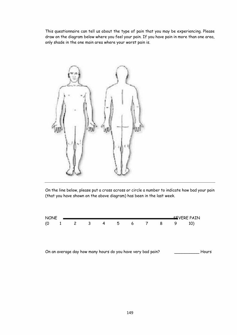
This questionnaire can tell us about the type of pain that you may be experiencing. Please

draw on the diagram below where you feel your pain. If you have pain in more than one area,

only shade in the one main area where your worst pain is.

On the line below, please put a cross across or circle a number to indicate how bad your pain

(that you have shown on the above diagram) has been in the last week.

NONE SEVERE PAIN

(0 1 2 3 4 5 6 7 8 9 10)

On an average day how many hours do you have very bad pain? __________ Hours

150

Below are 7 questions about your pain (the one in the diagram). Think about how your pain

that you showed in the diagram has felt over the last week. Put a tick against the

descriptions that best match your pain. These descriptions may, or may not, match your pain

no matter how severe it feels. Only tick the responses that describe your pain in any

question.

1. In the area where you have pain, do you also have 'pins and needles', tingling or prickling

sensations?

------a) NO - I don't get these sensations

------b) YES - I get these sensations often

2. Does the painful area change colour (perhaps looks mottled or more red) when the pain is

particularly bad?

------a) NO - The pain does not affect the colour of my skin

------b) YES - I have noticed that the pain does make my skin look different from normal

3. Does your pain make the affected skin abnormally sensitive to touch? Getting unpleasant

sensations or pain when lightly stroking the skin might describe this.

------a) NO - The pain does not make my skin in that area abnormally sensitive to touch

------b) YES - My skin in that area is particularly sensitive to touch

4. Does your pain come on suddenly and in bursts for no apparent reason when you are

completely still? Words like 'electric shocks', jumping and bursting might describe this.

------a) NO - My pain doesn't really feel like this

------b) YES - I get these sensations often

5. In the area where you have pain, does your skin feel unusually hot like a burning pain?

------a) NO - I don't have burning pain

------b) YES - I get burning pain often

6. Gently rub the painful area with your index finger and then rub a non-painful area (for

example, an area of skin further away or on the opposite side from the painful area). How

does this rubbing feel in the painful area?

------a) The painful area feels no different from the non-painful area

------b) I feel discomfort, like pins and needles, tingling or burning in the painful area that

is different from the non-painful area

7. Gently press on the painful area with your finger tip then gently press in the same way

onto a non-painful area (the same non-painful area that you chose in the last question). How

does this feel in the painful area?

------a) The painful area does not feel different from the non-painful area

------b) I feel numbness or tenderness in the painful area that is different from the non-

painful area.

151

Appendix 2. SHORT FORM-36 (SF36) SURVEY

Please answer the following questions about your health. Select ONLY ONE ANSWER for each

question

1. In general, would you say your health is:

1. Excellent

2. Very Good

3. Good

4. Fair

5. Poor

2. Compared to one year ago, how would you rate your health in general now?

1. Much better now than one year ago

2. Somewhat better now than one year ago

3. About the same as one year ago

4. Somewhat worse now than one year ago

5. Much worse than one year ago

3. Does your health now limit you in this activity? If so, how much? Vigorous activities, such as

running, lifting heavy objects, participating in strenuous sports.

1. Yes, limited a lot

2. Yes, limited a little

3. No, not limited at all

4. Does your health now limit you in this activity? If so, how much? Moderate activities, such

as moving a table, pushing a vacuum cleaner, bowling or playing golf.

1. Yes, limited a lot

2. Yes, limited a little

3. No, not limited at all

152

5. Does your health now limit you in this activity? If so, how much? Lifting or carrying

groceries.

1. Yes, limited a lot

2. Yes, limited a little

3. No, not limited at all

6. Does your health now limit you in this activity? If so, how much? Climbing several flights of

stairs.

1. Yes, limited a lot

2. Yes, limited a little

3. No, not limited at all

7. Does your health now limit you in this activity? If so, how much? Climbing one flight of

stairs.

1. Yes, limited a lot

2. Yes, limited a little

3. No, not limited at all

8. Does your health now limit you in this activity? If so, how much? Bending, kneeling, or

stooping.

1. Yes, limited a lot

2. Yes, limited a little

3. No, not limited at all

9. Does your health now limit you in this activity? If so, how much? Walking more than a mile.

1. Yes, limited a lot

2. Yes, limited a little

3. No, not limited at all

153

10. Does your health now limit you in this activity? If so, how much? Walking several blocks.

1. Yes, limited a lot

2. Yes, limited a little

3. No, not limited at all

11. Does your health now limit you in this activity? If so, how much? Walking one block.

1. Yes, limited a lot

2. Yes, limited a little

3. No, not limited at all

12. Does your health now limit you in this activity? If so, how much? Bathing or dressing

yourself.

1. Yes, limited a lot

2. Yes, limited a little

3. No, not limited at all

13. During the past 4 weeks, have you had the following problem with your work or other

regular daily activities as a result of your physical health? Cut down the amount of time you

spent on work or other activities.

1. Yes

2. No

14. During the past 4 weeks, have you had the following problem with your work or other

regular daily activities as a result of your physical health? Accomplished less than you would

like.

1. Yes

2. No

15. During the past 4 weeks, have you had the following problem with your work or other

regular daily activities as a result of your physical health? Were limited in the kind of work or

other activities.

1. Yes

154

2. No

16. During the past 4 weeks, have you had the following problem with your work or other

regular daily activities as a result of your physical health? Had difficulty performing the work or

other activities (for example, it took extra effort).

1. Yes

2. No

17. During the past 4 weeks, have you had the following problem with your work or other

regular daily activities as a result of any emotional problems (such as feeling depressed or

anxious). ?Cut down the amount of time you spent on work or other activities.

1. Yes

2. No

18. During the past 4 weeks, have you had the following problem with your work or other

regular daily activities as a result of any emotional problems (such as feeling depressed or

anxious) ?Accomplished less than you would like.

1. Yes

2. No

19. During the past 4 weeks, have you had the following problem with your work or other

regular daily activities as a result of any emotional problems (such as feeling depressed or

anxious)?Didn't do work or other activities as carefully as usual.

1. Yes

2. No

20. During the past 4 weeks, to what extent has your physical health OR emotional problems

interfered with your normal social activities with family, friends, neighbors, or groups?

1. Not at all

2. Slightly

3. Moderately

4. Quite a bit

5. Extremely

155

21. How much bodily pain have you had during the past 4 weeks?

1. None

2. Very mild

3. Mild

4. Moderate

5. Severe

6. Very severe

22. During the past 4 weeks how much did pain interfere with your normal work (including

both work outside the home and housework)?

1. Not at all

2. A little bit

3. Moderately

4. Quite a bit

5. Extremely

23. How much of the time during the past 4 weeks: Did you feel full of pep?

1. All of the time

2. Most of the time

3. A good bit of the time

4. Some of the time

5. A little of the time

6. None of the time

24. How much of the time during the past 4 weeks: Have you been a very nervous person?

1. All of the time

2. Most of the time

3. A good bit of the time

4. Some of the time

5. A little of the time

156

6. None of the time

25. How much of the time during the past 4 weeks: Have you felt so down in the dumps that

nothing could cheer you up?

1. All of the time

2. Most of the time

3. A good bit of the time

4. Some of the time

5. A little of the time

6. None of the time

26. How much of the time during the past 4 weeks: Have you felt calm and peaceful?

1. All of the time

2. Most of the time

3. A good bit of the time

4. Some of the time

5. A little of the time

6. None of the time

27. How much of the time during the past 4 weeks: Did you have a lot of energy?

1. All of the time

2. Most of the time

3. A good bit of the time

4. Some of the time

5. A little of the time

6. None of the time

28. How much of the time during the past 4 weeks: Have you felt downhearted and blue?

1. All of the time

2. Most of the time

3. A good bit of the time

157

4. Some of the time

5. A little of the time

6. None of the time

29. How much of the time during the past 4 weeks: Did you feel worn out?

1. All of the time

2. Most of the time

3. A good bit of the time

4. Some of the time

5. A little of the time

6. None of the time

30. How much of the time during the past 4 weeks: Have you been a happy person?

1. All of the time

2. Most of the time

3. A good bit of the time

4. Some of the time

5. A little of the time

6. None of the time

31. How much of the time during the past 4 weeks: Did you feel tired?

1. All of the time

2. Most of the time

3. A good bit of the time

4. Some of the time

5. A little of the time

6. None of the time

158

32. During the past 4 weeks, how much of the time has your physical health or emotional

problems interfered with your social activities (like visiting with friends, relatives, etc.)?

1. All of the time

2. Most of the time

3. Some of the time

4. A little of the time

5. None of the time

33. How true or false is the following statement? I seem to get sick a little easier than other

people.

1. Definitely true

2. Mostly true

3. Don't know

4. Mostly false

5. Definitely false

34. How true or false is the following statement? I am as healthy as anybody I know.

1. Definitely true

2. Mostly true

3. Don't know

4. Mostly false

5. Definitely false

35. How true or false is the following statement? I expect my health to get worse.

1. Definitely true

2. Mostly true

3. Don't know

4. Mostly false

5. Definitely false

36. How true or false is the following statement? My health is excellent.

1. Definitely true

159

2. Mostly true

3. Don't know

4. Mostly false

5. Definitely false

37. Are you ...?

1. Male

2. Female

38. How old were you on your last birthday?

Age:

160

Appendix 3: Hospital Anxiety and Depression Scale Scoring Sheet

Yes Yes No No

definitely sometimes not much not at all

1) I wake early and then sleep badly for the rest of the night 3 2 1 0

2) I get very frightened or have panic feelings for 3 2 1 0 apparently no reason

3) I feel miserable and sad 3 2 1 0

4) I feel anxious when I go out of the house on my own 3 2 1 0

5) I have lost interest in things 3 2 1 0

6) I get palpitations, or sensations of ‘butterflies’ in my 3 2 1 0 stomachor chest

7) I have a good appetite 0 1 2 3

8) I feel scared or frightened 3 2 1 0

9) feel life is not worth living 3 2 1 0

10) I still enjoy the things I used to 0 1 2 3

11) I am restless and can’t keep still 3 2 1 0

12) I am more irritable than usual 3 2 1 0

13) I feel as I have slowed down 3 2 1 0

14) Worrying thoughts constantly go through my mind 3 2 1 0

Anxiety 2,4,6,8,11,12,14

Depression 1,3,5,7,9,10,13

Scoring 3,2,1,0 (for item 7 & 10 the scoring is reversed)

GRADING: 0-7 =Non-case 8 and above +ve

161

Appendix 4: McGill (SF) Pain Assessment Form

Name:

Hospital No:

DOB:

Tick the level of pain for each word or tick none if it does not apply to you.

No Type of pain None Mild Moderate Severe

1 Throbbing

2 Shooting

3 Stabbing

4 Sharp

5 Cramping

6 Gnawing

7 Hot-burning

8 Aching

9 Heavy

10 Tender

11 Splitting

12 Tiring-Exhausting

13 Sickening

14 Fearful

15 Cruel-punishing

Put a cross on this line to show how bad your pain is. At the left end of line means no pain at all, at right

end means worst-pain possible.

………………………………………………………

Date:……………

No

Pain

Worst Possible

Pain Please do not write in this box:

S -------- / 33 A -------- / 12 VAS -------------- /10

162

Presentations and Publications

163

Currently in press in the “International journal of diabetes and metabolism”

Diagnosis and treatment of atypical painful neuropathy due to “Insulin neuritis” in

patients with diabetes

Amir Aslam1, Satyan Rajbhandari1,2 and Jaipaul Singh2

1Department of Diabetes and Endocrinology, Lancashire Teaching Hospital NHS Trust,

UK and 2School of Pharmacy and Biomedical Sciences and School of Forensic and

Investigative Sciences, University of Central Lancashire, Preston, PR1 2HE, Lancashire,

UK

Running title: Insulin neuritis

Correspondence

Professor Jaipaul Singh

2School of Pharmacy and Biomedical Sciences and School of Forensic and Investigative

Sciences,

University of Central Lancashire,

Preston, PR1 2HE,

Lancashire,

England, UK

Email:[email protected]

Tel: 00 44 1772 893515

164

Abstract

Diabetes is very common and its global prevalence is rising day by day. As a result we

are seeing more complications related to diabetes. In order to prevent micro vascular and

macro vascular complications such as retinopathy, nephropathy, erectile dysfunction,

neuropathy, myocardial infarction and stroke health care professionals are keen to have

better glycaemic control. When dealing with newly diagnosed or poorly controlled

diabetes patients are encouraged to bring down glycated haemoglobin (HbA1c). Diabetic

painful neuropathy (DPN) is one of the well-known complications associated with long-

term poor glycaemic control. However, on the other hand rapid control of high blood

sugar can precipitate painful neuropathy known as “insulin neuritis”. The rapid tight

glycaemic control with either insulin or oral hypoglycaemic agents on poorly controlled

diabetic patients cause flux of blood glucose and metabolic shift resulting in structural

changes at nerve endings (endoneural blood vessels) which resemble the retinopathy

changes in retina. It causes steal effect and hypoxia in the nerves and hence precipitates

neuropathic pain. It lasts for about 6 months and responds well to standard treatment of

painful neuropathy. Health professionals need to be aware of this condition and consider

gentle glycaemic control when aiming for Target HbA1c. This review outlines the

disease, the symptoms, the types and treatment.

Words for index: insulin neuritis, diabetes mellitus, glycaemic control, neuropathy,

retinopathy, blood glucose

165

Introduction

Diabetes mellitus (DM) is the commonest metabolic disease currently affecting more that

250 million people worldwide and it costs the Governments of the world more than £800

billion to diagnose, treat and care for diabetic patients. DM is associated with numerous

long-term complications including cardiomyopathy, nephropathy, neuropathy and

retinopathy. This review addresses diabetic painful neuropathy (DPN) which is one of the

well-known complications of diabetes and it affects up to 53% of diabetic population1. It

is the most common form of painful neuropathy2. It manifests with varying description

from mild pins and needle sensation to the stabbing pain, burning, unremitting or even

described as electric shock. The most common feature is cutaneous hypersensitivity

leading to acute distress on contact with an external stimulus, such as clothing3. The

pathogenesis of DPN is mainly caused by inflammatory process4 and strongly correlates

with longer duration of the diabetes and poor glycaemic control5-18

Treatment induces acute neuropathy due to rapid glycaemic control has been reported in

literature as ‘insulin neuritis’ that usually manifests with severe excruciating neuropathic

pain in the first month of initiation of insulin or oral hypoglycaemic agents. Symptoms

usually last up to 6 months and respond to treatment that is usually needed up to 6

months3. Insulin neuritis was first described by Caravati in 1933. He reported a diabetic

woman with numbness, tingling, and shooting pains in the lower extremities that appeared

four weeks after the initiation of insulin. The pain increased despite the use of analgesics

and sedatives, but resolved within 3 days of stopping insulin concurrent with severe

hyperglycaemia. Further attempts at the use of insulin resulted in similar levels of pain.

He called the condition “insulin neuritis”19. The word insulin neuritis is a misnomer, as it

can also be induced by oral hypoglycaemic agent20. The cause is not directly by insulin

166

but mainly due to the change in flux of blood glucose caused by rapid change in blood

glucose level following pharmacological treatment21.

Symptoms

There are several studies and case reports in the literature about insulin neuritis with

varying presentation after starting insulin or oral hypoglycaemic agents. These reports

described the most common features as generalized pain bilaterally mainly distally in feet

with burning sensation, hypersensitivity and contact discomfort of the skin within 2 to 4

weeks20,22,23. It may present with truncal neuropathy24,25,26,27, autonomic neuropathy28,

worsening of retinopathy29 and even with profound weight loss22,30,31,32

Generalize pain mainly distally.

The most common presentation of Insulin neuritis is symmetrical and bilateral distal

neuropathic pain mainly involving feets3. In one observational study on 6 patients with

diabetes, all experienced severe excruciating bilateral neuropathic pain mainly in feet

after 2-4 weeks of insulin treatment with rapid reduction of blood glucose up to one fifth

of initial levels. This improved in all cases with symptomatic treatment allowing

discontinuation of therapy in 3-8 months20. A case report on a newly diagnosed type 1

diabetes patient described development of severe pain in his feet, which prevented him

from walking, after initiation of insulin. The HbA1c of that patient dropped from 14.1 to

7.6%, and 3 months after presentation, the patient showed dramatic improvement and

regained his ability to walk33. There is another similar case report of painful neuropathy

on 15th day of treatment with intense insulin therapy following poor glycaemic control

period of 8 years. He responded well on symptomatic treatment on day 3 on venlafaxine.34

167

Diabetic neuropathic cachexia

Painful neuropathy is sometimes associated with profound weight loss and called

“Diabetic neuropathic cachexia”. This has also been reported with insulin neuritis that

could last up to a year. The exact mechanism and cause is unknown3. It is observed that

constant pain and discomfort can cause loss of appetite and low mood which results in

patients not eating enough and start losing weight. Most patients respond well with

neuropathic pain treatment which gives pain relief and regain weight. In one observational

study, 9 diabetic patients experienced painful neuropathy with constant burning pain

mainly in the legs, especially distally. There was marked troublesome allodynia

associated with profound weight loss along with depression with impotence. These severe

manifestations subsided in most cases in 6 months and in all cases in 10 months 22. There

is another case report in which patient presented with painful neuropathy, profound

weight loss after initiation of insulin therapy within 3 months31

Truncal neuropathy

Insulin neuritis may precipitate focal neuropathic pain called “Truncal neuropathy” on

specific dermatome region. Truncal neuropathy in diabetes presents with neuropathic

pain such as a hypoesthesia, regional hyperalgesia, allodynia and sometime focal

weakness in specific dermatome region. It usually presents with unilateral abdominal or

thoracic wall pain.3,25 There was one case of insulin neuritis which presented with painful

neuropathy with paraesthesia and hyperesthesia restricted to the abdomen and this was

associated with profound weight loss. The haemoglobin A (1c) had dropped from 12% to

7.5% within 5 months, following rapid improvement in glycaemic control. On

168

investigation, there was no indication of disease in intra-abdominal area. The symptoms

improved dramatically within 4 months after symptomatic treatment30

It is not uncommon that these patients have to undergo a number of investigations to

determine the cause of pain before having the diagnosis of truncal neuropathy24-27,30.

There are several cases of truncal neuropathy that were misdiagnosed initially as for

example hernia due to focal weakness on abdominal wall26, angina due to left sided chest

wall pain25 and painless gall stones due to focal sensory deficit complicated with painless

jaundice secondary gall stone27. The diagnosis of truncal neuropathy is essentially clinical

and positive recognition of neuropathic element of pain is the key factor. Most people

respond well on neuropathic treatment and usually settle in 3 to 12 months.

Autonomic neuropathy

Autonomic dysfunction is one of the complications of diabetes3. It manifests with one or

more of the following: erectile dysfunction, gatsroparesis, neurogenic bladder, dry feet,

depressed cough reflex, postural hypotension or high blood flow to foot35,36 . Insulin

neuritis has been reported to precipitate autonomic neuropathy. In one prospective study

on 16 diabetic patients followed up for 18 months, all the patients develop severe painful

neuropathy in 8 weeks of intense glycaemic treatment. All individuals with treatment for

induced neuropathy had evidence of autonomic dysfunction on testing and exhibited

symptoms of autonomic impairment. Approximately, 69% of cohort had systolic blood

pressure falls > 20 mmHg. Symptoms of autonomic dysfunction were more prevalent and

more severe in subjects with type 1 diabetes, particularly with respect to symptoms of

orthostatic intolerance and gastrointestinal function. Urinary frequency, nocturia and

anhidrosis were reported more frequently in individuals with type 2 diabetes28.

169

Retinopathy

Retinopathy is a well-known complication of diabetes and directly related with poor

glycaemia and duration of diabetes37. It is also proven that better glycaemic control

prevent worsening of retinopathy38. Insulin neuritis with rapid flux of blood glucose

causes structural changes at endoneural blood vessels of nerves which resemble with

retinopathic changes in retina39. Rapid drop in blood glucose in poorly controlled diabetes

may exert the same changes in retina, thus worsening the retinopathy. In one large

observational study, 87 patients were divided in 3 groups of varying glycaemic control.

These included a group of poor glycaemic control corrected rapidly, poor glycaemic

control not corrected and good control group. The progression rate of diabetic

maculopathy was significantly higher in the group that underwent rapid control than in

the other 2 groups (P <02). Patients with moderate to severe non-proliferative diabetic

retinopathy preoperatively in the rapid control group had significantly higher progression

rates of diabetic retinopathy and maculopathy (P <002 and p<008, respectively) 40.

Pathogenesis

In 1992 Boulton first described the observation that acute painful neuropathy might

follow sudden change in glycaemia control suggesting that blood glucose flux could

precipitate pain. Sudden changes in glycaemia may contribute to the generation of

impulses or even induce relative hypoxia in nerve fibres, indicating that it is the

combination of structural and functional changes in peripheral nerves which cause the

pain21. This observation was experimentally tested by Kihara et al in 1994 on rats. In their

study, they infused insulin under non- hypoglycaemic conditions and evaluated its effect

on endoneurial oxygen tension, nerve blood flow, and the oxy-haemoglobin dissociation

170

curve of peripheral nerves in normal and diabetic rats. Their results showed that insulin

administration could cause a reduction in nerve nutritive blood flow and an increase in

arterio-venous shunt flow. When the latter was eliminated by the closure of arterio-venous

shunts (infusion of 5-hydroxytryptamine), endoneurial oxygen reverted to normal. These

findings clearly indicate a deleterious vasoactive effect of insulin and may explain the

development of insulin neuritis41.

In 1996 Tesfaye et al observed neurovascular changes in vivo in five human diabetic

patients with insulin neuritis. These patients presented with severe sensory symptoms

but clinical examination and electrophysiological tests were normal except with one

subject who had severe autonomic neuropathy and all tests were abnormal. On sural

nerve exposure in vivo, epineural blood vessels showed severe structural abnormalities

resembling the retinopathy changes normally seen in the retina, including arteriolar

attenuation, tortuosity and aterio-venous shunting and proliferating new vessels

formation. They hypothesized that the structural abnormalities with new vessels

formation in epineural blood vessels cause steal effect and hence results in hypoxia and

neuropathic pain39. It can now be postulated that sudden change in glycaemic control

can cause flux effect resulting in structural and functional changes at the epineural blood

vessels of nerves which in turn can lead to neuropathic pain “Insulin neuritis (see figure

1)”21,39.

Treatment

Management of neuropathic pain in “insulin neuritis” is symptomatic including first line

medication tricyclic antidepressants (Amitriptyline) or selective serotonin uptake

inhibitor (Duloxetine). Second line medications include anti-epileptic medications

171

(Gabapentin, Pregabalin, Carbamazepine and Topiramate) and Opioids. Most patients

recover within 6 months of onset of insulin neuritis3

Conclusion

The flow diagram in Figure 2 summarises the pathogenesis of insulin neuritis. With

increasing prevalence of diabetes and its complications, both health professional and

patients are keen to have good glycaemic control in order to prevent long term

complications42. Most of the time it is not a problem but on several occasions intense

treatment for rapid glycaemic control may cause insulin neuritis. This is presumed to be

caused by change in glucose flux which can result in structural and functional changes

at the nerves leading to hypoxia. This in turn can precipitate neuropathic pain and the

whole phenomenon is called “insulin neuritis”. It usually manifests distally in feet and is

bilateral with burning sensation, hypersensitivity and allodynia. It could affect focally –

truncal neuritis and may present with neuropathic pain and/or weakness in dermatomal

region. Similarly, it may present with autonomic symptoms. Constant pain may cause

cachexia and loss of appetite which can result in significant weight loss. Most patients

respond well with neuropathic treatment and recover within 6 months. It is very

important to be aware that treatment induced insulin neuritis can have significant impact

on the quality of the life of the diabetic patient. This can be easily prevented by gradual

glycaemic control and by symptomatic treatment as necessary. Healthcare professionals

need to be aware of this condition when managing poorly controlled diabetic patients

and should consider gradual titration of the pharmacological agents employed to treat

the patients.

172

References

1. Mimi O, Teng CL and Chia YC, The prevalence of diabetic peripheral

neuropathy in an outpatient setting. The Medical Journal of Malaysia 2003;

58 (4): 533-538.

2. Chong MS and Hester J, Diabetic painful neuropathy: current and future

treatment options, Drugs 2007; 67 (4): 38-45.

3. Larsen, Kronenberg. et al. Williams Textbook of Endocrinology. Tenth

edition Saunders, Elsevier Science. Philadelphia 2002,

4. Said G, Diabetic neuropathy – A Review. National Clinical Practice and

Neurology 2007; 3 (6):331-340

5. Shaw JE, Hodge AM, de Courten M, Dowse GK, Gareeboo H, Tuomilehto J,

Alberti KG and Zimmet PZ. Diabetic neuropathy in Mauritius: prevalence

and risk factors. Diabetes Research and Clinical Practice 1998; 42 (2): 131-

139.

6. Börü UT, Alp R, Sargin H, Koçer A, Sargin M, Lüleci A, and Yayla A.

Prevalence of peripheral neuropathy in type 2 diabetic patients attending a

diabetes centre in Turkey. Endocrine Journal 2004; 51 (6): 563-567

7. Walters DP, Gatling W, Mullee MA and Hill RD, The prevalence of diabetic

distal sensory neuropathy in an English community. Diabetic Medicine 1992;

9 (4): 349-353.

8. Abbott CA, Malik RA, van Ross ER, Kulkarni J and Boulton AJ, Prevalence

and characteristics of painful diabetic neuropathy in a large community-based

diabetic population in the UK. Diabetes care 2011; 34 (10): 34-48.

9. Tesfaye S, Boulton A, Diabetic neuropathy, Oxford University Press, United

States, first edition 2009, pp 8-56

10. Erbas T, Ertas M, Yucel A, Keskinaslan A and Senocak M, Prevalence of

peripheral neuropathy and painful peripheral neuropathy in Turkish diabetic

patients. Journal of Clinical Neurophysiology 2011; 28 (1): 51-55.

11. Jambart S, Ammache Z, Haddad F, Younes A, Hassoun A, Abdalla K, Selwan

CA, Sunna N, Wajsbrot D and Youseif E. Prevalence of painful diabetic

peripheral neuropathy among patients with diabetes mellitus in the Middle

East region. Journal of International Medical Research 2011; 39 (2): 366-377.

12. Kästenbauer T, Irsigler P, Sauseng S, Grimm A and Prager R, The prevalence

of symptoms of sensorimotor and autonomic neuropathy in Type 1 and Type

2 diabetic subjects. Journal of Diabetes and Its Complications 2004; 18 (1):

27-31

13. Dyck PJ, Kratz KM, Karnes JL, Litchy WJ, Klein R, Pach JM, Wilson DM,

O'Brien PC, Melton LJ 3rd and Service FJ. The prevalence by staged severity

of various types of diabetic neuropathy, retinopathy, and nephropathy in a

population-based cohort: the Rochester Diabetic Neuropathy Study.

Neurology 1993; 43 (4): 817-824

173

14. Maser RE, Steenkiste AR, Dorman JS, Nielsen VK, Bass EB, Manjoo Q,

Drash AL, Becker DJ, Kuller LH and Greene DA, Epidemiological correlates

of diabetic neuropathy. Report from Pittsburgh Epidemiology of Diabetes

Complications Study. Diabetes 1989; 38 (11): 1456-1461

15. Pradeepa R, Rema M, Vignesh J, Deepa M, Deepa R and Mohan V,

Prevalence and risk factors for diabetic neuropathy in an urban south Indian

population: the Chennai Urban Rural Epidemiology Study (CURES-55).

Diabetic Medicine 2008; 25 (4): 407-412.

16. Fedele D, Comi G, Coscelli C, Cucinotta D, Feldman EL, Ghirlanda G, Greene

DA, Negrin P and Santeusanio F, A multicenter study on the prevalence of

diabetic neuropathy in Italy. Italian Diabetic Neuropathy Committee. Diabetes

Care 1997; 20 (5): 836-843.

17. Hsu WC, Yen AM, Liou HH, Wang HC and Chen TH. Prevalence and risk

factors of somatic and autonomic neuropathy in prediabetic and diabetic

patients. Neuroepidemiology 2009; 33 (4): 344-349

18. Morkrid K, Ali L and Hussain A, Risk factors and prevalence of diabetic

peripheral neuropathy: A study of type 2 diabetic outpatients in Bangladesh.

International Journal of Diabetes in Developing Countries 2010; 30 (1): 11-

17.

19. Caravati CM, Insulin neuritis: a case report. Va Med Monthly 1933. 59: 745-

746

20. Dabbya R, Sadeha M, Yair Lampla et al. Acute painful neuropathy induced

by rapid correction of serum glucose levels in diabetes patients. Biomedicine

and Pharmacotherapy 2009; 63 (10): 707-709

21. Boulton A, What causes neuropathic pain? Journal of Diabetes and its

Complication 1992; 6 (1): 58-63

22. Archer A, Watkins P J, Thomas P K, The natural history of acute painful

neuropathy in diabetes mellitus. Journal of Neurology Neurosurgery

Psychiatry 1983; 46 (6) :491-499

23. Leow MKS, Wyckoff J. Under-recognised paradox of neuropathy from rapid

glycaemic control. Postgraduate Medical Journal 2005; 81:103-107

24. Takayama S, Takahashi Y, Osawa M. Acute Painful Neuropathy Restricted to

the Abdomen Following Rapid Glycaemic Control in Type 2 Diabetes. The

Journal of International Medical Research 2004; 32(5): 558-562

25. Ibitoye R, Rajbhandari SM. Neuropathic truncal pain – a case series. Q J Med

2012. doi:10.1093/qjmed/hcs128

26. Parry G, Floberg J. Diabetic truncal neuropathy presenting as abdominal

hernia. Neurology 1989; 39: 1488

27. Gentile S, Asymptomatic choledocholithiasis associated with diabetic

neuropathy. Report of a case. La Clinica Terapeutica 1994; 144 (5):461-465

28. Gibbons CH,Freeman R, Treatment-induced diabetic neuropathy: a

reversible painful autonomic neuropathy. Annals of Neurology 2010; 67(4):

534-541

29. Suto C, Hori S, Kato S, Muraoka K, Kitano S. Effect of Perioperative

Glycemic Control in Progression of Diabetic Retinopathy and Maculopathy.

Archives of Ophthalmology. 2006;124 (1): 38-45.

174

30. Van Heel DA, Levitt NS, Winter TA. Diabetic neuropathic cachexia: the

importance of positive recognition and early nutritional support. International

Journal Clinical Practice 1998; 52: 591-592.

31. Yuen1 KCJ, Day JL, Flannagan DW, Rayman G. Diabetic neuropathic

cachexia and acute bilateral cataract formation following rapid glycaemic

control in a newly diagnosed Type 1 diabetic patient. Diabetic Medicine 2001;

18 (10): 854–857

32. Knopp M, Rajabally YA. Common and Less Common Peripheral Nerve

Disorders Associated with Diabetes. Current Diabetes Reviews 2012; 8 (3):

229-236(8)

33. Wilson JL,Sokol DK,Smith LH,Snook RJ,Waguespack SG,Kincaid JC,

Acute painful neuropathy (insulin neuritis) in a boy following rapid glycemic

control for type 1 diabetes mellitus. Journal of Child Neurology, 2003; 18/5:

365-367

34. Guldiken S,Guldiken B,Arikan E,Altun Ugur B,Kara M,Tugrul A, Complete

relief of pain in acute painful diabetic neuropathy of rapid glycaemic control

(insulin neuritis) with venlafaxine HCL. Diabetes, Nutrition and Metabolism

- Clinical and Experimental 2004; 17 (4): 247-259

35. Watkins (2003), ABC of Diabetes, BMJ Publishing Group London, Fifth

Edition 2003, pp 32-37

36. Galer, Gianas and Jensen, Painful diabetic polyneuropathy: epidemiology,

pain description, and quality of life.. Diabetes Research and Clinical Practice

2000; 47 (2): 123-128

37. Stratton IM, Kohner EM, Aldington SJ, Turner RC, Holman RR, Manley

SE, Matthews DR. UKPDS 50: risk factors for incidence and progression of

retinopathy in Type II diabetes over 6 years from diagnosis Diabetologia 2001; 44(2):156-63.

38. Holman R, Paul S, Bethel A, Matthews D, Neil A, 10-Year Follow-up of

Intensive Glucose Control in Type 2 Diabetes. New England Journal of

Medicine 2008; 359:1577-1589

39. Tesfaye S, Malik R, Harris N, Jakubowski JJ, Mody C, Rennie IG, Ward JD.

Arterio-venous shunting and proliferating new vessels in acute painful

neuropathy of rapid glycaemic control (insulin neuritis). Diabetologia 1996;

39 (3): 329-335

40. Suto C, Hori S, Kato S, Muraoka K, Kitano S. Effect of Perioperative

Glycemic Control in Progression of Diabetic Retinopathy and Maculopathy.

Arch Ophthalmol. 2006; 124 (1):38-45.

41. Kihara M, Zollman PJ, Smithson L,Lagerlund TD,Low PA, Hypoxic effect

of exogenous insulin on normal and diabetic peripheral nerve. American

Journal of Physiology 1994;266 (6 Pt 1):E980–E985.

42. Williams J, Alling K, A Program for Normal Glycemic Control of Insulin

dependent Diabetes. Diabetes Care 1980; 3: 160-162

175

PATHOGENESIS OF INSULIN NEURITIS

Poorly control diabetes patient

Intense hypoglycaemic treatment

with insulin or oral hypoglycaemic medication

Rapid flux of blood glucose

Structural changes at nerve endings (endoneural blood vessels)

resembles changes in retina

(Aterio-venous shunting, Attenuation, tortuosity and proliferating new vessels formation)

Steal effect and Hypoxia at Nerve endings

Neuropathic Pain (Insulin Neuritis)

Figure 1: A flow diagram showing the pathogenesis of insulin neuritis

176

Figure 2: Arteriolar attenuation, tortuosity and aterio-venous shunting and proliferating

new vessels formation of vasanervosum seen in sural nerve of patient with insulin

neuritis (photo courtesy of Tesfaye and Boulton 9)

The impact of painful diabetic neuropathy on quality of life: An observational study

Amir Aslam, Jaipaul Singh, Satyan M Rajbhandari

Article

Citation: Aslam A, Singh J, Rajbhandari S (2014) The impact of painful diabetic neuropathy on quality of life. Diabetes & Primary Care 16: XX–X

Article points1. Painful diabetic neuropathy

(PDN) is a common and potentially very serious complication of diabetes.

2. There is relatively little research aimed at quantifying the impact of PDN on quality of life (QoL) and mental health.

3. Here the authors report data from north-west England suggesting that PDN is associated with a negative impact on QoL and anxiety.

Key words– Anxiety

– Depression

– Painful diabetic neuropathy

– Quality of life

AuthorsAmir Aslam is a Clinical Research Fellow, Lancashire Hospitals NHS Trust, Chorley and South Ribble District General Hospital, Chorley. Jaipaul Singh is a Professor of Physiology, School of Pharmacy and Biomedical Sciences and School of Forensic and Investigative Sciences, University of Central Lancashire, Preston Satyan M Rajbhandari is a Consultant in Diabetology and Endocrinology, Lancashire Hospitals NHS Trust, Chorley and South Ribble District General Hospital, Chorley, and a Clinical Professor, University of Central Lancashire, Preston.

About a third of people with diabetes experience PDN at some point in their lives, and it

is a distressing condition affecting individuals both physically and emotionally. The aim of

the study reported here was to assess quality of life, anxiety and depression in people with

PDN using the 36-item Short Form Health Survey and the Hospital Anxiety and Depression

Scale questionnaires, comparing these results against those in with people with diabetes

who did not have PDN. The findings are presented in this article.

Diabetes & Primary Care Vol 16 No 4 2014 XX

Currently, over 380 million people worldwide are living with diabetes and it is estimated that this figure will rise

up to 592 million in the year 2035 (International Diabetes Federation, 2013). The prevalence of diabetes-related complications is also rising. Painful diabetic neuropathy (PDN) is a common complication of diabetes, affecting about a third of all people with diabetes (Tesfaye, 2009). It is characterised by bilateral symmetrical distal neuropathic pain in the lower extremities with varied symptoms including mild pins and needles, a tingling sensation, a shooting pain similar to electric shock, a constant burning sensation with nocturnal exacerbation, and contact hyper-sensitivity (allodynia; Larsen et al, 2002). Relentless pain and allodynia can affect people both physically and mentally and can cause disturbance in sleep, low mood, impotence and social withdrawal. In some extreme cases, the affected individual is unable to walk (Quattrini and Tesfaye, 1996; Galer et al, 2000; Gardner and Shoback, 2007). PDN can significantly alter – and, moreover, has a huge impact on – individuals’ quality of life (QoL).

Currently, there are only a few studies that have been performed specifically to measure the physical and mental impact of PDN on QoL. The study reported here was designed to assess QoL, anxiety and depression in people with PDN (PDN group) compared with those

with diabetes not known to have PDN (control group).

There are several health-related questionnaires available to assess QoL and physical and mental wellbeing (Healthmeasurement.org, 2014). Typically, researchers use the 36-item Short Form Health Survey (SF-36®) for the assessment of QoL and the Hospital Anxiety and Depression Scale (HADS) for the assessment of mood and anxiety. Ware and Sherbourne (1992) introduced SF-36, which was designed for use in clinical practice and research, health policy evaluations and general population surveys. SF-36 includes 36 subjective questions that assess eight health concepts of QoL from the patient’s point of view:1 Limitations in physical activities because of

health problems.2 Limitations in social activities because of

physical or emotional problems.3 Limitations in usual role activities because of

physical health problems.4 Bodily pain.5 General mental health (psychological distress

and wellbeing).6 Limitations in usual role activities because of

emotional problems.7 Vitality (energy and fatigue). 8 General health perceptions.

SF-36 is a practical, reliable and valid measure of physical and mental health and has been

The impact of painful diabetic neuropathy on quality of life: An observational study

XX Diabetes & Primary Care Vol 16 No 4 2014

used in a variety of chronic health conditions including diabetic neuropathic pain (Garratt, 1993; Ware et al, 1994; Rosenstock et al, 2004; Vinik et al, 2013) and published in more than 4000 documents, as of 2002 (Turner-Bowker et al, 2002).

The HADS questionnaire was originally developed by Zigmond and Snaith (1983) for psychometric evaluation. Since then, it has been widely used worldwide by health professionals, in both the community and hospital settings, and it has been found to be both a reliable and a valid measure of anxiety and depression (el-Rufaie and Absood, 1987; Nortvedt et al, 2006). The HADS questionnaire is based on a total of 14 questions, seven for anxiety assessment and seven for depression. HADS provides clear cut-off scores for severity of anxiety and depression. We felt that HADS would serve as an ideal tool for screening and thus adopted it in our study.

MethodsStudy designThis was an observational study. The SF-36 and HADS questionnaires were used for data collection, based on the rationale described above. It takes approximately 15 minutes to fill in the SF-36 questionnaire and 5 minutes to fill in the HADS questionnaire, which meant that participants were able to fill these in while waiting for their appointment or to post them back to the research team after completing them at home.

ParticipantsThe PDN group was formed from attendees at the diabetic neuropathic pain clinic at Chorley and South Ribble District General Hospital, while the control group (comprising people with diabetes not known to have neuropathic pain) was formed from individuals visiting the Aston Healthcare GP surgery at Whiston (Merseyside) for diabetes review. Each group consisted of 25 consecutive consenting patients at the respective sites. Individuals under 16 or over 80 years of age were excluded from participation. All individuals gave consent for participation. Institutional approvals were obtained at both centres for the study.

Assessment of QoL, anxiety and depressionSF-36 (used for QoL assessment)The SF-36 questions were scored from 0 (worst possible functioning) to 100 (highest level of function). The average scores from those questions that addressed each specific area of a functional health domain provided the final score for the domain. Aggregate scores were compiled as a percentage of the total points possible, using the RAND scoring system (RAND Health, 2014).

Of the eight domains (described earlier), four relate to physical health (physical functioning, physical health limitation, pain and general health) and four to mental health (social functioning, emotional wellbeing, fatigue and emotional problem limitation). Aggregate scores for physical health domains and for mental health domains were also calculated.

HADS questionnaire (used for the assessment of anxiety and depression)Each HADS question was scored from 0 (excellent mental health) to 3 (worst mental health). Aggregate scores (with a maximum of 21) were calculated for the seven anxiety questions and the seven depression questions. Scores between 0 to 7 were considered “normal”, for both anxiety and depression assessment. Scores of 8 and above were considered to be significant for the diagnosis of anxiety or depression (el-Rufaie and Absood, 1987; Nortvedt et al, 2006).

Statistical analysisData were analysed using GraphPad software (GraphPad Software Inc, 2014). For the normally distributed continuous variables from SF-36 and HADS, means (± standard deviation [SD]) were calculated and analysed using the unpaired Student’s t-test. Categorical data were also calculated, as a percentage of participants. The categorical data from HADS were analysed as a 2x2 table using Fisher’s exact test.

For the purpose of visually summarising the data, box-plots were also created, using Minitab (2014) statistical software, and these represented median, minimum and maximum values, as well as the lower and upper quartiles.

Page points1. In this observational study, the

36-item Short Form Health Survey was used the assessment of quality, while the Hospital Anxiety and Depression Scale was employed to explore specific aspects of mental health.

2. The painful diabetic neuropathy group was formed from attendees at the diabetic neuropathic pain clinic at Chorley and South Ribble District General Hospital, while the control group (comprising people with diabetes not known to have neuropathic pain) was formed from individuals visiting the Aston Healthcare GP surgery at Whiston (Merseyside) for diabetes review.

3. Each group consisted of 25 consecutive consenting patients at the respective sites.

Diabetes & Primary Care Vol 16 No 4 2014 XX

ResultsThe two groups were similarly distributed (P>0.05) in age and also in sex (PDN group, 60% male; control group, 56% male). Participants in the PDN group had significantly (P<0.05) lower scores in seven out of eight domains of SF-36 compared with the control group (Table 1). The exception was emotional wellbeing. Both physical health and mental health summary scores were significantly lower in the PDN group than the control group (Figure 1).

Individuals in the PDN group had significantly higher HADS anxiety scores, but HADS depression scores were not statistically significantly different from those in the control group (Figure 2).

Fourteen individuals (56%) out of 25 had anxiety in the PDN group (the mean score was 7.32 ± 3.42). In the control group, five individuals (20%) met the criterion for a diagnosis of anxiety (the mean score was 4.72 ± 4.34). The P-values calculated from comparisons of the continuous data and of the categorical data were 0.023 and 0.018, respectively (both statistically significant).

Fifteen people (60%) out of 25 had depression in PDN group (the mean score was 8.36 ± 4.05). In the control group, 11 people (44%) met the criterion for a diagnosis of depression (the mean score was 6.6 ± 4.16). The P-values calculated from comparisons of the continuous data and of the categorical data were 0.136 and 0.396, respectively (neither being statistically significant).

Discussion Few studies have specifically reported the impact of PDN on QoL and psychological wellbeing of people with diabetes (Benbow et al, 1998; Quattrini and Tesfaye, 1996; Galer et al, 2000; Gore et al, 2005; Argoff et al, 2006; Van Acker et al, 2009). Our data reveal a significant association of PDN with poor QoL and anxiety symptoms but not with depression. This last observation could be because a number of people with PDN were treated with antidepressants for their neuropathic pain, and the underlying symptoms of depression might have thus been reduced to some extent, or it could be down to insufficient power.

Comparison with existing dataThe data from our study hint at a significant impairment of QoL associated with PDN within both the physical and mental health areas of the SF-36 questionnaire. The results are consistent with similar research reported using a shorter (12-item) version of the questionnaire. Van Acker et al (2009) found significant impairment in both the physical and mental health components of QoL. In another study, by Benbow et al (1998), the Nottingham Health Profile questionnaire was used, and it was found that there were significant impairments in QoL in five of the six domains (emotional reaction, energy, pain, physical mobility and sleep). The exception was the social isolation domain. Similarly, in the present study, the data showed significant impairment in all of the domains but one (emotional wellbeing).

Page points1. Few studies have specifically

reported the impact of painful diabetic neuropathy (PDN) on quality of life (QoL) and psychological wellbeing of people with diabetes

2. The authors’ data hint at an association of PDN with poor QoL and anxiety symptoms.

The impact of painful diabetic neuropathy on quality of life: An observational study

SF-36 domainMean score in

PDN groupMean score in control group

95% confidence interval

P-value

Physical functioning 28.4 65.2 18.9 to 54.7 <0.0001*

Physical health limitation 17.0 61.0 22.0 to 66.1 <0.0002*

Pain 29.3 59.9 14.2 to 47.0 <0.0005*

General health 31.1 52.0 7.3 to 34.6 0.0034*

Social functioning 48.8 68.0 2.0 to 36.4 0.0292*

Emotional wellbeing 61.4 69.3 -7.0 to 22.6 0.292

Fatigue 25.4 42.4 4.8 to 29.3 0.0073*

Emotional problem limitation 41.3 72.0 5.3 to 56.0 0.0188*

*P<0.05.

PDN=painful diabetic neuropathy.

Table 1. Data for the eight domains of the 36-item Short Form Health Survey (SF-36®) in the study groups.

As mentioned earlier, there are reports of severe PDN with constant unrelenting neuropathic pain, disturbance of sleep and even the loss of the ability to walk, owing to the severity of pain (Quattrini and Tesfaye, 1996; Galer et al, 2000; Gardner and Shoback, 2007). This can in turn lead to withdrawal from routine activity of life, including employment, and can also affect emotional wellbeing and contribute to social isolation. The data for the emotional wellbeing domain in our study and the social isolation

domain of Benbow et al (2000) study were not significant, perhaps owing to the presence of only a small number of the severe type of PDN case associated with extreme symptoms.

HADS data in the present study showed that more than half (56%) of the participants in the PDN group had anxiety symptoms, with this proportion (and the summarised continuous data) being statistically significantly different from those of the control group. The data were broadly consistent with those reported by Gore et al (2005), using the HADS questionnaire. They reported that 35% of their participants had anxiety symptoms. However, they used a threshold score on HADS of 11 or above (moderate-to-severe symptoms), while we used a threshold score of 8 and above. Our data for depression symptoms showed that more than half (60%) of the individuals in the PDN group had symptoms of depression (a score above 7). However, comparisons of the differences from the control group were not statistically significant. In contrast, Gore et al (2005) showed a significant association between PDN and depression. In their study, the prevalence of depression in people with PDN was 28% (a score of 11 or above).

A large systematic review and meta-analysis reported the prevalence of depression in people with diabetes to be around 17.5% (Ali et al, 2006). In our study, the control group of people with diabetes was found to have an unusually high prevalence of depression (44%). This may be down to random factors or could have resulted from the control group having been taken from an area of relatively low socioeconomic status.

Strengths and limitations of the studyThe study population was well defined, and both groups of participants had a 100% response in completing the two questionnaires. The groups were similar in age and in the ratio of males to females.

Recall bias could potentially exist when participants are completing questionnaire. However, most questions from both questionnaires used were based on current or recent physical and mental wellbeing of the person, and hence recall bias is considered to have been minimal.

The impact of painful diabetic neuropathy on quality of life: An observational study

XX Diabetes & Primary Care Vol 16 No 4 2014

Figure 1. A box-plot of the overall physical and mental health scores from the 36-item Short Form Health Survey in the painful diabetic neuropathy (PDN) and control (C) groups (boxes for median and lower and upper quartile values [with bars for minimum and maximum score]; n=25).

PDN C

Physical health

PDN C

Mental health

Agg

rega

te p

erce

ntag

e sc

ore

Figure 2. A box-plot of the Hospital Anxiety and Depression Scale (HADS) scores in the painful diabetic neuropathy (PDN) and control (C) groups (boxes for median and lower and upper quartile values [with bars for minimum and maximum score]; n=25).

PDN C

Anxiety score

PDN C

Depression score

HA

DS

scor

e

Diabetes & Primary Care Vol 16 No 4 2014 XX

A major limitation of the study relates to the selection of the control group. As mentioned above, the GP surgery from which the control group data were taken lies in an area of north-west England with a low socioeconomic status. It is known that low socioeconomic community status has a positive association with prevalence of depression (Murali and Oyebode, 2004). Furthermore, the two groups were selected from healthcare settings of a different nature. These discrepancies, and the lack of randomisation in the study, could thus have led to selection bias, which in turn could have had an impact on outcomes. Data were not collected to compare factors other than age and sex (duration of diabetes and the presence of other complications are among the other potential confounding factors). As with any non-randomised study, it is not possible to infer a causal relationship and thus our conclusions can only be tentative at most.

ConclusionOverall, we believe our study tentatively suggests that, in a population in north-west England, PDN has a clinically significant impact on QoL and is also associated with symptoms of anxiety. Further research would be needed to shed more light on depression and to draw firmer conclusions on the potential causal nature of the association observed.

In light of our findings, we suggest that, when caring for people with PDN, clinicians should consider exploring psychosocial wellbeing and the overall impact of the condition on QoL. n

Declaration of competing interestsThe authors reported no conflict of interests regarding the publication of this paper.

Ali S, Stone MA, Peters JL et al (2006) The prevalence of co-morbid depression in adults with Type 2 diabetes: a systematic review and meta-analysis. Diabet Med 23: 1165–73

Argoff CE, Cole BE, Fishbain DA, Irving GA (2006) Diabetic peripheral neuropathic pain: clinical and quality-of-life issues. Mayo Clin Proc 81(4 Suppl): S3–11

Benbow SJ, Wallymahmed ME, MacFarlane IA (1998) Diabetic peripheral neuropathy and quality of life. QJM 91: 733–7

el-Rufaie OE, Absood G (1987) Validity study of the Hospital Anxiety and Depression Scale among a group of Saudi patients. Br J Psychiatry 151: 687–8

Galer B, Gianas A, Jensen M (2000) Painful diabetic polyneuropathy: epidemiology, pain description, and quality of life. Diabet Res Clin Pract 47: 123–8

Gardner D, Shoback D (2007) Greenspan’s Basic and Clinical Endocrinology (8th edition). McGraw-Hill Medical, New York, NY, USA

Garratt AM (1993) The SF36 health survey questionnaire: an outcome measure suitable for routine use within the NHS? BMJ 306: 1440–4

Gore M, Brandenburg NA, Dukes E et al (2005) Pain severity in diabetic peripheral neuropathy is associated with patient functioning,symptom levels of anxiety and depression, and sleep. J Pain Symptom Manage 30: 374–85

GraphPad Software Inc (2014) QuickCalcs. GraphPad Software Inc, La Jolla, CA, USA. Available at: http://www.graphpad.com/quickcalcs (accessed 22.07.14)

Healthmeasurement.org (2014) Measures. Available at: http://www.healthmeasurement.org/Measures.html (accessed 05.04.14)

International Diabetes Federation (2013) IDF Diabetes Atlas (6th edition). IDF, Brussels, Belgium. Available at: http://www.idf.org/diabetesatlas (accessed 22.07.14)

Larsen PR, Kronenberg H, Melmed S, Polonsky K (2002) Williams Textbook of Endocrinology (10th edition). Elsevier Health Sciences, Philadelphia, PA, USA

Minitab (2014) Powerful tools for improving quality. Available at: http://www.minitab.com/en-us/products (accessed 22.07.14)

Murali V, Oyebode F (2004). Poverty, social inequality and mental health. Advances in Psychiatric Treatment 10: 216–24

Nortvedt MW, Riise T, Sanne B (2006) Are men more depressed than women in Norway? Validity of the Hospital Anxiety and Depression Scale. J Psychosom Res 60: 195–8

Quattrini C, Tesfaye S (1996) Understanding the impact of painful diabetic neuropathy. Diabetes Metab Res Rev 19: S2–S8

RAND Health (2014) Medical Outcomes Study: 36-Item Short Form Survey Scoring Instructions. RAND Health, Santa Monica, CA, USA. Available at: http://www.rand.org/health/surveys_tools/mos/mos_core_36item_scoring.html (accessed 22.07.14)

Rosenstock J, Tuchman M, LaMoreaux L, Sharma U (2004) Pregabalin for the treatment of painful diabetic peripheral neuropathy: a double-blind, placebo-controlled trial. Pain 110: 628–38

Tesfaye S (2009) Assessment and management of painful diabetic neuropathy. In: Tesfaye S, Boulton A (eds). Diabetic Neuropathy. Oxford University Press, New York, NY, USA, pp 37–52.

Turner-Bowker DM, Bartley PJ, Ware JE (2002) SF-36® Health Survey and “SF” Bibliography (3rd edition). Quality Metric Inc, Lincoln, RI, USA

Van Acker K, Bouhassira D, De Bacquer D et al (2009). Prevalence and impact on quality of life of peripheral neuropathy with or without neuropathic pain in type 1 and type 2 diabetic patients attending hospital outpatients clinics. Diabetes Metabolism 35: 206–13

Vinik A, Emir B, Cheung R, Whalen E (2013) Relationship between pain relief and improvements in patient function/quality of life in patients with painful diabetic peripheral neuropathy or post-therapeutic neuralgia treated with pregabalin. Clin Ther 35: 612–23

Ware JE Jr, Kosinski M, Keller SK (1994) SF-36® Physical and Mental Health Summary Scales: A User’s Manual. The Health Institute Boston, MA, USA

Ware JE Jr, Sherbourne CD (1992) The MOS 36-item short-form health survey (SF-36). I. Conceptual framework and item selection. Med Care 30: 473–83

Zigmond AS, Snaith RP (1983) The hospital anxiety and depression scale. Acta Psychiatr Scand 67: 361–70

The impact of painful diabetic neuropathy on quality of life: An observational study

“When caring for people with painful diabetic neuropathy, clinicians should consider exploring psychosocial wellbeing and the overall impact of the condition on quality of life.”

Review ArticlePathogenesis of Painful Diabetic Neuropathy

Amir Aslam,1 Jaipaul Singh,2 and Satyan Rajbhandari1,2

1 Department of Diabetes, Lancashire Teaching Hospital NHS Trust, Chorley and South Ribble District General Hospital,Preston Road, Chorley PR7 1PP, UK

2 School of Pharmacy and Biomedical Sciences and School of Forensic and Investigative Sciences,University of Central Lancashire, Preston, Lancashire PR1 2HE, UK

Correspondence should be addressed to Amir Aslam; [email protected]

Received 4 February 2014; Revised 31 March 2014; Accepted 15 April 2014; Published 6 May 2014

Academic Editor: Sulayman D. Dib-Hajj

Copyright © 2014 Amir Aslam et al. This is an open access article distributed under the Creative Commons Attribution License,which permits unrestricted use, distribution, and reproduction in any medium, provided the original work is properly cited.

The prevalence of diabetes is rising globally and, as a result, its associated complications are also rising. Painful diabetic neuropathy(PDN) is a well-known complication of diabetes and themost common cause of all neuropathic pain. About one-third of all diabetespatients suffer fromPDN. It has a huge effect on a person’s daily life, both physically andmentally. Despite huge advances in diabetesand neurology, the exact mechanism of pain causation in PDN is still not clear.The origin of pain could be in the peripheral nervesof the central nervous system. In this review, we discuss various possible mechanisms of the pathogenesis of pain in PDN. Wediscuss the role of hyperglycaemia in altering the physiology of peripheral nerves. We also describe central mechanisms of pain.

1. Introduction

Diabetes affects 382 million people wordlwide and its preva-lence is expected to increase to 592 million by the year2035 [1]. Diabetic neuropathy, a well-known, long-termcomplication of diabetes, can affect almost half of the diabeticpopulation [2] and is associated with higher morbidity andmortality [3]. Diabetic neuropathy encompasses a variety ofclinical or subclinical presentations. Painful diabetic neu-ropathy (PDN) is a common type of diabetic neuropathyand the most common cause of neuropathic pain [4]. Thereported prevalence of PDN varied from 11% in Rochester,Minnesota, USA [5], to 53.7% in the Middle East [6]. OneUK study published in 2011 reported that the prevalence ofPDNwas 21.5% in type 2 diabetes patients and 13.4% in type 1diabetes patients, resulting in an overall prevalence of 21% [7].In the large, prospective EURODIAB study in 16 Europeancountries, almost one-quarter of type 1 patients developednew onset painful diabetic neuropathy over a seven-yearperiod [8]. A prospective study in Finland followed newlydiagnosed diabetes patients between the ages of 45 and 64years for 10 years. It found a 6% prevalence at the timeof diagnosis of diabetes and a 26.4% prevalence at the 10-year follow-up [9]. In a large UK-based community diabetic

population, Abbot et al. [7] observed that increasing age wasdirectly related to painful symptoms of neuropathy. Moststudies found no significant difference in gender; however,Abbot et al. [7] reported a slightly higher prevalence ofpainful symptoms of neuropathy in females (38%) thanmales (31%). The same study also found a higher prevalenceof painful symptoms in South Asians (38%) compared toEuropeans (32%).

Painful diabetic neuropathy (PDN) symptoms exhibit asymmetrical “stocking and gloves” distribution and are oftenassociated with nocturnal exacerbation. It can be presentedfrom a mild pins and needle sensation to stabbing, burning,unremitting, or even unpleasant electric shock sensation.There can be allodynia in the form of cutaneous hypersen-sitivity leading to acute distress on contact with an externalstimulus, such as clothing [10]. The pain is often worseat night and often disturbs sleep, causing tiredness duringthe day. Some patients present with distressing allodyniaand severe pain in the legs. This may be so painful that itprevents them from performing their daily activities, therebyimpacting their employment and social life. The constant,unremitting pain and withdrawal from social life often resultin depression [11]. In extreme cases, patients lose theirappetite and experience significant weight loss, which is

Hindawi Publishing CorporationPain Research and TreatmentVolume 2014, Article ID 412041, 7 pageshttp://dx.doi.org/10.1155/2014/412041

2 Pain Research and Treatment

reported in the literature as “diabetic neuropathic cachexia”[10].

2. Physiology of Pain

Pain is the body’s perception of actual or potential damage tothe nerve or tissue by noxious stimuli. The sensory afferentnerves carry sensations from the skin, joints, and visceravia large and small fibres. Large fibres, such as A-alpha, areresponsible for limb proprioception and A-beta fibres carrysensations of limb proprioception, pressure, and vibration.Large A-delta myelinated fibres and small C unmyelinatedfibres are mainly responsible for carrying nociceptive sen-sations. Superficial pain is often a sharp or pricking sen-sation and is transmitted by A-delta fibres. A deep-seated,burning, itching, aching type of pain is often accompa-nied with hyperalgesia and allodynia and is transmitted viaslow, unmyelinated C fibres. Tissue damage results in therelease of inflammatory chemicals, such as prostaglandins,bradykinins, and histamines, at the site of inflammation,which triggers the depolarization of nociceptors, therebygenerating an action potential.The action potential transmitsthe nociceptive sensation, via the dorsal root ganglion (DRG),to the dorsal horn of the spinal cord.The release of glutamateand substance P results in the relay of nociceptive sensationsto the spinothalamic tract, thalamus, and, subsequently, thecortex, where pain is interpreted and perceived [12].

Nociceptive pain is the normal response to noxious insultor injury of tissues such as skin, muscles, visceral organs, andjoints. Nociceptive pain usually subsides upon the healing ofthe tissue injury. On the other hand, neuropathic pain arisesas a direct consequence of a lesion or disease affecting thesomatosensory system without any noxious stimuli. Neuro-pathic pain is caused by damage or pathological change andis characterised by the activation of abnormal pathways ofpain at the peripheral nerves and posterior roots (peripheralneuropathic pain) or spinal cord and brain (central pain) [13].Neuropathic pain manifestation can be focal, multifocal, orgeneralized depending on the involvement of peripheral orcentral origin and cause of the disease. A few examples ofneuropathic pain are listed in Table 1.

3. Neuropathic Pain Generation Pathogenesis

Theorigin of pain in PDN is not fully understood.The abnor-malities in the peripheral or central nervous system couldbe related to hyperglycaemia, as this is the key metabolicabnormality of diabetes.There aremany other conditions thatproduce pain similar to that of PDNand theymay also aid ourunderstanding of the pathophysiology of PDN.

3.1. Ectopic Electrical Impulses. Chronic hyperglycemic dam-age to the nerves can cause regeneration of nerve sprouts,called neuromas, at the stump. The sprouting of the newnerves in all directions causes collateral damage of otherwiseundamaged nerves and expands the sensitized area [14].Hyperexcitability in the neuroma generates ectopic impulsesthat affect neighbouring intact afferents and the cell bodies

Table 1: Examples of neuropathic pain.

Origin of pain Structure ExamplePeripheral nervoussystem

Central nervous system

Nerve

Dorsal root

Spinal cord

Thalamus

Diabetic painfulneuropathyNeuromaPhantom limb painTrigeminal neuralgiaLumbosacral plexopathy

Postherpetic neuralgiaBrachial plexus avulsion

Spinal cord injurySpinal cord infarctionMultiple sclerosis

InfarctTumourParkinson’s disease

of the DRG. It leads to spontaneous, exaggerated, abnormalhyperexcited responses, along with increased sensitivity toa given stimulus [15]. This phenomenon is called peripheralsensitization. Electrical impulses from the axon of small fibresat the dorsal horn of the spinal cord are increased and, hence,it alters the “gate” (described below) and causes the release ofsubstance P and glutamate.This causes a relay of the impulsesto the ascending track, which is perceived as pain [16].

3.2. Change in Glucose Flux and Pain. Treatment of inducedacute neuropathy due to rapid glycemic control in the firstmonth of the initiation of insulin or oral hypoglycemicagents has been reported in the literature as “insulin neuritis.”In 1992, Boulton [17] first described the observation thatacute painful neuropathy might follow a sudden change inglycaemia control, suggesting that blood glucose flux couldprecipitate pain. This observation was experimentally testedin rats by Kihara et al. in 1994 [18]. In their study, they infusedinsulin under nonhypoglycemic conditions and evaluated itseffect on endoneurial oxygen tension, nerve blood flow, andthe oxyhaemoglobin dissociation curve of peripheral nervesin normal and diabetic rats. Their results showed that insulinadministration caused a reduction in nerve nutritive bloodflow and an increase in arteriovenous shunt flow. When thearteriovenous shunts were obliterated by the infusion of 5-hydroxytryptamine, endoneurial oxygen reverted to normal.Sudden changes in glycaemia may induce relative hypoxia innerve fibres, which contributes to the generation of impulses,thereby indicating that it is the combination of structural andfunctional changes in peripheral nerves that cause the pain.

In 1996, Tesfaye et al. [19] observed neurovascularchanges in vivo in five human diabetic patients with insulinneuritis. These patients presented with severe sensory symp-toms but clinical examination and electrophysiological testswere normal, except in one subject who had severe autonomicneuropathy. On sural nerve exposure in vivo, epineuralblood vessels showed severe structural abnormalities resem-bling the retinopathy changes normally seen in the retina,

Pain Research and Treatment 3

B

B

A

A

D

D

D

C

Figure 1: Arteriolar attenuation (A), tortuosity (B), arteriovenousshunting (C), and proliferation of newly formed vessels (D) of thevasa nervosum seen in the sural nerve of a patient with insulinneuritis (photo courtesy of Tesfaye).

including arteriolar attenuation, tortuosity, arteriovenousshunting, and the proliferation of newly formed vessels.Theyhypothesized that the structural abnormalities in epineuralblood vessels, together with the formation of new vessels,caused a steal effect and, hence, resulted in hypoxia andneuropathic pain. It can now be postulated that a suddenchange in glycemic control can cause flux effects that resultin structural and functional changes in the epineural bloodvessels of nerves, which, in turn, can lead to neuropathicpain or “insulin neuritis” [17, 19] (Figure 1). Symptoms can bemild and often go unreported but may present with severe,excruciating neuropathic pain. Symptoms usually last up tosix months and respond to treatment that is usually neededfor up to six months [10].

3.3. Role of the Dorsal Root Ganglion in Neuropathic Pain.The expression of voltage-gated sodium and calcium chan-nels and voltage-independent potassium channels in theDRG has a significant role in the generation of nociceptivesensation and peripheral sensitization that leads to centralsensitization. Hyperexcited ectopic impulses are generatedby the expression of various voltage-gated sodium channels,such as Nav1.3, Nav1.7, and Nav1.8 [20]. The voltage-gatedsodium channel Nav1.3 probably plays a key role in thedevelopment of neuropathic pain [21]. Amir et al. describedafter nerve injury, in theDRG, the fact that there is a sustainedphasic discharge that results in repeated firing [22]. Thevoltage-dependent sodium channel alternates with a voltage-independent potassium leak to oscillate membrane poten-tials. When these oscillations reach the threshold amplitude,they result in the generation of ectopic impulses and, hence,lead to sustained peripheral sensitization [23]. In addition tothe voltage-gated sodium channels, the expression of voltage-gated calcium channels was also found in neuropathic pain[24]; specifically subtype Cav 3.2 is highly expressed in

DRG neurons and showed strong correlation with allodynia[25]. Calcium entry through voltage-gated calcium channelscauses the release of substance P and glutamate, which resultsin the modulation of pain at the dorsal horn [26]. Theupregulation of transient receptor potential expression is alsofound to be associated with neuropathic pain. Studies founda direct relationship between TRPV1 (transient receptorpotential vanilloid 1) and neuropathic pain. A few animalstudies suggest that hyperalgesia does not develop in TRPV1-deficient mice and TRPV1 antagonists reduce pain behaviourin mice [27, 28].

3.4. Methylglyoxal and Pain. Methylglyoxal (MG) is a reac-tive intracellular by-product of several metabolic pathways.However, the most important source of MG is glycolysis andhyperglycaemia [29]. Studies found that PDN patients hadsignificantly higher concentration of plasma MG (>600 nM)compared to healthy control or diabetes patients without pain[30, 31]. MG depolarizes the sensory neuron by activatingTRPV1 in the DRG [32] and also induces posttranslationalmodification of the voltage-gated sodium channel Nav1.8[30]. These changes are associated with increased electricalexcitability and facilitate firing of nociceptive neurons.

3.5. Sympathetic Modulation of Pain. Nociceptive A-deltaand C fibres are normally not directly connected tosympathetic nervous system. Several experiments using𝛼-adrenoreceptor agonists found that it did not activatesympathetic neurons at nociceptor fibres under normal con-ditions [33, 34]. It is widely accepted that the sympatheticnervous system does not activate the sensory nervous systemunder normal conditions.

Neuropathy causes hypersensitivity in nerves as a resultof an abnormal epinephrine-mediated transmission fromone axon to another. This unusual connection is calledephaptic transmission or cross-talk [35]. It was also notedthat damaged nerves in the periphery also cause basketformation, called sympathetic sprouting in the DRG, whichresults in the release of noradrenaline [36]. Both sympatheticsprouting and ephaptic transmission release adrenaline andcause sympathetic-sensory coupling.This leads to an increasein ectopic and spontaneous firing.This unusual connection iscalled sympathetically maintained pain.

Several studies proved this hypothesis and showed dra-matic improvement in pain relief after sympathetic block-age [37], sympathectomy [38], or temporary blockage with𝛼-adrenergic antagonists with intravenous phentolamine[39].

3.6. Gate Control Theory. In 1965, Melzak and Wall [40]described, for the first time, the fact that nervous connectionsfrom the peripheral to central nervous system and to thebrain are not a seamless transmission of information. Theydescribed the gate mechanism at the dorsal horn of the spinalcord, which inhibits or facilitates the flow of afferent impulsesfromperipheral nerves to the spinal cord before it evokes painperception.The activity at the gate is primarily dependent onthe transmission of impulses along small or large nerve fibres.

4 Pain Research and Treatment

Small nerve fibres, unmyelinated C fibres, and myelinated A-delta fibres tend to open the gate and large A-beta fibres tendto close the gate. Opening and closing of the gate depend onthe number of input impulses.Thus, if nociceptive input fromC- and A-delta fibres exceeds A-beta fibre input, then the gateis open and nociceptive impulses ascend to the spinal cord.On the other hand, if A-beta fibre input (touch, vibration,and pressure) exceeds that of C- and A-delta fibre input(pain), then the gate is closed; nociceptive impulses only passthrough when the gate is open. The classic example of thisphenomenon is the rubbing of an injured site immediatelyafter suffering from trauma, which results in gate closure.

3.7. Central Sensitization. Central sensitization was firstdescribed by Woolf in 1983. Nonnoxious stimuli transmittedfrom the periphery with A-beta fibres (touch) were perceivedas painful by patients with allodynia [41]. A-delta fibres andC fibres are innervated in laminae I-II and A-delta fibres alsoare innervated in laminaV of the dorsal horn.Themajority ofspinal cord neurons that express the substance P receptor arelocated in lamina I or have their cell bodies in laminae III-IVbut extend their dendrites to lamina I. The pain mediationof noxious stimuli occurs by releasing substance P, mainlyin lamina I of the dorsal horn. A-beta fibres are innervateddeep in laminae III to V and are responsible for touchmediation [42–44]. Peripheral sensitization and sustainedhyperexcited impulses at the dorsal horn cause an increase inresponsiveness to noxious and nonnoxious stimulation. Thiswas believed to be due to the structural plasticity of sproutingof A-beta fibres, which leads to “rewiring” of the dorsal hornlaminae in the central nervous system (CNS) [44]. As a result,the CNS pathway, which is responsible for transmitting onlynonnoxious stimuli (touch), was replaced by sprouting A-beta fibres that transmit nonnoxious impulses and releasesubstance P in the dorsal horn, thereby mediating allodynia[45]. This hypothesis was mainly based on experimentsthat showed that the uptake of the cholera toxin B (CTB)subunit, which is a selective tracer for large myelinated A-fibres, terminated in lamina II [46]. The selectivity of thistoxin after peripheral nerve injury is somewhat controversial.Experiments demonstrated that uptake of the CTB tracerwas not selective, that CTB was found in axons of all types,including A-delta fibres and C fibres, and that the CTBtracer incorporated in C fibres that terminated in lamina II[47]. This contradicts the hypothesis of structural plasticityand A-beta fibres sprouting in lamina II. However, studieswith immunostaining and electrophysiological recordingshave clearly established that peripheral nerve injury causeslarge myelinated fibres to begin to drive nociceptive neu-rons in superficial lamina [48, 49]. The persistent incomingnerve impulses lead to activation of N-methyl-D-aspartate(NMDA) receptors on postsynaptic membranes in the dorsalhorn of the spinal cord. This leads to the release and bindingof glutamate (an excitatory neurotransmitter), which causesan influx of sodium and calcium and an efflux of potassium.This generates a larger postsynaptic action potential andaugments the perception of normal stimuli, thereby resultingin allodynia [50].

3.8. Central Inhibition. Impulses from the brainstem nucleidescend to the spinal cord and influence the transmissionof pain signals at the dorsal horn. The periaqueductal greymatter (PAG), locus coeruleus, the nucleus raphe magnus,and several bulbar nuclei of reticular formation give rise todescending modulatory pathways. These pathways dampenor enhance the pain signal. The projections from the nucleusraphe magnus to the spinal cord are the major source ofserotonin in the spinal cord. Exogenous opioids imitate theendogenous opioids and induce analgesia by acting upon thePAG, reticular formation, and the spinal dorsal horn [12].The antidepressant serotonin and norepinephrine reuptakeinhibitors (SNRIs) [51] and opioids [52] have been found tobe beneficial in the treatment of neuropathic pain as thesemedications increase the availability of these neurotrans-mitters and, hence, increase inhibition at the spinal cord.Psychological factors, such as fear and anxiety, can influencethe inhibitory mechanism through the modulatory system.Cognitive behavioural therapies are thought to be beneficialin modulating the pain by reducing the fear and anxiety [53].

3.9. Thalamic Abnormalities. The nociceptive hyperexcitedimpulse generated within primary afferent nerves is modu-lated and amplified not only at the DRG-spinal cord level butalso at the thalamic ventral posterolateral (VPL) level, beforebeing relayed to the cerebral cortex. This was experimentallyproved in streptozosin rat model with PDN. The experimentdemonstrated hyperexcitability in thalamic VPL neurons,with increased responses to phasic brush, press, and pinchstimuli applied to peripheral receptive fields. VPL neuronsfrom diabetic rats also displayed enhanced spontaneousactivity, independent of ascending afferent impulses, andenlarged receptive fields [54]. Selvarajah et al. [55] investi-gated this further in humans using a magnetic resonance(MR) perfusion scan in patients with PDN. This studydemonstrated increased thalamic vascularity and sluggishblood flow. Similar vascular perfusion findings were alsoobserved at the sural nerve in patients with PDN [56]. It wassuggested that increased perfusion at thalamus VPL neuronsin PDN patients causes an increase in neuronal activity and,hence, further modulates pain and central sensitization.

3.10. Chronic Neuropathic Pain and Plasticity of Brain. Neu-roplasticity or plasticity of the brain is the term used todescribe the adaptive change in structure, chemical bal-ance, and function of the brain in response to changeswithin the body or in the external environment. In responseto chronic neuropathic pain, neuroplasticity is associatedwith somatosensory cortex remodelling, reorganization, andhyperexcitability in the absence of external stimuli. A study ofpatients with chronic neuropathic and nonneuropathic painusing functional and anatomicalmagnetic resonance imagingfound cortical reorganization and changes in somatosensorycortex activity only in the neuropathic pain group [57].Provoked pain and spontaneous stimuli may reverse theremodelling and reorganization at the somatosensory cortex.Studies have shown a beneficial effect of pain relief withtranscranial magnetic stimulation (TMS) and transcranial

Pain Research and Treatment 5

direct current stimulation (tDCS), which suggests a reversalof plasticity [58, 59].

4. Conclusion

In summary, the exact mechanism of pain in PDN is farfrom being clear. The source of pain could be anywhere inthe pathway from the damaged nerves to the somatosensorycortex or it could be due to a combination of pathologies.PDN is a distressing condition and, as a result, adverselyaffects a patient’s quality of life, both physically and mentally.Despite significant advances in therapeutics, the treatmentof chronic symptoms of pain in PDN is still suboptimaland challenging for clinicians [11, 60]. This may be due toa poor understanding of the pathogenesis of PDN. There isan increasing body of evidence that suggests that the centralnervous system is primarily responsible for maintainingpainful symptoms. In recent years, there have been significantadvances in the neuroimaging of pain. Further research isneeded to have a better understanding of the disease processof PDN, which will help to tackle this enormous challenge.

Conflict of Interests

The authors declare that there is no conflict of interestsregarding the publication of this paper.

References

[1] International Diabetes Federation, IDF Diabetes Atlas, Interna-tional Diabetes Federation, Brussels, Belgium, 6th edition, 2013.

[2] R. Tapp and J. Shaw, “Epidemiology of diabetic neuropathy,” inDiabetic Neuropathy, S. Tesfaye and A. Boulton, Eds., OxfordUniversity Press, Oxford, UK, 2009.

[3] A. I. Vinik, B. D. Mithcell, and S. B. Leichter, “Epidemiology ofthe complications of diabetes,” in Diabetes: Clinical Science inPractice, R. D. G. Leslie and D. C. Robbins, Eds., pp. 221–287,Cambridge University Press, Cambridge, UK, 1994.

[4] M. S. Chong and J. Hester, “Diabetic painful neuropathy:current and future treatment options,” Drugs, vol. 67, no. 4, pp.569–585, 2007.

[5] P. J. Dyck, K. M. Kratz, J. L. Karnes et al., “The prevalenceby staged severity of various types of diabetic neuropathy,retinopathy, and nephropathy in a population-based cohort: theRochester Diabetic Neuropathy Study,”Neurology, vol. 43, no. 4I, pp. 817–824, 1993.

[6] S. Jambart, Z. Ammache, F.Haddad et al., “Prevalence of painfuldiabetic peripheral neuropathy among patients with diabetesmellitus in the Middle East region,” Journal of InternationalMedical Research, vol. 39, no. 2, pp. 366–377, 2011.

[7] C. A. Abbott, R. A. Malik, E. R. van Ross, J. Kulkarni, and A.J. Boulton, “Prevalence and characteristics of painful diabeticneuropathy in a large community-based diabetic population inthe UK,” Diabetes Care, vol. 34, no. 10, pp. 2220–2224, 2011.

[8] S. Tesfaye, L. K. Stevens, J. M. Stephenson et al., “Prevalenceof diabetic peripheral neuropathy and its relation to glycaemiccontrol and potential risk factors: the EURODIAB IDDMComplications Study,” Diabetologia, vol. 39, no. 11, pp. 1377–1384, 1996.

[9] J. Partanen, L. Niskanen, J. Lehtinen, E. Mervaala, O. Siitonen,and M. Uusitupa, “Natural history of peripheral neuropathyin patients with non-insulin-dependent diabetes mellitus,” TheNewEngland Journal ofMedicine, vol. 333, no. 2, pp. 89–94, 1995.

[10] P. R. Larsen, H. M. Kronenberg, S. Melmed et al., WilliamsTextbook of Endocrinology, Saunders, Philadelphia, Pa, USA,10th edition, 2002.

[11] C. Quattrini and S. Tesfaye, “Understanding the impact ofpainful diabetic neuropathy,”Diabetes/MetabolismResearch andReviews, vol. 19, supplement 1, pp. S2–S8, 2003.

[12] W. D. Willis and K. N. Westlund, “Neuroanatomy of the painsystem and of the pathways that modulate pain,” Journal ofClinical Neurophysiology, vol. 14, no. 1, pp. 2–31, 1997.

[13] R. D. Treede, T. S. Jensen, J. N. Campbell et al., “Neuropathicpain: redefinition and a grading system for clinical and researchpurposes,” Neurology, vol. 70, no. 18, pp. 1630–1635, 2008.

[14] M. Devor, P. Lomazov, and O. Matzner, “Sodium channelaccumulation in injured axons as a substrate for neuropathicpain,” in Touch, Temperature and Pain in Health and Disease:Mechanisms and Assessments, J. Boivie, P. Hansson, and U.Lindblom, Eds., pp. 207–230, IASP Press, Seattle, Wash, USA,1994.

[15] R. E. Study and M. G. Kral, “Spontaneous action potentialactivity in isolated dorsal root ganglion neurons from rats witha painful neuropathy,” Pain, vol. 65, no. 2-3, pp. 235–242, 1996.

[16] J. N. Campbell, S. N. Raja, R. A. Meyer, and S. E. Mackinnon,“Myelinated afferents signal the hyperalgesia associated withnerve injury,” Pain, vol. 32, no. 1, pp. 89–94, 1988.

[17] A. J. M. Boulton, “What causes neuropathic pain?” Journal ofDiabetes and Its Complications, vol. 6, no. 1, pp. 63–68, 1992.

[18] M. Kihara, P. J. Zollman, I. L. Smithson, T. D. Lagerlund,and P. A. Low, “Hypoxic effect of exogenous insulin on nor-mal and diabetic peripheral nerve,” The American Journal ofPhysiology—Endocrinology and Metabolism, vol. 266, no. 6, pp.E980–E985, 1994.

[19] S. Tesfaye, R. Malik, N. Harris et al., “Arterio-venous shuntingand proliferating new vessels in acute painful neuropathy ofrapid glycaemic control (insulin neuritis),”Diabetologia, vol. 39,no. 3, pp. 329–335, 1996.

[20] J. A. Black, L. Nikolajsen, K. Kroner, T. S. Jensen, and S. G.Waxman, “Multiple sodium channel isoforms and mitogen-activated protein kinases are present in painful human neuro-mas,” Annals of Neurology, vol. 64, no. 6, pp. 644–653, 2008.

[21] T. R. Cummins, F. Aglieco, M. Renganathan, R. I. Herzog,S. D. Dib-Hajj, and S. G. Waxman, “Nav1.3 sodium channels:rapid repriming and slow closed-state inactivation displayquantitative differences after expression in a mammalian cellline and in spinal sensory neurons,” Journal of Neuroscience, vol.21, no. 16, pp. 5952–5961, 2001.

[22] R. Amir, M. Michaelis, and M. Devor, “Membrane potentialoscillations in dorsal root ganglion neurons: role in normalelectrogenesis and neuropathic pain,” Journal of Neuroscience,vol. 19, no. 19, pp. 8589–8596, 1999.

[23] R. Amir, C.-N. Liu, J. D. Kocsis, and M. Devor, “Oscillatorymechanism in primary sensory neurones,” Brain, vol. 125, no.2, pp. 421–435, 2002.

[24] E. A. Matthews and A. H. Dickenson, “Effects of spinallydelivered N- and P-type voltage-dependent calcium channelantagonists on dorsal horn neuronal responses in a rat modelof neuropathy,” Pain, vol. 92, no. 1-2, pp. 235–246, 2001.

6 Pain Research and Treatment

[25] E. Bourinet, A. Alloui, A.Monteil et al., “Silencing of the Cav3.2T-type calcium channel gene in sensory neurons demonstratesits major role in nociception,” EMBO Journal, vol. 24, no. 2, pp.315–324, 2005.

[26] D. M. White and M. Zimmermann, “The bradykinin-inducedrelease of substance P from nerve fibre endings in the rat saphe-nous nerve neuronma is not related to electrophysiologicalexcitation,”Neuroscience Letters, vol. 92, no. 1, pp. 108–113, 1988.

[27] M. J. Caterina, A. Leffler, A. B. Malmberg et al., “Impairednociception and pain sensation in mice lacking the capsaicinreceptor,” Science, vol. 288, no. 5464, pp. 306–313, 2000.

[28] L. J. Hudson, S. Bevan, G. Wotherspoon, C. Gentry, A. Fox,and J. Winter, “VR1 protein expression increases in undamagedDRG neurons after partial nerve injury,” European Journal ofNeuroscience, vol. 13, no. 11, pp. 2105–2114, 2001.

[29] Y. Inoue and A. Kimura, “Methylglyoxal and regulation ofits metabolism in microorganisms,” Advances in MicrobialPhysiology, vol. 37, pp. 177–227, 1995.

[30] A. Bierhaus, T. Fleming, S. Stoyanov et al., “Methylglyoxalmodification of Nav1.8 facilitates nociceptive neuron firing andcauses hyperalgesia in diabetic neuropathy,” Nature Medicine,vol. 18, no. 6, pp. 926–933, 2012.

[31] Y. Han, E. Randell, S. Vasdev et al., “Plasma methylglyoxal andglyoxal are elevated and related to early membrane alterationin young, complication-free patients with Type 1 diabetes,”Molecular and Cellular Biochemistry, vol. 305, no. 1-2, pp. 123–131, 2007.

[32] D. A. Andersson, C. Gentry, E. Light et al., “Methylglyoxalevokes pain by stimulating TRPA1,” PLoS ONE, vol. 8, no. 10,Article ID e77986, 2013.

[33] M. Elam and V. G. Macefield, “Does sympathetic nerve dis-charge affect the firing of myelinated cutaneous afferents inhumans?” Autonomic Neuroscience: Basic and Clinical, vol. 111,no. 2, pp. 116–126, 2004.

[34] S. Zahn, S. Leis, C. Schick, M. Schmelz, and F. Birklein, “No𝛼-adrenoreceptor-induced C-fiber activation in healthy humanskin,” Journal of Applied Physiology, vol. 96, no. 4, pp. 1380–1384,2004.

[35] W. Janig, J. D. Levine, and M. Michaelis, “Interactions ofsympathetic and primary afferent neurons following nerveinjury and tissue trauma,” Progress in Brain Research, vol. 113,pp. 161–184, 1996.

[36] T. Kanno, T. Yaguchi, and T. Nishizaki, “Noradrenaline stimu-lates ATP release from DRG neurons by targeting 𝛽3 adreno-ceptors as a factor of neuropathic pain,” Journal of CellularPhysiology, vol. 224, no. 2, pp. 345–351, 2010.

[37] H. S. Yoo, F. S. Nahm, P. B. Lee, and C. J. Lee, “Early thoracicsympathetic block improves the treatment effect for upperextremity neuropathic pain,” Anesthesia and Analgesia, vol. 113,no. 3, pp. 605–609, 2011.

[38] M. Sekiguchi, H. Kobayashi, Y. Sekiguchi, S.-I. Konno, andS.-I. Kikuchi, “Sympathectomy reduces mechanical allodynia,tumor necrosis factor-alpha expression, and dorsal root gan-glion apoptosis following nerve root crush injury,” Spine, vol.33, no. 11, pp. 1163–1169, 2008.

[39] S. N. Raja, R.-D. Treede, K. D. Davis, and J. N. Campbell,“Systemic alpha-adrenergic blockade with phentolamine: adiagnostic test for sympathetically maintained pain,” Anesthe-siology, vol. 74, no. 4, pp. 691–698, 1991.

[40] R. Melzack and P. D. Wall, “Pain mechanisms: a new theory,”Science, vol. 150, no. 3699, pp. 971–979, 1965.

[41] C. J. Woolf, “Evidence for a central component of post-injurypain hypersensitivity,” Nature, vol. 306, no. 5944, pp. 686–688,1983.

[42] C. J. Woolf, P. Shortland, and R. E. Coggeshall, “Peripheralnerve injury triggers central sprouting of myelinated afferents,”Nature, vol. 355, no. 6355, pp. 75–78, 1992.

[43] H. R. Koerber, K. Mirnics, P. B. Brown, and L. M. Mendell,“Central sprouting and functional plasticity of regeneratedprimary afferents,” Journal of Neuroscience, vol. 14, no. 6, pp.3655–3671, 1994.

[44] D. Bouhassira, “Mechanisms of pain in peripheral neuropathy,”Acta Neurologica Scandinavica, vol. 173, pp. 12–29, 1999.

[45] A. J. Harris, “Cortical origin of pathological pain,” The Lancet,vol. 354, no. 9188, pp. 1464–1466, 1999.

[46] H. A. Lekan, S. M. Carlton, and R. E. Coggeshall, “Sproutingof A𝛽 fibers into lamina II of the rat dorsal horn in peripheralneuropathy,” Neuroscience Letters, vol. 208, no. 3, pp. 147–150,1996.

[47] D. I. Hughes, D. T. Scott, A. J. Todd, and J. S. Riddell, “Lackof evidence for sprouting of A𝛽 afferents into the superficiallaminas of the spinal cord dorsal horn after nerve section,”Journal of Neuroscience, vol. 23, no. 29, pp. 9491–9499, 2003.

[48] H. Bester, S. Beggs, and C. J. Woolf, “Changes in tactile stimuli-induced behavior and c-Fos expression in the superficial dorsalhorn and in parabrachial nuclei after sciatic nerve crush,”Journal of Comparative Neurology, vol. 428, pp. 45–61, 2000.

[49] C. J. Woodbury, F. A. Kullmann, S. L. McIlwrath, and H. R.Koerber, “Identity of myelinated cutaneous sensory neuronsprojecting to nocireceptive laminae following nerve injury inadult mice,” Journal of Comparative Neurology, vol. 508, no. 3,pp. 500–509, 2008.

[50] L. Chen and L. Y. Huang, “Protein kinase C reduces Mg2+ blockof NMDA-receptor channels as a mechanism of modulation,”Nature, vol. 356, no. 6369, pp. 521–523, 1992.

[51] D. J. Goldstein, Y. Lu, M. J. Detke, T. C. Lee, and S. Iyengar,“Duloxetine vs. placebo in patients with painful diabetic neu-ropathy,” Pain, vol. 116, no. 1-2, pp. 109–118, 2005.

[52] Y. Harati, C. Gooch, M. Swenson et al., “Double-blind random-ized trial of tramadol for the treatment of the pain of diabeticneuropathy,” Neurology, vol. 50, no. 6, pp. 1842–1846, 1998.

[53] J. D. Otis, K. Sanderson, C. Hardway et al., “A randomized con-trolled pilot study of a cognitive-behavioral therapy approachfor painful diabetic peripheralneuropathy,” Journal of Pain, vol.14, no. 5, pp. 475–482.

[54] T. Z. Fischer, A. M. Tan, and S. G. Waxman, “Thalamic neuronhyperexcitability and enlarged receptive fields in the STZmodelof diabetic pain,” Brain Research, vol. 1268, pp. 154–161, 2009.

[55] D. Selvarajah, I. D. Wilkinson, R. Gandhi, P. D. Griffiths,and S. Tesfaye, “Microvascular perfusion abnormalities of thethalamus in painful but not painless diabetic polyneuropathy:a clue to the pathogenesis of pain in type 1 diabetes,” DiabetesCare, vol. 34, no. 3, pp. 718–720, 2011.

[56] S. E. M. Eaton, N. D. Harris, S. Ibrahim et al., “Increased suralnerve epineurial blood flow in human subjects with painfuldiabetic neuropathy,” Diabetologia, vol. 46, no. 7, pp. 934–939,2003.

[57] S. M. Gustin, C. C. Peck, L. B. Cheney, P. M. Macey, G. M.Murray, and L. A. Henderson, “Pain and plasticity: is chronicpain always associated with somatosensory cortex activity andreorganization?”The Journal of Neuroscience, vol. 32, no. 43, pp.14874–14884, 2012.

Pain Research and Treatment 7

[58] H. Knotkova and R. A. Cruciani, “Non-invasive transcranialdirect current stimulation for the study and treatment ofneuropathic pain,” Methods in Molecular Biology, vol. 617, pp.505–515, 2010.

[59] R. Treister, M. Lang, M. M. Klein, and A. L. Oaklander, “Non-invasive TranscranialMagnetic Stimulation (TMS) of themotorcortex for neuropathic pain-at the tipping point?” RambamMaimonides Medical Journal, vol. 4, no. 4, Article ID e0023,2013.

[60] A. G. Archer, P. J. Watkin, and P. K. Thomas, “The natural his-tory of acute painful neuropathy in diabetesmellitus,” Journal ofNeurology Neurosurgery and Psychiatry, vol. 46, no. 6, pp. 491–499, 1983.

Submit your manuscripts athttp://www.hindawi.com

Stem CellsInternational

Hindawi Publishing Corporationhttp://www.hindawi.com Volume 2014

Hindawi Publishing Corporationhttp://www.hindawi.com Volume 2014

MEDIATORSINFLAMMATION

of

Hindawi Publishing Corporationhttp://www.hindawi.com Volume 2014

Behavioural Neurology

EndocrinologyInternational Journal of

Hindawi Publishing Corporationhttp://www.hindawi.com Volume 2014

Hindawi Publishing Corporationhttp://www.hindawi.com Volume 2014

Disease Markers

Hindawi Publishing Corporationhttp://www.hindawi.com Volume 2014

BioMed Research International

OncologyJournal of

Hindawi Publishing Corporationhttp://www.hindawi.com Volume 2014

Hindawi Publishing Corporationhttp://www.hindawi.com Volume 2014

Oxidative Medicine and Cellular Longevity

Hindawi Publishing Corporationhttp://www.hindawi.com Volume 2014

PPAR Research

The Scientific World JournalHindawi Publishing Corporation http://www.hindawi.com Volume 2014

Immunology ResearchHindawi Publishing Corporationhttp://www.hindawi.com Volume 2014

Journal of

ObesityJournal of

Hindawi Publishing Corporationhttp://www.hindawi.com Volume 2014

Hindawi Publishing Corporationhttp://www.hindawi.com Volume 2014

Computational and Mathematical Methods in Medicine

OphthalmologyJournal of

Hindawi Publishing Corporationhttp://www.hindawi.com Volume 2014

Diabetes ResearchJournal of

Hindawi Publishing Corporationhttp://www.hindawi.com Volume 2014

Hindawi Publishing Corporationhttp://www.hindawi.com Volume 2014

Research and TreatmentAIDS

Hindawi Publishing Corporationhttp://www.hindawi.com Volume 2014

Gastroenterology Research and Practice

Hindawi Publishing Corporationhttp://www.hindawi.com Volume 2014

Parkinson’s Disease

Evidence-Based Complementary and Alternative Medicine

Volume 2014Hindawi Publishing Corporationhttp://www.hindawi.com

26 Volume 32, Number 1, 2014 • CliniCal Diabetes

c a s e s t u d i e s

Abdominal Pain and Weight Loss in New-Onset Type 1 Diabetes

Amir Aslam, MBBS, MRCP, MRCGP, Joanne Byrne, BSc, DSN, and Satyan M. Rajbhandari, MBBS, MD, FRCP London, FRCP Edin

Patients who are newly diag-nosed with type 1 diabetes are routinely counseled by their

health care professionals to make lifestyle changes and take other measures to improve their glyce-mic control and prevent long-term complications. However, the rapid achievement of metabolic control can lead to unforeseen consequences. We recently identified one such case, in which rapid improvement in metabolic control precipitated insulin neuritis with associated weight loss, resulting in extensive and unnecessary investigations.

PRESENTATION A 31-year-old man presented to our hospital emergency department with new-onset type 1 diabetes compli-cated by ketoacidosis. He responded to intravenous fluids and insulin and was discharge on day 2 on a basal-bolus insulin regimen using premeal insulin aspart three times daily and insulin detemir at bedtime.

Under the supervision of a diabetes specialist nurse, his metabolic control improved, with self-monitoring of blood glucose results between 90 and 126 mg/dl. The patient did, how-ever, develop severe right-side lower abdominal pain that was associated with weight loss and was accordingly referred to a gastroenterologist. Testing was performed to exclude celiac dis-ease, and the patient also underwent numerous tests including urine culture, abdominal ultrasound, CT scan of the abdomen, colonoscopy, and barium

meal follow-through, all of which yielded normal or negative results.

The patient was seen in the diabe-tes clinic 2 months after the episode of ketoacidosis and still complained of sharp pain over his right side. There was no aggravating factor, but he reported nocturnal exacerbation of pain that disturbed his sleep.

It transpired that his appetite had reduced, and he had lost ~ 15 lb in weight since his diagnosis, despite having good glycemic control. His A1C had fallen from 14.4 to 7.1% within that 2-month period. During the consultation, he denied body image problems or excessive exercise or self-induced vomiting. On further examination, he had mild tenderness over the right iliac fossa region and had altered sensation in the derma-tome supplied by the right T12 and L1 nerve root. He was diagnosed with “truncal neuropthy” due to “insulin neuritis” causing pain and cachexia.

The patient was prescribed ami-triptyline, 25 mg at night, with the dose gradually increasing to 75 mg. His pain and appetite improved, and he gained 13 lb within a month.

QUESTIONS1. Should blood glucose be lowered

gradually in all cases to avoid “insulin neuritis?”

2. Is there any association between weight loss and “truncal neuropathy?”

3. Could the patient in this case have had an underlying behavior disorder?

4. Were all of the invasive investi-gations performed in this case necessary for a 31-year-old man?

COMMENTARYAcute neuropathy resulting from rapid glycemic control has been reported in literature as “insulin neuritis” that usually manifests with severe excruciating neuropathic pain in the first month of insulin therapy. Symptoms can last up to 6 months and respond to treatment, which is usually required for a similar period.1 In one observational study,2 six patients with diabetes experienced severe neuropathic pain, mostly in their feet. The pain started within 2–4 weeks of initiation of intensive diabetes therapy, during which blood glucose levels dropped up to one-fifth of initial levels.

The patient in this case developed localized pain in his abdominal wall within 4 weeks of rapid correction of blood glucose. Similar abdomi-nal wall pain has been reported after rapid reduction of A1C from 12 to 7.5% in a patient with type 2 diabetes.3

Development of acute painful neuropathy after rapid glycemic con-trol suggests that blood glucose flux is responsible for the pain.4 Tesfaye et al.5 elegantly demonstrated several structural abnormalities in the sural nerve, including arteriolar attenua-tion, tortuosity, and arterio-venous shunting with new vessel formation in patients with insulin neuritis. The combination of structural and

©

27CliniCal Diabetes • Volume 32, Number 1, 2014

c a s e s t u d i e s

functional changes in the nerves is possibly the cause of neuropathic pain in insulin neuritis.4

Our patient experienced weight loss associated with neuropathic pain, which resulted in a number of clinical investigations. Weight loss associated with painful diabetic neuropathy has been reported in the literature as “diabetic neuropathic cachexia,” which can last up to 1 year. Most patients respond well to neuropathic pain treatment, which provides pain relief and assists in increasing weight. The exact mecha-nism and cause are unknown.1

In one observational study,6 nine patients with diabetes reported to have painful neuropathy with constant discomfort, and profound weight loss was noted, along with depression and impotence. The severe manifestation subsided in all cases within 10 months and in most cases within 6 months. One case has been reported7 in which the patient presented with profound weight loss associated with painful neuropathy in the abdomen, as was the case with our patient.

The abdominal pain in our case resulted from truncal neuropathy, a condition that manifests with neuro-pathic pain such as a hypoesthesia, regional hyperalgesia, allodynia, and sometime focal weakness in a spe-cific dermatome region.1,8 The onset is sub-acute, and symptoms are usu-ally unilateral but can be bilateral.

There are many possible causes of pain in the abdominal or tho-racic wall; thus, patients with such symptoms often undergo numerous investigations.8–10 There are several

cases in which investigations led to a misdiagnosis of hernia, angina, or choledocholithiasis, with patients subsequently failing to respond to treatment for those conditions.8,9,11

The diagnosis of truncal neuropa-thy is made on clinical grounds with a good history and physical exami-nation. The pain is neuropathic in character (i.e., burning and stab-bing), localized to a dermatome, and often associated with altered sensa-tion.8 Most people respond well to neuropathic treatment within 3–12 months.

CLINICAL PEARLS• Rapid correction of blood glucose

can cause insulin neuritis that can presents as neuropathic pain.

• Neuropathic pain can be associ-ated with weight loss.

• Neuropathic pain localized to the thoracic or abdominal wall on one side is due to truncal neuritis. This is often missed and leads to extensive investigations, the results of which are usually normal.

• Most patients with insulin neuritis and truncal neuropathy respond well to specific treatment for pain-ful neuropathy.

REfERENCES1Larsen P, Kronenberg H, Melmed

S, Polonsky K: Williams Textbook of Endocrinology. 10th ed. Philadelphia, Pa., Saunders Elsevier Science, 2002

2Dabbya R, Sadeha M, Lampla Y, Gilad R, Watemberg N: Acute painful neuropa-thy induced by rapid correction of serum glucose levels in diabetes patients. Biomed Pharmacother 63:707–709, 2009

3Takayama S, Takahashi Y, Osawa M: Acute painful neuropathy restricted to the abdomen following rapid glycemic control

in type 2 diabetes. J Int Med Res 32:558–562, 2004

4Boulton A: What causes neuropathic pain? J Diabet Complications 6:58–63, 1992

5Tesfaye S, Malik R, Harris N, Jakubowski JJ, Mody C, Rennie IG, Ward JD: Arterio-venous shunting and proliferat-ing new vessels in acute painful neuropathy of rapid glycaemic control (insulin neuritis). Diabetologia 39:329–335, 1996

6Archer A, Watkins PJ, Thomas PK: The natural history of acute painful neuropathy in diabetes mellitus. J Neurol Neurosurg Psychiatry 46:491–499, 1983

7Van Heel DA, Levitt NS, Winter TA: Diabetic neuropathic cachexia: the importance of positive recognition and early nutritional support. Int J Clin Pract 52:591–592, 1998

8Ibitoye R, Rajbhandari SM: Neuropathic truncal pain: a case series. Q J Med 105:1027–1031, 2012

9Parry G, Floberg J: Diabetic truncal neuropathy presenting as abdominal hernia. Neurology 39:1488–1490, 1989

10Leow MKS, Wyckoff J: Under-recognized paradox of neuropathy from rapid glycemic control. Postgrad Med J 81:103–107, 2005

11Gentile S: Asymptomatic choledocholi-thiasis associated with diabetic neuropathy: report of a case. Clin Ter 144:461–465, 1994

Dr. Amir Aslam, MBBS, MRCP, MRCGP, is a clinical research fellow, and Joanne Byrne, BSc, DSN, is a diabetes specialist nurse in the Diabetes Department, Lancashire Hospitals NHS Trust, Chorley & South Ribble Hospital in Chorley, U.K. Satyan M. Rajbhandari, MBBS, MD, FRCP London, FRCP Edin, is a consultant in diabetes and endocrinology at the same institution and a professor at the University of Central Lancashire in Preston, U.K.

©

Depression is more common in subjects with diabetes amongst general practice attendees

A Aslam1,2,3, J Singh2, SM Rajbhandari1,21Lancashire Hospitals NHS Trust, Chorley District General Hospital, Chorley, UK. 2University of Central Lancashire, Preston, UK. 3Aston Healthcare, Whiston Primary Care Resource Centre, Whiston, UK

Globally the mortality from heart attack and stroke has declined due to better treatments as a result we are living longer with chronic health conditions (CHC). Diabetes is a major chronic health condition currently affecting more than 350 million people worldwide and its prevalence is rising day by day. The main aim of this study was to compare the frequency of depression between subjects attending diabetes clinic (DM group) and other clinic (C group) in a busy general practice.

This was a prospective audit to study prevalence of depression using Hospital Anxiety and Depression Scale (HADS) in general practice setting. 25 adult subjects with diabetes [mean age 51 +/-14 years standard deviation (SD); 56%: Males], who attended for diabetes review and 25 adult subjects who attended other clinic [mean Age 49, +/- 13 years (SD); 52% Males] self-completed HADS questionnaire. The results were analysed using Fisher’s exact test.

11 subjects (44%) out of the 25 were diagnosed with depression in DM group (mean score 6.60 and SD 4.16) compared to only 3 subjects (12%) in the C group (mean score 3.20 and SD 3.06 (P < 0.0255).

It is estimated about 1/3rd of CHC people are suffering from underlying depression. The frequency of depression was significantly higher in DM group and found to be more than 3 times compared to C group. Clinicians should consider screening for underlying depression when diabetes patients attend surgery.

Background and Aim:

Method:

Results:

Conclusions:

Comparison of frequency of depression HADS score comparison

DM Group(n=25)

C Group(n=25)

Percentage of subjects with depression

P < 0.0255

6.6SD 4.16

3.20SD 3.06

C Group (n=25)DM Group (n=25)

Mean age 51 +/- 14 years (SD)Male 56%, Female 44%

Mean age 49 +/- 13 years (SD)Male 52%, Female 48%

Mean Score

Lancashire HospitalsNHS Trust

Other Clinic(C group)

Diabetes Clinicsubject

(DM group)

44%

12%

60 PRACTICAL DIABETES VOL. 30 NO. 2 COPYRIGHT © 2013 JOHN WILEY & SONS

Case report

Case historyA 66-year-old woman with childhoodonset type 1 diabetes, complicatedby blindness due to retinopathy andearly vascular dementia followingcerebrovascular accident, had chal-lenging behaviour towards healthcare professionals. She lived alonewith family support and was inde-pendently mobile. She managed herown blood glucose testing andinsulin injections, but had an inap-propriately fixed idea about the dose and type of insulin. She wasadmitted to the hospital six timeswith diabetic ketoacidosis (DKA) inone year. On one occasion whenadmitted due to DKA, she neededintensive treatment in the criticalcare unit.

After one of her admissions forDKA treatment she was dischargedwith an increased package of care,which included diabetes specialistnurse input in the community anddistrict nurses administering long-acting insulin on a daily basis.Unfortunately, she was admittedagain with DKA because she refusedto open the door to district nurses

and so missed her insulin injections.Following that episode, she was discharged to a care home for allinsulin to be administered by staff.In the care home, she became ver-bally abusive and screamed, wantingto go home. She was assessed by apsychiatrist and it was found that shehad some degree of dementia, withno insight regarding diabetes, butwas deemed to have capacity tomake her own decision about goinghome. She was therefore allowedhome, and it took only a few daysbefore she was readmitted with DKA.After recovering, she wanted to gohome against medical advice.

On questioning, she was found tohave no capacity to understandabout the life-threatening conse-quences of not taking insulin. In herbest interests, she had to be deprivedof her liberty and was started ononce-daily treatment with long-act-ing insulin against her wishes. Inview of this, the treating teamapplied to the local primary caretrust (PCT) for authorisation fordeprivation of liberty safeguards(DoLS) assessment. She had the

Deprivation of liberty to safeguardagainst recurrent ketoacidosis

AbstractAdvances in medical treatment have resulted in prolonged survival of people with diabetes,with multiple complications. Vascular dementia is one of these and is increasingly seen due toa reduction in mortality from cardiovascular causes. People suffering from dementia are oftennot capable of weighing up the advantages and disadvantages of proposed treatment in orderto give an informed decision. In most cases, this incapacity does not cause problems aspatients and their carers agree with the recommendation made by their health careprofessionals. However, we encountered a challenging case where we had to apply fordeprivation of liberty safeguards (DoLS) to treat in the patient’s best interests.

We report the case of a patient with vascular dementia who had repeated admissionswith life-threatening diabetic ketoacidosis (DKA) as she refused to comply with the insulintreatment because of her lack of insight regarding her diabetes care. In order to prevent harmto her, an application was successfully made for DoLS. This allowed treatment with once-daily,long-acting analogue insulin under supervision even against her wishes. This prevented furtheradmission to hospital with DKA.

DoLS was introduced in the UK in April 2009 to safeguard some of the most vulnerablepeople in our society for their own safety. People with type 1 diabetes are increasingly survivinglonger and may suffer from dementia. The majority will manage with some help from family orhealth care worker, but in a small proportion DoLS may be needed, as in our case, to preventrecurrent life-threatening complications. Copyright © 2013 John Wiley & Sons.

Practical Diabetes 2013; 30(2): 60–62

Key wordsDoLS; dementia; type 1 diabetes; recurrent DKA; deprivation of liberty safeguards

Dr A Aslam1

MRCP, MRCGP, Clinical Research Fellow

Professor SM Rajbhandari1,2

FRCP, Consultant Diabetologist and Honorary Clinical Professor

1Department of Diabetes, Lancashire TeachingHospital, Chorley, UK2University of Central Lancashire, Preston, UK

Correspondence to: Dr Amir Aslam, Clinical Research Fellow, Department of Diabetes, Chorley & South RibbleHospital, Preston Road, Chorley PR7 1PP, UK; email: [email protected]

Received: 11 September 2012Accepted in revised form: 29 November 2012

DoLS assessment including mentalcapacity and best interests assess-ment, and, later on, a best interestsmeeting with the medical team, thenursing team, a family representa-tive, PCT representative, socialworker, best interests assessor, andgeneral practitioner. It was agreedthat she had no capacity to makedecisions about her treatment andcare needs. The local authorityauthorised the DoLS in the patient’sbest interests for once-daily, fixed-dose, long-acting analogue insulinalong with finger prick blood test forglucose even against her wishes. Shewas transferred to a care homewhere nursing staff injected dailyinsulin which she could not refuse,and she did not have any furtheradmissions with DKA.

Deprivation of libertyDepriving liberty from someone wholacks the capacity to consent to thearrangements made for their care ortreatment is a serious matter. TheDoLS makes it clear that a personmay only be deprived of their liberty:in their own best interests to protectthem from harm if it is a propor-tionate response to the likelihoodand seriousness of the harm; and ifthere is no less restrictive alternative.DoLS must not be used as a form ofpunishment, or for the convenienceof professionals, carers or anyoneelse. It should not be extended dueto delays in moving people betweencare or treatment settings. DoLSdoes not occur in every case wherean individual lacks capacity to makedecisions. In deciding whether ornot application is necessary, themanaging authority (hospital orcare home) should carefully con-sider whether any restrictions thatwill be needed to provide ongoingcare or treatment amount to a deprivation of liberty.1

DoLS was introduced to preventbreaches of the ECHR (EuropeanConvention on Human Rights) after a court case of HL vs UnitedKingdom.2 HL, an autistic man withlearning disability, had no capacity tomake any decision about his treat-ment or hospital admission. He wasadmitted to a psychiatric hospital onan informal basis under common lawbut was prevented from leaving thehospital with his carers. His carers

challenged this in the EuropeanCourt of Human Rights; the judge-ment held that the informal hospitaladmission constituted a deprivationof HL’s liberty and, further, that thedeprivation of liberty had not beenin accordance with the procedureprescribed by law, therefore inbreach of Article 5(1) of the ECHR.3This led to the introduction of DoLSin the UK in April 2009.4

Deprivation of liberty safeguards:application processThe managing authority (hospital orcare home) has to apply to the super-visory body (PCT, local authority orWelsh minister) for the assessment toget lawful authorisation to deprive liberty. A standard authorisation canbe applied for when the managingauthority feels that it is highly likely that the patient’s liberty will be deprived in the next 28 days.However, in circumstances wherethere is no time to wait for standardauthorisation, the managing author-ity can issue urgent authorisationthemselves, which lasts for seven days,and at the same time apply for standard authorisation. This shouldbe assessed within the timeframe ofurgent authorisation. Standard autho-risation assessment must be com-pleted by the supervisory body within21 days of application, and urgentauthorisation assessment should becompleted before its expiry. Thesupervisory body only authorises deprivation of liberty when they aresatisfied with the following:1• The person should be at least 18years of age or older.• The person should have a mentaldisorder – including dementia,learning disability, or certain neuro-logical brain disorders (e.g. as aresult of brain injury).• The person lacks capacity todecide treatment or residence.5• It should be in the best interests ofthe person to deprive liberty in orderto prevent the likelihood and serious-ness of the harm. The best interestsassessor should seek the views ofthose interested in the care and welfare of the person such as familycarers or close relatives; if no-one canrepresent on the patient’s behalf,then the managing authority shouldapply for an Independent MentalCapacity Advocate to represent and

provide help about continued use of safeguards.• The person is not eligible for deprivation of liberty authorisationif they need treatment for mentalhealth for which they should bedetained under the Mental HealthAct 1983.• There is no existing authority fordecision making for that personwhich would conflict with depriva-tion of liberty authorisation such asan advanced directive made by theperson for refusal of particulartreatment.

If all of the above assessments sup-port the authorisation of DoLS, thenthe best interests assessor recom-mends authorisation to the person’sappointed representative. If there isany doubt or contradiction regard-ing the decisions, there is the rightto apply for Court of Protectionwhich has the power to terminateauthorisation or vary conditions. Ifthere is no conflict, the managingauthority implements DoLS. Themaximum duration of authorisationis 12 months. Reapplication by themanaging authority is necessarybefore the authorisation expires if itis still deemed to be necessary.1

DiscussionPeople with diabetes have a 2.5 timeshigher risk of developing dementia.6In one prospective study of 1262patients followed up for 4.3 years,the adjusted relative risk of stroke-associated dementia in patients with diabetes was 3.4 times higher.7Various neurophysiological andstructural changes have beendescribed in subjects with type 1 dia-betes;8,9 however, there is a paucityof literature regarding an associa-tion between type 1 diabetes anddementia. The Rotterdam study on6330 participants found a 3.2 timeshigher prevalence of dementia indiabetes subjects treated withinsulin.10 This problem is likely toincrease as the survival of peoplewith type 1 diabetes is improv-ing.11,12 Patients with dementia often fail to remember to take theirprescribed medications. One of theconsequences of missing insulin intype 1 diabetes could be life-threat-ening DKA. This can be preventedby special reminders, supervision by

PRACTICAL DIABETES VOL. 30 NO. 2 COPYRIGHT © 2013 JOHN WILEY & SONS 61

Deprivation of liberty to safeguard against recurrent ketoacidosis

Case report

62 PRACTICAL DIABETES VOL. 30 NO. 2 COPYRIGHT © 2013 JOHN WILEY & SONS

Deprivation of liberty to safeguard against recurrent ketoacidosis

Case report

family members or administrationby health care professionals. Most ofthe time, dementia patients andtheir families concur with the treat-ment plan; however, if a situationarises when either the patient ortheir family disagree, the treatingteam needs to consider applyingDoLS in order to prevent harm inthe best interests of the patient.Therefore, health care professionalsmanaging type 1 diabetes need to be aware of DoLS and related legal issues.

We applied for DoLS in ourpatient as she was neither taking herinsulin nor allowing anyone to give itto her, which resulted in multipleepisodes of life-threatening DKA.Due to vascular dementia, she didnot have any insight into the dangers of not taking insulin. Boththe treating team and her familyagreed on DoLS, and there were noadvanced directives. Consequently,DoLS was authorised for the use oflong-acting insulin once a day alongwith daily blood glucose monitoring,

which prevented further admissionswith diabetic ketoacidosis.

Declaration of interestsThere are no conflicts of interestdeclared.

References1. Ministry of Justice. Mental Capacity Act 2005,

Deprivation of liberty safeguards, Code of Practice to supplement the main Mental Capacity Act 2005. Code of Practice. London: The StationeryOffice, 2008.

2. HL v United Kingdom (App No 45508/99) (2005) 40EHRR 32.

3. Council of Europe. European Convention onFundamental Human Rights and Freedoms. Rome:Council of Europe, 1950.

4. Mental Health Act 2007, http://webarchive.nationalarchives.gov.uk/+/www.dh.gov.uk/en/Healthcare/Mentalhealth/DH_089882 [accessed 7June 2012].

5. The Mental Capacity Act, www.justice.gov.uk/protecting-the-vulnerable/mental-capacity-act[accessed 7 June 2012].

6. Cheng G, et al. Diabetes as a risk factor for dementiaand mild cognitive impairment: a meta-analysis oflongitudinal studies. Intern Med J 2012;42(5):484–91.

7. Luchsinger JA, et al. Diabetes mellitus and risk ofAlzheimer’s disease and dementia with stroke in amulti-ethnic cohort. Am J Epidemiol 2001;154(7):635–41.

8. Brands AM, et al. The effects of type 1 diabetes oncognitive performance: a meta-analysis. DiabetesCare 2005;28(3):726–35.

9. Pell GS, et al. Age-related loss of brain volume andT2 relaxation time in youth with type 1 diabetes.Diabetes Care 2012;35(3):513–9.

10. Ott A, et al. Association of diabetes mellitus anddementia: the Rotterdam Study. Diabetologia1996;39(11):1392–7.

11. Secrest AM, et al. All-cause mortality trends in alarge population-based cohort with long-standingchildhood-onset type 1 diabetes: the AlleghenyCounty type 1 diabetes registry. Diabetes Care2010;33(12):2573–9.

12. Harjutsalo V, et al. Time trends in mortality in patientswith type 1 diabetes: nationwide population basedcohort study. BMJ 2011;343:d5364.

l Patients with type 1 diabetes areincreasingly surviving longer anddeveloping complications such asvascular dementia

l Vascular dementia makes it difficult forthe patient to understand the need forinsulin to prevent diabetic ketoacidosis

l Deprivation of liberty safeguards (DoLS)can be applied for from the localauthority in order to ensure thesepatients take insulin under supervision,thus preventing diabetic ketoacidosis

Key points

Diary❚ 5th International Conference on Advances in Diabetes andInsulin Therapy11–14 April 2013Sofia, BulgariaEmail: [email protected] Website: http://adit-conf.org

❚ PsychoSocial Aspects of Diabetes(PSAD) Spring Meeting12–14 April 2013CroatiaWebsite: www.psad-easd.eu

❚ Association of British ClinicalDiabetologists (ABCD) SpringMeeting18–19 April 2013St John’s Hotel, Solihull, West Midlands, UKEmail: [email protected] Website: www.diabetologists.org.uk

❚ 5th International Congress on Prediabetes and the Metabolic Syndrome18–20 April 2013Vienna, AustriaEmail: [email protected] Website: www2.kenes.com/prediabetes/

❚ British Renal SocietyConference14–16 May 2013Manchester, UKEmail: [email protected] Website: www.britishrenal.org

❚ Diabetic Foot and Lower Limb Care in the 21st Century:What every patient has the right to expect18 May 2013Weston Education Centre, King’s College London Email: [email protected] or [email protected]

❚ The Royal College ofOphthalmologists Annual Congress21–23 May 2013Arena and Convention Centre, Liverpool, UKEmail: [email protected] Website: www.rcophth.ac.uk

❚ Hot Topics in Obesity23 May 2013Postgraduate Medical Centre, Derriford Hospital,Plymouth, UKEmail: [email protected]

❚ ADA 73rd Scientific Sessions21–25 June 2013Chicago, USAWebsite: www.diabetes.org

❚ Heart UK 27th Annual Conference5–7 July 2013UWE Exhibition and Conference Centre, Bristol, UKWebsite: www.heart.org.uk

❚ Foundation of European Nursesin Diabetes Annual Conference20–21 September 2013Barcelona, SpainWebsite: www.fend.org

❚ EASD Annual Meeting23–27 September 2013Barcelona, SpainEmail: [email protected]: www.easd.org

❚ International Society forPaediatric and AdolescentDiabetes Annual Meeting16–19 October 2013Gothenburg, SwedenEmail: [email protected] Website: www.ispad.org


Recommended