+ All Categories
Home > Documents > Prevalence of Entamoeba species in captive primates in ... · Table 1The prevalence of...

Prevalence of Entamoeba species in captive primates in ... · Table 1The prevalence of...

Date post: 04-May-2019
Category:
Upload: vanquynh
View: 216 times
Download: 0 times
Share this document with a friend
16
Submitted 12 June 2014 Accepted 30 June 2014 Published 29 July 2014 Corresponding author Hany M. Elsheikha, [email protected] Academic editor Erika Braga Additional Information and Declarations can be found on page 13 DOI 10.7717/peerj.492 Copyright 2014 Regan et al. Distributed under Creative Commons CC-BY 4.0 OPEN ACCESS Prevalence of Entamoeba species in captive primates in zoological gardens in the UK Carl S. Regan, Lisa Yon, Maqsud Hossain and Hany M. Elsheikha School of Veterinary Medicine and Science, University of Nottingham, Loughborough, Leicestershire, UK ABSTRACT The aim of this study was to determine the prevalence of amoebic infection in non- human primates (NHPs) from six Zoological gardens in the United Kingdom. Ini- tially, 126 faecal samples were collected from 37 individually identified NHPs at Twycross Zoo, UK, and were subjected to microscopic examination. A subsequent, nationwide experiment included 350 faecal samples from 89 individually identified NHPs and 73 unidentified NHPs from a number of UK captive wildlife facilities: Twycross Zoo (n = 60), Colchester Zoo (n = 3), Edinburgh Zoo (n = 6), Port Lympne Wild Animal Park (n = 58), Howletts Wild Animal Park (n = 31), and Cotswold Wildlife Park (n = 4). Samples were examined by PCR and sequencing using four specific primer sets designed to dierentiate between the pathogenic E. histolytica, the non-pathogenic E. dispar, and non-pathogenic uninucleate cyst- producing Entamoeba species. In the first experiment, Entamoeba was detected in 30 primates (81.1%). Six (16.2%) primates were infected with E. histolytica species complex. The highest carriage of Entamoeba species was found in Old World Colobi- nae primates. In the nationwide experiment, molecular analysis of faecal samples revealed notable rates of Entamoeba infection (101 samples, 28.9%), including one sample infected with E. histolytica, 14 samples with E. dispar, and 86 samples with uninucleated-cyst producing Entamoeba species. Sequences of positive uninucleated- cyst producing Entamoeba samples from Twycross Zoo clustered with the E. polecki reference sequences ST4 reported in Homo sapiens, and are widely separated from other Entamoeba species. These findings suggest a low prevalence of the pathogenic Entamoeba infection, but notable prevalence of non-pathogenic E. polecki infection in NHPs in the UK. Subjects Parasitology, Taxonomy, Epidemiology, Infectious Diseases, Public Health Keywords Entamoeba, Homo sapiens, Zoonosis, Public health, Phylogenetics, Prevalence, Zoos, Nonhuman primates INTRODUCTION Entamoeba (family Entamoebidae) is a genus of diverse intestinal protists found in humans, nonhuman primates (NHPs) and other animals. It encompasses several species, including E. histolytica, E. dispar, E. moshkovskii, E. polecki, E. nutalli, E. chattoni, E. coli, E. hartmanni, E. ecuadoriensis and E. Bangladeshi. NHPs harbour a number of Entamoeba spp. of varied importance to human and domestic animal health. E. histolytica species How to cite this article Regan et al. (2014), Prevalence of Entamoeba species in captive primates in zoological gardens in the UK. PeerJ 2:e492; DOI 10.7717/peerj.492
Transcript

Submitted 12 June 2014Accepted 30 June 2014Published 29 July 2014

Corresponding authorHany M. Elsheikha,[email protected]

Academic editorErika Braga

Additional Information andDeclarations can be found onpage 13

DOI 10.7717/peerj.492

Copyright2014 Regan et al.

Distributed underCreative Commons CC-BY 4.0

OPEN ACCESS

Prevalence of Entamoeba species incaptive primates in zoological gardens inthe UKCarl S. Regan, Lisa Yon, Maqsud Hossain and Hany M. Elsheikha

School of Veterinary Medicine and Science, University of Nottingham, Loughborough,Leicestershire, UK

ABSTRACTThe aim of this study was to determine the prevalence of amoebic infection in non-human primates (NHPs) from six Zoological gardens in the United Kingdom. Ini-tially, 126 faecal samples were collected from 37 individually identified NHPs atTwycross Zoo, UK, and were subjected to microscopic examination. A subsequent,nationwide experiment included 350 faecal samples from 89 individually identifiedNHPs and 73 unidentified NHPs from a number of UK captive wildlife facilities:Twycross Zoo (n = 60), Colchester Zoo (n = 3), Edinburgh Zoo (n = 6), PortLympne Wild Animal Park (n = 58), Howletts Wild Animal Park (n = 31), andCotswold Wildlife Park (n = 4). Samples were examined by PCR and sequencingusing four specific primer sets designed to differentiate between the pathogenicE. histolytica, the non-pathogenic E. dispar, and non-pathogenic uninucleate cyst-producing Entamoeba species. In the first experiment, Entamoeba was detected in30 primates (81.1%). Six (16.2%) primates were infected with E. histolytica speciescomplex. The highest carriage of Entamoeba species was found in Old World Colobi-nae primates. In the nationwide experiment, molecular analysis of faecal samplesrevealed notable rates of Entamoeba infection (101 samples, 28.9%), including onesample infected with E. histolytica, 14 samples with E. dispar, and 86 samples withuninucleated-cyst producing Entamoeba species. Sequences of positive uninucleated-cyst producing Entamoeba samples from Twycross Zoo clustered with the E. poleckireference sequences ST4 reported in Homo sapiens, and are widely separated fromother Entamoeba species. These findings suggest a low prevalence of the pathogenicEntamoeba infection, but notable prevalence of non-pathogenic E. polecki infectionin NHPs in the UK.

Subjects Parasitology, Taxonomy, Epidemiology, Infectious Diseases, Public HealthKeywords Entamoeba, Homo sapiens, Zoonosis, Public health, Phylogenetics, Prevalence, Zoos,Nonhuman primates

INTRODUCTIONEntamoeba (family Entamoebidae) is a genus of diverse intestinal protists found in

humans, nonhuman primates (NHPs) and other animals. It encompasses several species,

including E. histolytica, E. dispar, E. moshkovskii, E. polecki, E. nutalli, E. chattoni, E. coli,

E. hartmanni, E. ecuadoriensis and E. Bangladeshi. NHPs harbour a number of Entamoeba

spp. of varied importance to human and domestic animal health. E. histolytica species

How to cite this article Regan et al. (2014), Prevalence of Entamoeba species in captive primates in zoological gardens in the UK. PeerJ2:e492; DOI 10.7717/peerj.492

complex (E. histolytica, E. dispar and E. moshkovskii) are morphologically indistinguish-

able, but have different virulence capabilities. E. histolytica is the most important zoonotic

pathogen (Sargeaunt, Williams & Jones, 1982; Verweij et al., 2003; Ekanayake et al., 2006),

and has been reported in NHPs, causing intra- and extra-intestinal disease (Solaymani-

Mohammadi et al., 2006; Ulrich et al., 2010). Also, E. histolytica is known to be responsible

for 50 million human cases of haemorrhagic colitis and extra-intestinal abcessation, and

100,000 deaths annually (World Health Organization, 1997). In contrast, E. dispar is able

to colonize the intestine, but is noninvasive. E. moshkovskii is primarily free-living and

the ability to cause disease in human is still unclear (Heredia, Fonseca & Lopez, 2012).

Also, human diseases linked to the uninucleated cyst-producing E. chattoni have been

attributed to contact with monkeys (Sargeaunt, Patrick & O’Keeffe, 1992). E. nuttalli and

E. histolytica species complex have previously been confused or misidentified on routine

examination due to their morphological similarity, but are now considered separate species

with restricted host specificity (Tachibana et al., 2013).

Microscopic examination of faecal samples has been traditionally the primary method

of Entamoeba detection; however, it does not allow the differentiation of the pathogenic

E. histolytica from the non-pathogenic Entamoeba spp (Kebede et al., 2003; Verweij et

al., 2003; Fotedar et al., 2007). Knowledge of Entamoeba epidemiology and evolution

has considerably progressed in recent years, with improved isolation, identification,

and genotyping methods (Levecke et al., 2010; Stensvold et al., 2011). These molecular

methods have detected considerable diversity within the genus, and enabled the detection

and distinction of species (including the so-called ribosomal lineages) that cannot be

differentiated by traditional parasitological methods. Despite the continued importance of

Entamoeba spp and the known susceptibility of NHPs to infection very little information

is available on the prevalence of Entamoeba infection in NHP populations in the United

Kingdom. Given its zoonotic potential and public health impact, including in a zoological

setting the present study assessed and compared Entamoeba prevalence in captive primates

in various zoological gardens throughout the United Kingdom using molecular methods of

Entamoeba detection.

MATERIALS & METHODSStudy areas and sampling designA preliminary study was performed to establish the prevalence of Entamoeba infection

in primates in a single zoological park in the United Kingdom, and to identify which

families of primates required the most focus during the subsequent nationwide study.

Hence, Twycross Zoo was chosen as the preliminary study site as it houses a wide variety

of primate species and families, and amoebic infection had been identified historically

and was suspected in their primates at the time of the study. Thirty-seven primates

were available for inclusion within the study, including six species of primates from 23

enclosures (Table 1). To identify individual primates in group enclosures a feed item for

each primate was impregnated with approximately 0.5 g of different coloured edible cake

glitter (Rainbow Dust Colours Limited, Lockstock Hall Preston, England) and fed during

Regan et al. (2014), PeerJ, DOI 10.7717/peerj.492 2/16

Table 1 The prevalence of Entamoeba spp. in non-human primates from Twycross Zoo.

Species Family E. histolytica complex E. hartmanni E. coli

Black Howler Monkey (Alouatta caraya; n = 17) Atelidae 11.8% (2) 35.3% (6) 70.6% (12)

Brown Woolley Monkey (Lagothrix lagotricha; n = 6) Atelidae 16.7% (1) 66.7% (4) 66.7% (4)

Eastern Javan Langur (Trachypithecus auratus auratus; n = 9) Colobinae 22.2% (2) 88.9% (8) 77.8% (7)

Dusky Leaf Monkey (Trachypithecus obscures; n = 1) Colobinae 100% (1) 100% (1) 0%

Golden Lion Tamarin (Leontopithecus rosalia; n = 2) Callitrichidae 0% 0% 0%

Golden-headed Lion Tamarin (Leontopithecus chrysomelas; n = 2) Callitrichidae 0% 0% 0%

Total (n = 37) 16.2% (6) 51.4% (19) 62.2% (23)

the morning by the keeper. Each group individual was assigned and fed a different glitter

colour from two days before sample collection until successful completion of sample

collection; this was typically two to five days. All stool samples in each enclosure were

collected separately on clean disposable paper plates during morning cleaning by keepers

until three stools per primate were identified from the glitter colour allocated for each

primate. Three samples per primate were collected to account for intermittently shed

Entamoeba. A representative 3–5 g stool sample for each animal (identified by different

glitter colours) was placed into a labeled clean 7 ml plastic bijoux, using a clean wooden

swab stick, containing 10% formalin, and was stored at 5 ◦C until further processing. Age,

sex, species, treatment with amoebicidal medication and enclosure identity were recorded.

Subsequent to the preliminary study, a nationwide epidemiological study was

conducted to identify the prevalence of Entamoeba infection, E. histolytica, E. dispar and

uninucleate cyst-producing species, within Colobinae primates at six different zoos in the

United Kingdom (Fig. 1). Primates from the Colobinae family (genus Semnopithecus, Tra-

chypithecus or Presbytis) were selected as the sample population for the nationwide study

based on two reasons. Firstly, the preliminary data from Twycross Zoo demonstrated the

highest prevalence of amoebiasis in the Old World Colobinae monkeys. This is in agree-

ment with results from other studies (Tachibana et al., 2001; Levecke et al., 2007). Secondly,

primates from the Colobinae family have specialised sacculated stomachs, an adaption to

their leaf-eating lifestyle, which provides favorable conditions for ingested Entamoeba cyst

excystation, and trophozoite tissue invasion (Matz-Rensing et al., 2004; Ulrich et al., 2010).

A total 350 samples were collected from 162 primates from six zoological parks within

the United Kingdom (Table 2), between July 2010 and August 2011. This sample group

included primarily primates from the Colobinae family, but also some New World

monkeys. All zoological parks housed primates and non-primate species. Primates

occupied indoor concrete enclosures with access to external grassed sections. Same

species primates occupied mixed sex group enclosures. Some primates were housed

alone for medical reasons, or due to social incompatibility with the rest of the group. It

was possible to collect repeat samples from four primates sampled in the preliminary

study; all other primates from Twycross Zoo were unavailable for sampling. The same

stool collection technique was used as for the preliminary study with one modification

Regan et al. (2014), PeerJ, DOI 10.7717/peerj.492 3/16

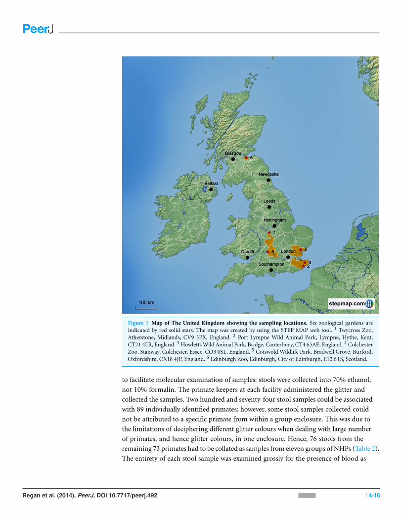
Figure 1 Map of The United Kingdom showing the sampling locations. Six zoological gardens areindicated by red solid stars. The map was created by using the STEP MAP web tool. 1 Twycross Zoo,Atherstone, Midlands, CV9 3PX, England. 2 Port Lympne Wild Animal Park, Lympne, Hythe, Kent,CT21 4LR, England. 3 Howletts Wild Animal Park, Bridge, Canterbury, CT4 65AE, England. 4 ColchesterZoo, Stanway, Colchester, Essex, CO3 0SL, England. 5 Cotswold Wildlife Park, Bradwell Grove, Burford,Oxfordshire, OX18 4JP, England. 6 Edinburgh Zoo, Edinburgh, City of Edinburgh, E12 6TS, Scotland.

to facilitate molecular examination of samples: stools were collected into 70% ethanol,

not 10% formalin. The primate keepers at each facility administered the glitter and

collected the samples. Two hundred and seventy-four stool samples could be associated

with 89 individually identified primates; however, some stool samples collected could

not be attributed to a specific primate from within a group enclosure. This was due to

the limitations of deciphering different glitter colours when dealing with large number

of primates, and hence glitter colours, in one enclosure. Hence, 76 stools from the

remaining 73 primates had to be collated as samples from eleven groups of NHPs (Table 2).

The entirety of each stool sample was examined grossly for the presence of blood as

Regan et al. (2014), PeerJ, DOI 10.7717/peerj.492 4/16

Table 2 Non-human primates sampled in the nationwide study.

Study site Species of primate Number of primates sampled

Individuallyidentified

Unidentified primates(no. of group)

Twycross Zoo Eastern Javan Langur (Trachypithecus auratus auratus) 9 4 (1)

Black Howler Monkey (Alouatta caraya) 15 11 (2)

Woolley Monkeys (Lagothrix lagotricha) 6 –

Dusky Leaf Monkey (Trachypithecus obscures) 1 –

Golden Lion Tamarin (Leontopithecus rosalia) 2 –

Golden-headed Lion Tamarin (Leontopithecus chrysomelas) 2 –

Francois Langur (Trachypithecus francoisi) 5 –

Dusky Leaf Monkey (Trachypithecus obscures) 5 –

Port Lympne Wild Animal Park Eastern Javan Langur (Trachypithecus auratus auratus) 9 39 (4)

Grizzled Leaf Monkey (Presbytis comata) – 7 (1)

Banded Leaf Monkey (Presbytis femoralis) – 3 (1)

Howletts Wild Animal Park Banded Leaf Monkey (Presbytis femoralis) 5 –

Dusky Leaf Monkey (Trachypithecus obscures) 14 –

Francois Langur (Trachypithecus francoisi) 2 –

Grizzled Leaf Monkey (Presbytis comata) 8 –

Eastern Javan Langur (Trachypithecus auratus auratus) 2 –

Colchester Zoo Silvery Langur (Trachypithecus cristatus cristatus) 3 –

Cotswold Wildlife Park Purple-faced Langur (Trachypithecus vetulus monticola) 1 3 (1)

Edinburgh Zoo Purple-faced Langur (Trachypithecus vetulus vetulus) – 6 (1)

Total 89 73 (11)

a possible indication of gastrointestinal illness and potential parasitism. Thirty two

(82.1%) of primates had been treated with a vitamin D3 supplement and 10 day course

of metronidazole (Flagyl) followed by 10 days of diloxinide furoate in the six months prior

to sample collection.

The study was approved by The University of Nottingham (UK) School of Veterinary

Medicine and Science (SVMS) Ethical Review Committee. The Committee reviews all

research studies involving School personnel and is chaired by Professor David Haig. The

committee passed this study as good to proceed, not requiring any further ethical review.

Parasite identificationAll formalin preserved samples were analysed microscopically using a modified Ridley’s

formol-ether concentration technique, which enhances microscopic sensitivity by produc-

ing ‘cleaner’ samples that are more efficient to examine. Following sedimentation, samples

were then examined microscopically for the presence of Entamoeba species from the

E. histolytica complex (E. histolytica, E. dispar and E. moshkovskii), E. coli and E. hart-

manni. Data was analyzed using Minitab 15. Binary logistic regression was used to demon-

strate statistical significance between prevalence of infection and primate demographics.

All prevalence data is derived using the total number of primates as the denominator.

Regan et al. (2014), PeerJ, DOI 10.7717/peerj.492 5/16

Table 3 Primer sets and PCR conditions used in the present study.

Forward primer Reverse primer Amplification reaction

Primer set 2* Primer 5.1: (5′′-AAG GAT AAC TCTTGT TAA TTG CAG-3′′)

Primer 3.2: (5′′-TGT CTA AAT TACCCC AAT TTC C-3′′)

30 cycles of 94 ◦C, 57 ◦C, and 72 ◦C each for 30 s,followed by a final 2 min at 72 ◦C

Primer set 3* Primer 5.2: (5′′-GGA ATA GCT TTTTGA GAA GAA GG-3′′)

Primer 3.2: (5′′-TGT CTA AAT TACCCC AAT TTC C-3′′)

30 cycles of 94 ◦C, 57 ◦C, and 72 ◦C each for 30 s,followed by a final 2 min at 72 ◦C

E. histolytica

RRH5: (5′′-GCG CCT TTT TAT TCAATA TAC TCC-3′′)

RRH3: (5′′-GGA TGA AGA TAT CTTCAC AGG G-3′′)

30 cycles of 94 ◦C, 59 ◦C , and 72 ◦C each for 30 s,followed by a final 2 min at 72 ◦C

E. dispar

RRD5: (5′′-CAT GAG GCG CCT TTTTAT CA-3′′)

RRD3: (5′′-AGG GGA TGA TGA TATTGA ACA CAC TC-3′′)

30 cycles of 94 ◦C, 59 ◦C , and 72 ◦C each for 30 s,followed by a final 2 min at 72 ◦C

Notes.* Two primer sets were used to target uninucleated cyst-producing Entamoeba species (E Victory, pers. comm., 2010).

Molecular analysesQIAamp DNA Stool Mini Kit (QIAgen, UK) was used according to the manufacturer’s

instructions to extract parasite DNA directly from faeces. Technique modifications to

improve the yield and purity of DNA extracts included increasing the lysis temperature to

95 ◦C and adding an extra wash prior to sample elution with Buffer AE. Concentration and

DNA purity in sample extracts was analyzed, using a Thermo Scientific NanoDropTM1000

Spectrophotometer, prior to PCR amplification. The strategy used for selection of PCR

primers (Table 3) was based on the use of previously published diagnostic primers for

the mononucleate Entamoeba species, E. histolytica and E. dispar (Ali, Zaki & Clark,

2005). Both species have been previously found in the faeces of NHPs. It is important

to discriminate E. histolytica from other nonpathogenic amoebas because E. histolytica

carries the risk of zoonosis (Rivera & Kanbara, 1999; Tachibana et al., 2000; Tachibana et al.,

2001; Verweij et al., 2003; Rivera, Yason & Adao, 2010; Feng et al., 2011). Also, we used two

species complex specific primers to amplify uninucleate cyst-producing species, but not

tetra- or octonucleate cyst-producing species. All PCR products were subjected to DNA

sequencing to identify the species/subtype of each amplicon including those amplified by

the species diagnostic primers.

Separate PCRs were performed with each primer pair in a reaction mixture of 40 µl

consisting of 4 µl of extracted DNA, 20 µl of Biomix (Bioline, UK), 15 µl of sterile distilled

water, and 0.5 µl of each forward and reverse primer. The amplification reactions were

performed using a Bioer Xp Cycler as described in Table 4. PCR products were separated

by electrophoresis in 1.2% agarose gels run at 100 V on a Thermo Scientific Easycast

B1 or D2 electrophoresis gel tank with a Thermo Scientific EC 1000 XL Power Pac for

approximately 60 min. A mix of 7 µl of PCR product and 3.5 µl of loading buffer (New

England Biolabs Ltd., UK) were applied to each well. A 1-kbp molecular size ladder (New

England Biolabs) was added to each gel for product size estimation. Gels were stained with

0.1 µg/ml ethidium bromide solution. Amplified DNA was visualized under UV light.

Regan et al. (2014), PeerJ, DOI 10.7717/peerj.492 6/16

Table 4 Details of purified amplicons of Entamoeba species from which nucleotide sequences were obtained.

Primate species Zoological park Primers Target

Banded Leaf Monkey (Presbytis femoralis) Howlett’s Wild Animal Park RRH3, RRH5 E. histolytica

Eastern Javan Langur (Trachypithecus auratus auratus) Twycross Zoo RRD3, RRD5 E. dispar

Dusky Leaf Monkey (Trachypithecus obscures) Twycross Zoo RRD3, RRD5 E. dispar

Dusky Leaf Monkey (Trachypithecus obscures) Howletts Wild Animal Park RRD3, RRD5 E. dispar

Eastern Javan Langur (Trachypithecus auratus auratus) Howletts Wild Animal Park RRD3, RRD5 E. dispar

Eastern Javan Langur (Trachypithecus auratus auratus) Twycross Zoo RRD3, RRD5 E. dispar

Eastern Javan Langur (Trachypithecus auratus auratus) Twycross Zoo RRD3, RRD5 E. dispar

Eastern Javan Langur (Trachypithecus auratus auratus) Twycross Zoo RRD3, RRD5 E. dispar

Eastern Javan Langur (Trachypithecus auratus auratus) Twycross Zoo RRD3, RRD5 E. dispar

Black Howler Monkey (Alouatta caraya) Twycross Zoo P3.2, P5.2 Uninucleates

Black Howler Monkey (Alouatta caraya) Twycross Zoo P3.2, P5.2 Uninucleates

Woolly Monkey (Lagothrix lagotricha) Twycross Zoo P3.2, P5.2 Uninucleates

Golden-headed Lion Tamarin (Leontopithecus chrysomelas) Twycross Zoo P3.2, P5.2 Uninucleates

Eastern Javan Langur (Trachypithecus auratus auratus) Twycross Zoo P3.2, P5.2 Uninucleates

Eastern Javan Langur (Trachypithecus auratus auratus) Twycross Zoo P3.2, P5.2 Uninucleates

Woolly Monkey (Lagothrix lagotricha) Twycross Zoo P3.2, P5.2 Uninucleates

Molecular phylogeneticsOne positive amplicon per primate (a total of 16 amplicons) was selected for sequencing,

based on visualization of PCR products (Table 4). Amplicons were purified, using

a QIAquick PCR Purification Kit (QIAgen, UK), according to the manufacturer’s

instructions and then subjected to sequencing on the Illumina platform by Source

BioScience (Nottingham, UK) using the primers from the PCR. Nucleotide sequences were

determined at least once on each DNA strand. Three representative Entamoeba nucleotide

sequences obtained in this study were deposited in GenBank under accession numbers

KJ149294, KJ149295, KJ149296.

Raw sequencing chromatograms were evaluated with Geneious (version 5.4) software.

Newly obtained Entamoeba sequences were compared with similar sequences available at

the GenBank database by using the Bl2Seq algorithm as implemented in BLASTn (Altschul

et al., 1990). Multiple alignments of all nucleotide sequences were obtained by using the

MUSCLE program (Edgar, 2004). The resulting alignments were adjusted manually when

necessary using CLUSTALX (Larkin et al., 2007). The unmatched ends were deleted to

obtain a homogeneous matrix of characters and thus increase the reliability of the tree

obtained. Phylogenetic trees were inferred from the nucleotide sequence alignments by

the maximum-likelihood (ML) method using the BIONJ algorithm (Gascuel, 1997) and

distance method with HKY85 model (Hasegawa, Kishino & Yano, 1985) of nucleotide

substitution implemented in PhyML-aLRT (Guindon & Gascuel, 2003). The reliability of

the branching order was assessed by performing 1,000 bootstrap replicates.

Regan et al. (2014), PeerJ, DOI 10.7717/peerj.492 7/16

RESULTSEntamoeba prevalence at Twycross ZooOne hundred and twenty-six stool samples were collected from 37 individual primates. No

primate demonstrated ill health at the time of sample collection and no samples contained

grossly visible blood. Microscopic examination demonstrated Entamoeba shedding in

81.1% of 37 primates sampled (Table 1). Entamoeba coli was the most prevalent Entamoeba

species shed (62.2%), with three of six primate species shedding this Entamoeba species.

Shedding of species from the E. histolytica complex was identified in 16.2% of primates

(6 primates). Co-infection with two or more Entamoeba species was identified in 14

primates. Old World Colobinae primates showed the highest prevalence of Entamoeba

infection. Entamoeba infection was significantly associated with species of primate (P <

0.05) and administration of metronidazole (P < 0.05). More specifically, infection with

E. coli was significantly associated with both parameters (both P < 0.05). Primates previ-

ously treated with metronidazole showed greater infection with E. coli (76.9%) compared

to those untreated (25.0%). No significant associations were identified between primate

demographic characteristics and infection with Entamoeba from the E. histolytica complex

or E. hartmanni. Eggs from Trichuris species were identified in samples from two primates.

Entamoeba prevalence at multiple zoosEntamoeba was present in 101 (28.9%) samples (Table 5), indicating a notable prevalence

of Entamoeba infection at the national level. No more than one species of Entamoeba was

identified per sample. Three Entamoeba species were detected by species-specific PCR and

confirmed with sequencing and BLAST: E. histolytica, E. dispar and E. polecki. E. histolytica

was detected in one sample (2.9%), E. dispar in 14 samples (4.0%) and uninucleated

cyst-producing Entamoeba species in 86 (24.6%) samples. E. histolytica and E. dispar were

identified in samples from Colobinae primates only, whilst uninucleated-cyst producing

Entamoeba species were identified in samples primarily from New World monkeys, but

also in primates from the Colobinae family. Entamoeba infection was only detected in

primates from three zoological parks (Table 5): E. histolytica was only identified at one

park, E. dispar in three parks, and uninucleated cyst-producing Entamoeba species in three

parks. No primate was found to harbor mixed Entamoeba species. All primates sampled

appeared clinically healthy at the time of sample collection.

To infer the phylogenetic relationship of the isolates detected in the present study,

E. histolytica, E. dispar and E. polecki, with previously characterized isolates, we used

maximum likelihood method. PCR amplicons from sixteen samples were purified and

submitted for sequencing (Table 4). The sequence from the sample that produced an

amplicon with the E. histolytica-specific primers was identical to the corresponding region

of the GenBank sequence for E. histolytica from monkey (AB197936) from a cynomolgus

monkey. Likewise, sequences obtained from eight samples that produced amplicons with

the E. dispar-specific primers were identical to the corresponding region of the GenBank

sequence (AB282661) for E. dispar from a rhesus monkey. Seven sequences were obtained

from uninucleate amplicons from Twycross Zoo and shared high sequence homology

Regan et al. (2014), PeerJ, DOI 10.7717/peerj.492 8/16

Table 5 The prevalence of Entamoeba species by species of primate and zoological park.

Species of primate E. histolytica E. dispar Uninucleates

% (number of primate)

Old world monkey

Banded Leaf Monkey (Presbytis femoralis; n = 20) 5.0 (1) 0 0

Dusky Leaf Monkey (Trachypithecus obscures; n = 60) 0 3.3 (2) 0

Eastern Javan Langur (Trachypithecus auratus auratus; n = 117) 0 10.3 (12) 29.1 (34)

Francois Langur (Trachypithecus francoisi; n = 19) 0 0 0

Grizzled Leaf Monkey (Presbytis comata; n = 27) 0 0 0

Silvery Langur (Trachypithecus cristatus cristatus; n = 9) 0 0 0

Purple-faced Langur (Trachypithecus vetulus vetulus; n = 11) 0 0 0

Subtotal (n = 263) 0.76 (1) 5.3 (14) 12.9 (34)

New world monkey

Black Howler Monkey (Alouatta caraya; n = 52) 0 0 70.9 (40)

Woolly Monkey (Lagothrix lagotricha; n = 23) 0 0 47.8 (11)

Golden-headed Lion Tamarin (Leontopithecus chrysomelas; n = 6) 0 0 16.7 (1)

Golden Lion Tamarin (Leontopithecus rosalia; n = 6) 0 0 0

Subtotal (n = 87) 0 0 59.8 (52)

Total (n = 350) 0.3 (1) 4.0 (14) 24.6 (86)

Zoological park

Colchester Zoo (n = 9) 0 0 0

Cotswold Wildlife Park and Gardens (n = 8) 0 0 0

Edinburgh Zoo (n = 3) 0 0 0

Howletts Wild Animal Park (n = 90) 1.1 (1) 2.2 (2) 0

Port Lympne Wild Animal Park (n = 72) 0 2.8 (2) 0

Twycross Zoo (n = 168) 0 6.0 (10) 51.2 (86)

Total (n = 350) 0.3 (1) 4.0 (14) 24.6 (86)

E. polecki. Two representative sequences were used to build a phylogenetic tree. As seen

in Fig. 2, E. polecki sequences obtained in the present study clustered with and formed a

monophyletic group with E. polecki subtype 4 isolates reported in Homo sapiens from Asia,

Africa and Europe.

DISCUSSIONNonhuman primates harbour a number of Entamoeba spp of varied importance to human

and domestic animal health. The prevalence and genetic identity of Entamoeba species was

investigated in primate collections at six major NHP zoos in the United Kingdom. Results

indicated a low prevalence of the pathogenic E. histolytica in the examined primates. This is

important to the primate population and also to the many thousands of human visitors of

these zoos each year. Higher prevalence of non-pathogenic Entamoeba species was however

identified in the primates sampled. Previous studies utilizing molecular methods to

identify carriage of Entamoeba species demonstrated a similar prevalence data to that seen

in the current study. Low carriage of E. histolytica and higher carriage of other Entamoeba

species in NHP populations has been demonstrated in both captive (Tachibana et al.,

Regan et al. (2014), PeerJ, DOI 10.7717/peerj.492 9/16

Figure 2 Phylogenetic tree based on partial 18SrDNA sequences, showing the relationships amongEntamoeba species. Phylogenetic analysis used two different approaches, distance-based analysis andmaximum-likelihood (ML), produced trees with identical topologies of which only ML tree is presented.GenBank accession numbers and host species are given in parentheses after the taxon name. Sequencesin bold face were obtained during this study. Numbers above branches are bootstrap values (%) from1,000 replicates. Nodes of the tree with bootstrap values of ≥95% are indicated by black closed circles.The node is not labeled where bootstrap support values is <50. Bar = estimated number of substitutionsper site.

Regan et al. (2014), PeerJ, DOI 10.7717/peerj.492 10/16

2000; Tachibana et al., 2001; Takano et al., 2005; Rivera, Yason & Adao, 2010) and free-living

NHP species (Rivera & Kanbara, 1999). However, the NHP populations examined in these

studies were based outside of Europe, with all of the captive populations investigated in

these studies existing in research facilities in Asia. Levecke et al. (2010) reported that 36%

of faecal samples collected from various primate species in zoological parks in Belgium

and The Netherlands contained E. histolytica, and identified Entamoeba species as the most

prevalent gastrointestinal parasite within the sampled population.

The lack of sex or age predisposition to infection with Entamoeba species in our study

is in agreement with other studies (Lilly, Mehlman & Doran, 2002; Jones-Engel et al., 2004;

Gillespie, Greiner & Chapman, 2005; Muehlenbein, 2005; Ekanayake et al., 2006; Teichroeb et

al., 2009). In the present study, the highest prevalence of Entamoeba infection was detected

in Old World monkeys; this finding is in agreement with reports from other studies in

Japan (Tachibana et al., 2001) and Belgium (Levecke et al., 2007). Primates from the

Colobinae family have specialised sacculated stomachs, an adaption to their leaf-eating

lifestyle, which provides favorable conditions for ingested Entamoeba cyst excystation,

and trophozoite tissue invasion (Matz-Rensing et al., 2004; Ulrich et al., 2010). The higher

carriage of uninucleated cyst-producing Entamoeba species, compared to other Entamoeba

species, identified at Twycross Zoo may be explained by the asymptomatic commensal

carriage of a non-pathogenic Entamoeba species. These non-pathogenic species are less

likely to be clinically identified; therefore, infected primates are less likely to receive

amoebicidal treatment. Administration of amoebicidal drugs might have been the cause of

the apparent increase in the prevalence of uninucleated cyst-producing Entamoeba species

and Entamoeba coli in primates at Twycross Zoo. Re-establishment of gastrointestinal

microflora, following treatment with amoebicidal agents, may have favoured the growth

of the commensal populations of the octonucleated cyst-producing Entamoeba species

(E. coli) in treated primates, as confirmed by microscopic examination of stool samples. In

line with this assumption is the reported high frequency of the commensal uninucleated

and octonucleated cyst-producing commensal Entamoeba species in primate populations

(Tachibana et al., 2000; Petrasova et al., 2010). Alternatively, this may be explained by

the development of metronidazole resistance in these uninucleated and octonucleated

cyst-producing Entamoeba species.

The difference in the prevalence of Entamoeba among zoos (Table 5) can be explained by

the differences in biosecurity and precautionary measures taken to prevent parasitic disease

transmission. All zoos participated in the study already implement routine disinfection

programmes (personal communication with Zoos). However, additional precautionary

measures are needed in order to prevent the transmission of infection between enclosures

including hygienic food preparation, provision of potable water, and disinfection of keeper

footwear, over-clothing, hands, and cleaning equipment between enclosures. Effective

drainage and water microfiltration within enclosures is also critical. Proactive pest control

measures reduce arthropod vectors transporting infective cysts between enclosures (Pang,

Chang & Chang, 1993; Denver, 2008). Additionally, avoiding mixed primate exhibits

reduces the transmission of amoebiasis between NHP of different susceptibility. The same

Regan et al. (2014), PeerJ, DOI 10.7717/peerj.492 11/16

measure may help to prevent zoonotic transmission to zoo visitors. Unfortunately limited

time and financial resources often result in deficiencies in one or more of these measures.

Methods for Entamoeba identification have been undergoing rapid change over the past

decade and molecular phylogenetic techniques are rapidly becoming the procedures of

choice (Levecke et al., 2010; Stensvold et al., 2011). PCR amplification of the 18S rDNA gene

directly from a sample of mixed microbiota alleviates the need for culturing Entamoeba

(Levecke et al., 2010), and once DNA is prepared, there are no biohazard dangers.

rDNA-based molecular phylogenetic techniques were used to identify the Entmaoeba

species detected in the faecal samples from NHP in the present study. Sequences from

E. histolytica and E. dispar obtained in the study were identical to previously reported

sequences in Genbank AB197936 and AB282661, respectively. Phylogenetic analysis of the

partial-length 18SrDNA sequence showed that the uninucleate amplicons from Twycross

Zoo were all E. polecki.

The uninucleated-cyst-producing Entamoeba infecting humans E. polecki species

complex has been found to encompass four subtypes (ST1–ST4) (Stensvold et al., 2011).

E. polecki ST1 (previously given to E. polecki in pigs); ST2 (E. chattoni from non-human

primates); ST3 (E. struthionis from pigs and ostriches); and ST4 (restricted to humans;

unlikely to be zoonotic); indicating low host specificity of ST1 and ST3. Comparison

between sequences obtained in the present study and reference sequences obtained from

GenBank for each of the four E. polecki subtypes indicated that sequences of E. polecki

obtained in the present study from NHPs [Woolly Monkey (Lagothrix lagotricha),

Eastern Javan Langur (Trachypithecus auratus auratus), Golden-headed Lion Tamarin

(Leontopithecus chrysomelas), and Black Howler Monkey (Alouatta caraya)] formed a

phylogenetic cluster (Fig. 2) with isolates of E. polecki subtype 4 reported in Homo sapiens

from Africa, Asia and Europe (Stensvold et al., 2011). Given the reported high specificity

of E. polecki subtype 4 to humans, the similarity between sequences obtained from

NHPs from Twycross zoo in the present study with E. polecki ST4 sequences obtained

from Homo sapiens suggest a zoonotic potential. However, more analysis is needed

before any suggestion about the zoonotic implication of the isolates obtained in this

study to be made. A group of uninucleate Entamoebas (referred to as Entamoeba RL3),

phylogenetically distant from the E. polecki complex, have been reported from Francois

Langur (Trachypithecus francoisi) from Twycross Zoo in England (Stensvold et al., 2011).

Interestingly, sequences of this Entamoeba RL3 group did not seem to share similarity

with the sequences of uninucleated-cyst-producing E. polecki obtained in the present study

from NHPs from the same Zoo.

Sequence data (Table 4) also suggest that a common source asymptomatic infection with

the uninucleated cyst-producing Entamoeba species, E. polecki, at Twycross Zoo may have

propagated through many primate enclosures. This study did not examine the prevalence

of E. nutalli, an emerging species currently seeming to be prevalent in NHPs (Tachibana

et al., 2013). Since E. nuttalli has been associated with symptomatic carriage, and appears

to be restricted in host distribution to NHPs, it would be interesting to know whether any

of the animals sampled in the present study harboured E. nuttalli. Thus, further studies

Regan et al. (2014), PeerJ, DOI 10.7717/peerj.492 12/16

are needed to establish the prevalence of this important species in NHPs in the United

Kingdom and its zoonotic risk to public health.

CONCLUSIONThis is the first study to report the prevalence of Entamoeba infection in captive NHPs

in the United Kingdom. Data collected from six zoos suggests a notable prevalence of

Entamoeba infection in NHPs in UK. DNA sequencing of positive stool samples revealed

three main species of Entamoeba, E. histolytica, E. dispar and E. polecki ST4 circulating in

the zoo’s environment in the UK. Some Entamoeba species can have zoonotic potential,

thus can constitute a risk for humans who are in close contact with primates.

ACKNOWLEDGEMENTSThe following persons and facilities provided support and samples: Nic Masters, primate

keepers, research collaborators and curators from all participating zoological parks.

John Williams and Emma Victory from Diagnostic Parasitology Laboratory, London

School of Hygiene and Tropical Medicine provided technical advice on PCR optimization

experiments.

ADDITIONAL INFORMATION AND DECLARATIONS

FundingSummer studentship was provided by the Society for General Microbiology, University of

Nottingham and Zebra Foundation to Carl Regan. The funders had no role in study design,

data collection and analysis, decision to publish, or preparation of the manuscript.

Grant DisclosuresThe following grant information was disclosed by the authors:

Society for General Microbiology Elective Research Project Grant.

Undergraduate research project grant, University of Nottingham - School of Veterinary

Medicine and Science.

Zebra Foundation student research grant.

Competing InterestsThe authors declare there are no competing interests.

Author Contributions• Carl S. Regan conceived and designed the experiments, performed the experiments,

wrote the paper, prepared figures and/or tables, reviewed drafts of the paper.

• Lisa Yon contributed reagents/materials/analysis tools, reviewed drafts of the paper.

• Maqsud Hossain analyzed the data, contributed reagents/materials/analysis tools,

reviewed drafts of the paper.

• Hany M. Elsheikha conceived and designed the experiments, performed the experi-

ments, analyzed the data, contributed reagents/materials/analysis tools, wrote the paper,

prepared figures and/or tables, reviewed drafts of the paper.

Regan et al. (2014), PeerJ, DOI 10.7717/peerj.492 13/16

Animal EthicsThe following information was supplied relating to ethical approvals (i.e., approving body

and any reference numbers):

The study was approved by The University of Nottingham (UK) School of Veterinary

Medicine and Science (SVMS) Ethical Review Committee.

The committee passed this study as not requiring any further ethical review.

DNA DepositionThe following information was supplied regarding the deposition of DNA sequences:

GenBank KJ149294, KJ149295, KJ149296.

Data DepositionThe following information was supplied regarding the deposition of related data:

Nottingham ePrints Archive (http://eprints.nottingham.ac.uk/).

Supplemental InformationSupplemental information for this article can be found online at http://dx.doi.org/

10.7717/peerj.492.

REFERENCESAli IKM, Zaki M, Clark CG. 2005. Use of PCR amplification of tRNA gene-linked short tandem

repeats for genotyping Entamoeba histolytica. Journal of Clinical Microbiology 43(12):5842–5847DOI 10.1128/JCM.43.12.5842-5847.2005.

Altschul SF, Gish W, Miller W, Myers EW, Lipman DJ. 1990. Basic local alignment search tool.Journal of Molecular Biology 215:403–410 DOI 10.1016/S0022-2836(05)80360-2.

Denver MC. 2008. Reptile protozoa. In: Fowler ME, Miller RE, eds. Zoo and wild animal medicinecurrent therapy, vol. 6. Missouri: Saunders Elsevier, 154–159.

Edgar RC. 2004. MUSCLE: multiple sequence alignment with high accuracy and high throughput.Nucleic Acids Research 32:1792–1797 DOI 10.1093/nar/gkh340.

Ekanayake DK, Arulkanthan A, Horadagoda NU, Sanjeevani GKM, Kieft R, Gunatilake S,Dittus WPJ. 2006. Prevalence of crytosporidium and other enteric parasites among wildnon-human parasites in Polonnaruwa, Sri Lanka. American Journal of Tropical Medicine andHygiene 74:322–329.

Feng M, Yang B, Yang L, Fu Y, Zhuang Y, Liang L, Xu Q, Cheng X, Tachibana H. 2011. Highprevalence of Entamoeba infections in captive long-tailed macaques in China. ParasitologyResearch 109:1093–1097 DOI 10.1007/s00436-011-2351-2.

Fotedar R, Stark D, Beebe N, Marriott D, Ellis J, Harkness J. 2007. Laboratory diagnostictechniques for Entamoeba species. Clinical Microbiology Reviews 20(3):511–532DOI 10.1128/CMR.00004-07.

Gascuel O. 1997. BIONJ: an improved version of the NJ algorithm based on a simple model ofsequence data. Molecular Biology and Evolution 14:685–695DOI 10.1093/oxfordjournals.molbev.a025808.

Gillespie TR, Greiner EC, Chapman CA. 2005. Gastrointestinal parasites of the Colobus Monkeyof Uganda. Journal of Parasitology 91(3):569–573 DOI 10.1645/GE-434R.

Regan et al. (2014), PeerJ, DOI 10.7717/peerj.492 14/16

Guindon S, Gascuel O. 2003. A simple, fast, and accurate algorithm to estimate large phylogeniesby maximum likelihood. Systematic Biology 52:696–704 DOI 10.1080/10635150390235520.

Hasegawa M, Kishino H, Yano T. 1985. Dating of the human-ape splitting by a molecular clock ofmitochondrial DNA. Journal of Molecular Evolution 22(2):160–174 DOI 10.1007/BF02101694.

Heredia RD, Fonseca JA, Lopez MC. 2012. Entamoeba moshkovskii perspectives of a newagent to be considered in the diagnosis of amebiasis. ACTA Tropica 123:139–145DOI 10.1016/j.actatropica.2012.05.012.

Jones-Engel L, Engel GA, Schillaci MA, Kyes K, Froehlich J, Paputungan U, Kyes RC. 2004.Prevalence of enteric parasites in pet macaques in Sulawesi, Indonesia. American Journal ofPrimatology 62:71–82 DOI 10.1002/ajp.20008.

Kebede A, Verweij J, Dorigo-Zetsma W, Sanders E, Messele T, van Lieshout L, Petros B,Polderman T. 2003. Overdiagnosis of amoebiasis in the absence of Entamoeba histolytica amongpatients presenting with diarrhoea in Wonji and Akaki, Ethiopia. Transactions of the RoyalSociety of Tropical Medicine and Hygiene 97:305–307 DOI 10.1016/S0035-9203(03)90153-2.

Larkin MA, Blackshields G, Brown NP, Chenna R, McGettigan PA, McWilliam H, Valentin F,Wallace IM, Wilm A, Lopez R, Thompson JD, Gibson TJ, Higgins DG. 2007. Clustal W andclustal X version 2.0. Bioinformatics 23:2947–2948 DOI 10.1093/bioinformatics/btm404.

Levecke B, Dorny P, Geurden G, Vercammen F, Vercruysse J. 2007. Gastrointestinal protozoain non-human primates of four zoological gardens in Belgium. Veterinary Parasitology148:236–246 DOI 10.1016/j.vetpar.2007.06.020.

Levecke B, Dreesen L, Dorny P, Verweij JJ, Vercammen F, Casaert S, Vercruysse J, Geldhof P.2010. Molecular identification of Entamoeba spp. in captive nonhuman primates. Journal ofClinical Microbiology 48(8):2988–2990 DOI 10.1128/JCM.00013-10.

Lilly AA, Mehlman PT, Doran D. 2002. Intestinal parasites in gorillas, chimpanzees, and humansat Mondika Research Site, Dzanga-Ndoki National Park, Central African Republic. InternationalJournal of Primatology 23(3):555–573 DOI 10.1023/A:1014969617036.

Matz-Rensing K, Floto A, Kaup FJ, Schaller K. 2004. Gastric amoebiasis due to Entamoebahistolytica infection a mandrill (Mandrillus sphinx). In: Proceedings of the fifth EuropeanAssociation of Zoo- and Wildlife Veterinarians (EAZWV). Ebeltoft, Denmark.

Muehlenbein MP. 2005. Parasitological analyses of the male chimpanzees (Pan troglodytesschweinfurthii) at Ngogo, Kibale National Park, Uganda. American Journal of Primatology65:167–179 DOI 10.1002/ajp.20106.

Pang VF, Chang CC, Chang WF. 1993. Concurrent gastric and hepatic amebiasis in a dusky leafmonkey (Presbytis obscurus). Journal of Zoo and Wildlife Medicine 24:204–207.

Petrasova J, Modry D, Huffman MA, Mapua MI, Bobakova L, Mazoch V, Singh J, Kaur T,Petrzelkova KJ. 2010. Gastrointestinal parasites of indigenous and introduced primate speciesof Rubondo Island National Park, Tanzania. International Journal of Primatology 31:920–936DOI 10.1007/s10764-010-9439-x.

Rivera WL, Kanbara H. 1999. Detection of Entamoeba dispar DNA in macaque feces bypolymerase chain reaction. Parasitology Research 85:493–495 DOI 10.1007/s004360050583.

Rivera WL, Yason JADL, Adao DEV. 2010. Entamoeba histolytica and E. dispar infectionsin captive macaques (Macaca fascicularis) in the Philippines. Primates 51:69–74DOI 10.1007/s10329-009-0174-x.

Sargeaunt PG, Patrick S, O’Keeffe D. 1992. Human infections of Entamoeba chattoni masqueradeas Entamoeba histolytica. Transactions of the Royal Society of Tropical Medicine and Hygiene86:633–634 DOI 10.1016/0035-9203(92)90162-6.

Regan et al. (2014), PeerJ, DOI 10.7717/peerj.492 15/16

Sargeaunt PG, Williams JE, Jones DM. 1982. Electrophoretic isoenzyme patterns of Entamoebahistolytica and Entamoeba chattoni in a primate survey. Journal of Protozoology 29:136–139DOI 10.1111/j.1550-7408.1982.tb02897.x.

Solaymani-Mohammadi S, Rezaian M, Babaei Z, Rajabpour A, Meamar AR, Pourbabai AA,Petri Jr WA. 2006. Comparison of a stool antigen detection kit and PCR for diagnosis ofEntamoeba histolytica and Entamoeba dispar infections in asymptomatic cyst passers in Iran.Journal of Clinical Microbiology 44(6):2258–2261 DOI 10.1128/JCM.00530-06.

Stensvold CR, Lebbad M, Victory EL, Verweij JJ, Tannich E, Alfellani M, Legarraga P, Clark CG.2011. Increased sampling reveals novel lineages of Entamoeba: consequences of geneticdiversity and host specificity for taxonomy and molecular detection. Protist 162(3):525–541DOI 10.1016/j.protis.2010.11.002.

Tachibana H, Cheng XJ, Kobayashi S, Fujita Y, Udono T. 2000. Entamoeba dispar, but notE. histolytica, detected in chimps. Parasitology Research 86:537–541DOI 10.1007/s004360000205.

Tachibana H, Cheng XJ, Kobayashi S, Matsubayashi N, Gotoh S, Matsubayashi K. 2001. Highprevalence of infection with Entamoeba dispar, but not E. histolytica, in captive macaques.Parasitology Research 87:14–17 DOI 10.1007/s004360000289.

Tachibana H, Yanagi T, Lama C, Pandey K, Feng M, Kobayashi S, Sherchand JB. 2013. Prevalenceof Entamoeba nuttalli infection in wild rhesus macaques in Nepal and characterization of theparasite isolates. Parasitology International 62:230–235 DOI 10.1016/j.parint.2013.01.004.

Takano J, Narita T, Tachibana H, Shimizu T, Komatsubara H, Terao K, Fujimoto K. 2005.Entamoeba histolytica and Entamoeba dispar infections in cynomolgus monkeys imported intoJapan for research. Parasitology Research 97:255–257 DOI 10.1007/s00436-005-1415-6.

Teichroeb JA, Kutz SJ, Parkar U, Thompson RCA, Sicotte P. 2009. Ecology of the gastrointestinalparasites of Colobus vellerosus at Boabeng-Fiema, Ghana: possible anthropozoonotictransmission. American Journal of Physical Anthropology 140:498–507 DOI 10.1002/ajpa.21098.

Ulrich R, Boer M, Herder V, Spitzbarth I, Hewicker-Trautwein M, Baumgartner W, Wohlsein P.2010. Epizootic fatal amebiasis in an outdoor group of Old World monkeys. Journal of MedicalPrimatology 39:160–165 DOI 10.1111/j.1600-0684.2010.00405.x.

Verweij JJ, Vermeer J, Brienen EAT, Blotkamp C, Laeijendecker D, van Lieshout L,Polderman AM. 2003. Entamoeba histolytica infections in captive primates. ParasitologyResearch 90:100–103.

World Health Organization. 1997. Amoebiasis. Weekly Epidemiology Record 72:97–99.

Regan et al. (2014), PeerJ, DOI 10.7717/peerj.492 16/16


Recommended