An-Najah National University
Faculty of Graduate Studies
Prevalence of Iron Deficiency Anemia among
School Children in Salfeet District
By
Mohammad Mahmoud Mohammad Odeh
Supervisor
Dr. Nael S. Abu-Hasan
Co- Supervisor
Dr. Riad Amer
Submitted in Partial Fulfillment for the Requirements for the Degree of
Master in Public Health, Faculty of Graduate Studies, at An-Najah
National University, Nablus, Palestine.
2006
III
Dedication
To My Beloved Wife, Parents for their Patience and
Encouragements with Love and Respect
IV
Acknowledgement
I would like to express my deep thanks for my supervisors Dr. Nael S.
Abu-Hasan and Dr. Riad A. Amar for the valuable discussions, efforts,
encouragements and their continuous support throughout this study.
Thanks to Mr. Yaseen Afaneh for his kind help and assistance in statistical
analysis. Thanks are also due to the Palestinian Ministry of Education,
Directorate of Education and School Headmasters at Salfeet district for
their help in sample collection. Thanks to administrative and lab.
technicians at Al-Watany Hospital for their kind help and assistance in
blood analysis.
Last but not least thanks are due to my beloved wife, parents and
family for their continuous support during my years of study.
V
List of Contents
page Contents
III Dedication Iv Acknowledgment V List of Contents VI List of Tables VII Abstract CHAPTER ONE: INTRODUCTION 2 General Background 1.1 3 Definition 1.2 3 Pathophysiology 1.3 6 Iron needs during infancy and childhood 1.4 8 Causes of iron deficiency anemia 1.5 9 Symptoms of iron deficiency anemia 1.6 10 Diagnosis of iron deficiency anemia 1.7 12 Treatment 1.8 13 Patient Education 1.9 13 Complications 1.10 14 Prevention 1.11 15 The prevalence and distribution of iron deficiency
worldwide 1.12
16 Iron deficiency anemia in Palestine 1.13 17 Objectives of the study 1.14 CHAPTER TWO: METHODOLOGY
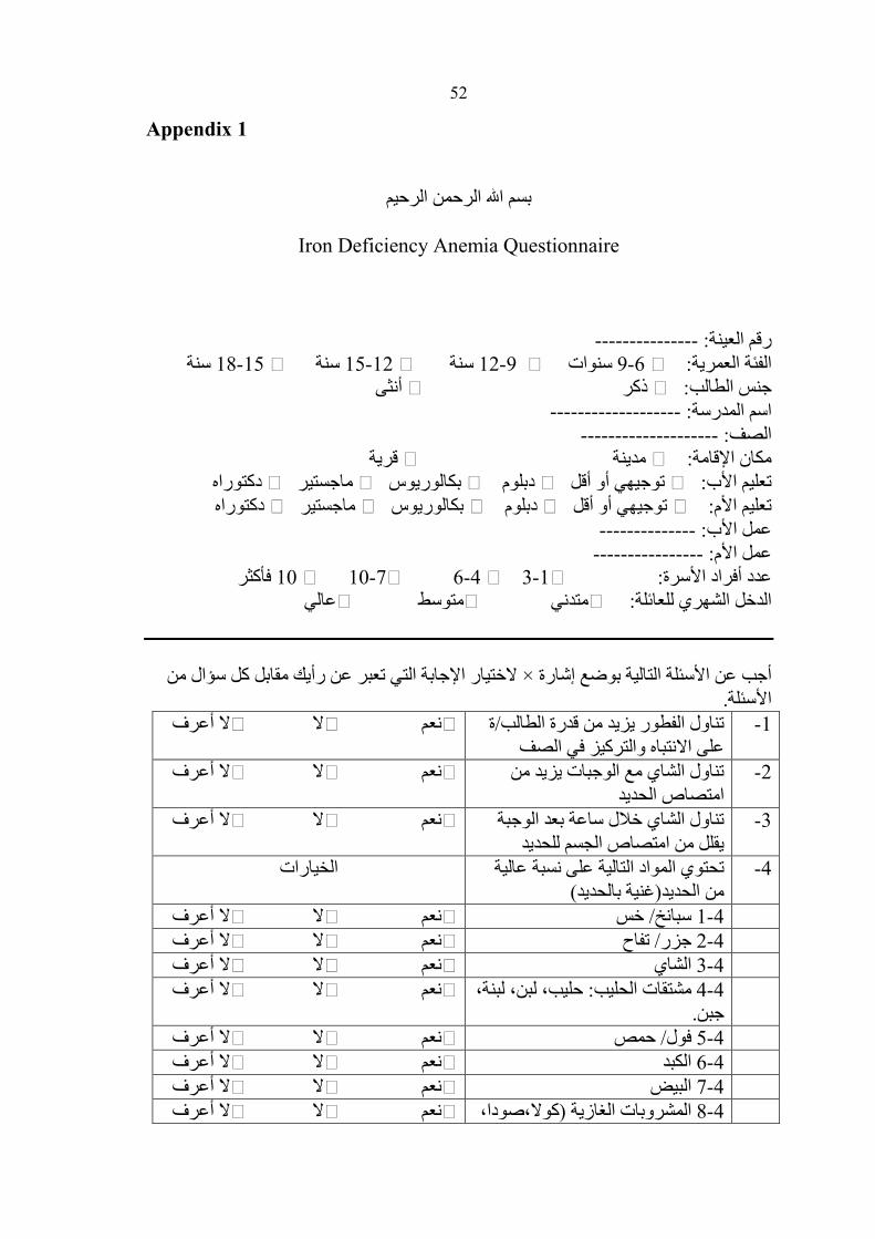
19 Study sample 2.1 20 Tools of study 2.2 20 Questionnaire 2.2.1 20 Blood tests 2.2.2 20 Procedure 2.3 21 Data analysis 2.4 22 CHAPTER THREE: RESULTS AND DISCUSSION 23 Prevalence of ID and iron deficiency anemia 3.1 29 Knowledge, awareness and practices of study
population towards iron deficiency 3.2
35 Healthy practices and iron deficiency 3.3 38 Consequences of iron deficiency 3.4 42 Recommendations and concluding remarks 3.5 43 References 51 Appendices Arabic abstract ب
VI
List of Tables
Table Page
Table 1.1 The normal values for the age-matched red cell indexes, and serum iron
11
Table 1.2 Estimated percentage of anemia prevalence (1990-1995) based on blood hemoglobin concentration
15
Table 1.3 Estimated prevalence of anemia (1990-1995), WHO regions based on blood hemoglobin concentration
16
Table 2.1 Distribution of the study sample 19 Table 2.2 Cutoff values for iron deficiency and anemia 20 Table 3.1 Prevalence of iron deficiency by demographic
patient characteristics 29
Table 3.2 Prevalence of iron deficiency according to family awareness regarding diet maintaining iron levels
34
Table 3.3 The prevalence of iron deficiency related to the practice (health profiles) of the study population
38
Table 3.4 Prevalence of iron deficiency according to consequences of the disease
41
VIIPrevalence of Iron Deficiency Anemia among
School Children in Salfeet District
By
Mohammad Mahmoud Mohammad Odeh
Supervisor
Dr. Nael S. Abu-Hasan
Abstract
A cross-sectional study conducted in the second semester of the
academic year 2005 to investigate the prevalence of iron deficiency anemia
in school children aged 6 to 18 years, who live in the district of Salfeet in
the West Bank area of Palestine. The study sample consisted of 144(49.7
%) male students, and 146 (50.3 %) female students. Complete blood
count (CBC) was performed and blood samples with main corpuscular
volume (MCV) value less than 80um³(FL) were subjected to serum iron
test. The prevalence of iron deficiency was 26.7% (12.7% with anemia,
and 14% without anemia). The prevalence of iron deficiency among
females was 30.5%, and among males was (21.6%). Iron deficiency was
apparent in all studied age groups. The prevalence of 32.4% was observed
among the age group 6- 8 years, 35.3% among age group 9-11 years,
25.9% among 12-14 years and 12.1% among 15-18 years old. Differences
in prevalence rates were statistically significant (P= 0.01 at α = 0.05).
According to place of residency, there was statistically significant
difference between the overall prevalence of iron deficiency among
children living in villages compared to children living in the city (22.8%
versus 32.6% respectively, P < 0.01). There was no clear link between
family size and iron deficiency. With respect to prevalence of iron
deficiency and family income, no significant difference was observed
(24.9% low income; 28.1% with medium and 30.2% with high income). In
general, improper daily healthy practices and poor knowledge regarding
VIII
iron rich nutrients and its absorption found. Previous history of other
diseases seems to contribute to the highly observed prevelance rate of IDA.
To effectively face these deficiencies it is necessary to think about the
possibilities and cost effectiveness of fortifying foodstuffs (floor, salt, milk)
and it is essential to carry out nutritional education activities to improve
children and parents awareness and knowledge regarding iron deficiency
anemia and its consequence.
2
1.1 General background
Iron deficiency is the most prevalent and common micronutrient
deficiency in the developing world today (Tatala et al., 1998; Asobayire et
al., 2001; Abalkhail and Shawky, 2002; Hashizume et al., 2003). The
public health effects of iron deficiency include anemia, decreased
intellectual and work performance as well as functional alterations of the
small bowel (Oski, 1993). Beside other vulnerable age groups, such as
infancy and early childhood, adolescence is placed at a high risk level for
developing iron deficiency, due to a combination of menstrual iron losses
in girls and a rapid physical growth, especially in boys (Fomon et al.,
2003).
Poor diet quality and low dietary iron bioavailability are the principal
factors that contribute to the increased incidence of iron deficiency (Tatala
et al., 1998). The bioavailability of haem iron, present in animal products,
is high with absorption rates of 20−30%, whereas the bioavailability of
nonhaem iron is determined by the presence of enhancing or inhibiting
factors (Hurrell, 1997). The main enhancers of nonhaem iron absorption
are meat (haem iron) and vitamin C (Cook & Reddy, 2001). Inhibitors
include phytate (nuts, bran and oat products, whole-wheat and brown
flour), polyphenols (tea, coffee, cocoa, some spices and vegetables),
calcium (milk products) and Phosphorous (Reddy et al., 2000).
In developing countries, low standards of living, low socio-economic
conditions, restricted access to food and lack of knowledge for good dietary
practices and personal hygiene contribute even more to a high occurrence
of iron deficiency and hence anemia (Hall et al., 2001; Islam et al., 2001;
Soekarjo et al., 2001). Intestinal parasitic infection, due to poor hygienic
3
conditions also interferes with iron absorption, thus expanding the
prevalence of iron deficiency anemia in the developing world (Olivares et
al., 1999; Musaiger, 2002).
1.2 Definition
Iron deficiency anemia is a decrease in the total hemoglobin levels
caused by a lack of sufficient iron (Goldenring, 2003). It is the most
common cause of anemia worldwide. Iron is needed to form hemoglobin
and is mostly stored in the body in the form of ferritin and hemosiderin.
About 30% of iron is stored as ferritin and hemosiderin in the bone
marrow, spleen, and liver. Iron-deficiency anemia does not develop
immediately. Instead, a person progresses through stages of iron
deficiency, beginning with iron depletion, in which the amount of iron in
the body reduced but the amount of iron in the red blood cells remains
constant. If iron depletion not corrected, it progresses to iron deficiency,
eventually leading to iron-deficiency anemia.
1.3 Pathophysiology
Iron is vital for all living organisms because it is essential for multiple
metabolic processes, including oxygen transport, DNA synthesis, and
electron transport. Iron equilibrium in the body regulated carefully to
ensure that sufficient iron is absorbed in order to compensate for body
losses of iron. While body loss of iron quantitatively is as important as
absorption in terms of maintaining iron equilibrium, it is a more passive
process than absorption. Consistent errors in maintaining this equilibrium
lead to either iron deficiency or iron overload (Conrad, 2000).
Iron balance usually achieved by regulation of iron absorption in the
proximal small intestine. Either diminished absorbable dietary iron or
4
excessive loss of body iron can cause iron deficiency. Diminished
absorption is usually due to an insufficient intake of dietary iron in the
absorbable form.
Iron uptake in the proximal small bowel occurs by three separate
pathways. These are the heme pathway, the ferric pathway and the ferrous
pathway. Heme iron not chelated and precipitated by numerous
constituents of the diet that renders nonheme iron nonabsorbable.
Examples are phytates, phosphates, tannates, oxalates, and carbonates.
Heme is maintained soluble and available for absorption by globin
degradation products produced by pancreatic enzymes. Heme iron and
nonheme iron are absorbed into the enterocyte noncompetitively. Heme
enters the cell as an intact metalloporphyrin, presumably by a vesicular
mechanism, degraded within the enterocyte by heme oxygenase with
release of iron so that it traverses the basolateral cell membrane in
competition with nonheme iron to bind transferrin in the plasma (Marcel,
2005).
Ferric iron utilizes a different pathway to enter cells than ferrous iron.
This shown by competitive inhibition studies, the use of blocking
antibodies against divalent metal transporter-1 (DMT-1) and beta3-integrin,
and transfection experiments using DMT-1 DNA. This indicated that ferric
iron utilizes beta3-integrin and mobilferrin, while ferrous iron uses DMT-1
to enter cells (Lee, 1999). Which pathway transports most nonheme iron in
humans is not known. Most non-heme dietary iron is the ferric iron. Iron
absorption in mice and rats may involve more ferrous iron because they
excrete moderate quantities of ascorbate in intestinal secretions. On the
contrary, humans are a scorbutic species and are unable to synthesize a
scorbate to reduce body ferric iron (Marcel, 2005).
5
There are other proteins, which appear to be involved in iron
absorption. These are stimulators of iron transport (SFT), which are
reported to increase the absorption of both ferric and ferrous iron, and
hephaestin, which is postulated to be important in the transfer of iron from
enterocytes into the plasma (Marcel,2005).
The iron concentration within enterocytes varies directly with the
body's requirement for iron. Absorptive cells in iron-deficient humans and
animals contain little stainable iron, whereas this increased significantly in
subjects who are replete in iron (Marcel, 2005). Untreated phenotypic
hemochromatosis creates little stainable iron in the enterocyte, similar to
iron deficiency. Iron within the enterocyte may operate by up-regulation of
a receptor, saturation of an iron-binding protein, or both. In contrast to
findings in iron deficiency, enhanced erythropoiesis, or hypoxia, endotoxin
rapidly diminishes iron absorption without altering enterocyte iron
concentration. This suggests that endotoxin and, perhaps, cytokines alter
iron absorption by a different mechanism (Marcel, 2005).
Most iron delivered to nonintestinal cells is bound to transferrin.
Transferrin iron is delivered into nonintestinal cells via 2 pathways, the
classical transferrin receptor pathway (high affinity, low capacity) and the
pathway independent of the transferrin receptor (low affinity, high
capacity) (Marcel, 2005). Otherwise, the non-saturability of transferrin
binding to cells cannot be explained. In the classical transferrin pathway,
the transferrin receptor complex enters the cell within an endosome.
Acidification of the endosome releases iron from transferrin so that it can
enter the cell. The apotransferrin is recycled back to plasma for
reutilization. The method by which the transferrin receptor–independent
pathway delivers iron to the cell is not known (Marcel, 2005). Non-
6
intestinal cells also possess the mobilferrin integrin and DMT-1 pathways.
Their function in the absence of an iron-saturated transferrin is uncertain;
however, their presence in nonintestinal cells suggests they may participate
in intracellular functions in addition to their ability to facilitate cellular
uptake of iron (Marcel, 2005).
1.4 Iron needs during infancy and childhood
To meet the needs of iron for growth and to replace normal losses, iron
intake must supplement the approximately 75 mg of iron per kilogram of
body weight that is present at birth (Widdowson, Spray, 1951). Iron losses
from the body are small and relatively constant except during episodes of
diarrhea or during the feeding of whole cow's milk, when iron losses may
be increased. About two thirds of iron losses in infancy occur when cells
are extruded from the intestinal mucosa and the remainder when cells are
shed from the skin and urinary tract. In the normal infant, these losses
average approximately 20µg per kilogram per day. An infant who weighs
3kg at birth and 10kg at one year of age will require approximately 270 to
280mg of additional iron during the first year of life to maintain normal
iron stores (Widdowson, 1951).
After one year of age, the diet becomes more varied and there is less
information from studies on which to base dietary recommendations. The
recommended dietary allowance decreases to 10mg per day for children
between 4 and 10 years of age and then increases to 18mg per day at the
age of 11 to provide for the accelerated growth that take place during
adolescence (Elk, 1985).
There are two broad types of dietary iron; about 90% of iron from food
is in the form of iron salts and referred to as non-heme iron. The extent to
7
which this type of iron is absorbed is highly variable and depends both on
the person's iron status and on the other components of the diet. The other
10% of dietary iron is in the form of heme iron, which derived primarily
from the hemoglobin and myoglobin of meat. Heme iron is well absorbed,
and its absorption less strongly influenced by the person's iron stores or the
other constituents of the diet. There is little meat in the diet of most infants;
therefore, most of their dietary iron is non-heme, and their intake is highly
influenced by other dietary factors. Ascorbic acid enhances the absorption
of non-heme iron, as do meat, fish, and poultry (Derman et al., 1980).
Inhibitors of absorption include bran, polyphenols, oxalates, phytates,
vegetable fiber, the tannins in tea, and phosphates (Charlton and Bothwell,
1989). Heme iron itself promotes the absorption of non-heme iron. For
example, adults absorb approximately four times as much non-heme iron
from a mixed meal when the principal protein source is meat, fish, or
chicken than when it is milk, cheese, other dairy products, or eggs. The
beverage is also important.
Breast milk and cow's milk both contain about 0.5 to 1.0mg of iron per
liter, but its bioavailability differs markedly. The absorption of iron from
breast milk is uniquely high, about 50 percent on average, and tends to
compensate for its low concentration. In contrast, only about 10% of the
iron in whole cow's milk is absorbed. About 4% of iron is absorbed from
iron-fortified cow's-milk formulas that contain 12mg of iron per liter
(Saarinen, 1977; McMillan et al., 1977). The reasons for the high
bioavailability of iron in breast milk are unknown, although it appears that
the high concentrations of calcium, phosphorus, and protein, in conjunction
with the low concentration of ascorbic acid, are responsible, in part, for the
poor absorption of iron from cow's milk.
8
1.5 Causes of iron deficiency anemia
Iron-deficiency anemia can be the consequence of several factors,
including:
� Insufficient iron in the diet
� Poor absorption of iron by the body
� Ongoing blood loss, most commonly from menstruation or from
gradual blood loss in the intestinal tract
� Periods of rapid growth
� Damage of intestines
� Infection and disturbance of mucosa
� Elevation of pancreatic secretions
A diet low in iron is most often behind iron-deficiency anemia in
infants, toddlers, and teens. Children who do not eat enough or who eat
foods that are poor sources of iron are at risk for developing iron-
deficiency anemia. Poverty is a contributing factor to iron-deficiency
anemia because families living at or below the poverty level usually do not
get enough iron-rich foods. Iron deficiency can also lead to better
absorption of lead, which increases the risk of lead poisoning in children,
especially those living in older homes. The combination of iron-deficiency
anemia and lead poisoning can make children very ill and can put them at
risk for learning and behavioral problems. During infancy and
adolescence, the body demands more iron. Children are at higher risk for
iron-deficiency anemia during periods of rapid growth when iron in their
diet is not sufficient to make up for the increased needs.
9
In infants, discontinuing iron-fortified formula and introducing cow's
milk before 12 months can lead to iron-deficiency anemia. Cow's milk is
low in iron necessary for the infant growth and development when it
replaces the consumption of iron-rich foods. Milk decreases the absorption
of iron and can irritate the lining of the intestine, causing small amounts of
bleeding. This slow, gradual loss of blood in the stool combined with low
iron intake may eventually result in iron deficiency and anemia. Pre-
maturity and low birth weights are other factors that put an infant at risk for
iron-deficiency anemia. Before birth, full-term, normal-weight babies have
developed iron stores that can last them 4 to 6 months. Because premature
babies do not spend enough time in the uterus getting nutrients from the
mother's diet, their iron stores are not as great and are often depleted in just
2 months (Christopher, 2003).
Children between 1 and 3 years of age are at risk of iron deficiency
and iron-deficiency anemia, even though it is not a period of exceptional
growth. Most toddlers are no longer consuming iron-fortified formula and
infant cereal, and they are not eating enough iron-rich foods to make up for
the difference. Toddlers also tend to drink a lot of cow's milk, often more
than 24 ounces a day. During the first stages of puberty, when a lot of
growth occurs, boys are at risk of iron-deficiency anemia. Adolescent girls
are at higher risk because of menstrual blood loss and smaller iron stores
compared with boys (Christopher, 2003).
1.6 Symptoms of iron deficiency anemia
Many people with iron deficiency anemia will not suffer from
additional symptoms, however several common symptoms of iron-
10
deficiency anemia are well defined but individuals may experience these
symptoms differently. The symptoms include:
� Headache
� Abnormal pallor or lack of color of the skin
� Irritability
� Lack of energy or tiring easily (fatigue)
� Increased heart rate (tachycardia)
� Sore or swollen tongue
� Enlarged spleen
� A desire to eat peculiar substances such as dirt or ice in large
amounts (a condition called pica).
1.7 Diagnosis of iron deficiency anemia
Iron-deficiency anemia develops as end result of a series of steps that
begins with depletion of stored iron. First, iron disappears from the bone
marrow, and the red-cell distribution width becomes abnormal. Next, there
is a loss of transport iron, reflected by a reduced serum iron level. Then
erythropoiesis becomes iron-deficient, as indicated by a reduced mean
corpuscular volume and an increased concentration of red-cell
protoporphyrin. The result is overt anemia.
Diagnosis of moderately or severe iron-deficiency anemia is easy. The
disease is characterized by low MCV, reduced serum ferritin level, reduced
serum iron level, increased serum iron-binding capacity, increased red-cell
protoporphyrin level, and increased red-cell distribution width. The
diagnosis of mild forms of iron-deficiency anemia may present a greater
challenge. The laboratory tests may be less reliable, and the values of iron-
11
deficient and iron-sufficient persons overlap considerably (Charlton, 1983;
Yip, 1984). The following represent general considerations:
• A complete blood count (CBC) may reveal low hemoglobin levels and
low hematocrit (the percentage of red blood cells in whole blood). The
CBC also gives information about the size of the red blood cells (RBCs).
RBCs with low hemoglobin tend to be smaller and less pigmented
(Microcytic and hypochromic).
• Serum iron directly measures the amount of iron in blood, but may not
accurately reflect iron concentrations in cells
• Serum ferritin reflects total body iron stores. It is one of the earliest
indicators of depleted iron levels, especially when used in conjunction with
other tests, such as (CBC).
The most useful single laboratory value for the diagnosis of iron
deficiency may be plasma ferritin. Ferritin is the cellular storage protein
for iron. Plasma ferritin differs from its cellular counterpart in several
respects, and appears to be a secreted protein of different origin (Arosio, et
al., 1977). Plasma ferritin values often falls under 10% of its baseline
levels with significant iron deficiency. The normal values for age-matched
red cell indexes and serum iron listed in Table 1-1.
Table 1.1 Normal values for age-matched red cell indexes and serum iron.
Age Hemoglobin
g/dl
MCV
Um³(FL)
Serum iron
µg/dl
7-12 yrs 11.5-15.5 80-100 12-18 y
Male
Female
12.5-15.5 12-16
80-100
50-100 70-160
Adopted from: Siberry and Iannone, 2000; Rodger, 1993 / MCV= mean corpuscular volume
12
1.8 Treatment
The response of iron deficiency anemia to adequate amounts of iron
supplements is an important diagnostic and therapeutic feature. Oral
administration of simple ferrous salts (sulfate, gluconate, and fumarate)
provides inexpensive and satisfactory therapy. No evidence that addition
of any trace metal, vitamin, or other hemantic substance significantly
increases the response to simple ferrous salts. The therapeutic dose
calculated in terms of elemental iron; ferrous sulfate is 20% elemental iron
by weight. A daily intake of 4-6 mg/kg of elemental iron in three divided
doses provides an optimal amount of iron for the stimulated bone marrow.
Intolerance to oral iron is uncommon in young children, although older
children and adolescents sometimes have gastrointestinal complaints. A
parenteral iron preparation (iron dextran) is an effective form of iron and is
usually safe when given in a properly calculated dose, but the response to
parenteral iron is no more rapid or complete than that obtained with proper
oral administration of iron, unless malabsorption is a factor (Richard et al.,
2004).
While adequate iron medication is given, reconsideration of patient's
diet is essential, and the consumption of milk should be limited to a
reasonable quantity, preferably 500ml/24 hours or less. This reduction has
a dual effect. The amounts of iron-rich foods is increased, and blood loss
from intolerance to cow's milk proteins is reduced. When re-education of
child and parents is not successful, parenteral iron medication may be
indicated (Richard et al., 2004).
Eating a diet with iron-rich foods can help treat iron-deficiency
anemia. Good sources of iron include the following (UMMC, 2004):
13
• Meats - beef, lamb, liver, and other organ meats
• Poultry - chicken, duck, turkey, liver (especially dark meat)
• Fish - shellfish, including clams, mussels, sardines and anchovies
• Leafy greens of the cabbage family and collards
• Legumes and Yeast-leavened whole-wheat bread and rolls
• Iron-enriched white bread, pasta, rice, and cereals
1.9 Patient education
Public health officials in geographic regions where iron deficiency is
prevalent need to be aware of the significance of iron deficiency, its effect
on work performance, and the importance of providing iron during
pregnancy and childhood. Addition of iron to basic foodstuffs usually
employed to solve this problem (Hoffman etal, 1998).
1.10 Complications of iron deficiency
Iron deficiency anemia diminishes work performance by forcing
muscles to depend mostly on anaerobic metabolism. This believed to be
due to deficiency in iron-containing respiratory enzymes in addition to
anemia. Severe anemia due to any cause may produce hypoxemia and
enhances the occurrence of coronary insufficiency and myocardial
ischemia. Likewise, it can worsen the pulmonary status of patients with
chronic pulmonary disease (Marcel, 2005).
Defective structure and function of epithelial tissues usually observed
in severe iron deficiency. Fingernails may become brittle or longitudinally
ridged with the development of spoon-shaped nails. The tongue may show
atrophy of the lingual papillae and develop a glossy appearance. Angular
stomatitis may occur with fissures at the corners of the mouth. Dysphagia
may occur with solid foods, with webbing of the mucosa at the junction of
14
the hypopharynx and the esophagus; this has been associated with
squamous cell carcinoma of the cricoid area. Atrophic gastritis occurs in
iron deficiency with progressive loss of acid secretion, pepsin, and intrinsic
factor and development of an antibody to gastric parietal cells (Marcel,
2005).
Cold intolerance develops in one fifth of patients with chronic iron
deficiency anemia and is manifested by vasomotor disturbances, neurologic
pain, or numbness and tingling. Rarely, severe iron deficiency anemia is
associated with papilledema, increased intracranial pressure, and the
clinical picture of pseudotumor cerebri. These manifestations corrected
with iron therapy. Impaired immune function reported in subjects, who are
iron deficient, and there are reports that these patients are prone to
infection; however, evidence that this is directly due to iron deficiency is
not convincing because of the presence of other factors. Children deficient
in iron may exhibit behavioral disturbances. Neurologic development is
impaired in infants and scholastic performance reduced in children of
school age. The IQ of schoolchildren deficient in iron reported as
significantly less than non-anemic peers in addition to behavioral
disturbances and growth impairment. All these manifestations improve
following iron therapy (Marcel, 2005).
1.11 Prevention
Eating foods rich in iron can help prevent iron deficiency anemia, as
part of a balanced diet. Eating plenty of iron-containing foods is
particularly important for people who have higher iron requirements. The
child's diet is the most important way to prevent and treat iron deficiency.
If the diet is deficient in iron, iron should be taken orally during periods of
15
increased requirements, such as during pregnancy and lactation to increase
dietary intake or using iron supplements.
1.12 The prevalence and distribution of iron deficiency worldwide
The prevalence of iron deficiency varies widely depending on the
criteria used to establish the diagnosis. Variables include age,
socioeconomic status, family size, nutritional status, and total income of the
family. According to UNICEF report two billion people suffer from
anemia worldwide and most of them have iron deficiency anemia,
especially in underdeveloped and developing countries, where 40-50% of
children are iron deficient (UNICEF, 1998). According to world health
organization (WHO), there are no current global figures for iron deficiency
anemia, but using anemia as an indirect indicator 39-48% children in non-
industrialized countries compared to 6-20% in industrialized countries are
iron deficient as shown in table 1.2 (WHO, 2001).
Table 1.2 Estimated percentage of anemia prevalence (1990-1995) based on blood hemoglobin concentration
Percentage of affected population
Age group/y Industrialized
countries
Non-industrialized
Countries
0-4 years 20.1 39 5-14 years 5.9 48.1 Females 15-59 y 10.3 42.3 Males 15-59 y 4.3 30
Data presented in table 1.3 shows regions with the numbers of anemic
cases in these regions as reported by WHO (WHO, 2001).
16Table 1.3 Estimated prevalence of anemia (1990-1995) by WHO regions
based on blood hemoglobin concentration
Total affected population in thousands
WHO regions Children
0-4 years
Children
5-14 years
Females
15-59 years
Males
15-59 years
Africa 45228 85212 57780 41925 Americas 14200 40633 53787 19443 South-East
Asia
11426 207802 214991 184752
Europe 12475 12867 27119 13318 Eastern
Mediterranean
33264 37931 60196 41462
Western
Pacific
29793 156839 158667 174400
Overall 245386 541284 572540 475300
1.13 Iron deficiency anemia in Palestine
Iron deficiency anemia recognized as an important health problem in
Palestine. Relatively, large number of children (50%) has iron deficiency
anemia (Hopkins-Al-Quds University, 2002). This survey reveals that the
nutritional status of the Palestinian children in the West Bank and Gaza is
seriously deteriorating due to the prevailing political situation in the area.
They suggested that impaired psychomotor development, coordination,
scholastic achievement, and decreased physical activity could be the result
of the deteriorating nutritional status. The authors developed a program
with the ministry of health and ministry of education to offer iron and
vitamins supplementation for schoolchildren. The results also indicated
that 60% of Palestinian families face various difficulties in acquiring
sufficient food including closure (60%), curfews (31%), and loss of income
(56%). In addition, 61% of families reported borrowing money to secure
food, 43% reported using savings, and 32% relying on food aid. Meat
consumption decreased by 68% and anemia prevalence reached 50%. The
constant restriction, closure, curfews reduce the availability or economical
17
access to fresh fruits and vegetables, as well as micronutrient dens foods,
such as poultry, meat, fish, and milk. Reduction in the consumption of
such food commodities puts the population at risk to suffer from iron, Vit
A, Folate, Zinc, Calcium, Vit B2, Vit B12, and Vit C deficiencies (John
Hopkins, 2002).
The Palestinian Ministry of health, WHO, and UNICEF conducted a
comprehensive review of nutrition situation among schoolchildren in the
West Bank and Gaza Strip in 2005. The findings of this study showed that
there is little information on the nutritional status and dietary habits of
schoolchildren. Moreover, it appears that food sold at some school
canteens are of low nutritional value and all regulations on the quality of
food available to students are not forced (WHO, 2005).
In Jenin district, 5% of secondary school children reported to suffer from
iron deficiency anemia (Khrewish, 2003). This study showed that 16% of
the anemic students were males, and 84% were females. The study
indicates that the main risk factors of iron deficiency anemia were age,
gender, type of diet and economic status.
1.14 Study objectives
1. To estimate the prevalence of iron deficiency among school aged
children in Salfeet district.
2. To evaluate the level of knowledge, awareness and practices of parents
of the study population concerning the significance of iron for children
health.
3. To identify the possible risk factors of iron deficiency among the study
population
19
2.1 Study sample
Out of 5761 students in Salfeet district, two hundred ninety students
randomly chosen in order to evaluate the prevalence of iron deficiency in
this group. The sample represents school children of all educational levels
and age ranged between 6-18 years. This cross sectional study represents
the total population through the used parameters of sample selection. The
study sample was collected through two stage stratified random sample
from seven towns and villages (Salfeet, Kafr-Aldeek, Bruqin, Farkhah,
Khirbet- Kais, Yasouf, and Skaka) having in mind educational level and
gender variations. Table 2.1 shows the distribution of the study sample
based on population size in each locality. For each town or village students
selected using the odd numbers from the students list and students were
selected from different schools within each village. Students number for
each village based on total population size.
Table 2.1 Distribution of the study sample
Study Level
Secondary Elementary
Total
Female Male Female Male
Student
No.
Living Area
123 31 34 29 29 2447 Salfeet
59 20 14 11 14 1196 Kafr-Aldeek
47 12 8 11 16 936 Bruqin
21 4 2 9 6 391 Farkhah
2 0 0 1 1 35 Khirbet- Kais
20 5 7 4 4 409 Yasouf
18 4 6 5 3 347 Skaka
290 76 71 70 73 5761 Total
The study sample consisted of 144(49.7 %) male students, and 146
(50.3%) female students. Elementary level was represented by 143
(49.3%) students (73M/ 70F) with an age range between 6-12 years, while
20
147 (50.7%) of students (71M/ 76F) were in the secondary level with an
age ranged between 12-18 years.
2.2 Tools of study
2.2.1 Questionnaire
A specially designed questionnaire was prepared for this purpose
[Appendix 1]. The questionnaire included personal demographic data, a set
of questions used to measure the level of awareness, knowledge, practices
and health profile.
2.2.2 Blood tests
Complete blood count (CBC) conducted for all participants. Based on
main corpuscular volume, all samples with a value less than 80um³(FL)
were considered to be at risk and were subjected to serum iron test. Blood
sample collection and blood tests performed as described later in the
procedure section. Table 2.2 represents the internationally adopted cutoff
values for the used blood tests.
Table 2.2 Cutoff values for iron deficiency and anemia
Age (Years) Hemoglobin g/dl MCV um³(FL) Serum iron
µg/dl
7-12 yrs 11.5-15.5 80-100 12-18 y
Male
Female
12.5-16 12-15.5
80-100
50-100
70-160
MCV= mean corpuscular volume.
2.3 Procedure
Permission from the Ministry of education obtained to carry out the
survey study [Appendix 2]. A consent form for blood collection obtained
21
from the parents [Appendix 3]. Data collected through home visits and
direct interview with the parents.
Samples collection and handling
1. Blood samples were obtained following standard methods by well-
trained nurses to prevent hemolysis and clot formation
2. Blood samples were then transferred under appropriate conditions,
avoiding exposure to high or low temperature, to Al-Watani Hospital
laboratory where blood tests were performed
3. CBC and Serum Iron tests were performed on the collected samples
within 17 hours, CBC done using Cell Dyne 1700 (Auto analyzer) and S.
iron was done using Kerawell 2900 (Diasystem).
4. All samples with MCV less than 80 femtoliter (Siberry and Iannone,
2000; Rodger, 1993) were processed for serum Iron evaluation
5. Samples with hemoglobin less than 11.5g/dl, MCV below 80 um³(FL),
and serum iron less than 50µg/dl were considered iron deficiency anemia.
Samples with MCV below 80 um³ (FL), serum iron below 50µg/dl, and
hemoglobin within normal value were considered iron deficiency (Siberry
and Iannone, 2000; Rodger, 1993).
2.4 Data analysis
Data of the questionnaire and blood test were analyzed using SPSS
software (Statistical Package for Social Sciences). Descriptive studies and
Chi-Square used. Calculated weighted mean were used to measure the
means as un weighted mean to avoid bias.
23
3.1 Prevalence of iron deficiency and iron deficiency anemia
Iron deficiency is a global nutritional problem, which mainly affects
infants, children, and women of childbearing age. Using anemia as an
indicator of iron deficiency, an estimated 30-60% of women and children
in developing countries are iron deficient. Even in developed countries,
iron deficiency warrants significant public health concern (Halileh and
Gordon, 2006). In developing countries, the main cause of iron deficiency
is low iron bioavailability in diet. The consequences of iron deficiency are
many and serious, affecting not only individuals' health but also the
development of societies and countries. Prevention and control of iron
deficiency and IDA in all age groups within societies with different iron
requirements, necessitates coordination of various intervention programs
(Halileh and Gordon, 2006).
In Palestine, studies on iron deficiency anemia are limited and none
directed mainly to school students. In addition, most of these studies
depended on complete blood count as a major diagnostic tool. The present
study represents is the first to focus on school-aged children at the various
educational levels in Salfeet locality using the most commonly adopted
diagnostic procedures for the determination of iron deficiency with or
without anemia (see Table 2.2).
Among the 5761 schoolchildren between 6 and 18 years, 26.7% were
with iron deficiency (12.7% ID, and 14% IDA). Other types of anemia
and students with transient infections or chronic inflammatory process
excluded as infections known to induce secondary iron deficiency anemia
(Yip and Dallman, 1988). Our findings with respect to prevalence of iron
deficiency anemia are much higher than that reported by Khrewish among
24
secondary school children (5% for those aged 14-18 years) in Jenin district
(Khrewish, 2003). It is important to note that Khrewish study was limited
to secondary school children and used CBC as the main diagnostic tool for
IDA. However, our findings are consistent with the results obtained among
first and ninth grade schoolchildren in which, an overall prevalence of
23.9% in Gaza and 14.7% in the West Bank was reported by UNRWA
(UNRWA, 2005). The UNRWA study also showed that the prevalence of
iron deficiency anemia in some pockets higher than others. Alarming rates
were reported among first grade schoolchildren (Khan Younis, 59.9%;
Jabalia, 52.3% and Rafah, 30.4%). Similar findings among first and ninth
grades students reported by the WHO in the West Bank area (15%), and
much higher rates reported in Gaza 29.5% (WHO, 2005).
The results of other studies that focused on pregnant women, infants,
and preschool children; showed that anemia is a common problem among
children aged 6-59 months (West Bank, 21%; Gaza, 19%) as reported by
Halileh and Gordon (Halileh and Gordon, 2006). Another study conducted
by Care committee reflects that despite the levels of malnutrition, the
prevalence of anemia among children 6-59 months of age varies little
between the West Bank (43.8%) and the Gaza Strip (44%). Four of every
five children in both areas have inadequate serum iron levels (Lucy, 2003).
Another study by UNRWA in 2004 on the prevalence of iron
deficiency anemia among children 6 to 36 months of age, pregnant women
and nursing mothers, revealed that anemia in Gaza Strip was fairly high
(54.7% among children, 35.7% among pregnant women and 45.7% among
nursing mothers. The corresponding rates in the West Bank were 34.3%
among children, 29.5% among pregnant women and 23.1% among nursing
mothers). The high prevalence of anemia for many children may cause
25
permanent negative effects on their physical and mental development. It is
worth mentioning that the prevalence of anemia among infants 6 to 12
months of age reached 75% in Gaza (WHO, 2004).
After reviewing the results of previous studies, it is clearly evident that
there is no programs are adopted to prevent or decrease the prevalence of
iron deficiency anemia like those taken in the neighboring countries. In
Jordan, a successful iodized salt program has helped to address iodine
deficiency and goiter. Moreover, in response to recent data indicating iron
deficiency anemia (22% for women and 10% for pre-school aged children),
a multi-sectoral effort based on lessons learned from the iodized salt
program led to the design of a iron flour fortification program.
Fortification estimated to cost 0.03 JD per capita per year, compared to
4.49J.D per capita per year to treat anemia (Mram project, 2004). A multi-
sectoral national committee involving representatives of the Ministries of
Health and Interior the Jordanian Royal Medical Society, flour millers and
food industry helped to ensure the program’s success (Maram project,
2004). On the other hand, there was a noticeable improvement in the
prevalence of iron deficiency anemia in Israel. The prevalence of IDA in
Jewish infants declined from 68% in 1946 to 50% in 1985 at an average
annual rate of 71.43% (Nitzan Kaluski1 et al., 2001). Following iron
supplementation directives, the average annual rate of decline increased to
74.0% and reached about 11% in 1996. IDA rates in Arab infants declined
by an annual average of 73.7%, and were consistently almost twice as high
as for Jewish infants (Nitzan Kaluski1 et al., 2001). Despite the
contribution of iron supplementation program to reduce IDA, the
persistently high rates indicate inadequate iron content in the diet. This
26
emphasizes the important role of a national food fortification program,
using staple foods commonly consumed (Nitzan et al., 2001).
Although there have been significant variations in the approaches and
findings of different nutrition studies conducted in the West Bank and Gaza
Strip, there is consensus that malnutrition and anemia pose significant
health threats to Palestinians, especially pregnant women and children, and
serious challenges to the health sector. Research results have been limited,
and had to limited influence on policy and program development.
Standardizing approaches, definitions, and reference points within the
nutrition research sector could improve that situation (Maram project,
2004).
Using the criteria in Table 2.2 to define iron deficiency and anemia,
the prevalence of iron deficiency with and without anemia was determined
for children with different age, gender, and demographic characteristics
(Table 3.1). For most groups considered, iron deficiency without anemia
was more prevalent than was iron deficiency with anemia. This is an
expected observation as young age groups represent periods of rapid
growth and depletion of blood iron, which deposited in bone tissue
(Looker, et al., 1997). On the other hand, adolescent girls also are more
susceptible to iron deficiency because of poor dietary intake in conjunction
with high iron requirements related to rapid growth and menstrual blood
loss. Our findings are consistent with that reported by (Looker, et al.,
1997). Iron deficiency in this case is most likely due to the fact that
adolescence may not be getting enough iron in their diet to make up for the
increased needs during these stages of life.
27
Iron deficiency was relatively common in all studied age groups. The
prevalence rates were (32.4%, 35.3%, 25.9%, and 12.1%) for children 6- 8
years old, 9 to 11 years old, 12 to 14 years old and above 15 years,
respectively. Differences in prevalence rates were statistically significant
(P = 0.01 at α = 0.05). These results clearly demonstrate the poor iron
dietary intake by these children.
Female's population had iron deficiency prevalence of 30.5%, which is
higher than that found among male population (21.6%). Again, one should
expect such variations between males and females due to poor food
consumption and blood loss during menstruation in old females.
Adolescents are vulnerable to iron deficiency because of increased
iron requirements related to rapid growth. Iron needs are highest in males
during peak pubertal development because of a greater increase in blood
volume, muscle mass and myoglobin (CDC, 1998; Provan, 1999; Beard,
2001). Iron needs continue to remain high in females because of menstrual
blood loss, which averages about 20mg of iron per month, but may be as
high as 58 mg in some individuals (CDC, 1998; Wharton, 1999).
According to place of residency, there was statistically significant
difference between the overall prevalence rate of iron deficiency in
children living in rural areas or villages compared to children in city
(22.8% versus 32.6%, P <0.01). This finding strongly indicates that
children living in rural areas and villages are getting more iron-rich foods
compared to residence of urban regions. This expected, as rural
communities are more self-dependent on their food supplies and their
eating habits and social beliefs may also contribute to this comparatively
lower prevalence of iron deficiency compared to city inhabitance. Our
28
finding (22.8% versus 32.6%, P <0.01) in this respect is inconsistent with
that of Zhonghua in which rural children in China reported to be more
prone to development of iron deficiency than urban (42.0% versus 39.5%,
P < 0.01) due to lower socioeconomic status of their families, poor
hygienic conditions, and poor family conditions (Zhonghua, 2004). It is
important to note that our comparison with such settings and communities
just to give a general idea about prevalence, which affected by several other
factors including social and behavioral believes.
With respect to family size, the prevalence of iron deficiency was
20.1%, 33.2%, 23.2% and 19.6% among families with 1-3 members, 4-6
members, 7-9 members and more than 10 members, respectively. Clearly
no link could be established between family size and iron deficiency as one
might speculate that increased prevalence of iron deficiency would
correlate with increased family size due to the fact that large families
require more income to support nutritional needs.
Interestingly, the findings in table 3.1 showed higher prevalence of
iron deficiency associated with increased family income. Our findings with
respect to prevalence of iron deficiency and family income (24.9% low;
28.1% medium and 30.2% high income) are contradictory to the believe
that poverty is a contributing factor to iron-deficiency anemia because
families living at or below the poverty level may not be getting enough
iron-rich foods. Again, one should mention that family behavior and
social habits regarding eating and food types might contribute to these
differences.
In fact previous studies in this field showed that socio-economic
status constitute an important factor in the development of iron deficiency
29
among children in other societies including Japan (Oski, 1993) and the
USA (Sargent, 1996).
Table 3.1 Prevalence of iron deficiency by demographic patient characteristics
Demographic
Characteristic N ID (%)
IDA
(%)
ID total
(%)
Normal
Total (%)
Age group 6-8 439 23.6 8.8 32.4 68.6 9-11 420 21.9 13.4 35.3 64.7 12-14 360 10.1 15.8 25.9 74.1 15 and more 138 5 7.1 12.1 87.9 Gender Female 845 16 14.5 30.5 69.5 Male 610 13.3 8.3 21.6 78.4 P. Residence Village 738 14.2 8.6 22.8 77.2 City 816 16.7 15.9 32.6 67.4 Family Size 1-3 60 13.4 6.7 20.1 79.9 4-6 938 20.5 12.7 33.2 66.8 7-9 578 12.1 11.2 23.2 76.8 10 and more 77 5.1 14.5 19.6 80.4 F. Income High 60 20.1 10.1 30.2 69.8 Medium 1096 14.3 13.8 28.1 71.9 Low 399 17.5 7.4 24.9 75.1 * P, place; F, family
3.2 Knowledge, awareness and practices of study population towards
iron deficiency
In the current study, we tried to search for factors that may contribute
to iron deficiency through measuring the level of knowledge, awareness
and practices of parents with respect to diets, daily habits and behaviors
that may improve or worsen the state of iron deficiency among their
children. Table 3.2 shows the various variables used to test knowledge and
30
awareness of the study population regarding ID. The numbers used in this
table represent the views of parents of ID children only.
For instant the habit of providing children with breakfast as one of the
main factors that maintain good health and improve the behavior of
children in school (Carroll, 1993), only 27.7% of the iron deficient
students' parents were aware that breakfast is responsible to improve their
children concentration and attention in school, while 22.4% of them have
no idea about the importance of breakfast and its effect on the health of
their children. This could be due to lack of knowledge reflected by low
level of education or due to limited time available for parents to pay
attention to their children.
Lack of knowledge is also clear from the effect of tea on iron
absorption since 31.5% of parents of iron deficient students' believed that
drinking tea with meals increase iron absorption, thus, reflecting poor
knowledge concerning the negative role of tea (tannin) to the pathogenesis
of iron deficiency especially with none heme diets (Disler et al., 1975).
Although, the adverse effects of tea especially among iron deficient
children well documented (Hamdauoui et al., 1995), our findings on the
effect of tea being consumed after meals also indicates the poor knowledge
of the study population as 33.8% of the parents of iron deficient students
believe that tea consumption dose not affect iron absorption and 18.2% of
them have no idea about the role of tea on iron absorption.
Poor knowledge with respect to iron rich foodstuff is also evident from
the findings among parents of iron deficient children, as 51.3% of them do
not know if green leafy plants generally are rich source of iron. It may be
important to know that spinach, although it contains some iron, is not a rich
31
choice for an iron boost. In fact, this is one of the most common
misconceptions. Rather, spinach tends to block the absorption of iron such
as do some substances found in coffee, chocolate, tea, and soy products.
Therefore, the consumption of these foods in hopes of obtaining iron is not
advisable and should be limited (Logan, 1997). Knowledge concerning
fruits and other vegetables as a non-heme source of iron that helps with
other heme products to compensate iron level in blood (TargetWoman.com,
2004-2006) was also limited. Evidently, 28.6% of the respondent parents
disagree with this fact and 33.5% do not know if carrots and apples are rich
sources for iron. The fact that, 100% of parents of students (iron
deficiency without anemia) believed that tea is a rich food source of iron is
another strong indication about the lack of knowledge concerning rich iron
food sources.
On the other hand, milk and other diary products constitute major food
sources in our area. These food items are poor in iron and may lead to iron
deficiency in infants and children (TargetWoman.com, 2004-2006). Milk
is low in iron (anemic children used to be called milk babies), it neither
enhances nor blocks iron absorption from other foods (McKesson Health
Solutions LLC, 2003). Our findings strongly indicates lack of knowledge
among the study population with respect to milk and its products as 25.3%
believed that milk is a rich source of iron and 16.9% have no idea about
iron contents in milk.
Liver is one of the most animal products that are very rich in heme
iron. For instant, liver from beef or chicken contains 3-8mg of iron per
ounce (BloodBook.com, 2000-2006). In our study only 25.8% of parents
agreed that liver is rich in iron, while 33.7% of them suggested that liver is
not rich in iron and 23.9% don't know if it is rich or not.
32
Similar findings found with respect to eggs since only 30.5% of
parents realize that eggs are rich source of iron and the rest either disagree
or do not know. Iron in egg yolk is poorly absorbed. In fact, unless you
have some vitamin C at the same time you eat an egg, egg yolk will keep
iron from being absorbed from other foods (McKesson Health Solutions
LLC, 2003).
Legumes, more commonly known as beans, are good sources of
protein and fiber and known for their relatively high content of iron.
Legumes are common diet components in the area used in soups, salads
and popular Middle Eastern dishes (Foul and Hummus). Chickpeas contain
about 2mg of iron in each ounce (Center for Young Women's Health,
Children's Hospital Boston, 1999-2006). However, knowledge concerning
legumes and their iron content seems to be limited as 54.6% of the study
population reported that they do not know.
Although, soft drinks known for their lack of iron and their inhibitory
role of iron absorption they are consumed in large quantities in our area.
This fact is well documented by the World Health Organization (WHO)
through its scientific group on nutritional anemia's which states that
"Certain foods have been found to interfere with iron absorption in the
gastrointestinal tract, and these foods should be avoided, including tea,
coffee, wheat bran, rhubarb, chocolate, soft drinks, red wine, ice cream
(WHO, 1998). In our study 29% of iron deficient students' parents
indicated that soft drinks are rich in iron, and 30% of them don't know if it
is rich or not, and only 26.9% of them answered that it is not rich in iron.
Natural fruit juice is not rich in iron, but is an important factor that
contains Vitamin C, which increases and improves the absorption of iron
33
(MCkinley, 2004-2006). Marked differences in enhancement of iron
absorption seen in the presence of ascorbic acid. Ascorbic acid promotes
iron absorption from the diet by reducing the negative effect on iron
absorption of certain ligands such as phytates and tannins present in the
diet. This interpretation is supported by observations that the most
pronounced effects of ascorbic acid were found in meals with a high
content of ligands known to inhibit iron absorption (Hallberg, Brune, and
Rossander, 1997). Our findings showed that 25.7% of the students' parents
do believe that fruit juice is rich in iron, 29.5% of them didn't agree, and
36% didn't know if it has iron or not.
Food habits of people deeply influenced by their culture, behavior and
life style. Some habits responsible for specific types of nutritional
disorders reported in different populations. In the current study, most
school children reported that they prefer to eat chips (poor source of iron)
from the school canteen. Such practice may affect their appetite for proper
food that improves their health and decrease the possibility of getting
diseases like iron deficiency. Clearly, 38.8% of iron deficient students'
parents indicated that chips is a rich in iron, 27% suggested that it is not
rich in iron, and 21% didn't know if it is rich or not. Moreover, 24.7% of
iron deficient students' parents answered that fish and sardines are rich in
iron, while 35.4% answered that they are not rich in iron, and 29.5% didn't
know if it is rich or not. These results again indicate poor knowledge
concerning fish and seafood as a rich source of heme iron, which contains
about 0.7-1mg/ounce (MCkinley, 2004-2006).
Finally, the parents suggested that biscuits are rich in iron with a
percentage of 26.2% of iron deficient students, 25.8% of iron deficient
students' parents suggested that it is not rich in iron, and 31.6% of them
34
didn't know if biscuits are rich in iron or not. Biscuits are produced from
wheat which is rich in iron, and according to other studies whole wheat,
biscuits have 1.9mg of iron in 6.5 inch of them (BloodBook.com, 2000-
2006).
In general we can evidently conclude that there is poor knowledge in
our population about the proper sources of iron and its absorption, an area
which needs special attention. Improvement in knowledge and life-style
can be enhanced through especially educational programs the curriculum
and through the media.
Table 3.2 Prevalence of iron deficiency according to family awareness regarding diet maintaining iron levels
Meals Answers n ID
(%)
IDA
(%)
ID %
Total
Yes 1513 15.7 12 27.7 No 0 0 0 0
Breakfast increases
ability to concentrate
and be alert Don’t know 40 11.2 11.2 22.4 Yes 934 16.1 15.4 31.5 No 600 14.9 8.5 23.4
Tea with meals increases
iron absorption
Don’t Know 0 0 0 0 Yes 856 12.1 11.9 24 No 538 20.1 13.7 33.8
Tea one hour after meal
deceases iron absorption
Don’t Know 120 15.2 3 18.2 The following types of food are rich in iron
Yes 1514 14.7 11.9 26.6 No 20 100 0 100
Spinach
Don’t know 20 51.3 0 51.3 Yes 714 12.1 12.1 24.2 No 461 17.5 11.1 28.6
Carrots / Apples
Don’t Know 360 20.5 13 33.5 Yes 20 100 0 100 No 1372 14.1 12.2 26.3
Tea
Don’t know 80 13.9 13.2 27.1 Yes 977 17.1 8.2 25.3 No 477 12.2 20.3 32.5
Milk and milk products
Don’t know 60 0 16.9 16.9 Yes 1273 14.7 12.8 27.5 Foul / hommus
No 139 23.4 0 23.4
35 Don’t Know 142 13.2 18 31.2
Yes 1192 13.8 12 25.8 No 141 28.9 4.8 33.7
Liver
Don’t Know 142 10.3 13.6 23.9 Yes 837 14.2 11.8 26 No 557 18.6 11.9 30.5
Eggs
Don’t Know 121 10.6 10.4 21 Yes 40 29 0 29 No 1414 14.4 12.5 26.9
Soft drinks
Don’t Know 100 24 6 30 Yes 1253 14.3 11.4 25.7 No 141 25.3 4.2 29.5
Natural fruit juice
Don’t Know 122 18 18 36 Yes 100 31 7.8 38.8 No 1354 14.7 12.3 27
Chips
Don’t Know 100 12.6 8.4 21 Yes 1339 16.6 8.1 24.7 No 236 9 26.4 35.4
Fish /Sardines
Don’t Know 140 8.4 21.1 29.5 Yes 140 22.6 3.6 26.2 No 1114 13.4 12.4 25.8
Biscuits
Don’t Know 220 20.1 11.5 31.6
3.3 Healthy practices and iron deficiency
Daily healthy practices that ensure rich sources of dietary iron studied
in order to evaluate the effect of such practices on iron deficiency anemia.
Data presented in table 3.3 shows the various tested variables used to
measure such healthy practices. The numbers used in this table represent
the views of parents of ID children only. Although breakfast is considered
as the most important meal of the day as it improve children health,
behavior, and school achievement (Carroll, 1993), only 50% of iron
deficient student's parents provide this meal. This reflects a poor practice
concerning their responsibilities towards the health of their children and
might be one of the reasons for their susceptibility to iron deficiency
anemia. Moreover, it is important to give children suitable type of food in
breakfast either to prevent iron deficiency or improve their iron level status.
36
The Center for Young Women's Health, Children's Hospital at Boston
showed that breakfast with cereals; whole wheat breads and legumes
contain iron, however this is non-heme iron which is not easily absorbed.
To increase iron absorption from these food sources, the center
recommends the addition of Vitamin C such as the introduction of orange
juice or other fruits (Center for Young Women's Health, Children's
Hospital Boston, 1999-2006). As we mentioned earlier non-hem sources of
iron such as legumes constitute a major source of iron and inclusion of
enough amounts of vitamin C is essential to insure a proper absorption of
iron. The observed practices of the parents do not reflect this attitude as
only 24.8% of the iron deficient students seems to take fruit juice.
Children also must have at least three regular meals through the day to
meet their needs of daily allowances of iron either from heme or non-heme
resources. So they must eat foods high in non heme iron and combine them
with heme iron foods to help absorption. In general, males need 12mg of
iron per day, while females need 15mg (Herbert, Victor and Genell, 1995).
Our findings showed that only 27% of the iron deficient students take three
meals, indicating bad practice that might contribute to their ID status.
Fruits and vegetables are one of the main available sources of non-
heme iron in our area; however, it must be taken with heme iron to
compensate the iron requirement for the children. Salads have different
types of vegetables, which are rich in iron or Vit C like tomato, so it is
important to involve these products in the main meals of children (Whitney
and Rolfes, 1996). In our study, we found that only 28.1% of the iron
deficient students are ingesting vegetables and salads, and 16.7% do not
take them regularly. We also found that 28.5% of the iron deficient
students do not always take fruits, and only 26.1% of them ingest these
37
products regularly. Despite the availability of fruits and vegetables
(reasonably cheap), it seems that parents were not aware of the importance
of such commodities as a non-heme rich source of iron. On the other hand,
the difficult economic situation constitute a major obstacle in maintaining
the proper health standard at all levels.
Tea influences the absorption of non-heme iron as heme iron is
relatively unaffected by tea (Doyle et al., 1999). Many studies reflect that
there is a higher risk of anemia amongst tea drinkers compared to none tea
drinkers (Gibson, 1999). On the contrary, other studies concluded that tea
consumption does not influence iron status in healthy individuals who are
eating a well balanced diet and have adequate iron stores. Only in
populations with marginal iron status seems to be a negative association
between tea consumption and iron status (Hamdauoui et al., 1995). Our
findings are consistent with these conclusions since 39.7% of iron deficient
students drink too much tea, and 24.3% of them drink it in moderate
amount, which make them more susceptible to iron deficiency.
Animal products mainly meat are rich sources of heme iron that is
absorbed more efficiently than nonheme iron, so eating red meat 2-3 times/
week ensures the absorption of adequate heme iron. Red meat also has a
special effect on iron absorption, when eaten together with the vegetables it
can boost the absorption of non-heme iron by up to four times (Denise,
2004). In our study, the fact that 26.8% and 28.2% of the iron deficient
students respectively eat animal product in little and middle amounts
strongly indicates the negative effects of these situations on IDA. Again,
one should emphasize the serious effects of the poor economic situation
and the burden on parents to provide the adequate amounts of such
expensive commodities to their growing children.
38Table 3.3 The prevalence of iron deficiency related to the practice (health
profiles) of the study population
Practice Answers n ID (%)
IDA
(%)
ID Total
(%)
Yes 1331 14.3 12.2 26.5 Make sure that
children have
breakfast daily
No 152 18.8 7.6 26.4
Yes 1255 14.6 12.4 27 Children provided
with three regular
meal daily
No 199 10.3 10.2 20.5
Yes 975 13.7 11.1 24.8 Fruit juice provided
for children No 518 16.4 13.9 30.3 Yes 1353 15.3 12.8 28.1 Vegetables provided as
salads No 139 9.6 7.1 16.7 Yes 1175 14.6 11.5 26.1 Fresh fruits provided
for children No 338 15.3 13.2 28.5 Much 336 23.5 16.2 39.7 Medium 617 11.7 12.6 24.3
Tea consumption
Little 601 16.3 9.3 25.6 Much 418 14 10.7 24.7
Medium 999 16.4 11.8 28.2
Availability of animal
products (meat, eggs,
milk,…etc) too Little 137 11.7 15.1 26.8
Much = more than tow cups daily; Medium = up to tow cups daily; Little = less than tow cups per week
3.4 Consequences of iron deficiency
Data presented in table 3.4 shows the different variables used in order
to evaluate the consequences of iron deficiency anemia on students
physical and or cognitive development. The numbers used in this table
represent the views of parents of ID children only. In our study, the finding
of 35.6% of IDA study population with previous history of other disease
especially gastrointestinal diseases which may contribute to the high
prevelance of IDA as the parents mentioned. A clear association between
IDA and other disease mainly gastrointestinal disease is well documented
39
(UMMC, 2005). These studies revealed that gastrointestinal tract
abnormalities can lead to iron deficiency, because any abnormalities in the
gastrointestinal tract could alter iron absorption and result in iron
deficiency. These abnormalities include, GI bleeding, and celiac disease
which is a part of the differential diagnosis in evaluating patients with iron
deficiency anemia (Audain, 1997). Other disease may be side by side with
iron deficiency like, blood disorders, hematuria, and hookwarm or parasite
diseases.
Poor study or school acheivement is one of the results of iron
deficiency in school children. Previous studies focused on this issue
through measuring the intellectual and cognitive performance and most of
these studies concentrated on test scores to study the effect of iron
deficiency on cognitive performance among school-aged children. The
majority of these studies raised the possibility that iron deficiency affects
academic performance (Webb and Oski, 1993). The finding of 23.8% of
iron deficiency students with low study achievements, as evaluated by the
parents, and 32.8% of them with low growth rates is consistent with these
studies. Childhood growth is often difficult to separate from overall
nutritional deficiency. The high prevalence of childhood iron deficiency
among less affluent people has linked deficiencies of iron and general
nutrients. When the two factors separated, correction of iron deficiency
improves growth independently of nutritional status. Iron deficiency, with
or without concomitant anemia, commonly impairs growth and intellectual
development in children. Studies of cognitive development in the setting of
iron deficiency produced disparate results for a time (Lozoff et al., 1991).
Around 25% of ID total deficiency reported to be under medication.
This reflects poor health status that might be affected by iron deficiency or
40
other diseases. On the other hand, 25.2% of the iron deficient students
reported history of bleeding disorders, which is most likely accused as a
main cause of iron deficiency. Bleeding reported to take place in different
sites including hematuria, hematemesis, hemoptysis and gastrointestinal
bleeding. One should also mention excessive menstrual losses that may be
overlooked. Two thirds of body iron is present in circulating red blood
cells as hemoglobin. Each gram of hemoglobin contains 3.47 mg of iron;
thus, each ml of blood lost from the body (hemoglobin 15 g/dl) results in a
loss of 0.5 mg of iron (Conrad, 2005). Hookworms also reported as other
causative bleeding agents as 35% of ID total reported to suffer from
hookworm infections. Necator americanus or Ancylostoma duodenale are
the most common parasitic species involved in bleeding, however, further
investigation is required for the identification of such parasites, which
known for their microscopic blood loss, which usually lead to iron
deficiency among children (Hopkins et al., 1997).
Regarding the symptoms of iron deficiency, definite signs and
symptoms were reported among the affected study population which is
consistent with iron deficiency. 12.5% were reported for headache, 37.6%
for tiredness and restlessness, 36.5% for drowsiness and fainting, 45.5% for
vision disturbances, and 19.4% for low concentration and attention,
respectively. The majority of these signs and symptoms are considered
common among students suffering from iron deficiency and iron
supplementation seems effective in relieve of symptoms (Mansson, 2005).
41Table 3.4 Prevalence of iron deficiency according to consequences of the
disease
Consequences Answers n ID
(%)
IDA
(%)
ID Total
(%)
Yes 172 22.4 13.2 35.6 Is child complaining of
any disease? No 1393 14.8 11.7 26.5 Yes 377 13.7 10.1 23.8 Do you think that your
child study achievement is
low? No 1158 16 12 28
Yes 222 18.2 14.6 32.8 Do you think growth level
of your child is low? No 1231 14.8 10.7 25.5 Did your child or /family members suffer from the following disorders?
Yes 39 0 49.4 49.4 Thalassemia
No 1494 15.6 11 27.6 Yes 62 16.4 34.4 50.8 Sickle Cell Anemia
No 1473 15.4 11 26.4 Yes 20 0 16.7 16.7 G6PD
No 1514 15.8 11.4 27.2 Yes 80 18.9 6.3 25.2 Does the child take any
kind of medication? No 1393 14.3 12 26.3 Yes 81 18.9 6.3 25.2 Did your child suffer from
bleeding disorders before? No 1433 14.3 12 26.3 Yes 341 20.6 14.4 35 Did the child complain
from worms disorders? No 1193 14.4 11 25.4
Is the child complaining from the following symptoms?
Much 80 5.6 16.9 12.5 Little 618 12.6 11.7 24.3
Headache
No 856 19 11.3 30.3 Much 120 18.8 18.8 37.6 Little 541 12.5 13.5 26
Tiredness and
Restlessness
No 854 16.6 9.8 26.4 Much 81 27.5 9 36.5 Little 260 13.1 11.2 25.3
Drowsiness and Fainting
No 1193 15.4 11.8 27.2 Much 100 18.2 27.3 45.5 Little 321 20.9 9.4 30.3
Vision Disturbances
No 1113 13.9 11.2 25.1 Much 99 15.5 3.9 19.4
Little 621 14.2 19.4 33.6
Low concentration and
follow up
No 794 16.1 7.8 23.9
42
3.5 Recommendations and concluding remarks
The effects of iron-deficiency anemia will depend on the duration and
severity of the situation. If left untreated, iron-deficiency anemia may lead
to behavioral or learning problems. These may not be reversible, even with
later iron supplementation in severe prolonged cases. However, in most
cases, iron-deficiency anemia is preventable by following some basic
recommendations. These include iron supplementation programs that
might include fortification of foodstuff; especially designed educational
programs through curriculum; other educational programs targeted both
children and parents and this might involve various media or channels.
Most of these activities can be run by both the Ministry of Education in
collaboration with the Ministry of Health or under the supervision of
various governmental and non-governmental organizations. Such programs
should focus on the needs of infants younger than 1 year (breast milk or an
infant formula supplemented with iron); the needs of children under 2 years
(requirements of cow's milk/day) and older age groups.
Finally, one should keep in mind that proper nutrition is not the only
factor that might affect the general health of young population. In our
community, emotional and psychological disturbances due to the prevailing
political situation are major factors influencing normal child development
at all levels and one should take these in consideration when planning
nutritional or educational programs concerning health and development of
young generations.
44
References
Abalkhail, B. and Shawky, S. (2002). Prevalence of daily breakfast intake,
iron deficiency anaemia and awareness of being anaemic among Saudi
school students. International Journal of Food Sciences and Nutrition,
53(6): 519−28.
Arosio, P., Yokota, M. and Drysdale, J. W. (1977). Characterization of
serum ferritin in iron overload: possible identity to natural apoferritin.
British Journal of Hematology, 36(2): 199-207.
Asobayire, F., Adou, P., Davidsson, L., Cook, J. and Hurrell, R. (2001).
Prevalence of iron deficiency with and without concurrent anemia in
population groups with high prevalences of malaria and other
infections. American Journal of Clinical Nutrition, 74(6): 776−782.
Beard, J.L. (2001). Iron biology in immune function, muscle metabolism
and neuronal functioning. Journal of Nutrition, 131(2S-2):568S-579S.
Carroll, J. (1993). A model of school learning. Teachers College Record,
83:723-44.
CDC. (1998). Recommendations to prevent and control iron deficiency in
the United States. MMWR Mortal Wkly Rep. P: 1-29. Retrieved from
World Wide Web: http://wonder.cdc.gov/wonder/PrevGuid
Charlton, R. W. and Bothwell, T. H. (1983). Iron absorption. Annual
Review of Medicine, 34:55-68.
Christopher, N. F. (2003). Iron deficiency anemia. Nemours Foundation
Center 1995-2005. Retrieved from the World Wide Web:
http://kidshealth.org/parent/misc/reviewers.html
45
Conrad, M.E. and Umbreit, J.N. (2000). Iron absorption and transport-an
update. American Journal of Hematology, 64: 287-298.
Cook, J. and Reddy, M. (2001). Effect of ascorbic acid intake on nonheme-
iron absorption from a complete diet. American Journal of Clinical
Nutrition, 73(1): 93−98.
Derman, D.P., Bothwell, T.H., MacPhail, A.P., et al., (1980). Importance
of ascorbic acid in the absorption of iron from infant foods. Scand
Journal of Hematology, 25(3): 193-201.
Disler, P.B., Lynch, S.R., Charlton, R.W., et al., (1975). The effect of tea
on iron absorption. CRC. USA. P: 193-200.
Doyle, W., Crawley, H., Robert, H. and Bates, C.J. (1999). Iron
deficiency in older people: interactions between food and nutrient
intakes with biochemical measures of iron; further analysis of the
National Diet and Nutrition Survey of people aged 65 years and over.
European Journal of Clinical Nutrition, 53(7): 552–559.
Elk, G. V. (1985). Iron deficiency. 2nd ed.: American Academy of
Pediatrics. USA. P: 213-220.
Fomon, S., Drulis, J., Nelson, S., Serfass, R., Woodhead, J. and Ziegler, E.
(2003). Inevitable iron loss by human adolescents, with calculations of
the requirement for absorbed iron. Journal of Nutrition, 133: 167−172.
Gibson, S.A. (1999). Iron intake and iron status of preschool children:
associations with breakfast cereals, vitamin C and meat. Public Health
Nutrition, 2(4): 521–528.
Goldenring, J. (2003). Iron deficiency anemia- children. Medline plus
Journal, 56(6): 152-156.
46
Hall, A., Bobrow, E., Brooker, S. et al., (2001). Anaemia in schoolchildren
in eight countries in Africa and Asia. Public Health Nutrition, 4(3):
749−756.
Hamdauoui, M. et al., (1995). Effect of different levels of an ascorbic acid
and tea mixture on non-haem iron absorption from a typical Tunisian
meal fed to healthy rats. Annals of Nutrition Metabolism, 48 (1):8-15.
Hashizume, M., Kunii, O., Sasaki, S., et al. (2003). Anemia and iron
deficiency among schoolchildren in the Aral Sea region, Kazakhstan.
Journal of Tropical Pediatric, 49 (3): 172−177.
Hoffman, R., Benz, E. J., Shattil, S. J. (1998). Hematology: Basic
principles and practice. New York, NY: Churchill Livingston. P:
397-427.
Hopkins, R.M., Gracey, M.S., Hobbs, R.P., Spargo, R.M., Yates, M. and
Thompson, R.C. (1997) The prevalence of hookworm infection, iron
deficiency and anemia in an aboriginal community in north-west
Australia. Medical Journal of Australia, 167(10): 554-5.
Hurrell, R. F. (1997). Bioavailability of iron. European Journal of Clinical
Nutrition, 51: S4−S8.
Islam, M.Z., Lamberg-Allardt, C., Bhuyan, M.A. and Salamatullah, Q.
(2001). Iron status of premenopausal women in two regions of
Bangladesh: prevalence of deficiency in high and low socio-economic
groups. European Journal of Clinical Nutrition, 55(7): 598−604.
Khrewish, I. M. (2003). Prevalence of Thalassemia Trait, Iron Deficiency
Anemia and Level of Thalassemia Awareness among Secondary
School Students in Jenin District. (Unpublished Master's Thesis) An-
Najah National University, Nablus, Palestine.
47
Linz A. (1997). Celiac disease in patients with iron deficiency anemia.
American Family Physician, 8(12): 146-149.
Looker, A.C., Dallman, P.R., Carroll, M.D., Bunter, E.W. and Johnson,
C.L. (1997). Prevalence of iron deficiency in the United States.
Journal of American Medical Association, 277. No.12: 973-976.
Lozoff, B., Jimenez, E. and Wolf, A. (1991). Long-term developmental
outcome of infants with iron deficiency. New England Journal of
Medicine, 325: 687-94.
The MARAM Conference (2004). Proceedings of the Maram Conference
on Data for Decision Making in Nutrition. Nutrition Programs:
Challenges and Opportunities. Amman, Jordan
Marcel, E., Conrad, M.D. (2005). Iron Deficiency Anemia. Distinguished
Professor of Medicine, University of South Alabama; Director Cancer
Center, Clinical Cancer Research Program, the Cancer Center, Mobile
Infirmary Medical. American Journal of the Medical Sciences,
318(4):213-215.
Musaiger, A.O. (2002). Iron deficiency anaemia among children and
pregnant women in the Arab Gulf countries. Nutrition and Health, Vol
16(3): 161−17.
Olivares, M., Walter, T., Hertrampf, E. and Pizarro, F. (1999). Anaemia
and iron deficiency disease in children. British Medical Bulletin, 55
(3): 534−543.
Oski, F. (1993). Iron deficiency in infancy and childhood. New England
Journal of Medicine, 329(3): 190−193.
Phyllis, G., Cooper, R.N., and McKesson 92005). Provider Technologies,
Anemia McKesson Corporation and/or one of its subsidiaries.
48
Provan, D. (1999). Mechanisms and management of iron deficiency
anaemia. British Journal of Haematology, 1: 19-26.
Reddy, M., Hurrell, R. and Cook, J. (2000). Estimation of nonheme-iron
bioavailability from meal composition. American Journal of Clinical
Nutrition, 71. No.4: 937−943.
Richard, E., Robert, M. and Hal, B. (2004). Nelson textbook of Pediatrics.
17th ed. Christian Book Distributors (CBD). USA.
Nanda, M.D. (2005). Glucose-6-phosphate dehydrogenase deficiency.
Department of Medicine, Section of Hematology/Oncology,
University of Chicago Medical Center, Chicago, IL
Rodger, L. (1993). Hematology Clinical and Laboratory Practice. Vol.
2, St. Louis: Mosby. USA.
Saarinen, U.M., Siimes, M.A. and Dallman, P.R. (1977). Iron absorption in
infants: high bioavailability of breast milk iron as indicated by the
extrinsic tag method of iron absorption and by the concentration of
serum ferritin. Journal of Pediatric, 91(1):36-39.
Sargent, J. D, Stukel, T. A., Dalton, M.A., Freeman, J. L. and Brown, M.J.
(1996). Iron deficiency in Massachusetts's communities: socio-
economic and demographic risk factors among children. American
Journal of Public Health, 95(2): 544-550.
Siberry, G.K. and Iannone, R. (2000). The Harriet Lane handbook. 15th
ed. St. Louis: Mosby. USA.
Soekarjo, D., Pee, S., Bloem, M., Tjiong, R., Yip, R., Schreurs, W. and
Muhilal. (2001). Socio-economic status and puberty are the main
factors determining anaemia in adolescent girls and boys in East Java,
Indonesia. European Journal of Clinical Nutrition, 55(11): 932−939.
49
Tatala, S., Svanberg, U. and Mduma, B. (1998). Low dietary iron
availability is a major cause of anemia. American Journal of Clinical
Nutrition, 69: 171−178.
University of Maryland Medical Center (UMMC). (2004). Iron-Deficiency
Anemia. Green street, Baltomore. USA.
Webb, T.E., Oski, F.A. (1993). Iron deficiency and scholastic achievement
in young adolescents. Journal of Pediatrics, 82(5):827-830.
Wharton, B.A. (1999). Iron deficiency in children: detection and
prevention. British Journal of Haematology, 106:270-280.
Whitney, E.N. and Rolfes, S.R. (1996). Understanding Nutrition. 10th
edition. Wadsworth Publishing. Kent state university.
WHO Scientific Group on Nutritional Anaemias. "Nutritional anaemias :
report of a WHO scientific group (meeting held in Geneva from 13 to
17 March 1967)". WHO. Geneva, 1968. Retrieved from World Wide
Web: http://en.wikipedia.org/wiki/Anemia.
WHO. (2005). State of Nutrition in West Bank and Gaza Strip. A
comprehensive review of nutrition situation of West Bank and Gaza
Strip. Retrieved from World Wide Web:
http://64.233.183.104/search?q=cache:1q3oe9SRBBYJ:www.who.int/
entity/hac/crises/international/wbgs/oPt
Widdowson, E.M. and Spray, C.M. (1951). Chemical development in
utero, Archives of Disease in Childhood, 77(1):205-214.
Yip, R., Dallman, P.R. (1988). The roles of inflammation and iron
deficiency as causes of anaemia. American Journal of Clinical
Nutrition, 48: 1295-1300.
50
Yip, R., Johnson, C. and Dallman, P.R. (1984). Age-related changes in
laboratory values used in the diagnosis of anemia and iron deficiency.
American Journal of Clinical Nutrition, 39(3):427-436.
Zhonghua, Er Ke Za Zhi. (2004). Prevalence of iron deficiency in children
aged 7 months to 7 years in China. Retrieved from World Wide Web:
www.ncbi.nlm.nih.gov/ PubMed- Abstracts.
52
Appendix 1
XYZ[\ا ^_Z[\ا` ا Xab
Iron Deficiency Anemia Questionnaire
cdYe\ا Xfر :--------------- ch[_e\ا cij\ا : 6-9 اتmdn 9-12 cdn 12-15 cdn 15-18cdn
o\ا pdqr\s : [ذآ vwxأ cnرz_\ا Xnا :-------------------
--------------------: ا\|}c}sf~ن اs�} : cdhz} ch[f دآ�mرا�m\s�b [Y�aqs} رmhس دm�bم ��Yqm� أو أXY�e� : �f ا�ب دآ�mرا� m\s�b [Y�aqs} رmhس دm�bم ��Yqm� أو أXY�e� : �f ا�م --------------: �_� ا�ب ----------------: �_� ا�م
��آz� :1-3 4-6 7-10 10[wد أ�]اد ا�n]ةc��se�\ ا\��]ي ��z\ا :�xz�} �nm�} �\s�
��sYر ا~cbsq ا\�� ��e] �^ رأ�bs�} �h آ� �nال {^ ×أrq �^ ا�c�in ا\���mb cY\s إ�sرة c�inا�.
1- r\so\رة اzf ^} zh¡h رmoj\ول اsd�/ ة ��v ا �s��x وا\�]آY¡ �� ا\|}
Xex أ�]ف
2- ^} zh¡h تs�qm\ي {� اs�\ول اsd�zhz£\ص اs|�}ا
Xex أ�]ف
3- c�qm\ا zeb c�sn ي �¥لs�\ول اsd�zhz£�\ Xa¦\ص اs|�}ا ^} ���h
Xex أ�]ف
mاد ا\�m�£� cY\s� c�ax v�� cY\sي ا\_ -4zhz£\ا ^})zhz£\sb cYd§(
ا\¨sYرات
4-1©xs�n /p� Xex أ�]ف أ�]ف sj� Xex ح/ q¡ر4-2 أ�]ف Xex ا\�sي4-3 4-4s���} rY�£\ت ا :rY�Z ،^�\ ،cd�\ ،
^�q. Xex أ�]ف
أ�]ف m� /¬_Z Xex ل4-5 4-6z��\ا Xex أ�]ف 4-7Y�\ا Xex أ�]ف 4-8 chزs¯\ت اsbا\_�]و ) mدا،آm° ، Xex أ�]ف
53
....) jn^ أب 4-9cYeY�o\اآ± اmj\ا [�s|� Xex أ�]ف 4-10s�}s�\وا p�Y�\ا Xex أ�]ف 4-11a\ا �_ /^hد[a\ا Xex أ�]ف 2-12²hm�a�\ا Xex أ�]ف أZ]ص ��v أن sd�hول ا�sj³ل ا\mojر -5
sY}mh Xex
6- c_´�d} تs�qل ¶¥ث وsj³ول ا�sd�h Xex �_� ��Xhz�� v ا\sj³·\ [�s|eل أ -7
�nsb_]ار Xex
أ�_� ��Xhz�� v ا\¨¸]وات �nsb_]ار -8 so�nت-
Xex
sd�h Xexول ا�sj³ل ا\mjاآ± �nsb_]ار -910- ���b يs�\ل اsj³ب ا�[�h {Yآ: [Yآ� �nm�} �Y�f آsd�h {Yول ا�sj³ل ا\_s¦�dت -11
�w} cYxاmY£\ا)X£�\ا ،Y�\ا ،rY�£\وا (���b:
[Yآ� �nm�} �Y�f
Xex ه� �xseh ا\�jo {^ {]ض {s؟ -1213- �xz�} �_�e\ا ��j³ �Y|£� ىm�a} أن z��e� ه� Xex 14- �xz�} ��j³ zd� m_d\ى اm�a} أن z��e� ه� Xex ه� �n½ وأن ���j³ vxs أو أي {^ أ�]اد ا�n]ة {^ -15
cY\s�\ا�{]اض ا:
)1( �nm�_\ا Ybا\�£] ا� sY_Yxأ)(Thalassemia )2( ��¦d_\م اz\ا [��(Sickle Cell Anemia) (G6PD)ا\�mjل )3(
Xex Xex Xex
Xex ه� sd�hول ا\�jo أدوcdYe} ch؟ -1617- Xex cbsq~² اxsج، إذا آ¥e\ع اmx [واء(أذآz\ا:( Xex ه� zZ ½�nوث أي zd� {h¡x ا\�jo؟ -18 Xex ض ا\zhzان؟ه� �vxs ا\�jo {^ ا~°�b cbs{]ا -19
^} �jo\ا �xseh ه�: ¥Y�fآYw]ا z°( اع(و�q رأس
¥Y�fآYw]ا ا\�re وا~رهsقcو�z\وار واz\ا ا[Ywآ ¥Y�f
shح ا\]ؤmم و�z� ا[Ywآ ¥Y�f
20-
cebs�_\وا ¡Yا\�]آ v�� رةz�\م اz� ا[Ywآ ¥Y�f
X�xوse� ^a£\ ا[��
54Appendix 2
������ ����� � ���
������� ���� �� � /����� ������� ���������
�� ��� �� ������ ����
� � ����� � ��� �� � � � !���� �� ����� ������� ���� / �"# � ��� ��$�� %�&
���� �� � ������� ' ������ ()� � %���� %��� �)# *� �+��� ,� ����� -� . ��
����� �/#� �# ���� �� ���� ��� '*� �� �01� ��$2� �3� �4�5� ��� .
� ���� 7���� ��8 ��� ������� �0� 9���� ' :�"� �����;� �0� �# <���� �� � ��� ���
�)# � ���� 7���� *��=5 1 ��>��� %���� ���� -)�� .
<���� �� ��?�5� -� ���@� -����� 5�� �&��� < �� A �����;� �# ' �)#�� �� -��� �
���> 1������ %�?��� � %� ��� !�� -� '���$�� �0� .��� A�&���� B�0�.
%����� ���� ���+
��� � �
� 5� ��� A�&��
������ ������ ���
����� ��������� ���
��� ������ ��� �� ������ ���� ��� �� ������ �� ����
��!�� "��� #� $����� ���
����%
&��� �� ��� ��
'���%
� .����� ��) #*�+ ,��
���� '��
� .��� ��� ����
+�� #� �������� ��� ������� -����� ����.� &/0 ��* ��1������ ���� ���� �
������ 2$���� #� ������ ������ ��� #� ������.
2006
ب
��� ������ ��� �� ������ ���� ��� �� ������ �� ����
��!�� "��� #� $����� ���
����%
&��� �� ��� ��
'���%
� . #*�+ ,����)�����
� .��� ��� ����
�3���
8 %� �)� ���� %��� �)#� ������ ()�� ��$@� ���� (�>+� :�1� ������ ������� C���
��� ��� �?��� � � ���� �� .��� ,�� ()��� �0� � D����6�18 ����"� �/#� �# ��� .
� ������ ���E�290 �"/#� �� �# ������ A �� �� ��4 ��?��+ ����� ����� ��� )144
'���0�� � 146 7�@� � .( %"� %4 � � ������ ���#� A� �� %��� �� + �$��# C���8 %��
"1�# �"�� ����� ������ �� 7�� %��� �)�� �+H � C�� ��� ��� ��� ������ %�� %��>���
� .&� � �)��80 %��� �# ������ I���� ������ 1 ��>��� %� <��� . ��� ���� ������ ()� �
������ ,��26.7 % 1�# ��12.7% ������ ()�� ����� ���� %��� �)# � ����� . �"���
7�@� ,�� ��$@� ����30.5 % ������ �0� �K�� ��� �#21.6 % ���0"�� ,�� . �"���
"?��� �0� ��� L����� ��� 7�� ����� �� ��� ��� �?��� A� � ,�� ��?+ ����� �� !���� �
��?$�8 � �& ��0� � (P= 0.01) . �)# 9��5� .>��� ���� � � &@� �� � L���� �# �
.>��� ���I� A ���I� ��$@� !�� �� ����)24.9 % '���� �� .>��� 9�0 �� 30.2 % ��
A��� �� .>��� 9�0 ( ���� �� ��� ,�� ��+��� �4�� B�0� 1���)32.6 (% ��� A ���) ��
:����)22.8 (% &����� ����� � � ��?$�8 %�& ��0 (P=0.01) . ���� B�0� /��� �&�
M$$� � �# <� � ��� ������ ���K�� ��$ ��� ��?�0K�� ��� �� .�� �#�� �� ,��� �# ����
<����� ��?�0K�� ��� �� !�= B�0� /���� . ��0 *�� 5� ��$@� N���� L���� �# �
(>"+5� �O?" ���#� ,�� *�� 5� �0� .4 � ��$@� ���� �# P���� /��� �)# �&O���
%��� �)�� ���$ �� . %�"�� �)�� ���$ �� �?# ,�� � ���� � ���� .�$���� ����� L���� �# �
������ ()� � D���� %��� �)�� ��&�� ������ �+H � ��3�� �&O ���� /��� �)# .