Research ArticlePrevalence and Genetic Relationship of Predominant Escherichiacoli Serotypes Isolated from Poultry, Wild Animals, andEnvironment in the Mekong Delta, Vietnam
1Department of Veterinary Medicine, College of Agriculture, Can �o University, Campus II, 3/2 Street, Ninh Kieu District,Can �o, Vietnam2Division of Animal Life Science, Institute of Agriculture, Tokyo University of Agriculture and Technology,3–5–8 Saiwai-Cho, Fuchu-Shi, Tokyo 183-8509, Japan
Avian pathogenic Escherichia coli (APEC) is the main causative agent of avian colibacillosis, which is an importantsystemic disease of profound economic and clinical consequences for the poultry industry worldwide. In this study, 975E. coli strains were isolated from 2,169 samples collected from cloacal swabs of chickens, in-farm wild animals (ants,geckos, flies, and rats), and environment. �e highest proportion of E. coli isolation was obtained from chicken cloacalswabs with 71.05% (95% confidence interval (CI) 66.69–75.05%) followed by the proportions of 38.15% (95% CI35.41–40.97%) and 38.11% (95% CI 34.15–42.24%) from wild animals or environment, respectively. Distribution ofO-antigen serotypes of the E. coli isolates, including O1, O2, O18, and O78, was determined by PCR. �e most pre-dominant serotype was O18 (10.56%) followed by O2 (9.44%), O1 (7.79%), and O78 (6.56%). Of note, serotype O18 wasmore likely distributed in the examined wild animals, especially in geckos. Polymorphic DNA fingerprints, generated byERIC-PCR, of representative E. coli strains of each serotype revealed genetic heterogeneity of the examined E. coli, and O18was more divergent with 63 clusters formed from 66 isolates. Furthermore, several E. coli strains from different samplesources shared high DNA fingerprint relatedness, suggesting that there exists complex transmission of E. coli fromchickens to wild animals and environment and vice versa in poultry husbandry settings. Although pathotypes of theexamined E. coli were not determined in this study, our results provided important findings of epidemiological and geneticcharacteristics of E. coli in the Mekong Delta and highlighted the prerequisite of stricter biocontainment to reduce theprevalence and consequences of APEC in poultry production.
1. Introduction
Escherichia coli (E. coli) is a ubiquitous organism in thegastrointestinal microbiota of human and animals. A ma-jority of E. coli are nonpathogenic and play an important rolein host metabolism, immunology, and nutrition [1]. How-ever, several E. coli strains can acquire specific virulencefactors and become pathogenic E. coli that are capable of
causing a wide range of diseases in human and animals [2].Concerning animal health, avian pathogenic E. coli (APEC)is the primary cause of avian colibacillosis; the disease ischaracterized by multiple systemic syndromes such as col-isepticemia, airsacculitis, perihepatitis, pericarditis, swollen-head syndrome, and fatal hemorrhagic septicemia, resultingin high morbidity, mortality, and carcass condemnation [2].�us, APEC is responsible for severe economic and clinical
HindawiVeterinary Medicine InternationalVolume 2021, Article ID 6504648, 12 pageshttps://doi.org/10.1155/2021/6504648
consequences to poultry production worldwide [3]. Besidesthe serious impacts on poultry health, many recent studieshave indicated that a subset of APEC might cause zoonoticinfections, posing a real threat to public health [4–7].
E. coli could be serotyped by differentiating its somatic O(a component of the surface lipopolysaccharide) and H(flagellar) antigens; thus, variability of the O-antigen pro-vides the basis for many serotyping schemes and that be-comes the current standard classification of taxonomy andepidemiology of E. coli [8, 9]. At present, 181 O-serotypes ofE. coli, numbered from O1 to O181, have been recognized[8–10], in which several specific O-serotypes of APEC havebeen frequently confirmed to be closely associated withserious diseases in human and animals. Notably, certainAPEC belonging to the serotypes O1, O2, O18, andO78 havebeen considered the most common pathogenic strains andthat is accounted for more than 80% of the APEC isolates[11–13].
Several studies have been carried to investigate themicrobiological characterization of APEC, including thedistribution of serotypes, attributed virulence, antimicrobialresistance, and phylogeny in several major poultry pro-ducing countries such as Spain [14], China [15], and SouthKorea [16]. However, little knowledge has been elucidatedabout epidemiological and genetic characteristics of APECin poultry production in Vietnam where there exists a rapidtransition to large and/or industrial-scale poultry enterprises[17]. �erefore, with this background, this study was con-ducted (i) to determine the prevalence and distribution ofcommon serotypes of APEC in poultry, in-farm wild ani-mals, and environment and (ii) to identify the genetic re-lationship of E. coli strains among these sample sources.
2. Materials and Methods
2.1. Ethical Approval. All experimental protocols were ap-proved by the Institutional Animal Care and Use Committeeof Can �o University, Vietnam. Capture and dissection ofwild animals were ethically performed following theguideline in accordance with the Regulation on AnimalExperimentation of Can �o University.
2.2. Sampling Site, Period, and Sample Collection. �esampling procedure was performed in backyards/smallpoultry farms in Vinh Long Province covering an area ofabout 1,526 km2 with approximately 9.6 million chickens[18], and this province is considered as a central division forpoultry production in the Mekong Delta, Vietnam (Sup-plementary Figure 1). From 2018 to 2020, a total of 2,169samples including cloacal swabs from healthy chickens, in-farm wild animals including ants, geckos (house gecko,Hemidactylus frenatus, flat-tailed house gecko,Hemidactylusplatyurus, and four-clawed gecko, Gehyra mutilata), flies(housefly,Musca domestica, and blowfly, Calliphoridae), andrats, and environmental samples were collected in this study.Details of sample types and distribution are described inSupplementary Table 1. All swab samples (cloaca feces; barnfloors) were put into a Carry-Blair transport medium
(Merck, Germany). Wild animals were trapped and putseparately in sterile bags with ventilation holes; feed (250 g)and drinking water (1,000mL) were collected directly in theflocks. All samples were cooled in an icebox and immediatelytransported to the laboratory within 24 hours for processingand identification.
In the laboratory, geckos were euthanized by freezing at−20°C in five minutes, while rats were euthanized by usingchloroform (Merck, Germany). Both were dissected at roomtemperature to collect cecal content individually andaseptically. Ants and flies were also inactivated by freezing at−20°C in five minutes, and all bodies were suspended in theenrichment broth of buffered peptone water medium (BPW,Merck, Germany).
2.3. E. coli Isolation and Identification. Isolation and iden-tification of E. coli were performed in accordance withVietnam National Standard of TCVN 7924-2:2008 [19],equivalent to the standard colony morphology and bio-chemical identification methods as previously described[20]. Briefly, collected samples were put into the enrichmentbroth and buffered peptone water medium (BPW, Merck,Germany) and incubated for 24 hours at 37°C. �en, theenriched suspension was inoculated onto MacConkey agar(MC, Merck, Germany) and incubated at 37°C overnight.From each positive sample, ten suspicious E. coli colonieswere selected for subculture on nutrient agar (NA, Merck,Germany). After incubation for 24 hours at 37°C, thoseisolates were individually examined for biochemical tests aspreviously described [21].
2.4. DNA Extraction. Total genomic DNA of a single E. colistrain was extracted by boiling method. A loopful of a colonyon the nutrient agar (NA, Merck, Germany) was dissolvedwith 1mL of sterile deionized distilled water and vortexedfor 10 s. �e suspension was placed on a thermal block at95°C for 10min and then was centrifuged at 10,000 rpm for5min.�e supernatant from this suspension was used as thetemplate for PCR [22].
2.5. SerotypingUsingMolecular PCRDetection. All the singleE. coli isolates were serotyped using conventional single PCRwith four primer pairs that were previously developed todetect and differentiate each of serotypes O1, O2, O18, andO78 [23]. Each 20 μl PCR reaction mixture comprises 1 μl ofgenomic DNA template and 18 μl of the 2x colorless Go-Taqmaster mix that included MgCl2, 10x PCR buffer, dNTPs, 10units of Taq DNA polymerase (Cat #M7132, Promega,Madison, WI, USA), and 0.5 μl of each 10 μM forward andreverse primer. PCR amplification was performed under thefollowing reaction conditions: initial denaturation at 94°Cfor 5min, followed by 30 cycles of 95°C for 35 s, 57°C for 30 s,72°C for 1min, a final extension at 72°C for 10min, and thenholding at 4°C. �e resulting PCR products were visualizedon 1.5% tris-acetate-EDTA agarose gels stained withethidium bromide alongside a 100 bp ladder (Bioline, UK)and photographed under UV transillumination.
2 Veterinary Medicine International
2.6. ERIC-PCR and Fingerprints Analyses. RepresentativeE. coli isolates of a single colony per sample were finger-printed using the enterobacterial repetitive intergenic con-sensus- (Eric-) PCR. A set of primers ERIC-1 (5′-ATG TAAGCTCCTGGGGAT TCAC-3′) and ERIC-2 (5′-AAG TAAGTG ACT GGG GTG AGC G-3′) was used to amplify theregions in the bacterial genome positioned between the Ericsequences [24]. �e component of the ERIC-PCR reactionmixture was prepared as mentioned above. �e ERIC-PCRwas performed in a thermocycler under the followingcondition: initial denaturation at 94°C for 5min followed by35 cycles consisting of denaturation at 94°C for 1min,annealing at 50°C for 1min, extension at 65°C for 8min, afinal extension step at 65°C for 8min, and final storage at4°C. �e products of ERIC-PCR were electrophoresed on a1.5% agarose gel, then stained with ethidium bromide, andvisualized by UV transillumination. �e image was capturedusing a gel documentation system for further analysis ofDNA fingerprinting.
Polymorphic DNA fingerprint was analyzed usingBionumerics software version 7.0 (Applied Maths, Sint-Martens-Latem, Belgium). Four dendrograms showingclustering of the APEC strains of each serotype were gen-erated based on the averaged similarity of thematrix with theuse of the algorithm of the Unweighted Pair Group Methodwith Arithmetic Mean (UPGMA) analysis and Dice simi-larity coefficient [24]. A cutoff value of ≥85% similaritycoefficient was applied to assign the clusters [25].
2.7. Data Execution and Statistical Analysis. Data collectionwas undertaken using Microsoft Excel. �e prevalenceproportion (expressed as a percentage) of E. coli isolationwas determined, and its confidence intervals (CI) werecalculated as 95% binomial proportions representingWilsonscore intervals. Statistical analyses, graphing, and visuali-zation were performed through computing platform Rversion 4.0.1 [26], contributed packages ggplot2 [27], binom[28], and epiR [29].
3. Results
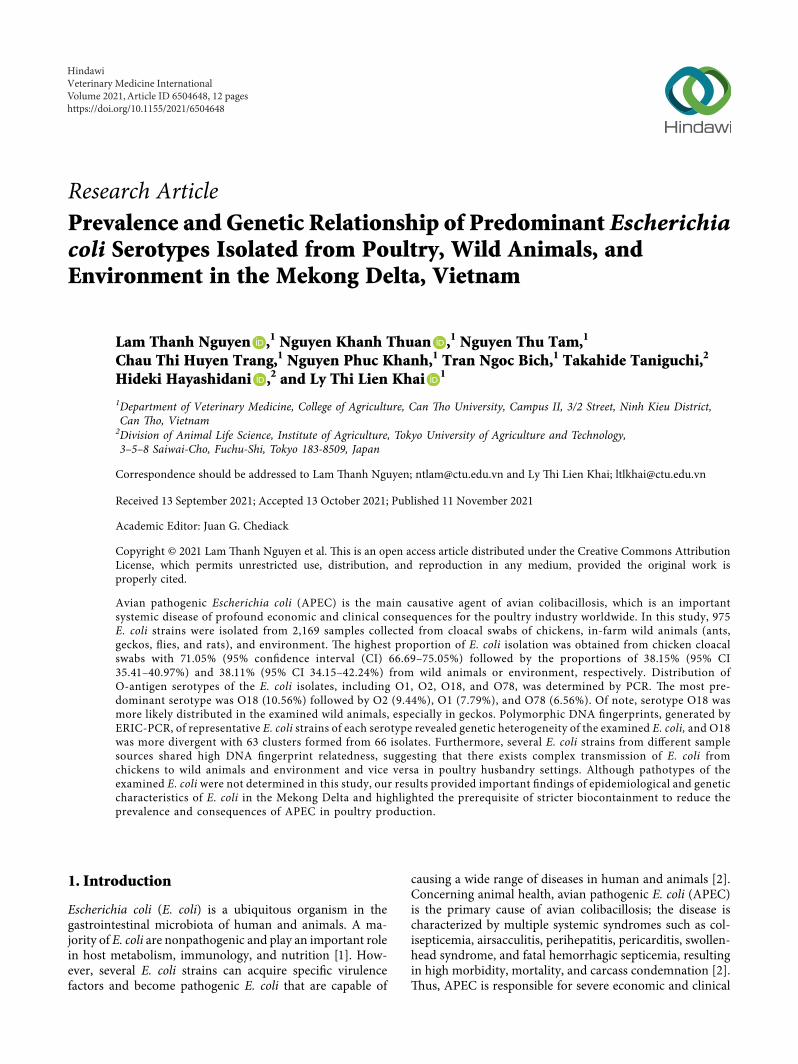
3.1. Prevalence of E. coli. A total of 975 E. coli strains wereisolated from 2,169 samples collected from cloacal swabs ofchickens, in-farm wild animals (ants, geckos, flies, and rats),and environment. �e proportions and the CIs of E. coliisolates per sample are presented in Figure 1. E. coli wasisolated from 319 of 449 (71.05% (95% CI 66.69–75.05%))cloacal swab samples of chickens, which was significantlyhigher than the number of E. coli either isolated from wildanimals 446 of 1,169 (38.15% (95% CI 35.41–40.97%)) andenvironment 210 of 551 (38.11% (95% CI 34.15–42.24%)),respectively (Figure 1(a)). Within the studied wild animals,the number of E. coli isolated from rats was 35 of 52 (67.30%(95% CI 53.76–78.48%)) that was the highest proportion incomparison with geckos 278 of 620 (44.84% (95% CI40.97–48.77%)), flies 108 of 297 (36.36% (95% CI31.10–41.98%)), and ants 25 of 200 (12.50% (95% CI8.61–17.80%)), respectively (Figure 1(b)).
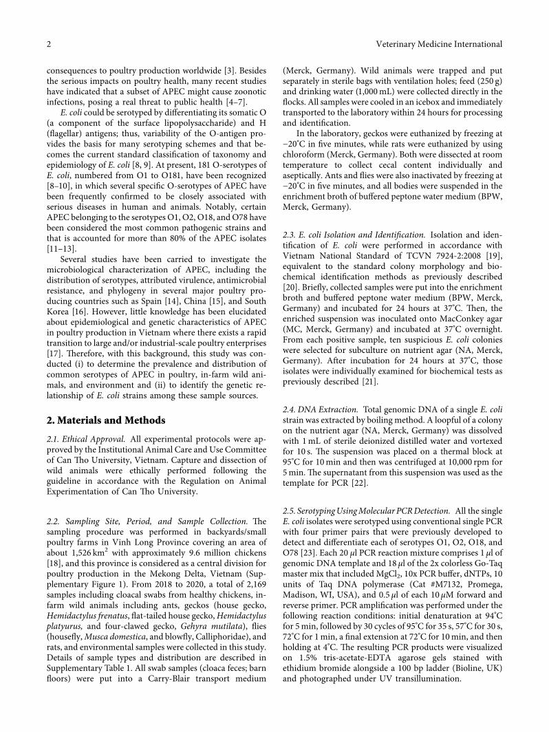
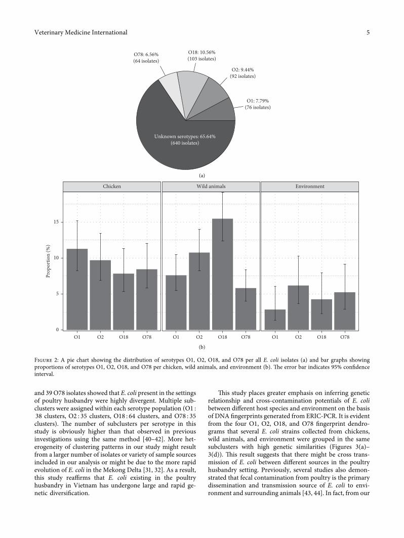
3.2. Distribution of Serotypes O1, O2, O18, and O78 in E. coliIsolates. All 975 E. coli strains were serotyped with O1, O2,O18, and O78 using conventional PCR (SupplementaryFigure 2). �e distribution of the examined O-serotypes isshown in Figure 2. Proportions of E. coli belonging to theserotypes O1, O2, O18, and O78 were 7.79%, 9.44%, 10.56%,and 6.56%, respectively. A large remaining proportion(65.64%) of E. coli was unknown serotypes (Figure 2(a)). Inchickens, the distribution of O1, O2, O18, and O78 E. coliwas almost equivalent with the proportions of 11.29% (95%CI 8.26–15.22%), 9.72% (95% CI 6.93–13.46%), 7.84% (95%CI 5.36–11.31%), and 8.46% (95% CI 5.88–12.03%), re-spectively. Similarly, the presence of these four serotypes wascomparable in the environment samples with proportionsranging from 2.86% (O1) to 6.19% (O2) (Figure 2(b)).Remarkably, O18 E. coli was accounted for a relatively largeproportion of 15.47% (95% CI 12.41–19.12%) of E. coliisolated from wild animals, and the majority of the O18E. coli were isolated from in-farm geckos (46.39%), 45 O18E. coli isolates from geckos out of total 97 O18 E. coli isolatesin all collected samples (data not shown).
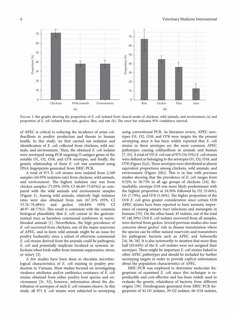
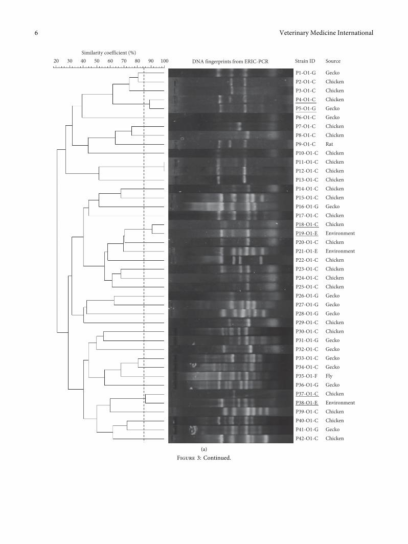
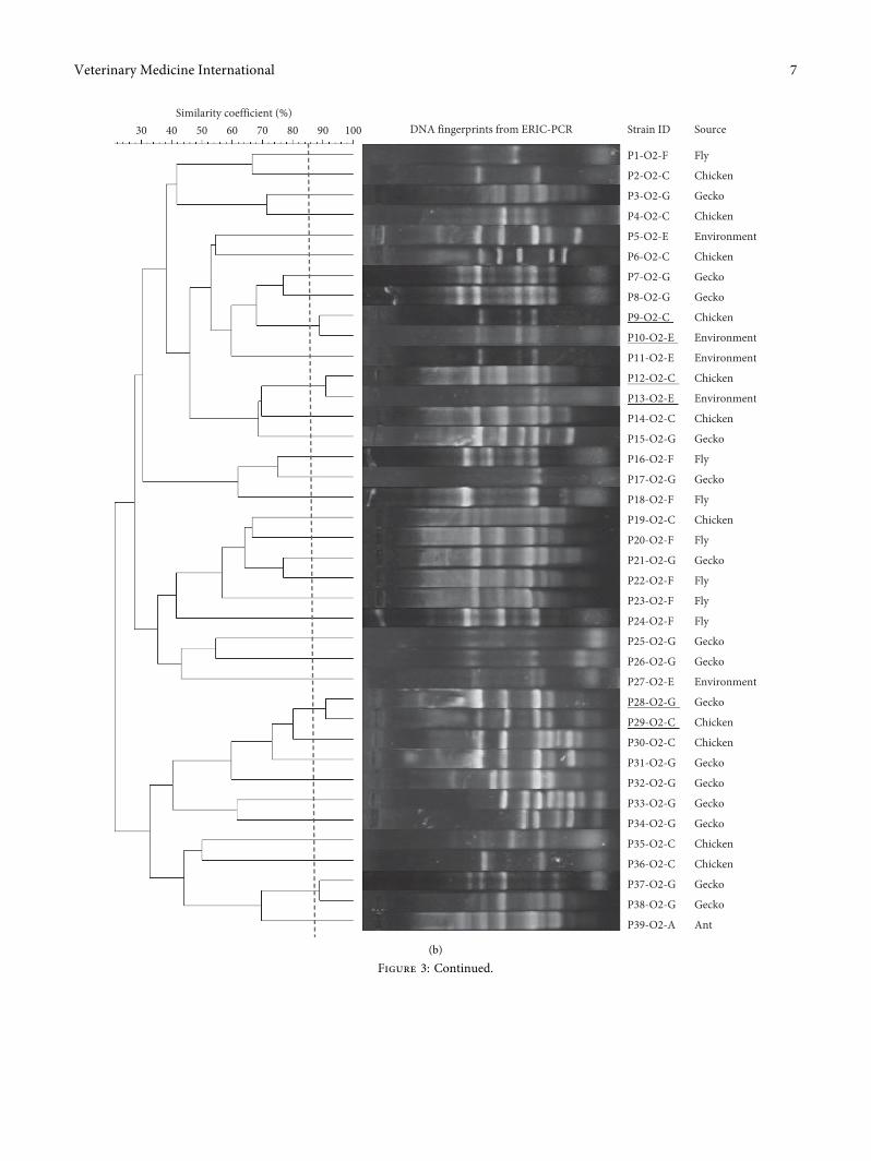
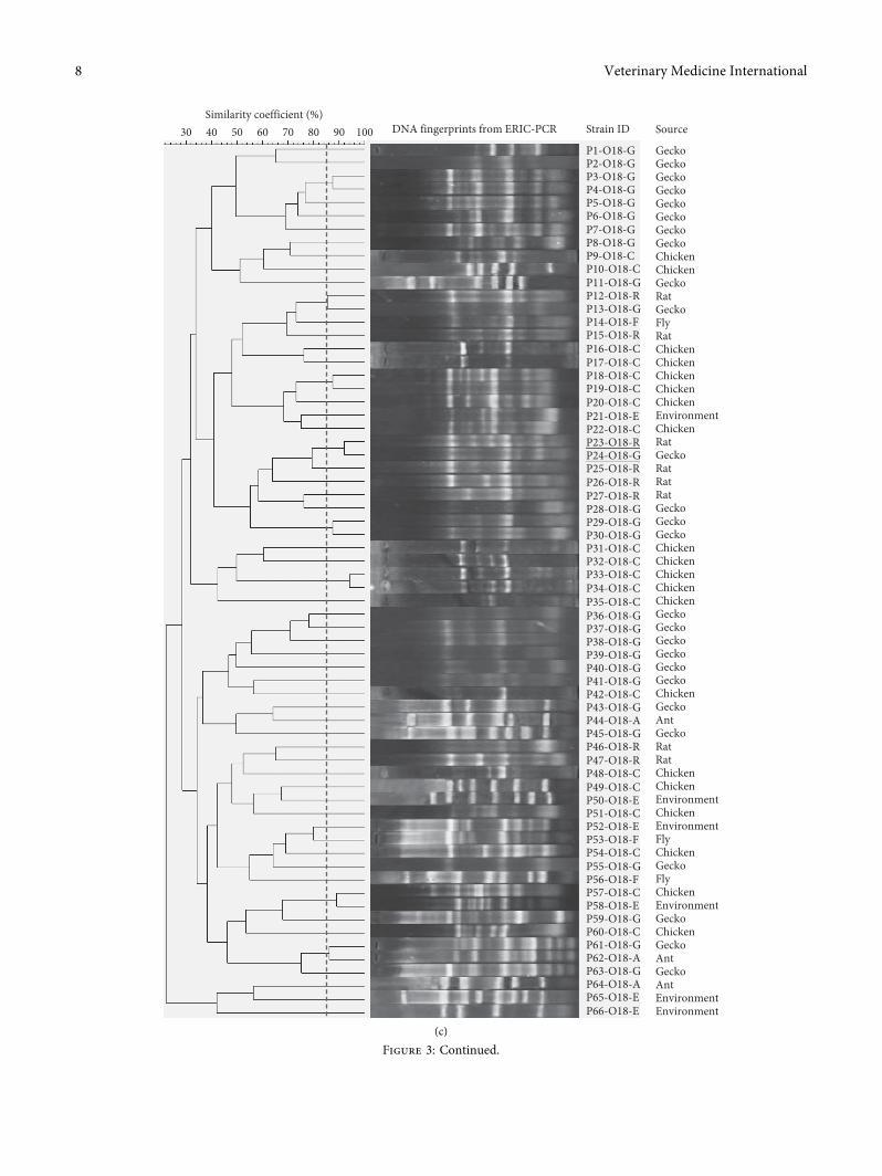
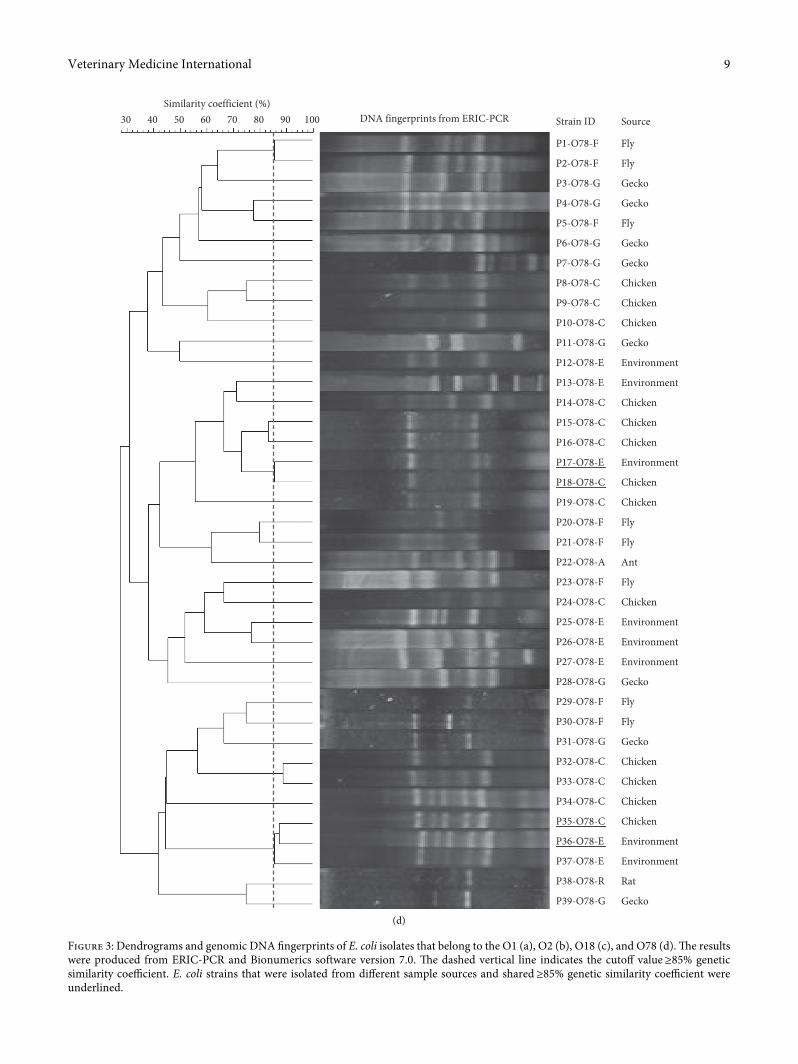
3.3. Genomic DNAFingerprinting andGenetic Relationship ofO1, O2, O18, and O78 E. coli. Representative E. coli isolatesof serotypes O1 (42 isolates), O2 (39 isolates), O18 (66isolates), and O78 (39 isolates) were selected based onsample types and space-time sources for ERIC-PCR toidentify genetic divergence and relationship among theE. coli isolates. Four dendrograms generated from genomicfingerprinting of E. coli isolates belonging to the examinedserotypes were, respectively, described in Figures 3(a)–3(d).�ese results showed that DNA fingerprinting patterns ofE. coli were heterogeneous, which was characterized byallocation of multiple genetic subclusters per serotype: O1(38 clusters out of 42 isolates), O2 (35 clusters out of 39isolates), O18 (64 clusters out of 66), and O78 (35 clustersout of 39 isolates) and all these subclusters were grouped intotwo (O78) to three (O1, O2, and O18) major clusters.Importantly, within the same serotype, several E. coli strainsisolated from different sample sources shared high geneticsimilarity (≥85%) and grouped together into distinct sub-clusters, for example, O1 serotypes (P4–O1–C/P5–O1-G,P18–O1–C/P19–O1-E, and P37–O1–C/P38–O1-E); O2 se-rotype (P9–O2–C/P10–O2-E, P12–O2–C/P13–O2-E, andP28–O2-G/P29–O2–C), O18 serotype (P23–O18-R/P24–O18-G), and O78 serotype (P17–O78-E/P18–O78–Cand P35–O78–C/P36–O78-E).
4. Discussion
Avian pathogenic E. coli is the major causative agent of aviancolibacillosis that is considered one of the most importantbacterial diseases in the global poultry industry due to itssubstantial economic impacts [2, 30]. In addition to causingserious consequences on poultry health and production,several APEC strains have been determined as potentialzoonotic pathogens [4–7]. �erefore, comprehensiveknowledge of biological and epidemiological characteristics
Veterinary Medicine International 3
of APEC is critical to reducing the incidence of avian col-ibacillosis in poultry production and threats to humanhealth. In this study, we first carried out isolation andidentification of E. coli collected from chickens, wild ani-mals, and environment. �en, the obtained E. coli isolateswere serotyped using PCR targeting O-antigen genes of thenotable O1, O2, O18, and O78 serotypes, and finally, thegenetic relationship of these E. coli was examined usingDNA fingerprints generated from ERIC-PCR.
A total of 975 E. coli strains were isolated from 2,169samples (44.95% isolation rate) from chickens, wild animals,and environment. �e highest isolation rate was fromchicken samples (71.05% (95% CI 66.69–75.05%)) as com-pared with the wild animals and environment samples(Figure 1). Among wild animals, relatively high isolationrates were also obtained from rats (67.31% (95% CI53.76–78.48%)) and geckos (44.84% (95% CI40.97–48.77%)). �is result is consistent with the commonbiological plausibility that E. coli coexist in the gastroin-testinal tract as harmless commensal symbionts in warm-blooded animals [1]. Nevertheless, the high prevalence ofE. coli recovered from chickens, one of the major reservoirsof APEC, and in-farm wild animals might be an issue forpoultry husbandry since a subset of otherwise commensalE. coli strains derived from the animals could be pathogenicE. coli and potentially implicate localized or systemic in-fections when birds suffer from immune suppression, stress,or injury [2].
A few studies have been done to elucidate microbio-logical characteristics of E. coli existing in poultry pro-duction in Vietnam. Most studies focused on investigatingvirulence attributes and/or antibiotics resistance of E. colistrains obtained from either poultry host species and en-vironment [31, 32]; however, information about the dis-tribution of serotypes of such E. coli remains elusive. In thisstudy, all 975 E. coli strains were subjected to serotyping
using conventional PCR. As literature review, APEC sero-types O1, O2, O18, and O78 were targets for the presentserotyping since it has been widely reported that E. colistrains in these serotypes are the most common APECpathotypes causing colibacillosis in animals and human[7, 33]. A total of 335 E. coli out of 975 (34.35%) E. coli strainswere defined as belonging to the serotypes O1, O2, O18, andO78 (Figure 2(a)).�ese serotypes were distributed at almostequivalent proportions among chickens, wild animals, andenvironment (Figure 2(b)). �is is in line with previousstudies showing that the prevalence of E. coli ranges from9.52% to 36.73% in all age groups of chickens [34]. Re-markably, serotype O18 was more likely predominant withthe highest proportion at 10.56% followed by O2 (9.44%),O1 (7.79%), and O78 (5.56%). �e higher proportion of theO18 E. coli gives greater consideration since certain O18APEC strains have been reported to have zoonotic impor-tance of causing urinary tract infections and meningitis inhumans [35]. On the other hand, 45 isolates, out of the total97 (46.39%) O18 E. coli isolates recovered from all samples,were derived from geckos. Several previous studies also raiseconcerns about geckos’ role in disease transmission wherethe species can be either natural reservoirs and transmittersfor pathogenic bacteria such as APEC and Salmonella[16, 36–38]. It is also noteworthy to mention that more thanhalf (65.64%) of the E. coli isolates were not assigned theirserotypes. �ere might be important E. coli strains linked toother APEC pathotypes and should be included for furtherserotyping targets in order to provide explicit informationabout the population characteristics of APEC.
ERIC-PCR was employed to determine molecular fin-gerprints of examined E. coli since this technique is re-producible and cost-effective and has been widely used toevaluate the genetic relatedness of bacteria from differentorigins [39]. Dendrograms generated from ERIC-PCR fin-gerprints of 42 O1 isolates, 39 O2 isolates, 66 O18 isolates,
0
10
20
30
40
50
60
70Pr
opor
tion
(%)
Wild animals EnvironmentChickens
(a)
0
10
20
30
40
50
60
70
Prop
ortio
n (%
)
Geckos Flies AntsRats
(b)
Figure 1: Bar graphs showing the proportion of E. coli isolated from cloacal swabs of chickens, wild animals, and environment (a) andproportion of E. coli isolated from ants, geckos, flies, and rats (b). �e error bar indicates 95% confidence interval.
4 Veterinary Medicine International
and 39 O78 isolates showed that E. coli present in the settingsof poultry husbandry were highly divergent. Multiple sub-clusters were assigned within each serotype population (O1 :38 clusters, O2 : 35 clusters, O18 : 64 clusters, and O78 : 35clusters). �e number of subclusters per serotype in thisstudy is obviously higher than that observed in previousinvestigations using the same method [40–42]. More het-erogeneity of clustering patterns in our study might resultfrom a larger number of isolates or variety of sample sourcesincluded in our analysis or might be due to the more rapidevolution of E. coli in the Mekong Delta [31, 32]. As a result,this study reaffirms that E. coli existing in the poultryhusbandry in Vietnam has undergone large and rapid ge-netic diversification.
�is study places greater emphasis on inferring geneticrelationship and cross-contamination potentials of E. colibetween different host species and environment on the basisof DNA fingerprints generated from ERIC-PCR. It is evidentfrom the four O1, O2, O18, and O78 fingerprint dendro-grams that several E. coli strains collected from chickens,wild animals, and environment were grouped in the samesubclusters with high genetic similarities (Figures 3(a)–3(d)). �is result suggests that there might be cross trans-mission of E. coli between different sources in the poultryhusbandry setting. Previously, several studies also demon-strated that fecal contamination from poultry is the primarydissemination and transmission source of E. coli to envi-ronment and surrounding animals [43, 44]. In fact, from our
O1: 7.79%(76 isolates)
O2: 9.44%(92 isolates)
O18: 10.56%(103 isolates)
O78: 6.56%(64 isolates)
Unknown serotypes: 65.64%(640 isolates)
(a)
Chicken Wild animals Environment
0
5
10
15
Prop
ortio
n (%
)
O2 O18 O78O1 O2 O18 O78O1 O2 O18 O78O1
(b)
Figure 2: A pie chart showing the distribution of serotypes O1, O2, O18, and O78 per all E. coli isolates (a) and bar graphs showingproportions of serotypes O1, O2, O18, and O78 per chicken, wild animals, and environment (b). �e error bar indicates 95% confidenceinterval.
Figure 3: Dendrograms and genomic DNA fingerprints of E. coli isolates that belong to the O1 (a), O2 (b), O18 (c), and O78 (d).�e resultswere produced from ERIC-PCR and Bionumerics software version 7.0. �e dashed vertical line indicates the cutoff value≥85% geneticsimilarity coefficient. E. coli strains that were isolated from different sample sources and shared≥85% genetic similarity coefficient wereunderlined.
Veterinary Medicine International 9
empirical observation, almost all small/backyard farms intheMekong Delta have relatively poor hygiene practices, andsuch wild animals as geckos, rats, flies, and ants commonlyexist in poultry farms. �us, cross transmission is almostcertain. �is study suggests that monitoring E. coli as well asAPEC contamination in environment and in-farms wildanimals could be considered as an indicator of interspeciesbarrier transposition in poultry production. In addition, theobserved high prevalence of E. coli belonging to the O1, O2,O18, and O78 serotypes from wild animals in poultry farmsis also concerning since these species also play importantroles in food-borne disease transmission but they are usuallyneglected in routine surveillance programs [36, 38].
�is study successfully examined in part the populationstructure of E. coli with the four common APEC serotypesand provided genetic evidence for cross transmission ofthese E. coli serotypes between different sources in thepoultry husbandry. However, there are some inevitablelimitations in the present study; for example, virulence,pathotypes, and antibiotics resistance characteristics of theobtained E. coli were not examined or a large proportion ofE. coli was not serotyped. Nevertheless, this study addedsignificant information on biological characteristics andepidemiological dynamics of E. coli which is necessary forbetter control of avian colibacillosis and prevention of thethreat of zoonotic diseases caused by APEC.
5. Conclusion
E. coli was predominantly isolated from small/backyardpoultry farms. Serotypes O1, O2, O18, and O78 wereaccounted for a high proportion of the E. coli population andO18 was the predominant serotype among the examinedE. coli. Chickens, in-farms geckos, and rats were identified asthe major sources of dissemination and transmission ofE. coli. Polymorphic DNA fingerprint analyses revealed thatAPEC from different sources of poultry husbandry exhibitlarge genetic heterogeneity and closed genetic relationship.�erefore, this study provides important information aboutepidemiological and genetic characteristics of E. coli andemphasizes the necessity of stricter biocontainment inpoultry production for prevention and control of aviancolibacillosis caused by APEC.
Data Availability
All data generated or analyzed during this study are includedin this published article and its supplementary informationfiles.
Conflicts of Interest
�e authors declare no conflicts of interest.
Authors’ Contributions
Lam �anh Nguyen and Nguyen Khanh �uan contributedequally to this work.
Acknowledgments
�is study was funded in part by the Can �o UniversityImprovement Project VN14-P6 and supported by the JapaneseODA Loan.
Supplementary Materials
Supplementary Table 1. Summary of sample types and theirdistribution. Supplementary Figure 1. Geographical mapshowing the location of sampling site: Vinh Long Province(black) is situated in the center of the Mekong Delta (grey)on the Vietnam map. Supplementary Figure 2. �e PCRproducts amplified using the allele-specific genes of O1 (3-A), O2 (3-B), O18 (3-C), and O78 (3-D) serotypes. Lane M:100 bp DNA ladder; lane P: positive control; lane N: negativecontrol (distilled water as template); lane 1 to lane 6: DNAsamples of E. coli tested. Arrows indicate sizes of amplifiedDNA fragments as follows: 263 bp (O1), 355 bp (O2), 459 bp(O18), and 623 bp (O78). . (Supplementary Materials)
References
[1] O. Tenaillon, D. Skurnik, B. Picard, and E. Denamur, “�epopulation genetics of commensal Escherichia coli,” NatureReviews Microbiology, vol. 8, no. 3, pp. 207–217, 2010.
[2] J. B. Kaper, J. P. Nataro, and H. L. T. Mobley, “PathogenicEscherichia coli,” Nature Reviews Microbiology, vol. 2, no. 2,pp. 123–140, 2004.
[3] L. K. Nolan, J. P. Vaillancourt, N. L. Barbieri, and C.M. Logue,“Colibacillosis,” Diseases of Poultry, 14th Edition, Chapter 18,pp. 770–830, John Wiley & Sons, Inc., Hoboken, NJ, USA,2020.
[4] K. E. Rodriguez-Siek, C. W. Giddings, C. Doetkott,T. J. Johnson, M. K. Fakhr, and L. K. Nolan, “Comparison ofEscherichia coli isolates implicated in human urinary tractinfection and avian colibacillosis,” Microbiology (Reading),vol. 151, no. 6, pp. 2097–2110, 2005.
[5] M. Moulin-Schouleur, M. Reperant, S. Laurent et al.,“Extraintestinal pathogenic Escherichia coli strains of avianand human origin: link between phylogenetic relationshipsand common virulence patterns,” Journal of Clinical Micro-biology, vol. 45, no. 10, pp. 3366–3376, 2007.
[6] M. Mellata, “Human and avian extraintestinal PathogenicE-scherichia coli: infections, zoonotic risks, and antibiotic re-sistance trends,” Foodborne Pathogens and Disease, vol. 10,no. 11, pp. 916–932, 2013.
[7] D. Kathayat, D. Lokesh, S. Ranjit, and G. Rajashekara, “Avianpathogenic Escherichia coli (APEC): an overview of virulenceand pathogenesis factors, zoonotic potential, and controlstrategies,” Pathogens, vol. 10, no. 4, p. 467, 2021.
[8] F. Kauffmann, “�e serology of the coli group,”�e Journal ofImmunology, vol. 57, pp. 71–100, 1947.
[9] B. Liu, A. Furevi, A. V. Perepelov et al., “Structure and ge-netics of Escherichia coli O antigens,” FEMS MicrobiologyReviews, vol. 44, no. 6, pp. 655–683, 2019.
[10] F. Scheutz, T. Cheasty, D. Woodward, and H. R. Smith,“Designation of O174 and O175 to temporary O groups OX3and OX7, and six new E. coli O groups that include Ver-ocytotoxin-producing E. coli (VTEC): O176, O177, O178,O179, O180 and O181,” Apmis, vol. 112, no. 9, pp. 569–584,2004.
[11] C. Ewers, G. Li, H. Wilking et al., “Avian pathogenic, uro-pathogenic, and newborn meningitis-causing Escherichia coli:how closely related are they?” International Journal of MedicalMicrobiology, vol. 297, no. 3, pp. 163–176, 2007.
[12] S. Huja, Y. Oren, E. Trost et al., “Genomic avenue to aviancolisepticemia,” MBio, vol. 6, 2015.
[13] J. W. Mehat, A. H. M. van Vliet, and R. M. La Ragione, “�eavian pathogenic Escherichia coli (APEC) pathotype iscomprised of multiple distinct, independent genotypes,”Avian Pathology, vol. 50, no. 5, pp. 402–416, 2021.
[14] J. E. Blanco, M. Blanco, A. Mora et al., “Serotypes ofEscherichia coli isolated from septicaemic chickens in Galicia(northwest Spain),” Veterinary Microbiology, vol. 61, no. 3,pp. 229–235, 1998.
[15] X. M. Wang, X.-P. Liao, W.-J. Zhang et al., “Prevalence ofserogroups, virulence genotypes, antimicrobial resistance, andphylogenetic background of avian PathogenicEscherichiacoliin South of China,” Foodborne Pathogens and Disease,vol. 7, no. 9, pp. 1099–1106, 2010.
[16] Y. B. Kim, M. Y. Yoon, J. S. Ha et al., “Molecular charac-terization of avian pathogenic Escherichia coli from broilerchickens with colibacillosis,” Poultry Science, vol. 99, no. 2,pp. 1088–1095, 2020.
[17] J.-D. Cesaro, H. Nguyen, and D. Guillaume, Atlas of livestocktransitions in Vietnam (1986–2016), IPSARD-CIRAD, Hanoi,65p. ISBN 978-2-87614-746-1, 2020.
[18] GSO, Statistical Yearbook of Vietnam. 2020, General Statis-tical Office of Vietnam, Hanoi, Vietnam, 2020.
[19] Vietnam National Standard (TCVN), Microbiology of Foodand Animal Feeding Stuffs-Horizontal Method for the Enu-meration of β-glucuronidase-positive Escherichia coli-Part 2:Colony-count Technique at 44°C Using 5-Bromo-4-Chloro-3-Indolyl β-D-glucuronide, Vietnam National Standard, Viet-nam, 2008.
[20] S. T. Cowan, Cowan and Steel’s Manual for the Identificationof Medical Bacteria, Cambridge University Press, Cambridge,UK, 2004.
[21] T. L. K. Ly, T. P. Tran, T. T. Nguyen, and T. Iwata, “Prevalenceof Escherichia coli O157 from cattle and foods in the Mekongdelta, Vietnam,” Journal of Veterinary Medical Science, vol. 13,pp. 107–113, 2009.
[22] T. T. Hong To, H. Yanagawa, N. Khanh �uan et al.,“Prevalence of Vibrio parahaemolyticus causing acute hep-atopancreatic necrosis disease of shrimp in shrimp,Molluscanshellfish and water samples in the Mekong Delta, Vietnam,”Biology (Basel), vol. 9, p. 312, 2020.
[23] S. Wang, Q. Meng, J. Dai et al., “Development of an allele-specific PCR assay for simultaneous sero-typing of avianpathogenic Escherichia coli predominant O1, O2, O18 andO78 strains,” PLoS One, vol. 9, Article ID e96904, 2014.
[24] J. Versalovic, T. Koeuth, and J. R. Lupski, “Distribution ofrepetitive DNA sequences in eubacteria and application tofingerprinting of bacterial genomes,” Nucleic Acids Research,vol. 19, pp. 6823–6831, 1991.
[25] E. A. Casarez, S. D. Pillai, and G. D. Di Giovanni, “Genotypediversity of Escherichia coli isolates in natural waters deter-mined by PFGE and ERIC-PCR,” Water Research, vol. 41,pp. 3643–3648, 2007.
[26] R Core Team, R: A Language and Environment for StatisticalComputing, R Foundation for Statistical Computing, Vienna,Austria, 2013.
[27] H. Wickham, Ggplot2: Elegant Graphics for Data Analysis,Springer, Berlin, Germany, 2016.
[28] S. Dorai-Raj, “Binom: Binomial Confidence Intervals forSeveral Parameterizations,” R package version, vol. 1, pp. 1–1,2014.
[29] M. Stevenson, M. M. Stevenson, and I. BiasedUrn, Package‘epiR’, 2021.
[30] R. Guabiraba and C. Schouler, “Avian colibacillosis: still manyblack holes,” FEMSMicrobiology Letters, vol. 362, no. 15, 2015.
[31] V. T. Nguyen, J. J. Carrique-Mas, T. H. Ngo et al., “Prevalenceand risk factors for carriage of antimicrobial-resistantEscherichia coli on household and small-scale chicken farmsin the Mekong Delta of Vietnam,” Journal of AntimicrobialChemotherapy, vol. 70, pp. 2144–2152, 2015.
[32] N. Nhung, N. Cuong, J. Campbell et al., “High levels ofantimicrobial resistance among Escherichia coli isolates fromlivestock farms and synanthropic rats and shrews in theMekong Delta of Vietnam,” Antimicrobial Agents and Che-motherapy, vol. 81, pp. 812–820, 2015.
[33] M. Dho-Moulin and J. M. Fairbrother, “Avian pathogenicEscherichia coli (APEC),” Veterinary Research, vol. 30,pp. 299–316, 1999.
[34] S. M. Lutful Kabir, “Avian colibacillosis and salmonellosis: acloser look at epidemiology, pathogenesis, diagnosis, controland public health concerns,” International Journal of Envi-ronmental Research and Public Health, vol. 7, pp. 89–114,2010.
[35] M.Moulin-Schouleur, C. Schouler, P. Tailliez et al., “Commonvirulence factors and genetic relationships between O18: K1:H7 Escherichia coli isolates of human and avian origin,”Journal of Clinical Microbiology, vol. 44, pp. 3484–3492, 2006.
[36] B. J. Morrison and J. E. Rubin, “Detection of multidrug-re-sistant Gram-negative bacteria from imported reptile andamphibian meats,” Journal of Applied Microbiology, vol. 129,pp. 1053–1061, 2020.
[37] C. Marin, L. Lorenzo-Rebenaque, O. Laso, J. Villora-Gon-zalez, and S. Vega, “Pet reptiles: a potential source oftransmission of multidrug-resistant Salmonella,” Frontiers inVeterinary Science, vol. 7, 2021.
[38] K. T. Nguyen, M. Hasegawa, T. M. T. Vo et al., “Wild geckosconsidered as the natural reservoir of Salmonella Weltevredenin Southeast Asian countries,” Zoonoses and Public Health,vol. 68, no. 7, pp. 815–822, 2021.
[39] K. J. Meacham, L. Zhang, B. Foxman, R. J. Bauer, andC. F. Marrs, “Evaluation of genotyping large numbers ofEscherichia coli isolates by enterobacterial repetitive inter-genic consensus-PCR,” Journal of Clinical Microbiology,vol. 41, pp. 5224–5226, 2003.
[40] B. R. Mohapatra, K. Broersma, and A. Mazumder, “Com-parison of five rep-PCR genomic fingerprinting methods fordifferentiation of fecal Escherichia coli from humans, poultryand wild birds,” FEMS Microbiology Letters, vol. 277,pp. 98–106, 2007.
[41] A. Hussain, S. Shaik, A. Ranjan et al., “Risk of transmission ofantimicrobial resistant Escherichia coli from commercialbroiler and free-range retail chicken in India,” Frontiers inMicrobiology, vol. 8, 2017.
[42] M. S. Sekhar, N. M. Sharif, T. S. Rao, and M. Metta, “Gen-otyping of virulent Escherichia coli obtained from poultry andpoultry farm workers using enterobacterial repetitive inter-genic consensus-polymerase chain reaction,” VeterinaryWorld, vol. 10, pp. 1292–1296, 2017.
[43] H. Duan, T. Chai, Y. Cai, Z. Zhong, M. Yao, and X. Zhang,“Transmission identification of Escherichia coli aerosol inchicken houses to their environments using ERIC-PCR,”
Veterinary Medicine International 11
Science in China-Series C: Life Sciences, vol. 51, pp. 164–173,2008.
[44] H. Laube, A. Friese, C. von Salviati, B. Guerra, and U. Rosler,“Transmission of ESBL/AmpC-producing Escherichia colifrom broiler chicken farms to surrounding areas,” VeterinaryMicrobiology, vol. 172, pp. 519–527, 2014.