CLASSIFICATION ARTICLEpublished: 08 November 2011
doi: 10.3389/fimmu.2011.00054
Primary immunodeficiency diseases: an update on theclassification from the International Union ofImmunological Societies Expert Committee for PrimaryImmunodeficiencyWaleed Al-Herz 1,2, Aziz Bousfiha3, Jean-Laurent Casanova4,5, Helen Chapel 6, Mary Ellen Conley 7,8*,
Charlotte Cunningham-Rundles9, Amos Etzioni 10, Alain Fischer 11, Jose Luis Franco12, Raif S. Geha13,
Lennart Hammarström14, Shigeaki Nonoyama15, Luigi Daniele Notarangelo13,16*, Hans Dieter Ochs17,
Jennifer M. Puck 18, Chaim M. Roifman19, Reinhard Seger 20 and Mimi L. K.Tang21,22,23
1 Department of Pediatrics, Kuwait University, Kuwait City, Kuwait2 Allergy and Clinical Immunology Unit, Department of Pediatrics, Al-Sabah Hospital, Kuwait City, Kuwait3 Clinical Immunology Unit, Casablanca Children Hospital Ibn Rochd Medical School, King Hassan II University, Casablanca, Morocco4 St. Giles Laboratory of Human Genetics of Infectious Diseases, Rockefeller Branch, The Rockefeller University, New York, NY, USA5 Laboratory of Human Genetics of Infectious Diseases, Necker Branch, Necker Medical School, University Paris Descartes and INSERM U980, Paris, France6 Clinical Immunology Unit, Nuffield Department of Medicine, University of Oxford, Oxford, UK7 Department of Pediatrics, University of Tennessee College of Medicine, Memphis, TN, USA8 Department of Immunology, St. Jude Children’s Research Hospital, Memphis, TN, USA9 Department of Medicine and Pediatrics, Mount Sinai School of Medicine, New York, NY, USA10 Meyer’s Children Hospital – Technion, Haifa, Israel11 Pediatric Hematology-Immunology Unit, Hôpital Necker Enfants-Malades, Assistance Publique–Hôpital de Paris, Necker Medical School, Paris Descartes
University, Paris, France12 Group of Primary Immunodeficiencies, University of Antioquia, Medellín, Colombia13 Division of Immunology, Children’s Hospital Boston, Boston, MA, USA14 Division of Clinical Immunology, Department of Laboratory Medicine, Karolinska Institute at Karolinska University Hospital Huddinge, Stockholm, Sweden15 Department of Pediatrics, National Defense Medical College, Saitama, Japan16 The Manton Center for Orphan Disease Research, Children’s Hospital Boston, Boston, MA, USA17 Department of Pediatrics, University of Washington and Seattle Children’s Research Institute, Seattle, WA, USA18 Department of Pediatrics, University of California San Francisco and UCSF Benioff Children’s Hospital, San Francisco, CA, USA19 Division of Immunology and Allergy, Department of Pediatrics, The Hospital for Sick Children and the University of Toronto, Toronto, ON, Canada20 Division of Immunology, University Children’s Hospital, Zürich, Switzerland21 Department of Allergy and Immunology, Royal Children’s Hospital Melbourne, Melbourne, VIC, Australia22 Murdoch Childrens Research Institute, Melbourne, Melbourne, VIC, Australia23 Department of Paediatrics, University of Melbourne, Melbourne, VIC, Australia
Edited by:
Eric Meffre, Yale University School ofMedicine, USA
Reviewed by:
Eric Meffre, Yale University School ofMedicine, USA
*Correspondence:
Mary Ellen Conley , Department ofPediatrics, University of TennesseeCollege of Medicine, Memphis, TN,USA; Department of Immunology MS351, St. Jude Children’s ResearchHospital, 262 Danny Thomas Place,Memphis, TN 38105, USA.e-mail: [email protected];
Luigi Daniele Notarangelo, Division ofImmunology and The Manton Centerfor Orphan Disease Research,Children’s Hospital Boston, KarpResearch Building, Room 20217, 1Blackfan Circle, Boston, MA 02115,USA.e-mail: [email protected]
We report the updated classification of primary immunodeficiency diseases, compiled bythe ad hoc Expert Committee of the International Union of Immunological Societies. Ascompared to the previous edition, more than 15 novel disease entities have been addedin the updated version. For each disorders, the key clinical and laboratory features are pro-vided. This updated classification is meant to help in the diagnostic approach to patientswith these diseases.
Keywords: primary immunodeficiency diseases
www.frontiersin.org November 2011 | Volume 2 | Article 54 | 1
Al-Herz et al. IUIS classification of primary immunodeficiencies
The International Union of Immunological Societies (IUIS)Expert Committee on Primary Immunodeficiency met in NewYork City, May 31–June 1, 2011 to update the classification ofhuman primary immunodeficiencies (PIDs). Novel developmentsin gene discovery and increased knowledge in the mechanisms thatgovern immune system development and function have resultedin the identification of several novel PIDs in the last 2 years.
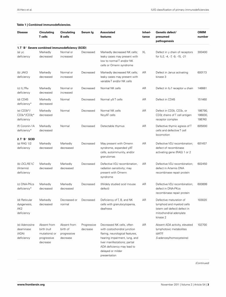
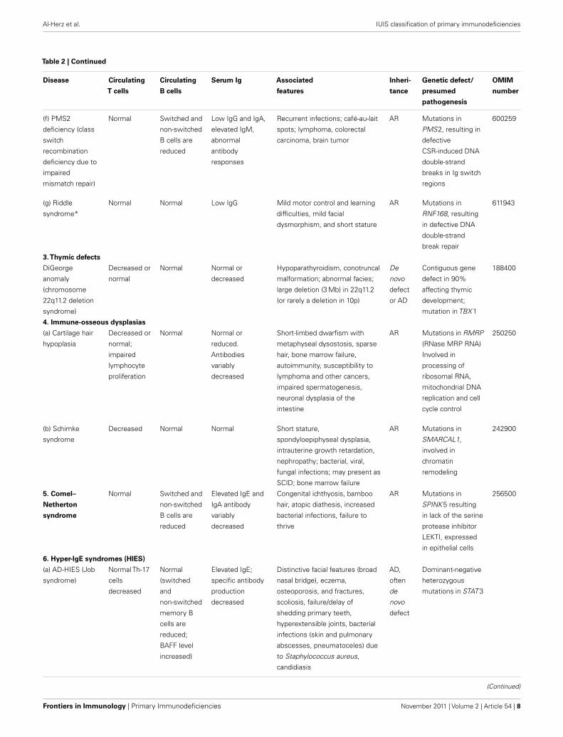
The classification of primary immunodeficiencies (PIDs) pro-vides a framework to help in the diagnostic approach to patients.As in recent classifications, eight major groups of PIDs have beenincluded in the Tables; however the order of the Tables has beenchanged with Table 2 now describing the “Well-defined syn-dromes with immunodeficiency” (previously Table 3) to reflectthe immunological similarity between the disorders included inthis Table and those in Table 1, “Combined immunodeficien-cies.”
Any classification of human disorders is somewhat arbitrary,and the classification of PIDs is no exception. Some disordersmight well belong to more than one group. CD40 ligand deficiency,for example, is reported both in Tables 1 and 3 (“Predominantlyantibody deficiencies”), to reflect the facts that failed B cell isotypeswitching was historically the most prominent feature of this con-dition (originally named Hyper-IgM syndrome) and that somepatients survive into adulthood without significant opportunisticinfections and do well with only immunoglobulin replacementtherapy. Explanatory notes provided after each Table offer addi-tional information (particularly where a condition appears inmore than one Table) and indicate which new disorders have beenadded to that Table.
Although this updated classification reports on the most typ-ical immunological findings and associated clinical and genetic
features for the various PIDs, there is extensive clinical, immuno-logical, and molecular heterogeneity that can not be easily recapit-ulated in a brief summary. To facilitate a more rigorous analysis ofeach disease,a column has been added on the right with a hyperlinkto refer to its catalog number in the Online Mendelian Inheritancein Man (OMIM) publicly accessible database (www.omim.org) ofhuman genetic disorders. It is suggested that the reader consultthis regularly updated and fully referenced resource.
The prevalence of the various PIDs varies in different coun-tries. For this reason, in this new classification, we have electedto avoid giving a comment on the relative frequency of PID dis-orders. However, an asterisk has been placed in the first column,after the disease name, to identify disorders for which fewer than10 unrelated cases have been reported in the literature. Some ofthese forms of PID can be considered extremely rare. Others haveonly recently been identified and it may be that more patients willbe detected over time.
Finally, it is increasingly recognized that different mutationsin the same gene may result in different phenotypes and maybe associated with different patterns of inheritance. This conceptof clinical, immunological, and genetic heterogeneity is assumingforemost importance. Notes in the text or in the footnotes identifysuch heterogeneity, when known.
The scope of the IUIS Expert Committee on Primary Immun-odeficiency is to increase awareness, facilitate recognition, andpromote optimal treatment for patients with Primary Immunod-eficiency disorders worldwide. For this reason, in addition to peri-odically revising the Classification of PIDs, the Expert Committeeis also actively involved in the development of diagnostic criteriaand in providing, upon request, advice with regard to therapeuticguidelines.
Frontiers in Immunology | Primary Immunodeficiencies November 2011 | Volume 2 | Article 54 | 2
Al-Herz et al. IUIS classification of primary immunodeficiencies
Table 1 | Combined immunodeficiencies.
Disease Circulating
T cells
Circulating
B cells
Serum Ig Associated
features
Inheri-
tance
Genetic defect/
presumed
pathogenesis
OMIM
number
1.T−B+ Severe combined immunodeficiency (SCID)
(a) γc
deficiency
Markedly
decreased
Normal or
increased
Decreased Markedly decreased NK cells;
leaky cases may present with
low to normal T and/or NK
cells or Omenn syndrome
XL Defect in γ chain of receptors
for IL-2, -4, -7, -9, -15, -21
300400
(b) JAK3
deficiency
Markedly
decreased
Normal or
increased
Decreased Markedly decreased NK cells;
leaky cases may present with
variable T and/or NK cells
AR Defect in Janus activating
kinase 3
600173
(c) IL7Rα
deficiency
Markedly
decreased
Normal or
increased
Decreased Normal NK cells AR Defect in IL-7 receptor α chain 146661
(d) CD45
deficiency*
Markedly
decreased
Normal Decreased Normal γ/δ T cells AR Defect in CD45 151460
(e) CD3δ*/
CD3ε*/CD3ζ*
deficiency
Markedly
decreased
Normal Decreased Normal NK cells
Noγ/δT cells
AR Defect in CD3δ, CD3ε, or
CD3ζ chains of T cell antigen
receptor complex
186790,
186830,
186740
(f) Coronin-1A
deficiency*
Markedly
decreased
Normal Decreased Detectable thymus AR Defective thymic egress of T
cells and defective T cell
locomotion
605000
2.T−B− SCID
(a) RAG 1/2
deficiency
Markedly
decreased
Markedly
decreased
Decreased May present with Omenn
syndrome, expanded γ/δT
cells, autoimmunity, and/or
granulomas
AR Defective VDJ recombination;
defect of recombinase
activating gene (RAG) 1 or 2
601457
(b) DCLRE1C
(Artemis)
deficiency
Markedly
decreased
Markedly
decreased
Decreased Defective VDJ recombination,
radiation sensitivity; may
present with Omenn
syndrome
AR Defective VDJ recombination;
defect in Artemis DNA
recombinase repair protein
602450
(c) DNA-PKcs
deficiency*
Markedly
decreased
Markedly
decreased
Decreased (Widely studied scid mouse
defect)
AR Defective VDJ recombination;
defect in DNA-PKcs
recombinase repair protein
600899
(d) Reticular
dysgenesis,
AK2
deficiency
Markedly
decreased
Decreased or
normal
Decreased Deficiency of T, B, and NK
cells with granulocytopenia,
deafness
AR Defective maturation of
lymphoid and myeloid cells
(stem cell defect) defect in
mitochondrial adenylate
kinase 2
103020
(e) Adenosine
deaminase
(ADA)
deficiency
Absent from
birth (null
mutations) or
progressive
decrease
Absent from
birth of
progressive
decrease
Progressive
decrease
Decreased NK cells, often
with costochondral junction
flaring, neurological features,
hearing impairment, lung, and
liver manifestations; partial
ADA deficiency may lead to
delayed or milder
presentation
AR Absent ADA activity, elevated
lymphotoxic metabolites
(dATP,
S-adenosylhomocysteine)
102700
(Continued)
www.frontiersin.org November 2011 | Volume 2 | Article 54 | 3
Al-Herz et al. IUIS classification of primary immunodeficiencies
Table 1 | Continued
Disease Circulating
T cells
Circulating
B cells
Serum Ig Associated
features
Inheri-
tance
Genetic defect/
presumed
pathogenesis
OMIM
number
3. Omenn
syndrome
Present;
restricted
heterogene-
ity
Normal or
decreased
Decreased,
except
increased
IgE
Erythroderma, eosinophilia,
adenopathies,
hepatosplenomegaly
AR Hypomorphic mutations in
RAG1/2, Artemis, IL7Rα,
RMRP, ADA, DNA Ligase IV,
γc, or associated with
DiGeorge syndrome; some
cases have no defined gene
mutation
603554
4. DNA ligase
IV deficiency
Decreased Decreased Decreased Microcephaly, facial
dysmorphisms, radiation
sensitivity; may present with
Omenn syndrome or with a
delayed clinical onset
AR DNA ligase IV defect,
impaired non-homologous
end joining (NHEJ)
601837
5. Cernunnos/
NHEJ1
deficiency*
Decreased Decreased Decreased Microcephaly, in utero growth
retardation, radiation
sensitivity
AR Cernunnos (NHEJ1) defect,
impaired non-homologous
end joining
611291
6. CD40
ligand
deficiency
Normal; may
progressively
decrease
IgM+ and
IgD+ B cells
present,
other
isotypes
absent
IgM
increased
or normal,
other
isotypes
decreased
Neutropenia,
thrombocytopenia; hemolytic
anemia, biliary tract, and liver
disease, opportunistic
infections
XL Defects in CD40 ligand
(CD40L) cause defective
isotype switching and
impaired dendritic cell
signaling
300386
7. CD40
deficiency*
Normal IgM+ and
IgD+ B cells
present,
other
isotypes
absent
IgM
increased
or normal,
other
isotypes
decreased
Neutropenia, gastrointestinal,
and liver/biliary tract disease,
opportunistic infections
AR Defects in CD40 cause
defective isotype switching
and impaired dendritic cell
signaling
109535
8. Purine
nucleoside
phosphory-
lase (PNP)
deficiency
Progressive
decrease
Normal Normal or
decreased
Autoimmune hemolytic
anemia, neurological
impairment
AR Absent PNP, T cell and
neurologic defects from
elevated toxic metabolites,
especially dGTP
164050
9. CD3γ
deficiency*
Normal, but
reduced TCR
expression
Normal Normal AR Defect in CD3 γ 186740
10. CD8
deficiency*
Absent CD8,
normal CD4
cells
Normal Normal AR Defects of CD8 α chain 186910
11. ZAP-70
deficiency
Decreased
CD8, normal
CD4 cells
Normal Normal AR Defects in ZAP-70 signaling
kinase
176947
12. Ca++ channel deficiency
(a) ORAI-I
deficiency*
Normal
number, but
defective
TCR-
mediated
activation
Normal Normal Autoimmunity, anhydrotic
ectodermic dysplasia,
non-progressive myopathy
AR Defect in ORAI-1, a Ca++
release-activated channel
(CRAC) modulatory
component
610277
(Continued)
Frontiers in Immunology | Primary Immunodeficiencies November 2011 | Volume 2 | Article 54 | 4
Al-Herz et al. IUIS classification of primary immunodeficiencies
Table 1 | Continued
Disease Circulating
T cells
Circulating
B cells
Serum Ig Associated
features
Inheri-
tance
Genetic defect/
presumed
pathogenesis
OMIM
number
(b) STIM-1
deficiency*
Normal
number, but
defective
TCR-
mediated
activation
Normal Normal Autoimmunity, anhydrotic
ectodermic dysplasia,
non-progressive myopathy
AR Defect in STIM-1, a stromal
interaction molecule
Ca++sensor
605921
13. MHC
class I
deficiency
Decreased
CD8, normal
CD4
Normal Normal Vasculitis AR Mutations in TAP1, TAP2 or
TAPBP (tapasin) genes giving
MHC class I deficiency
604571
14. MHC
class II
deficiency
Normal
number,
decreased
CD4 cells
Normal Normal or
decreased
Failure to thrive, diarrhea,
respiratory tract infections
AR Mutation in transcription
factors for MHC class II
proteins (CIITA, RFX 5,
RFXAP, RFXANK genes)
209920
15. Winged
helix
deficiency
(nude)*
Markedly
decreased
Normal Decreased Alopecia, abnormal thymic
epithelium, impaired T cell
maturation (widely studied
nude mouse defect)
AR Defects in forkhead box N1
transcription factor encoded
by FOXN1, the gene mutated
in nude mice
600838
16. Complete
DiGeorge
syndrome
Profoundly
decreased
Low to
normal
Decreased Lymphoproliferation
(lymphadenopathy,
hepatosplenomegaly),
autoimmunity (may resemble
IPEX syndrome), impaired T
cell proliferation
AD Deletion of chromosome
22q11.2 or in a minority of
cases other chromosomal
regions, including 10p;
heterozygous defects in
transcription factor TBX1
188400
17. Cartilage
hair
hypoplasia
Decreased or
normal;
impaired
lymphocyte
proliferation
Normal Normal or
reduced.
Antibodies
variably
decreased
Short-limbed dwarfism with
metaphyseal dysostosis,
sparse hair, bone marrow
failure, autoimmunity,
susceptibility to lymphoma
and other cancers, impaired
spermatogenesis, neuronal
dysplasia of the intestine
AR Mutations in RMRP (RNase
MRP RNA) Involved in
processing of ribosomal RNA,
mitochondrial DNA replication
and cell cycle control
250250
18. IKAROS
deficiency*
Normal, but
impaired
lymphocyte
proliferation
Absent Presumably
decreased
Anemia, neutropenia,
thrombocytopenia
AD de
novo
Mutation in IKAROS, a
hematopoietic specific
zinc-finger protein and a
central regulator of lymphoid
differentiation
19. STAT5b
deficiency*
Modestly
decreased
Normal Normal Growth-hormone insensitive
dwarfism, dysmorphic
features, eczema,
lymphocytic interstitial
pneumonitis, autoimmunity
AR Defects of STAT5b, impaired
development and function of
γδT cells, Treg, and NK cells,
impaired T cell proliferation
604260
20. ITK
deficiency*
Modestly
decreased
Normal Normal or
decreased
AR Defects in ITK, EBV
associated
lymphoproliferation
613011
21. MAGT1
deficiency*
Decreased
CD4 cells
Normal Normal EBV infection, lymphoma;
viral infections, respiratory
and GI infections
XL Mutations in MAGT1,
impaired Mg++ flux leading to
impaired TCR signaling
300715
(Continued)
www.frontiersin.org November 2011 | Volume 2 | Article 54 | 5
Al-Herz et al. IUIS classification of primary immunodeficiencies
Table 1 | Continued
Disease Circulating
T cells
Circulating
B cells
Serum Ig Associated
features
Inheri-
tance
Genetic defect/
presumed
pathogenesis
OMIM
number
22. DOCK8
deficiency
Decreased Decreased Low IgM,
increased
IgE
Low NK cells,
hypereosinophilia, recurrent
infections; severe atopy,
extensive cutaneous viral, and
bacterial (staph.) infections,
susceptibility to cancer
AR Defect in DOCK 8 243700
XL, X-linked inheritance; AR, autosomal recessive inheritance; AD, autosomal dominant inheritance; SCID, severe combined immune deficiency; EBV, Epstein Barr
virus; Ca++, calcium; MHC, major histocompatibility complex.
*Ten or fewer unrelated cases reported in the literature.
Three disorders have been added toTable 1: DOCK8 deficiency, IKAROS deficiency, and MAGT1 deficiency.
Infants with SCID who have maternal T cells engraftment may have T cells that do not function normally; these cells may cause autoimmune cytopenias or graft
versus host disease. Hypomorphic mutations in several of the genes that cause SCID may result in Omenn syndrome (OS), or “leaky” SCID. Both of these disorders
can be associated with higher numbers of T cells and reduced rather than absent activation responses when compared with typical SCID caused by null mutations.
A spectrum of clinical findings including typical SCID, OS, leaky SCID, and granulomas with T lymphopenia can be found in patients with RAG gene defects. RAC2
deficiency is a disorder of leukocyte motility and is reported inTable 5; however, one patient with RAC2 deficiency was found to have absent T cell receptor excision
circles (TRECs) by newborn screening, but T cell numbers and mitogen responses were not impaired. For additional syndromic conditions with T cell lymphopenia,
such as DNA repair defects, cartilage hair hypoplasia, IKAROS deficiency, and NEMO syndrome, see Tables 2 and 6; however, it should be noted that individuals
with the most severe manifestations of these disorders could have clinical signs and symptoms of SCID. Severe folate deficiency (such as with malabsorption due to
defects in folate carrier or transporter genes SLC10A1 or PCFT) and some metabolic disorders, such as methylmalonicaciduria, may present with reversible profound
lymphopenia in addition to their characteristic presenting features.
Frontiers in Immunology | Primary Immunodeficiencies November 2011 | Volume 2 | Article 54 | 6
Al-Herz et al. IUIS classification of primary immunodeficiencies
Table 2 | Well-defined syndromes with immunodeficiency.
Disease Circulating
T cells
Circulating
B cells
Serum Ig Associated
features
Inheri-
tance
Genetic defect/
presumed
pathogenesis
OMIM
number
1.Wiskott–Aldrich
syndrome (WAS)
Progressive
decrease,
abnormal
lymphocyte
responses to
anti-CD3
Normal Decreased IgM:
antibody to
polysaccharides
particularly
decreased; often
increased IgA and
IgE
Thrombocytopenia with small
platelets; eczema; lymphoma;
autoimmune disease; IgA
nephropathy; bacterial and viral
infections. XL thrombocytopenia
is a mild form of WAS, and XL
neutropenia is caused by
missense mutations in the
GTPase binding domain of WASP
XL Mutations in WAS;
cytoskeletal and
immunologic
synapse defect
affecting
hematopoietic
stem cell
derivatives
301000
2. DNA repair defects (other than those inTable 1)
(a) Ataxia–
telangiectasia
Progressive
decrease
Normal Often decreased
IgA, IgE, and IgG
subclasses;
increased IgM
monomers;
antibodies
variably
decreased
Ataxia; telangiectasia; pulmonary
infections; lymphoreticular and
other malignancies; increased
alpha fetoprotein and X-ray
sensitivity; chromosomal
instability
AR Mutations in ATM ;
disorder of cell
cycle checkpoint
and DNA double
strand break repair
208900
(b) Ataxia–
telangiectasia-like
disease (ATLD)*
Progressive
decrease
Normal Antibodies
variably
decreased
Moderate ataxia; pulmonary
infections; severely increased
radiosensitivity
AR Hypomorphic
mutations in
MRE11; disorder of
cell cycle
checkpoint and
DNA double-
strand break repair
604391
(c) Nijmegen
breakage
syndrome
Progressive
decrease
Variably
reduced
Often decreased
IgA, IgE, and IgG
subclasses;
increased IgM;
antibodies
variably
decreased
Microcephaly; bird like face;
lymphomas; solid tumors;
ionizing radiation sensitivity;
chromosomal instability
AR Hypomorphic
mutations in NBS1
(Nibrin); disorder of
cell cycle
checkpoint and
DNA double-strand
break repair
251260
(d) Bloom
syndrome
Normal Normal Reduced Short stature; bird like face;
sun-sensitive erythema; marrow
failure; leukemia; lymphoma;
chromosomal instability
AR Mutations in BLM ;
RecQ like helicase
210900
(e) Immunodefi-
ciency with
centromeric
instability and
facial anomalies
(ICF)
Decreased or
normal;
Responses
to PHA may
be decreased
Decreased or
normal
Hypogamma-
globulinemia;
variable antibody
deficiency
Facial dysmorphic features;
macroglossia;
bacterial/opportunistic infections;
malabsorption; cytopenias;
malignancies; multiradial
configurations of chromosomes
1, 9, 16; no DNA breaks
AR Mutations in DNA
methyltransferase
DNMT 3B (ICF1)
resulting in
defective DNA
methylation; or in
ZBTB24 (ICF2)
242860
(Continued)
www.frontiersin.org November 2011 | Volume 2 | Article 54 | 7
Al-Herz et al. IUIS classification of primary immunodeficiencies
Table 2 | Continued
Disease Circulating
T cells
Circulating
B cells
Serum Ig Associated
features
Inheri-
tance
Genetic defect/
presumed
pathogenesis
OMIM
number
(f) PMS2
deficiency (class
switch
recombination
deficiency due to
impaired
mismatch repair)
Normal Switched and
non-switched
B cells are
reduced
Low IgG and IgA,
elevated IgM,
abnormal
antibody
responses
Recurrent infections; café-au-lait
spots; lymphoma, colorectal
carcinoma, brain tumor
AR Mutations in
PMS2, resulting in
defective
CSR-induced DNA
double-strand
breaks in Ig switch
regions
600259
(g) Riddle
syndrome*
Normal Normal Low IgG Mild motor control and learning
difficulties, mild facial
dysmorphism, and short stature
AR Mutations in
RNF168, resulting
in defective DNA
double-strand
break repair
611943
3.Thymic defects
DiGeorge
anomaly
(chromosome
22q11.2 deletion
syndrome)
Decreased or
normal
Normal Normal or
decreased
Hypoparathyroidism, conotruncal
malformation; abnormal facies;
large deletion (3 Mb) in 22q11.2
(or rarely a deletion in 10p)
De
novo
defect
or AD
Contiguous gene
defect in 90%
affecting thymic
development;
mutation in TBX 1
188400
4. Immune-osseous dysplasias
(a) Cartilage hair
hypoplasia
Decreased or
normal;
impaired
lymphocyte
proliferation
Normal Normal or
reduced.
Antibodies
variably
decreased
Short-limbed dwarfism with
metaphyseal dysostosis, sparse
hair, bone marrow failure,
autoimmunity, susceptibility to
lymphoma and other cancers,
impaired spermatogenesis,
neuronal dysplasia of the
intestine
AR Mutations in RMRP
(RNase MRP RNA)
Involved in
processing of
ribosomal RNA,
mitochondrial DNA
replication and cell
cycle control
250250
(b) Schimke
syndrome
Decreased Normal Normal Short stature,
spondyloepiphyseal dysplasia,
intrauterine growth retardation,
nephropathy; bacterial, viral,
fungal infections; may present as
SCID; bone marrow failure
AR Mutations in
SMARCAL1,
involved in
chromatin
remodeling
242900
5. Comel–
Netherton
syndrome
Normal Switched and
non-switched
B cells are
reduced
Elevated IgE and
IgA antibody
variably
decreased
Congenital ichthyosis, bamboo
hair, atopic diathesis, increased
bacterial infections, failure to
thrive
AR Mutations in
SPINK 5 resulting
in lack of the serine
protease inhibitor
LEKTI, expressed
in epithelial cells
256500
6. Hyper-IgE syndromes (HIES)
(a) AD-HIES (Job
syndrome)
Normal Th-17
cells
decreased
Normal
(switched
and
non-switched
memory B
cells are
reduced;
BAFF level
increased)
Elevated IgE;
specific antibody
production
decreased
Distinctive facial features (broad
nasal bridge), eczema,
osteoporosis, and fractures,
scoliosis, failure/delay of
shedding primary teeth,
hyperextensible joints, bacterial
infections (skin and pulmonary
abscesses, pneumatoceles) due
to Staphylococcus aureus,
candidiasis
AD,
often
de
novo
defect
Dominant-negative
heterozygous
mutations in STAT 3
(Continued)
Frontiers in Immunology | Primary Immunodeficiencies November 2011 | Volume 2 | Article 54 | 8
Al-Herz et al. IUIS classification of primary immunodeficiencies
Table 2 | Continued
Disease Circulating
T cells
Circulating
B cells
Serum Ig Associated
features
Inheri-
tance
Genetic defect/
presumed
pathogenesis
OMIM
number
(b) AR-HIES No skeletal and connective
tissue abnormalities; no
pneumatoceles
AR
(i) TYK2
deficiency*
Normal, but
multiple
cytokine
signaling
defect
Normal (±) Elevated IgE Susceptibility to intracellular
bacteria (mycobacteria,
Salmonella), fungi, and viruses
Mutation in TYK 2 611521
(ii) DOCK8
deficiency
Reduced Reduced (±) Elevated IgE,
low IgM
Recurrent respiratory infections;
extensive cutaneous viral and
staphylococcal infections,
increased risk of cancer, severe
atopy with anaphylaxis
Mutation in
DOCK 8
611432
(iii) Unknown
origin
Normal Normal Elevated IgE CNS hemorrhage, fungal, and
viral infections
Unknown
7. Hepatic
veno-occlusive
disease with
immunodefi-
ciency
(VODI)
Normal
(decreased
memory T
cells)
Normal
(decreased
memory B
cells)
Decreased IgG,
IgA, IgM absent
germinal centers
absent tissue
plasma cells
Hepatic veno-occlusive disease;
Pneumocystis jiroveci
pneumonia; susceptibility to
CMV, candida;
thrombocytopenia;
hepatosplenomegaly
AR Mutations in SP110 235550
8. Dyskeratosis congenita (DKC)
(a) XL-DKC
(Hoyeraal-
Hreidarsson
syndrome)
Progressive
decrease
Progressive
decrease
Variable Intrauterine growth retardation,
microcephaly, nail dystrophy,
recurrent infections, digestive
tract involvement, pancytopenia,
reduced number and function of
NK cells
XL Mutations in
dyskerin (DKC1)
305000
(b) AR-DKC* Abnormal Variable Variable Pancytopenia, sparse scalp hair
and eyelashes, prominent
periorbital telangiectasia, and
hypoplastic/dysplastic nails
AR Mutation in NOLA2
(NHP2) or in
NOLA3 (NOP10)
224230
(c) AD-DKC Variable Variable Variable Reticular hyperpigmentation of
the skin, dystrophic nails,
osteoporosis, premalignant
leukokeratosis of the mouth
mucosa, palmar hyperkeratosis,
anemia, pancytopenia
AD Mutation in TERC 127550
Mutation in TERT
Mutation in TINF2
(Continued)
www.frontiersin.org November 2011 | Volume 2 | Article 54 | 9
Al-Herz et al. IUIS classification of primary immunodeficiencies
Table 2 | Continued
Disease Circulating
T cells
Circulating
B cells
Serum Ig Associated
features
Inheri-
tance
Genetic defect/
presumed
pathogenesis
OMIM
number
9. IKAROS
deficiency*
Normal, but
impaired
lymphocyte
proliferation
Absent Presumably
decreased
Anemia, neutropenia,
thrombocytopenia
AD de
novo
Mutation in
IKAROS, a
hematopoietic
specific zinc-finger
protein and a
central regulator of
lymphoid
differentiation
SCID, severe combined immune deficiency; XL, X-linked inheritance; AR, autosomal recessive inheritance; AD, autosomal dominant inheritance; MSMD, Mendelian
susceptibility of mycobacterial disease.
*Ten or fewer unrelated cases reported in the literature.
Four disorders listed inTable 2, complete DiGeorge anomaly, cartilage hair hypoplasia, IKAROS deficiency, and AR-HIES caused by DOCK8 deficiency, are also included
inTable 1 as they are characterized by striking T and B cell abnormalities. While not all DOCK8 deficient patients have elevated serum IgE, most have recurrent viral
infections and malignancies as a result of combined immunodeficiency. AR-HIES due to TYK2 deficiency is also described inTable 6, because of its association with
atypical mycobacterial disease resulting in MSMD. Because Riddle syndrome is caused by mutations in a gene involved in DNA double-strand break repair and is
associated with hypogammaglobulinemia, we have added this rare syndrome to Table 2. Chronic mucocutaneous candidiasis (CMC) has been moved to Table 6.
Autosomal dominant and autosomal recessive forms of Dyskeratosis congenita, caused by mutations of recently identified genes, have been included in this table.
Finally, we added IKAROS deficiency, observed in a single case, a prematurely born infant, who died at the age of 87 days. He had absent B and NK cells and
non-functional T cells, suggesting combined immunodeficiency.
Frontiers in Immunology | Primary Immunodeficiencies November 2011 | Volume 2 | Article 54 | 10
Al-Herz et al. IUIS classification of primary immunodeficiencies
Table 3 | Predominantly antibody deficiencies.
Disease Serum Ig Associated features Inheritance Genetic defect/
presumed
pathogenesis
OMIM
number
1. Severe reduction in all serum immunoglobulin isotypes with profoundly decreased or absent B cells
(a) BTK deficiency All isotypes decreased in
majority of patients; some
patients have detectable
immunoglobulins
Severe bacterial infections;
normal numbers of pro-B cells
XL Mutations in BTK, a
cytoplasmic tyrosine
kinase activated by
crosslinking of the BCR
300300
(b) μ Heavy chain
deficiency
All isotypes decreased Severe bacterial infections;
normal numbers of pro-B cells
AR Mutations in μ heavy
chain
147020
(c) λ5 deficiency* All isotypes decreased Severe bacterial infections;
normal numbers of pro-B cells
AR Mutations in λ5; part of
the surrogate light chain
in the pre-BCR
146770
(d) Igα deficiency* All isotypes decreased Severe bacterial infections;
normal numbers of pro-B cells
AR Mutations in Igα
(CD79a); part of the
pre-BCR and BCR
112205
(e) Igβ deficiency* All isotypes decreased Severe bacterial infections;
normal numbers of pro-B cells
AR Mutations in Igβ
(CD79b); part of the
pre-BCR and BCR
147245
(f) BLNK deficiency* All isotypes decreased Severe bacterial infections;
normal numbers of pro-B cells
AR Mutations in BLNK ; a
scaffold protein that
binds to BTK
604615
(g) Thymoma with
immunodeficiency
One or more isotypes may
be decreased
Bacterial and opportunistic
infections; autoimmunity;
decreased number of pro-B cells
None Unknown
(h) Myelodysplasia with
hypogammaglobuline-
mia
One or more isotypes may
be decreased
Infections; decreased number of
pro-B cells
Variable May have monosomy 7,
trisomy 8, or
dyskeratosis congenita
2. Severe reduction in at least 2 serum immunoglobulin isotypes with normal or low number of B cells
(a) Common variable
immunodeficiency
disorders
Low IgG and IgA and/or
IgM
Clinical phenotypes vary: most
have recurrent infections, some
have polyclonal
lymphoproliferation,
autoimmune cytopenias, and/or
granulomatous disease
Variable Unknown
(b) ICOS deficiency* Low IgG and IgA and/or
IgM
AR Mutations in ICOS 604558
(c) CD19 deficiency* Low IgG and IgA and/or
IgM
May have glomerulonephritis AR Mutations in CD19;
transmembrane protein
that amplifies signal
through BCR
107265
(d) CD81 deficiency* Low IgG, low or normal
IgA, and IgM
May have glomerulonephritis AR Mutations in CD81;
transmembrane protein
that amplifies signal
through BCR
186845
(e) CD20 deficiency* Low IgG, normal or
elevated IgM, and IgA
AR Mutations in CD20 112210
(Continued)
www.frontiersin.org November 2011 | Volume 2 | Article 54 | 11
Al-Herz et al. IUIS classification of primary immunodeficiencies
Table 3 | Continued
Disease Serum Ig Associated features Inheritance Genetic defect/
presumed
pathogenesis
OMIM
number
(f) TACI deficiency Low IgG and IgA and/or
IgM
Variable clinical expression AD or AR or
complex
Mutations in
TNFRSF13B (TACI)
604907
(g) BAFF receptor
deficiency*
Low IgG and IgM Variable clinical expression AR Mutations in
TNFRSF13C (BAFF-R)
606269
3. Severe reduction in serum IgG and IgA with normal/elevated IgM and normal numbers of B cells
(a) CD40L deficiency IgG and IgA decreased;
IgM may be normal or
increased; B cell numbers
may be normal or
increased
Opportunistic infections,
neutropenia, autoimmune
disease
XL Mutations in CD40LG
(also called TNFSF5 or
CD154)
300386
(b) CD40 deficiency* Low IgG and IgA; normal
or raised IgM
Opportunistic infections,
neutropenia, autoimmune
disease
AR Mutations in CD40
(also called TNFRSF5)
109535
(c) AID deficiency IgG and IgA decreased;
IgM increased
Enlarged lymph nodes and
germinal centers
AR Mutations in AICDA
gene
605257
(d) UNG deficiency IgG and IgA decreased;
IgM increased
Enlarged lymph nodes and
germinal centers
AR Mutations in UNG 191525
4. Isotype or light chain deficiencies with normal numbers of B cells
(a) Ig heavy chain
mutations and deletions
One or more IgG and/or
IgA subclasses as well as
IgE may be absent
May be asymptomatic AR Mutation or
chromosomal deletion
at 14q32
(b) κ chain deficiency* All immunoglobulins have
lambda light chain
Asymptomatic AR Mutations in κ constant
gene
147200
(c) Isolated IgG
subclass deficiency
Reduction in one or more
IgG subclass
Usually asymptomatic; a
minority may have poor antibody
response to specific antigens
and recurrent viral/bacterial
infections
Variable Unknown
(d) IgA with IgG
subclass deficiency
Reduced IgA with
decrease in one or more
IgG subclass
Recurrent bacterial infections in
majority
Variable Unknown
(e) Selective IgA
deficiency
IgA decreased/absent Usually asymptomatic; may have
recurrent infections with poor
antibody responses to
carbohydrate antigens; may have
allergies or autoimmune disease.
A very few cases progress to
CVID, others coexist with CVID
in the family
Variable Unknown
5. Specific antibody
deficiency with
normal Ig
concentrations and
normal numbers of
B cells
Normal Reduced ability to make
antibodies to specific antigens
Variable Unknown
(Continued)
Frontiers in Immunology | Primary Immunodeficiencies November 2011 | Volume 2 | Article 54 | 12
Al-Herz et al. IUIS classification of primary immunodeficiencies
Table 3 | Continued
Disease Serum Ig Associated features Inheritance Genetic defect/
presumed
pathogenesis
OMIM
number
6.Transient hypogam-
maglobulinemia of
infancy with normal
numbers of B cells
IgG and IgA decreased Normal ability to make
antibodies to vaccine antigens,
usually not associated with
significant infections
Variable Unknown
XL, X-linked inheritance; AR, autosomal recessive inheritance; AD, autosomal dominant inheritance; BTK, Bruton tyrosine kinase; BLNK, B cell linker protein; AID,
activation-induced cytidine deaminase; UNG, uracil-DNA glycosylase; ICOS, inducible costimulator; Ig(κ), immunoglobulin, or κ light chain type.
*Ten or fewer unrelated cases reported in the literature.
Two new autosomal recessive disorders that might previously have been called CVID have been added to Table 3. CD81 is normally co-expressed with CD19 on
the surface of B cells. Like CD19 mutations, mutations in CD81 result in normal numbers of peripheral blood B cells, low serum IgG, and an increased incidence of
glomerulonephritis. A single patient with a homozygous mutation in CD20 has been reported.
Common Variable Immunodeficiency Disorders (CVID) include several clinical and laboratory phenotypes that may be caused by distinct genetic and/or environ-
mental factors. Some patients with CVID and no known genetic defect have markedly reduced numbers of B cells as well as hypogammaglobulinemia. Alterations
in TNFRSF13B (TACI) and TNFRSF13C (BAFF-R) sequences may represent disease modifying mutations rather than disease causing mutations. CD40L and CD40
deficiency are included in Table 1 as well as this table. A small minority of patients with XLP (Table 4), WHIM syndrome (Table 6), ICF (Table 2), VOD1 (Table 2),
thymoma with immunodeficiency (Good syndrome) or myelodysplasia are first seen by an immunologist because of recurrent infections, hypogammaglobulinemia,
and normal or reduced numbers of B cells. Patients with GATA2 mutations (Table 5) may have markedly reduced numbers of B cells, as well as decreased monocytes
and NK cells and a predisposition to myelodysplasia but they do not have an antibody deficiency.
www.frontiersin.org November 2011 | Volume 2 | Article 54 | 13
Al-Herz et al. IUIS classification of primary immunodeficiencies
Table 4 | Diseases of immune dysregulation.
Disease Circulating
T cells
Circulating
B cells
Serum Ig Associated
features
Inheri-
tance
Genetic defect/
presumed
pathogenesis
OMIM
number
1. Immunodeficiency with hypopigmentation
(a) Chediak–
Higashi
syndrome
Normal Normal Normal Partial albinism, recurrent
infections, late-onset primary
encephalopathy, increased
lymphoma risk. Neutropenia,
Giant lysosomes, low NK, and
CTL activities, elevation of acute
phase markers
AR Mutations in LYST, impaired
lysosomal trafficking
214500
(b) Griscelli
syndrome,
type2
Normal Normal Normal Partial albinism, elevation of
acute phase markers,
encephalopathy in some
patients. Low NK and CTL
activities
AR Mutations in RAB27A
encoding a GTPase that
promotes docking of
secretory vesicles to the cell
membrane
607624
(c) Hermansky–
Pudlak
syndrome,
type 2*
Normal Normal Normal Partial albinism, increased
bleeding. Neutropenia, low NK,
and CTL activity
AR Mutations in the AP3B1
gene, encoding for the β
subunit of the AP-3 complex
608233
2. Familial hemophagocytic lymphohistiocytosis (FHL) syndromes
(a) Perforin
deficiency,
FHL2
Normal Normal Normal Severe inflammation, persistent
fever, cytopenias, splenomegaly.
Hemophagocytosis, decreased
to absent NK and CTL activities
AR Mutations in PRF1; perforin,
a major cytolytic protein
603553
(b) UNC13D
(Munc13-4)
deficiency,
FHL3
Normal Normal Normal Severe inflammation, persistent
fever, splenomegaly,
hemophagocytosis, decreased
NK and CTL activities
AR Mutations in UNC13D*
required to prime vesicles for
fusion (*as named in OMIM).
Note that also in OMIM the
“official” name is UNC13D
deficiency with the alternative
title of MUNC13D deficiency
608898
(c) Syntaxin 11
deficiency,
FHL4
Normal Normal Normal Severe inflammation, persistent
fever, splenomegaly.
Hemophagocytosis, decreased
to absent NK activity
AR Mutations in STX 11, required
for fusion of secretory
vesicles with the cell
membrane and release of
contents
603552
(d) STXBP2
(Munc 18-2)
deficiency,
FHL5
Normal Normal Normal or
low
Severe inflammation, fever,
splenomegaly,
hemophagocytosis possible
bowel disease. Decreased NK
and CTL activities with partial
restoration after IL-2 stimulation
AR Mutations in STXBP2,
required for fusion of
secretory vesicles with the
cell membrane and release of
contents
613101
(Continued)
Frontiers in Immunology | Primary Immunodeficiencies November 2011 | Volume 2 | Article 54 | 14
Al-Herz et al. IUIS classification of primary immunodeficiencies
Table 4 | Continued
Disease Circulating
T cells
Circulating
B cells
Serum Ig Associated
features
Inheri-
tance
Genetic defect/
presumed
pathogenesis
OMIM
number
3. Lymphoproliferative syndromes
(a) SH2D1A
deficiency, XLP1
Normal Normal or
reduced
Normal or
low
Clinical and immunologic
abnormalities triggered by
EBV infection, including
hepatitis, hemophagocytic
syndrome, aplastic anemia,
and lymphoma.
Dysgammaglobulinemia or
hypogammaglobulinemia, low
to absent NKT cells
XL Mutations in SH2D1A
encoding an adaptor protein
regulating intracellular signals
308240
(b) XIAP
deficiency, XLP2
Normal Normal or
reduced
Normal or
low
Clinical and immunologic
abnormalities triggered by
EBV infection, including
splenomegaly, hepatitis,
hemophagocytic syndrome
colitis
XL Mutations in XIAP encoding
an inhibitor of apoptosis
300635
4. Syndromes with autoimmunity
(a) Autoimmune
lymphoprolifera-
tive syndrome
(ALPS)
(i) ALPS-FAS Increased
CD4− CD8−
double
negative
(DN) T cells
Normal, but
increased
number of
CD5+ B cells
Normal or
increased
Splenomegaly, adenopathies,
autoimmune cytopenias,
increased lymphoma risk.
Defective lymphocyte
apoptosis
AD (AR
cases
are
rare
and
severe)
Mutations in TNFRSF6, cell
surface apoptosis receptor; in
addition to germline
mutations, somatic mutations
cause a similar phenotype
(ALPS-sFAS)
601859
(ii) ALPS-
FASLG
Increased
DN T cells
Normal Normal Splenomegaly, adenopathies,
autoimmune cytopenias, SLE
defective lymphocyte
apoptosis
AD AR Mutations in TNFSF6, ligand
for CD95 apoptosis receptor
134638
(iii) ALPS-
CASP10*
Increased
DN T cells
Normal Normal Adenopathies, splenomegaly,
autoimmunity. Defective
lymphocyte apoptosis
AD Mutations in CASP10,
intracellular apoptosis
pathway
603909
(iv) Caspase 8
defect*
Slightly
increased
DN T cells
Normal Normal or
decreased
Adenopathies, splenomegaly,
recurrent bacterial, and viral
infections. Defective
lymphocyte apoptosis and
activation,
hypogammaglobulinemia
AD Mutations in CASP8,
intracellular apoptosis and
activation pathways
607271
(v) Activating
N-RAS defect,
activating
K-RAS defect*
Increased or
normal DN T
cells
Elevation of
CD5 B cells
Normal Adenopathies, splenomegaly,
leukemia, lymphoma.
Defective lymphocyte
apoptosis following IL-2
withdrawal
Sporadic Somatic mutations in NRAS
encoding a GTP binding
protein with diverse signaling
functions; activating
mutations impair
mitochondrial apoptosis
164790
(Continued)
www.frontiersin.org November 2011 | Volume 2 | Article 54 | 15
Al-Herz et al. IUIS classification of primary immunodeficiencies
Table 4 | Continued
Disease Circulating
T cells
Circulating
B cells
Serum Ig Associated
features
Inheri-
tance
Genetic defect/
presumed
pathogenesis
OMIM
number
(vi) FADD
defi-
ciency*
Increased DN T
cells
Normal Normal Functional hyposplenism,
recurrent bacterial, and viral
infections, recurrent episodes
of encephalopathy and liver
dysfunction. Defective
lymphocyte apoptosis
AR Mutations in FADD encoding
an adaptor molecule
interacting with FAS, and
promoting apoptosis,
inflammation and innate
immunity
613759
(b) APECED
(APS-1),
autoimmune
polyendocrino-
pathy with
candidiasis
and
ectodermal
dystrophy
Normal Normal Normal Autoimmunity, particularly of
parathyroid, adrenal, and
other endocrine organs,
chronic candidiasis, dental
enamel hypoplasia, and other
abnormalities
AR Mutations in AIRE, encoding
a transcription regulator
needed to establish thymic
self-tolerance
240300
(c) IPEX,
immune dys-
regulation,
polyendo-
crinopathy,
enteropathy
(X-linked)
Lack of (and/or
impaired
function of)
CD4+ CD25+
FOXP3+
regulatory T
cells
Normal Elevated
IgA, IgE
Autoimmune enteropathy,
early onset diabetes,
thyroiditis hemolytic anemia,
thrombocytopenia, eczema
XL Mutations in FOXP3,
encoding a T cell transcription
factor
304790
(d) CD25
deficiency
Normal to
modestly
decreased
Normal Normal Lymphoproliferation,
autoimmunity. Impaired T cell
proliferation
AR Mutations in IL-2Rα chain 606367
(e) ITCH
deficiency*
Not assessed
(Th2 skewing in
Itch-deficient
mice)
Not
assessed (B
cells are
dysfunctional
in
Itch-deficient
mice)
Not
assessed
(elevated in
Itch-
deficient
mice)
Multi-organ autoimmunity,
chronic lung disease, failure
to thrive, developmental
delay, macrocephaly
AR Mutations in ITCH, an E3
ubiquitin ligase
613385
XL, X-linked inheritance; AR, autosomal recessive inheritance; AD, autosomal dominant inheritance; DN, double negative; SL, systemic lupus erythematosus.
*Ten or fewer unrelated cases reported in the literature.
STXBP2/Munc18-2 deficiency has been added as the cause of “FHL5,” a new form of FHL. Of note, “FHL1” has not yet received a genetic/molecular identification.
FADD deficiency is classified among the causes of ALPS. It should be stressed however that FADD deficiency is a more complex syndrome that encompasses
hyposplenism, hence bacterial infections, as well as a brain and liver primary dysfunction. EBV-driven lymphoproliferation is also observed in ITK deficiency and in
MAGT1 deficiency (Table 1).
Frontiers in Immunology | Primary Immunodeficiencies November 2011 | Volume 2 | Article 54 | 16
Al-Herz et al. IUIS classification of primary immunodeficiencies
Table 5 | Congenital defects of phagocyte number, function, or both.
Disease Affected
cells
Affected
function
Associated
features
Inheritance Genetic
defect/presumed
pathogenesis
OMIM
number
1. Defects of neutrophil differentiation
(a) Severe
congenital
neutropenia1
(ELANE
deficiency)
N Myeloid
differentiation
Subgroup with
myelodysplasia
AD ELANE: misfolded protein
response
202700
(b) SCN2* (GFI 1
deficiency)
N Myeloid
differentiation
B/T lymphopenia AD GFI1: loss of repression of
ELANE
613107
(c) SCN3
(Kostmann
disease)
N Myeloid
differentiation
Cognitive and neurological
defects in some patients
AR HAX 1:control of apoptosis 610738
(d) SCN4 (G6PC3
deficiency)
N + F Myeloid
differentiation,
chemotaxis,
O−2 production
Structural heart defects,
urogenital abnormalities, and
venous angiectasis of trunks
and limbs
AR G6PC3: abolished enzymatic
activity of
glucose-6-phosphatase,
aberrant glycosylation, and
enhanced apoptosis of
neutrophils and fibroblasts
612541
(e) Glycogen
storage disease
type 1b
N + M Myeloid
differentiation,
chemotaxis,
O−2 production
Fasting hypoglycemia, lactic
acidosis, hyperlipidemia,
hepatomegaly
AR G6PT 1: glucose-6-phosphate
transporter 1
232220
(f) Cyclic
neutropenia
N ? Oscillations in the number of
other leukocytes and platelets
AD ELANE : misfolded protein
response
162800
(g) X-linked
neutropenia/*
myelodysplasia
N + M Mitosis Monocytopenia XL WAS: regulator of actin
cytoskeleton (loss of
autoinhibition)
300299
(h) P14
deficiency*
N + L
Mel
Endosome
biogenesis
Neutropenia
Hypogammaglobulinemia
↓CD8 cytotoxicity partial
albinism growth failure
AR ROBLD3: endosomal adaptor
protein 14
610389
(i) Barth
syndrome
N Myeloid
differentiation
Cardiomyopathy, growth
retardation
XL Tafazzin (TAZ ) gene: abnormal
lipid structure of
mitochondrial membrane
302060
(j) Cohen
syndrome
N Myeloid
differentiation
Retinopathy, developmental
delay, facial dysmorphisms
AR COH1 gene: Pathogenesis
unknown
216550
(k) Poikiloderma
with neutropenia
N Myeloid
differentiation,
O−2 production
Poikiloderma, MDS AR C16orf 57 gene: Pg unknown 604173
2. Defects of motility
(a) Leukocyte
adhesion
deficiency type 1
(LAD1)
N + M +L + NK
Adherence,
chemotaxis,
endocytosis,
T/NK
cytotoxicity
Delayed cord separation, skin
ulcers periodontitis
leukocytosis
AR INTGB2: adhesion protein
(CD18)
116920
(Continued)
www.frontiersin.org November 2011 | Volume 2 | Article 54 | 17
Al-Herz et al. IUIS classification of primary immunodeficiencies
Table 5 | Continued
Disease Affected
cells
Affected
function
Associated
features
Inheritance Genetic
defect/presumed
pathogenesis
OMIM
number
(b) Leukocyte
adhesion
deficiency type 2
(LAD2)*
N + M Rolling,
chemotaxis
Mild LAD type 1 features plus
hh-blood group plus mental
and growth retardation
AR FUCT 1: GDP-Fucose transporter 266265
(c) Leukocyte
adhesion
deficiency type 3
(LAD3)
N + M +L + NK
Adherence,
chemotaxis
LAD type 1 plus bleeding
tendency
AR KINDLIN3:
Rap1-activation of β1-3 integrins
612840
(d) Rac 2
deficiency*
N Adherence,
chemotaxis
O−2 production
Poor wound healing,
leukocytosis
AD RAC2: Regulation of actin
cytoskeleton
602049
(e) β-actin
deficiency*
N + M Motility Mental retardation, short
stature
AD ACTB: cytoplasmic actin 102630
(f) Localized
juvenile
periodontitis
N Formyl peptide
induced
chemotaxis
Periodontitis only AR FPR1: chemokine receptor 136537
(g) Papillon–
Lefèvre
syndrome
N + M Chemotaxis Periodontitis, palmoplantar
hyperkeratosis in some
patients
AR CTSC : cathepsin C: abnormal
activation of serine proteases
245000
(h) Specific granule
deficiency*
N Chemotaxis Neutrophils with bilobed
nuclei
AR C/EBPE : myeloid transcription
factor
245480
(i) Shwachman–
Diamond
syndrome
N Chemotaxis Pancytopenia, exocrine
pancreatic insufficiency,
chondrodysplasia
AR SBDS: defective ribosome
synthesis
260400
3. Defects of respiratory burst
(a) X-linked chronic
granulomatous
disease (CGD)
N + M Killing (faulty
O−2
production)
McLeod phenotype in
patients with deletions
extending into the contiguous
Kell locus
XL CYBB: electron transport protein
(gp91phox)
306400
(b-e) Autosomal
CGD’s
N + M Killing (faulty
O−2
production)
AR CYBA: electron transport protein
(p22phox)
NCF1: adapter protein (p47phox)
NCF2: activating protein
(p67phox)
NCF4: activating protein (p40
phox)
233690
233700
233710
601488
4. MSMD
(a) IL12 and IL23
receptor β1 chain
deficiency
L + NK IFN-γ
secretion
Susceptibility to Mycobacteria
and Salmonella
AR IL12RB1: IL12 and IL23 receptor
β1 chain
601604
(b) IL12p40
deficiency
M IFN-γ
secretion
Susceptibility to Mycobacteria
and Salmonella
AR IL12B: subunit of IL12/IL23 161561
(c) IFN-γ
receptor 1
deficiency
M + L IFN-γ binding
and signaling
Susceptibility to Mycobacteria
and Salmonella
AR, AD IFNGR1: IFN-γR ligand binding
chain
107470
(Continued)
Frontiers in Immunology | Primary Immunodeficiencies November 2011 | Volume 2 | Article 54 | 18
Al-Herz et al. IUIS classification of primary immunodeficiencies
Table 5 | Continued
Disease Affected
cells
Affected
function
Associated
features
Inheritance Genetic
defect/presumed
pathogenesis
OMIM
number
(d) IFN-γ
receptor 2
deficiency
M + L IFN-γsignaling Susceptibility to Mycobacteria
and Salmonella
AR IFNGR2: IFN-γR accessory
chain
147569
(e) STAT1
deficiency (AD
form)*
M + L IFN-γsignaling Susceptibility to
Mycobacteria, Salmonella
AD STAT 1 600555
(f) Macrophage
gp91 phox
deficiency*
Mφ only Killing (faulty
O2-
production)
Isolated susceptibility to
mycobacteria
XL CYBB: electron transport
protein (gp 91 phox )
306400
(g) IRF8
deficiency (AD
form)*
CD1c+ MDC Differentiation
of
CD1c+ MDC
subgroup
Susceptibility to Mycobacteria AD IRF8: IL12 production by
CD1c+ MDC
601565
5. Other defects
(a) IRF
8-deficiency (AR
form)*
Monocytes
periph-
eral
DC
Cytopenias Susceptibility to
Mycobacteria, Candida,
myeloproliferation
AR IRF8: IL12 production
(b) GATA2
deficiency (Mono
MAC Syndrome)
Monocytes
periph-
eral
DC + NK + B
Multilineage
cytopenias
Susceptibility to
Mycobacteria, Papilloma
Viruses, Histoplasmosis,
Alveolar proteinosis,
MDS/AML/CMML
AD GATA2: loss of stem cells 137295
(c) Pulmonary
alveolar
proteinosis*
Alveolar
macro-
phages
GM-CSF
signaling
Alveolar proteinosis Biallelic
mutations in
pseudoauto-
somal
gene
CSF2RA 306250
XL, X-linked inheritance; AR, autosomal recessive inheritance; AD, autosomal dominant inheritance; ACTB, actin beta; B, B-lymphocytes; CEBPE, CCAAT/enhancer-
binding protein epsilon; CMML, chronic myelomonocytic leukaemia; CTSC, cathepsin C; CYBA, cytochrome b alpha subunit; CYBB, cytochrome b beta subunit;
DC, dendritic cells; ELANE, elastase neutrophil-expressed; GATA2, GATA binding protein 2; IFN, interferon; IFNGR1, interferon-gamma receptor subunit 1; IFNGR2,
interferon-gamma receptor subunit 2; IL12B, interleukin-12 beta subunit; IL12RB1, interleukin-12 receptor beta 1; IFR8, interferon regulatory factor 8; F, fibroblasts;
FPR1, formyl peptide receptor 1; FUCT1, fucose transporter 1; GFI1, growth factor independent 1; HAX1, HLCS1-associated protein X1; ITGB2, integrin beta-2; L,
lymphocytes; M, monocytes–macrophages; MDC, myeloid dendritic cells; MDS, myelodysplasia; Mel, melanocytes; Mφ, macrophages; MSMD, Mendelian suscepti-
bility to mycobacterial disease; N, neutrophils; NCF1, neutrophil cytosolic factor 1; NCF2, neutrophil cytosolic factor 2; NCF4, neutrophil cytosolic factor 4; NK, natural
killer cells; ROBLD3, roadblock domain containing 3; SBDS, Shwachman–Bodian–Diamond syndrome; STAT, signal transducer and activator of transcription.
*Ten or fewer unrelated cases reported in the literature.
Table 5 includes seven newly described genetic defects of phagocyte number and/or function including Barth syndrome, Cohen syndrome and Poikiloderma with
neutropenia. In these three clinically well-known diseases the genetic defects have been elucidated, although their molecular pathogenesis remains ill-defined. A
new cause of autosomal recessive chronic granulomatous disease, namely a deficiency of the cytosolic activating protein p40 phox, has now been found in two CGD
patients and is included under defects of respiratory burst. Under the heading of Mendelian susceptibility of mycobacterial disease (MSMD) two new entities were
added: a) a subgroup of X-linked gp91 phox deficiency with isolated susceptibility to mycobacteria and a defect of the respiratory burst in macrophages only; b) an
autosomal dominant form of IRF8 deficiency, resulting from a lack of CD1c+ myeloid dendritic cells that would normally secrete IL12.The clinical phenotype of MSMD
may vary, depending on the nature of the genetic defect. Finally GATA2 deficiency was recently identified as the cause of the Mono MAC syndrome, with multilineage
cytopenias (of monocytes, peripheral dendritic cells, NK- and B-lymphocytes) resulting in opportunistic infections (including mycobacteria), alveolar proteinosis and
malignancy.
www.frontiersin.org November 2011 | Volume 2 | Article 54 | 19
Al-Herz et al. IUIS classification of primary immunodeficiencies
Table 6 | Defects in innate immunity.
Disease Affected cell Functional
defect
Associated features Inheritance Genetic defect/presumed
pathogenesis
OMIM
number
1. Anhidrotic ectodermal dysplasia with immunodeficiency (EDA-ID)
(a) EDA-ID,
X-linked
(NEMO
deficiency)
Lymphocytes +monocytes
NFκB signaling
pathway
Anhidrotic ectodermal
dysplasia + specific antibody
deficiency (lack of Ab
response to
polysaccharides) + various
infections (mycobacteria and
pyogenes)
XL Mutations of NEMO (IKBKG),
a modulator of NF-κB
activation
300291,
300584,
300301
(b) EDA-ID,
autosomal
dominant*
Lymphocytes +monocytes
NFκB signaling
pathway
Anhidrotic ectodermal
dysplasia +T cell
defect + various infections
AD Gain-of-function mutation of
IKBA, resulting in impaired
activation of NF-κB
612132
2. IRAK4
deficiency
Lymphocytes +monocytes
TIR-IRAK
signaling pathway
Bacterial infections
(pyogenes)
AR Mutation of IRAK 4, a
component of TLR- and
IL-1R-signaling pathway
607676
3. MyD88
deficiency
Lymphocytes +monocytes
TIR-MyD88
signaling pathway
Bacterial infections
(pyogenes)
AR Mutation of MYD88, a
component of the TLR and
IL-1R-signaling pathway
612260
4. WHIM
(warts,
hypogamma-
globulinemia,
infections,
myelokathexis)
syndrome
Granulocytes +lymphocytes
Increased
response of the
CXCR4
chemokine
receptor to its
ligand CXCL12
(SDF-1)
Hypogammaglobulinemia,
reduced B cell number,
severe reduction of neutrophil
count, warts/HPV infection
AD Gain-of-function mutations of
CXCR4, the receptor for
CXCL12
193670
5. Epidermo-
dysplasia
verruciformis
Keratinocytes and
leukocytes
Human Papilloma virus (group
B1) infections and cancer of
the skin
AR Mutations of EVER1, EVER2 226400
6. Herpes simplex encephalitis (HSE)*
(a) TLR3
deficiency*
Central nervous
system (CNS)
resident cells and
fibroblasts
TLR3-dependent
IFN-α, -β, and -λ
induction
Herpes simplex virus 1
encephalitis
AD Mutations of TLR3 613002
(b) UNC93B1
deficiency
CNS resident
cells and
fibroblasts
UNC-93B-
dependent IFN-α,
-β, and -λ
induction
Herpes simplex virus 1
encephalitis
AR Mutations of UNC93B1 610551
(c) TRAF3
deficiency
CNS resident
cells and
fibroblasts
TRAF3-dependent
IFN-α, -β, and -λ
induction
Herpes simplex virus 1
encephalitis
AD Mutation of TRAF3
7. Predisposi-
tion to fungal
diseases*
Mononuclear
phagocytes
CARD9 signaling
pathway
Invasive candidiasis and
peripheral dermatophytosis
AR Mutations of CARD9 212050
8. Chronic mucocutaneous candidiasis (CMC)
(a) IL-17RA
deficiency*
Epithelial cells,
fibroblasts,
mononuclear
phagocytes
IL-17RA signaling
pathway
CMC AR Mutation in IL-17RA 605461
(b) IL-17F
deficiency*
T cells IL-17F-containing
dimers
CMC AD Mutation in IL-17F 606496
(Continued)
Frontiers in Immunology | Primary Immunodeficiencies November 2011 | Volume 2 | Article 54 | 20
Al-Herz et al. IUIS classification of primary immunodeficiencies
Table 6 | Continued
Disease Affected cell Functional
defect
Associated features Inheritance Genetic defect/presumed
pathogenesis
OMIM
number
(c) STAT1 gain-
of-function
T cells Gain-of-function
STAT1 mutations
that impair the
development of
IL-17-producing T
cells
CMC AD Mutations in STAT 1 614162
9.Trypanoso-
miasis*
APOL-I Trypanosomiasis AD Mutation in APOL-I 603743
XL, X-linked inheritance; AR, autosomal recessive inheritance; AD, autosomal dominant inheritance; NF-κB, nuclear factor κ B;TIR, toll and interleukin 1 receptor; IFN,
interferon; HP, human papilloma virus; TLR, toll-like receptor; IL: interleukin.
*Ten or fewer unrelated cases reported in the literature.
Four new disorders have been added to Table 6. AD TRAF3 deficiency is a new genetic etiology of HSE that has been diagnosed in a single patient. A new entry
in the Table is CMC, for which three genetic etiologies have been discovered. AR IL-17RA deficiency and AD IL-17F deficiency have been found in one kindred each.
Gain-of-function mutations in STAT1 have been found in over 50 patients with AD CMC. The mechanism of CMC in these patients involves impaired development of
IL-17-producing T cells, due to the hyperactivity of STAT1-dependent signals.
XR-EDA-ID is highly heterogeneous clinically, both in terms of developmental features (some patients display osteopetrosis and lymphedema, in addition to EDA,
while others do not display any developmental features) and infectious diseases (some display multiple infections, viral, fungal, and bacterial, while others display a
single type of infection). The various OMIM entries correspond to these distinct clinical diseases.
www.frontiersin.org November 2011 | Volume 2 | Article 54 | 21
Al-Herz et al. IUIS classification of primary immunodeficiencies
Table 7 | Autoinflammatory disorders.
Disease Affected cells Functional
defects
Associated
Features
Inheritance Genetic defect/
presumed
pathogenesis
OMIM
number
1. Defects effecting the inflammasome
(a) Familial
Mediterranean fever
Mature
granulocytes,
cytokine-
activated
monocytes
Decreased production of pyrin
permits ASC-induced IL-1
processing and inflammation
following subclinical serosal
injury; macrophage apoptosis
decreased
Recurrent fever, serositis
and inflammation
responsive to colchicine.
Predisposes to vasculitis
and inflammatory bowel
disease
AR Mutations of
MEFV
249100
(b) Hyper IgD
syndrome
Mevalonate kinase deficiency
affecting cholesterol
synthesis; pathogenesis of
disease unclear
Periodic fever and
leukocytosis with high IgD
levels
AR Mutations of
MVK
260920
(c) Muckle–Wells
syndrome
PMNs
monocytes
Defect in cryopyrin, involved
in leukocyte apoptosis and
NFkB signaling and IL-1
processing
Urticaria, SNHL,
amyloidosis
AD Mutations of
CIAS1(also called
PYPAF1 or
NALP3)
191900
(d) Familial cold
autoinflammatory
syndrome
PMNs,
monocytes
same as above Non-pruritic urticaria,
arthritis, chills, fever, and
leukocytosis after cold
exposure
AD Mutations of
CIAS1 Mutations
of NLRP12
120100
(e) Neonatal onset
multisystem
inflammatory
disease (NOMID) or
chronic infantile
neurologic
cutaneous and
articular syndrome
(CINCA)
PMNs,
chondrocytes
same as above Neonatal onset rash,
chronic meningitis, and
arthropathy with fever and
inflammation
AD Mutations of
CIAS1
607115
2. Non-inflammasome-related conditions
(a) TNF
receptor-associated
periodic syndrome
(TRAPS)
PMNs,
monocytes
Mutations of 55-kD TNF
receptor leading to
intracellular receptor retention
or diminished soluble cytokine
receptor available to bind TNF
Recurrent fever, serositis,
rash, and ocular or joint
inflammation
AD Mutations of
TNFRSF1A
142680
(b) Early onset
inflammatory bowel
disease
Monocyte/
macrophage,
activated T
cells
Mutation in IL-10 or IL-10
receptor leads to increase of
TNFγ and other
proinflammatory cytokines
Early onset enterocolitis
enteric fistulas, perianal
abscesses, chronic
folliculitis
AR Mutations in
IL10, IL10RA, or
IL10RB
146933
(c) Pyogenic sterile
arthritis, pyoderma
gangrenosum, acne
(PAPA) syndrome
Hematopoietic
tissues,
upregulated in
activated T
cells
Disordered actin
reorganization leading to
compromised physiologic
signaling during inflammatory
response
Destructive arthritis,
inflammatory skin rash,
myositis
AD Mutations of
PSTPIP1 (also
called C2BP1)
604416
(Continued)
Frontiers in Immunology | Primary Immunodeficiencies November 2011 | Volume 2 | Article 54 | 22
Al-Herz et al. IUIS classification of primary immunodeficiencies
Table 7 | Continued
Disease Affected cells Functional
defects
Associated
Features
Inheritance Genetic defect/
presumed
pathogenesis
OMIM
number
(d) Blau syndrome Monocytes Mutations in nucleotide
binding site of CARD15,
possibly disrupting
interactions with
lipopolysaccharides and NF-κB
signaling
Uveitis, granulomatous
synovitis, camptodactyly,
rash, and cranial
neuropathies, 30%
develop Crohn’s disease
AD Mutations of
NOD2 (also called
CARD15)
186580
(e) Chronic recurrent
multifocal
osteomyelitis and
congenital
dyserythropoietic
anemia (Majeed
syndrome)*
Neutrophils,
bone marrow
cells
Undefined Chronic recurrent
multifocal osteomyelitis,
transfusion-dependent
anemia, cutaneous
inflammatory disorders
AR Mutations of
LPIN2
609628
(f) DIRA (Deficiency
of the interleukin 1
receptor
antagonist)*
PMNs,
monocytes
Mutations in the IL1 receptor
antagonist allows unopposed
action of interleukin 1
Neonatal onset of sterile
multifocal osteomyelitis,
periostitis, and pustulosis
AR Mutations of
IL1RN
612852
AR, autosomal recessive inheritance; AD, autosomal dominant inheritance; PMN, polymorphonuclear cells; ASC, apoptosis-associated speck-like protein with a cas-
pase recruitment domain; CARD, caspase recruitment domain; CD2BP1, CD2 binding protein 1; PSTPIP1, proline/serine/threonine phosphatase-interacting protein
1; SNHL, sensorineural hearing loss; CIAS1, cold-induced autoinflammatory syndrome 1.
*Ten or fewer unrelated cases reported in the literature.
Autoinflammatory diseases are clinical disorders marked by abnormally increased inflammation, mediated predominantly by the cells and molecules of the innate
immune system, with a significant host predisposition. While the genetic defect of one of the most common autoinflammatory conditions, PFAPA, is not known,
recent studies suggest that it is associated with activation of IL-1 pathway and response to IL-1 beta antagonists.
Muckle–Wells syndrome, familial cold autoinflammatory syndrome, and neonatal onset multisystem inflammatory disease (NOMID) which is also called chronic
infantile neurologic cutaneous and articular syndrome (CINCA) are caused by similar mutations in CIAS1 mutations. The disease phenotype in any individual appears
to depend on modifying effects of other genes and environmental factors.
www.frontiersin.org November 2011 | Volume 2 | Article 54 | 23
Al-Herz et al. IUIS classification of primary immunodeficiencies
Table 8 | Complement deficiencies.
Disease Functional
defect
Associated
features
Inheritance Genetic defect/
presumed
pathogenesis
OMIM
number
C1q deficiency Absent CH50 hemolytic
activity, defective MAC, faulty
dissolution of immune
complexes, faulty clearance
of apoptotic cells
SLE-like syndrome, rheumatoid
disease, infections
AR Mutations in C1QA, C1QB,
C1QC, and loss of early
complement activation
120550;
601269;
120575
C1r deficiency Absent CH50 hemolytic
activity, defective MAC, faulty
dissolution of immune
complexes
SLE-like syndrome, rheumatoid
disease, multiple autoimmune
diseases, infections
AR Mutations in C1r and loss of
early complement activation
216950
C1s deficiency Absent CH50 hemolytic
activity
SLE-like syndrome; multiple
autoimmune diseases
AR Mutations in C1s and loss of
early complement activation
120580
C4 deficiency Absent CH50 hemolytic
activity, defective MAC, faulty
dissolution of immune
complexes, defective humoral
immune response to
carbohydrate antigens in
some patients
SLE-like syndrome, rheumatoid
disease, infections C4A; homozygous;
SLE, type I diabetes C4B:
homozygous: bacterial meningitis
AR Mutations in C4A and C4B
and loss of early complement
activation
120810;
120820
C2 deficiency Absent CH50 hemolytic
activity, defective MAC, faulty
dissolution of immune
complexes
SLE-like syndrome, vasculitis,
atherosclerosis, polymyositis, pyogenic
infections; glomerulonephritis
AR Mutations in C2 and loss of
early complement activation
217000
C3 deficiency Absent CH50 and AP50
hemolytic activity, defective
MAC, defective bactericidal
activity, defective humoral
immune response
Life threatening pyogenic infections;
SLE-like disease; glomerulonephritis;
atypical hemolytic–uremic syndrome;
selected SNPs with age related
macular degeneration
AR Mutations in C3 and loss of
complement activation by
classical and alternative
pathways
120700
C5 deficiency Absent CH50 and AP50
hemolytic activity, defective
MAC, defective bactericidal
activity
Neisserial infections, SLE AR Mutations in C5α? or C5β?
and loss of complement
activation
120900
C6 deficiency Absent CH50 and AP50
hemolytic activity, defective
MAC, defective bactericidal
activity
Neisserial infections, SLE AR Mutations in C6 and loss of
complement activation
217050
C7 deficiency Absent CH50 and AP50
hemolytic activity, defective
MAC, defective bactericidal
activity
Neisserial infections, SLE, vasculitis AR Mutations in C7 and loss of
terminal complement
activation
217070
C8a deficiency Absent CH50 and AP50
hemolytic activity, defective
MAC, defective bactericidal
activity
Neisserial infections, SLE AR Mutations in C8α and loss of
terminal complement
activation
120950
C8b deficiency Absent CH50 and AP50
hemolytic activity, defective
MAC, defective bactericidal
activity
Neisserial infections, SLE AR Mutations in C8β and loss of
terminal complement
activation
120960
C9 deficiency Reduced CH50 and AP50
hemolytic activity, defective
MAC, defective bactericidal
activity
Neisserial infections, weaker
association than in C5, C6, C7, or C8
deficiency
AR Mutations in C9 and loss of
terminal complement
activation
613825
(Continued)
Frontiers in Immunology | Primary Immunodeficiencies November 2011 | Volume 2 | Article 54 | 24
Al-Herz et al. IUIS classification of primary immunodeficiencies
Table 8 | Continued
Disease Functional
defect
Associated
features
Inheritance Genetic defect/
presumed
pathogenesis
OMIM
number
C1 inhibitor
deficiency
Spontaneous activation of the
complement pathway with
consumption of C4/C2,
spontaneous activation of the
contact system with
generation of bradykinin from
high molecular weight
kininogen
Hereditary angioedema AD Mutations in C1 inhibitor and
loss of regulation of
proteolytic activities of
complement C1
138470
Factor D
deficiency
Absent AP50 hemolytic
activity
Severe neisserial infection AR Mutations in factor D (CFD),
impairing alternative
complement activation
134350
Properdin
deficiency
Absent AP50 hemolytic
activity
Severe neisserial infection XL Mutations in properdin (PFC ),
impairing alternative
complement activation
312060
Factor I
deficiency
Spontaneous activation of the
alternative complement
pathway with consumption of
C3
Recurrent pyogenic infections,
glomerulonephritis, SLE;
hemolytic–uremic syndrome; selected
SNPS: severe pre-eclampsia
AR Mutations in factor I(CFI),
leading to accelerated
catabolism of C3
610984
Factor H
deficiency
Spontaneous activation of the
alternative complement
pathway with consumption of
C3
Hemolytic–uremic syndrome,
membranoproliferative
glomerulonephritis; neisserial
infections; selected SNPS: severe
pre-eclampsia
AR Mutations in factor H(CFH ),
leading to continuous
activation of the alternative
complement pathway and C3
deposition in tissues
609814
MASP1
deficiency
Potential loss of embryonic
cell migration signals
A developmental syndrome of facial
dysmorphism, cleft lip, and/or palate,
craniosynostosis, learning disability
and genital, limb and vesicorenal
anomalies
AR Mutations in MASP1 leading
to impaired complement
pathway through the
mannan-binding lectin serine
proteases
600521
3MC syndrome
COLEC11
deficiency
Potential loss of embryonic
cell migration signals
A developmental syndrome of facial
dysmorphism, cleft lip and/or palate,
craniosynostosis, learning disability
and genital, limb and vesicorenal
anomalies
AR Gene product CL-K1, a C-type
lectin that may serve as a
chemoattractant
612502
MASP2
deficiency*
Absent hemolytic activity by
the lectin pathway
Pyogenic infections; inflammatory lung
disease
AR Mutations in MASP2 leading
to impaired complement
pathway through the
mannan-binding lectin serine
proteases
605102
Complement
receptor 3 (CR3)
deficiency
See LAD1 inTable 5 AR Mutations in INTGB2 116920
Membrane
cofactor protein
(CD46) deficiency
Inhibitor of complement
alternate pathway, decreased
C3b binding
Glomerulonephritis, atypical
hemolytic–uremic syndrome; selected
SNPS: severe pre-eclampsia
AD Mutations in MCP leading to
loss of the cofactor activity
needed for the factor
I-dependent cleavage of C3B
and C4B
120920
Membrane attack
complex inhibitor
(CD59) deficiency
Erythrocytes highly
susceptible to
complement-mediated lysis
Hemolytic anemia, thrombosis AR Mutations in CD59 leading to
loss of this membrane
inhibitor of the membrane
attack complexes
107271
(Continued)
www.frontiersin.org November 2011 | Volume 2 | Article 54 | 25
Al-Herz et al. IUIS classification of primary immunodeficiencies
Table 8 | Continued
Disease Functional
defect
Associated
features
Inheritance Genetic defect/
presumed
pathogenesis
OMIM
number
Paroxysmal
nocturnal
hemoglobinuria
Complement-mediated
hemolysis
Recurrent hemolysis; hemoglobinuria,
abdominal pain, smooth muscle
dystonias, fatigue, and thrombosis
Acquired
X-linked
mutation
Disease results from the
expansion of hematopoietic
stem cells bearing mutations
in PIGA and subsequent loss
of biosynthesis of
glycosylphosphatidylinositol
(GPI) a moiety that attaches
proteins to the cell surface.
300818
Immunodeficiency
associated with
Ficolin 3
deficiency*
Absence of complement
activation by the Ficolin 3
pathway.
Recurrent severe pyogenic infections
mainly in the lungs; necrotizing
enterocolitis in infancy; selective
antibody defect to pneumococcal
polysaccharides
AR Mutations in FCN3, leading to
impaired complement
deposition
604973
XL, X-linked inheritance; AR, autosomal recessive inheritance; AD, autosomal dominant inheritance; MAC, membrane attack complex; SLE, systemic lupus erythe-
matosus; MBP, Mannose binding Protein; MASP2, MBP associated serine protease 2.
*Ten or fewer unrelated cases reported in the literature.
New entities added toTable 8 demonstrate the important role of complement regulators in a group of well-described inflammatory disorders. In particular, we have
added mutations in membrane bound as well as surface attached soluble complement regulatory proteins recognized in hemolytic–uremic syndrome, age related
macular degeneration and pre-eclampsia. The connecting theme of these otherwise unrelated clinical events is excessive activation or insufficient regulation of C3;
these events lead to recruitment of leukocytes and permit secretion of inflammatory and anti-angiogenic mediators that disrupt the vascular bed of the target organ.
Alterations in the genes for factor B (CFB), factor I (CFI), factor H (CFH), and CD46 act as susceptibility genes rather than disease causing mutations. Population
studies reveal no detectable increase in infections in MBP (also known at mannose binding lectin – MBL) deficient adults. The 3MC syndrome, a developmental
syndrome, has been variously called Carnevale, Mingarelli, Malpuech, and Michels syndrome.
Conflict of Interest Statement: Theauthors declare that the research wasconducted in the absence of anycommercial or financial relationshipsthat could be construed as a potentialconflict of interest.
Received: 25 August 2011; accepted: 20September 2011; published online: 08November 2011.
Citation: Al-Herz W, Bousfiha A,Casanova J-L, Chapel H, Conley ME,Cunningham-Rundles C, Etzioni A,Fischer A, Franco JL, Geha RS, Ham-marström L, Nonoyama S, NotarangeloLD, Ochs HD, Puck JM, RoifmanCM, Seger R and Tang MLK (2011)Primary immunodeficiency diseases: anupdate on the classification from theInternational Union of Immunological
Societies Expert Committee for PrimaryImmunodeficiency. Front. Immun. 2:54.doi: 10.3389/fimmu.2011.00054This article was submitted to Frontiers inPrimary Immunodeficiencies, a specialtyof Frontiers in Immunology.Copyright © 2011 Al-Herz, Bous-fiha, Casanova, Chapel, Conley,Cunningham-Rundles, Etzioni,Fischer, Franco, Geha, Hammarström,
Nonoyama, Notarangelo, Ochs, Puck,Roifman, Seger and Tang. This is anopen-access article subject to a nonex-clusive license between the authors andFrontiers Media SA, which permitsuse, distribution and reproduction inother forums, provided the originalauthors and source are credited andother Frontiers conditions are compliedwith.
Frontiers in Immunology | Primary Immunodeficiencies November 2011 | Volume 2 | Article 54 | 26