Primary Recovery of Biologically Active Compounds Using Macroporous Monolithic Hydrogels Hanora, Amro 2005 Link to publication Citation for published version (APA): Hanora, A. (2005). Primary Recovery of Biologically Active Compounds Using Macroporous Monolithic Hydrogels. Biotechnology, Lund University. Total number of authors: 1 General rights Unless other specific re-use rights are stated the following general rights apply: Copyright and moral rights for the publications made accessible in the public portal are retained by the authors and/or other copyright owners and it is a condition of accessing publications that users recognise and abide by the legal requirements associated with these rights. • Users may download and print one copy of any publication from the public portal for the purpose of private study or research. • You may not further distribute the material or use it for any profit-making activity or commercial gain • You may freely distribute the URL identifying the publication in the public portal Read more about Creative commons licenses: https://creativecommons.org/licenses/ Take down policy If you believe that this document breaches copyright please contact us providing details, and we will remove access to the work immediately and investigate your claim.
Transcript
LUND UNIVERSITY
PO Box 117221 00 Lund+46 46-222 00 00
Primary Recovery of Biologically Active Compounds Using Macroporous MonolithicHydrogels
Hanora, Amro
2005
Link to publication
Citation for published version (APA):Hanora, A. (2005). Primary Recovery of Biologically Active Compounds Using Macroporous MonolithicHydrogels. Biotechnology, Lund University.
Total number of authors:1
General rightsUnless other specific re-use rights are stated the following general rights apply:Copyright and moral rights for the publications made accessible in the public portal are retained by the authorsand/or other copyright owners and it is a condition of accessing publications that users recognise and abide by thelegal requirements associated with these rights. • Users may download and print one copy of any publication from the public portal for the purpose of private studyor research. • You may not further distribute the material or use it for any profit-making activity or commercial gain • You may freely distribute the URL identifying the publication in the public portal
Read more about Creative commons licenses: https://creativecommons.org/licenses/Take down policyIf you believe that this document breaches copyright please contact us providing details, and we will removeaccess to the work immediately and investigate your claim.
PRIMARY RECOVERY OF BIOLOGICALLY ACTIVE COMPOUNDSUSING MACROPOROUS MONOLITHIC HYDROGELS
Amro HanoraDepartment of BiotechnologyLund Institute of Technology
Lund University
2005
Akademisk avhandling som för avläggande av doktorsexamen vid tekniska fakulteten vid LundsUniversitet kommer att offentlingen försvaras Onsdagen den 9th November, kl. 10:30 i hörsal A påKemicentrum, Sölvegatan 39, Lund
Academic thesis which, by due permission of the Faculty of Engineering at Lund University, will bepublicly defended on Wednesday the 9th of November, in Lecture Hall A, in the Center for Chemistry andChemical Engineering, Sölvegatan 39, Lund, for the degree of Doctor of Philosophy.
Faculty opponent: Associate Professor Timothy John Hobley, BioCentrum, Technical University ofDenmark (TUD), Denmark.
Doctoral Dissertation 2005Department of BiotechnologyLund UniversitySweden
Table of ContentsLIST OF PAPERS........................................................................................................................................................................5
AIM OF THE PRESENT INVESTIGATION...........................................................................................................................8
3.2. PREPARATIVE BIOSEPARATION: CAPTURE OF PLASMID DNA USING MACROPOROUS CRYOGEL.....................19
4. REMOVAL OF CONTAMINANTS ...................................................................................................................................26
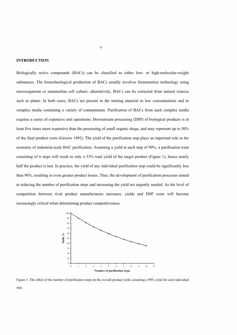
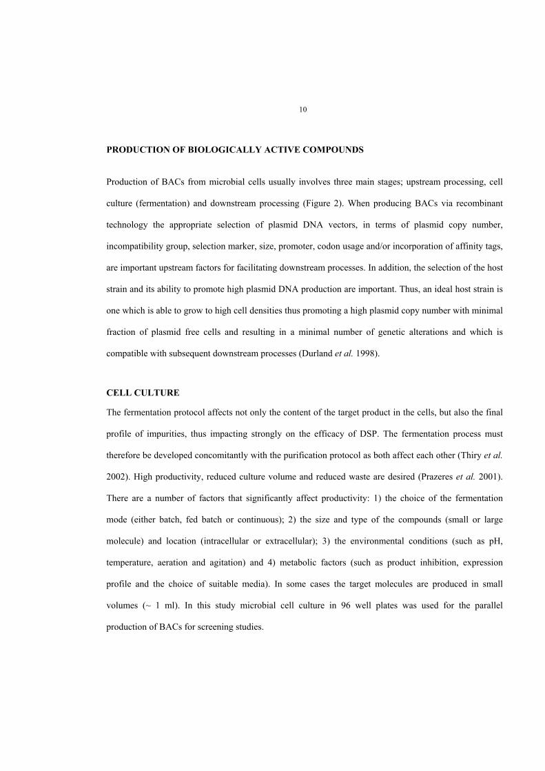
Figure 2. Schematic representation of the three primary steps used during the biotechnological production of biologicallyactive compounds (BACs); upstream processing, cell culture and downstream processing.
DOWNSTREAM PROCESS
At the end of the fermentation phase, large volumes of cell broth with relatively low concentrations of
product are obtained. The product arrives at the DSP stage in a relatively dilute form, and is contaminated
with numerous, closely related species (Lightfoot et al. 2004). DSP usually involves the following steps
or unit operations: cell harvest, cell lysis, primary recovery followed by a primary purification step,
polishing and formulation. The specific DSP depends largely on whether the product is extracellular or
intracellular. In both cases, the first step is to separate the cells from the suspension broth; this can be
achieved with one of several typical cell harvesting methods such as centrifugation, flocculation or
filtration. If the product is intracellular, including inclusion bodies, soluble proteins and plasmid DNA,
12
cell disruption is required. Four different methods are predominantly employed for cell disruption:
mechanical, physical, chemical or enzymatic. The choice of method depends on the nature of the cell-
producer used, the stability and activity of the product, and the nature of the subsequent DSP unit
operations. Integration of the cell/cell debris separation with a capture step can be achieved by using
expanded-bed adsorption chromatography (EBAC) (Ameskamp et al. 1999; Beck et al. 1999; Chase
1994; Chase et al. 1992; Pai et al. 1999; Varley et al. 1999) or high gradient magnetic fishing (HGMF)
(Hubbuch et al. 2001; Meyer et al. 2005). The selection of bioseparation techniques is largely dependent
on the target product (small or large molecule, its stability, sensitivity to degradation and extracellular or
intracellular localization), its intended use (commodity, analytical, biotechnological or pharmaceutical
application) and the demand for the final product.
1. BIOSEPARATION OF LOW-MOLECULAR-WEIGHT SUBSTANCES
The ever increasing demand for the production of low-molecular-weight substances places an economic
pressure on industrial biotechnology with respect to cost reduction, increasing productivity and reducing
processing time (Schügerl et al. 2005). Reducing the number of DSP unit operations in the purification
train increases the yield and decreases the cost. Lactic acid (α-hydroxypropionic acid) is usually produced
in the chemical industry from petroleum sources as a racemic mixture; however, production of pure
isomer of D (-) or L (+) lactic acid for food processes and biomedical applications is achieved by
microbial fermentation. When producing lactic acid in microbial cells, one faces the problem that
accumulating lactic acid inhibits the growth of the cells and subsequently its own formation. (Iyer et al.
1999; Schügerl 2000). Traditionally, lactic acid is purified either by precipitation with calcium salt
(Atkinson et al. 1991), extraction in an aqueous two-phase systems (Planas et al. 1999), extraction with
organic solvents (Dai Y et al. 1996) or by adsorption to ion exchange resins in a batch process (Vaccari et
al. 1993). To avoid the product inhibiting its own production, in situ recovery of lactic acid is favored.
Ion exchange chromatography is the method traditionally used in industrial bioprocessing. Packed-bed
13
chromatography using an anion exchange chromatography has been used for lactic acid purification;
however, it is not suitable for processing particulate containing cells feeds due to increased pressure drops
and eventual clogging of the column. Hence primary processing of feed is necessary.
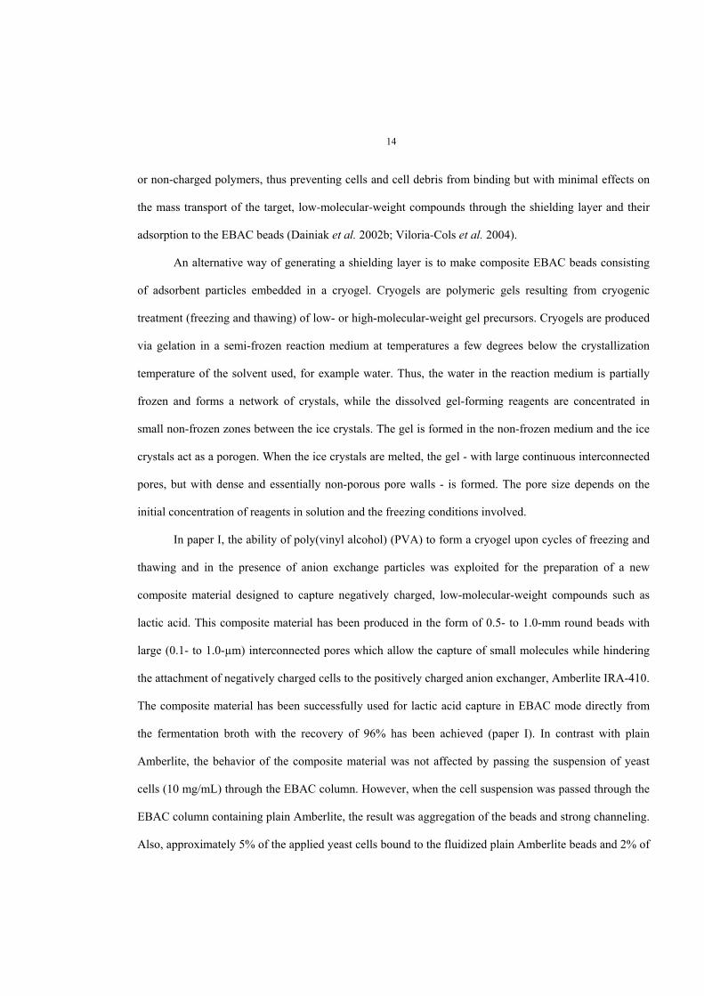
2. IN SITU RECOVERY OF LOW-MOLECULAR-WEIGHT-SUBSTANCES USING EXPANDED-BEDADSORPTION CHROMATOGRAPHY
Expanded-bed adsorption chromatography (EBAC) has been used for the purification of bio-molecules
directly from cell broth containing particulate matter. EBAC allows the integration of solid/liquid
separation, volume reduction and partial purification in a one-unit operation at high flow rate (Anspach et
al. 1999). In EBAC, the beaded particles with heterogeneous bead sizes (typically ranging from 50 to 400
µm) and densities are lifted by the up-flow of the mobile phase. The bed expands, giving rise to large
void volumes between the beads. The larger particles with higher densities populate the lower portion of
the expanded bed while the smaller particles, with lower densities, populate the upper portion. When non-
clarified cell broth is introduced onto an EBAC column, the particulate material and cell debris move
freely around the resin beads and eventually exit through the top of the column. The compound of interest
specifically binds to the beads while the non-bound and weakly-bound materials are washed out. The
expanded bed is allowed to settle, and the flow is reversed, allowing the target compound to be eluted
from the beads as in conventional packed-bed chromatography (Figure 3). A variety of functional groups
have been used as the ‘capture’ ligand immobilized on EBAC resins such as affinity, hydrophobic and ion
exchange ligands as well as immobilized metal affinity chromatography (IMAC) ligands and dyes.
Nevertheless, the feedstock composition may be critical due to the potential interaction of cells and cell
debris with adsorbent beads leading to their aggregation and resulting in bed instability and channeling
and a subsequently dramatic decrease in column performance (Anspach et al. 1999). In order to overcome
these limitations, beads have been specifically modified to prevent or reduce binding of the cells or cell
debris components without significantly affecting their binding capacity with the target molecule. For
example, ion-exchange EBAC beads have been covered with thin, shielding layers of oppositely charged
14
or non-charged polymers, thus preventing cells and cell debris from binding but with minimal effects on
the mass transport of the target, low-molecular-weight compounds through the shielding layer and their
adsorption to the EBAC beads (Dainiak et al. 2002b; Viloria-Cols et al. 2004).
An alternative way of generating a shielding layer is to make composite EBAC beads consisting
of adsorbent particles embedded in a cryogel. Cryogels are polymeric gels resulting from cryogenic
treatment (freezing and thawing) of low- or high-molecular-weight gel precursors. Cryogels are produced
via gelation in a semi-frozen reaction medium at temperatures a few degrees below the crystallization
temperature of the solvent used, for example water. Thus, the water in the reaction medium is partially
frozen and forms a network of crystals, while the dissolved gel-forming reagents are concentrated in
small non-frozen zones between the ice crystals. The gel is formed in the non-frozen medium and the ice
crystals act as a porogen. When the ice crystals are melted, the gel - with large continuous interconnected
pores, but with dense and essentially non-porous pore walls - is formed. The pore size depends on the
initial concentration of reagents in solution and the freezing conditions involved.
In paper I, the ability of poly(vinyl alcohol) (PVA) to form a cryogel upon cycles of freezing and
thawing and in the presence of anion exchange particles was exploited for the preparation of a new
composite material designed to capture negatively charged, low-molecular-weight compounds such as
lactic acid. This composite material has been produced in the form of 0.5- to 1.0-mm round beads with
large (0.1- to 1.0-µm) interconnected pores which allow the capture of small molecules while hindering
the attachment of negatively charged cells to the positively charged anion exchanger, Amberlite IRA-410.
The composite material has been successfully used for lactic acid capture in EBAC mode directly from
the fermentation broth with the recovery of 96% has been achieved (paper I). In contrast with plain
Amberlite, the behavior of the composite material was not affected by passing the suspension of yeast
cells (10 mg/mL) through the EBAC column. However, when the cell suspension was passed through the
EBAC column containing plain Amberlite, the result was aggregation of the beads and strong channeling.
Also, approximately 5% of the applied yeast cells bound to the fluidized plain Amberlite beads and 2% of
15
the yeast cells were irreversibly retained by the plain Amberlite resin, even after regeneration with 0.5 M
NaOH.
3. BIOSEPARATION OF HIGH-MOLECULAR-WEIGHT COMPOUNDS
3.1. ANALYTICAL BIOSEPARATIONS
The need for analytical methods to characterize new drug candidates has boosted research into novel
techniques for the rapid screening of large numbers of biological compounds. Assays that facilitate the
detection of biological substances and biological activities are thus highly desirable. Such systems could
also be applied to the analysis of biomolecular recognition at cellular membranes, ligand–receptor
binding events and other processes of biotechnological significance (Jelinek et al. 2001).
Increased demand for the parallel processing of large numbers of biological samples has
stimulated new developments in sample handling and processing. Traditionally, samples have been
processed and analyzed in 96-well plates. Parallel sample processing involved sample clarification from
cells and/or cell debris and capture of target biomolecules followed by their identification and
quantification (Galaev et al. 2005; Rossi et al. 2000). Clarification of large sets of biological samples
containing target biomolecules is both time-consuming and laborious. Employing the selectivity of
affinity chromatography as a capture step with the integration of a clarification step is thus desirable. An
ideal matrix for this purpose should have interconnected macropores with a pore size of 10-100 µm,
allowing cells and cell debris to pass through without being retained. On the other hand, the matrix should
be capable of efficient capture of the target molecule via strong affinity interactions.
Cryogels produced from polyacrylamide are mechanically strong, elastic matrices with a pore size
in the range of 10-100 µm. Besides the advantage of large pore size, the cryogel monoliths are used for
processing particulate (cells or cell debris) containing solutions (Arvidsson et al. 2002; Arvidsson et al.
2003; Dainiak et al. 2002a; Dainiak et al. 2004; Galaev et al. 2005; Kumar et al. 2003; Plieva et al. 2004,
paper numbers II, III and IV). Moreover, cryogel monoliths are elastic, allowing them to be slightly
16
Figure 3. Lactic acid capture from a microbial cell broth by expanded-bed adsorption chromatography.A) Capture of lactic acid by composite beads in an expanded-bed mode from non-clarified Lactobacillus delbrueckiifermentation broth. Non-clarified broth was applied to the column comprising composite beads (4-ml settled volume) in anexpanded-bed mode at a linear flow rate of 160 cm/h. The column was washed with de-ionized water in an expanded-bedmode. The flow was interrupted to allow the adsorbent to settle, and the elution was performed with 0.1 M HCl in a packed-bed mode.B) The chromatographic profile for lactic acid adsorption and elution. Lactic acid was analyzed using HPLC (closed rhombus).The cell content was analyzed by measuring the turbidity at 620 nm (open square).
compressed and easily placed inside a chromatographic column. When expanded, cryogel monoliths fill
the column tightly with no risk of leakage between the monolith and the walls of the column. Capillary
forces keep the liquid inside the pores of the cryogel monolith, rendering the columns drainage-protected.
Applying a certain volume of liquid to the top of a cryogel monolith column results in the displacement of
exactly the same volume of liquid from the column (Galaev et al. 2005).
Traditional 96-well plates with mini-columns packed with beaded particles require either vacuum
or pressure to drive the flow of the solution through the mini-columns. By contrast, gravity is sufficient
for liquid flow in the 96-well plates with mini-columns containing cryogel monoliths. Alternative
techniques for parallel processing of numerous samples, such as the use of magnetic beads with
immobilized affinity ligands, require special equipment and expensive materials (Ko et al. 1992).
Immobilized metal affinity chromatography (IMAC), was first introduced by Porath et al. (1975).
IMAC involves the formation of a complex between an immobilized chelating agent (e.g., tridentate
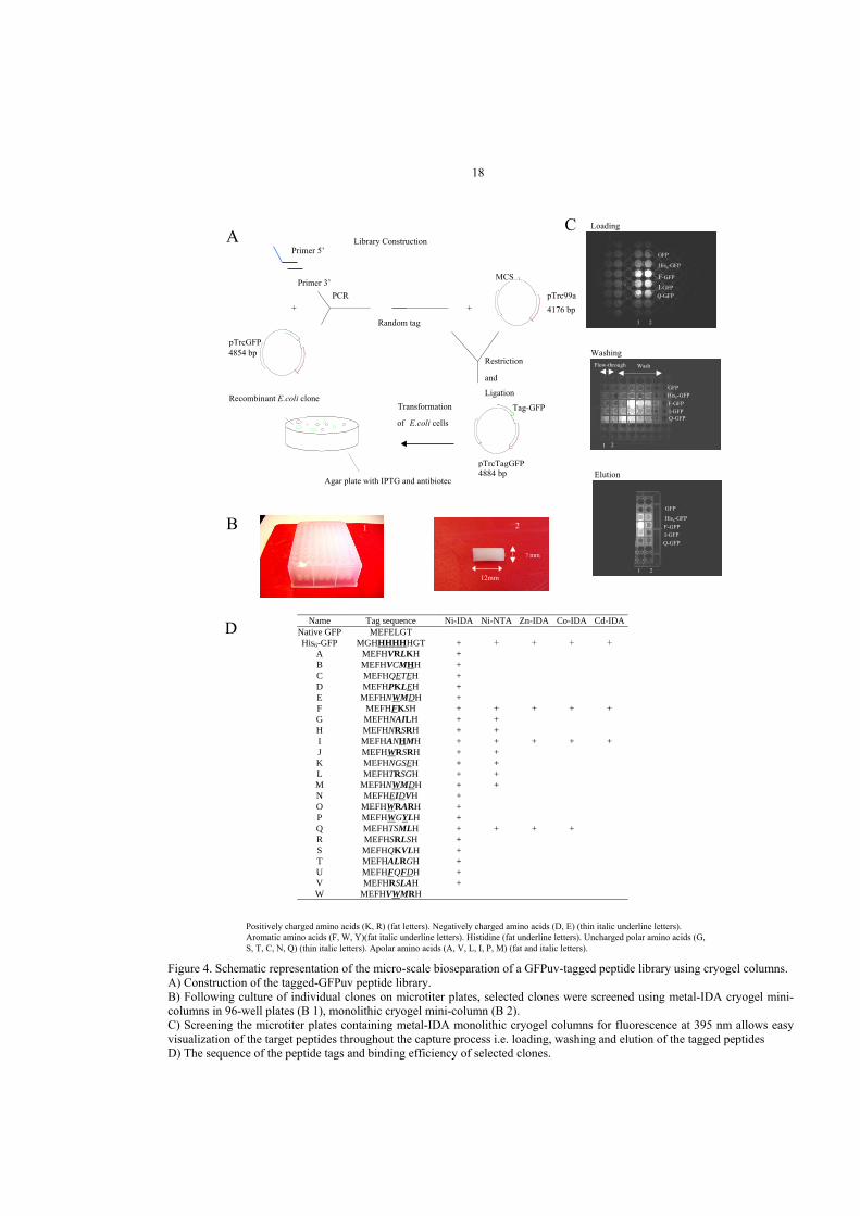
Figure 4. Schematic representation of the micro-scale bioseparation of a GFPuv-tagged peptide library using cryogel columns.A) Construction of the tagged-GFPuv peptide library.B) Following culture of individual clones on microtiter plates, selected clones were screened using metal-IDA cryogel mini-columns in 96-well plates (B 1), monolithic cryogel mini-column (B 2).C) Screening the microtiter plates containing metal-IDA monolithic cryogel columns for fluorescence at 395 nm allows easyvisualization of the target peptides throughout the capture process i.e. loading, washing and elution of the tagged peptidesD) The sequence of the peptide tags and binding efficiency of selected clones.
CPrimer 5’
Primer 3’
+GFP
Random tag
+pTrc99a4176 bp
Amp
MCS
lac lq
pTrcTagGFP4884 bp
Amp
Tag-GFP
lac lq
GFP
pTrcGFP4854 bp
Amp
lac lq
GFP
Recombinant E.coli clone Transformation
of E.coli cells
Agar plate with IPTG and antibiotec
PCR
Restriction
and
Ligation
Library Construction
1
GFP
His6-GFP
F-GFP
I-GFPQ-GFP
2
GFPHis6-GFPF-GFPI-GFPQ-GFP
WashFlow-through
1 2
1
GFP
His6-GFPF-GFPI-GFPQ-GFP
2
ALoading
Elution
Washing
B 1
7 mm
2
12mm
19
3.2. PREPARATIVE BIOSEPARATION: CAPTURE OF PLASMID DNA USING MACROPOROUS CRYOGEL
There is an increasing demand for pure preparations of plasmid DNA. Traditionally, plasmid DNA is
produced from E. coli cells. The production of plasmid DNA involves three steps: upstream processing,
fermentation of E. coli cells, then purification. Purification of plasmid DNA involves the following unit
operations: cell harvest by centrifugation or filtration, cell lysis, primary recovery and adsorption
followed by polishing and formulation. At the laboratory scale, cell harvesting can be achieved with
centrifugation. However, at the industrial scale, the cells are normally harvested using well-developed
continuous centrifugation or cross-flow filtration. The bacterial cell paste can be processed immediately
or frozen for future use (Durland and Eastman 1998). Traditionally, for plasmid purification, alkaline
lysis is the most common technique used for cell rupture. Alkaline conditions, in the presence of
ethylenediamine tetraacetic acid (EDTA) and the detergent, sodium dodecyl sulfate (SDS), are
traditionally employed for cell rupture and denaturation of host cell proteins with partial denaturation of
the genomic DNA. This procedure usually results in a large volume of viscous, particulate-containing
solution. Plasmid DNA accounts for less than 1% (w/w) of bacterial cell lysate while RNA, a major
contaminant in plasmid DNA preparation, accounts for 6% (w/w) (Prazeres et al. 2001).
Cesium chloride/ethidium bromide (CsCl/EtBr) buoyant density gradient separation followed by
ultracentrifugation (Prather et al. 2003) and alcohol precipitation using ethanol or isopropanol, are
frequently used for laboratory-scale plasmid purification. However for large-scale plasmid purification,
other techniques such as triple helix affinity precipitation (Costioli et al. 2003) and fractional precipitation
from cell lysates using cetyltrimethylammonium bromide (CTAB) (Lander et al. 2002) have been
developed. Selective precipitation using compaction agents such as spermine and spermidine (Murphy et
al. 1999), precipitation by forming polyelectrolyte complexes (Wahlund et al. 2004b), and partitioning in
thermoseparating aqueous two-phase polymer systems (Kepka et al. 2004) have also been used. Super-
paramagnetic nano-particles coated with polyethyleneimine (PEI) were developed for plasmid DNA
20
purification (Chiang et al. 2005). However, a variety of chromatographic techniques are predominantly
used for plasmid DNA purification such as ion exchange chromatography (Tseng et al. 2003), size
exclusion chromatography (Horn et al. 1995), triple helix affinity chromatography (Wils et al. 1997),
thiophilic interaction chromatography (Sandberg et al. 2004), hydrophobic interaction chromatography
(Diogo et al. 1999), reverse phase chromatography (Green et al. 1997) and hydroxyapatite
chromatography (Giovannini et al. 2002). EBAC has also been used for the capture of plasmid DNA
(Varley et al. 1999) from filtered and centrifuged cell lysates (Ferreira et al. 2000; Theodossiou et al.
2001). More recently, monolithic columns were used for plasmid DNA purification from clarified cell
lysate (Bencina et al. 2004; Branovic et al. 2004; Urthaler et al. 2005).
Ion exchange chromatography remains the method most commonly used in the DSP of
biomolecules due to its robustness, rapid separation, organic solvent-free process, its ability to withstand
sanitation with sodium hydroxide and a wide selection of industrial media. Phosphate groups located in
the backbone of nucleic acids carry a negative charge at pH values above 4; hence, interactions between
these groups and the positively charged anion exchanger groups may occur. The strength of this
interaction depends on the density and the conformation of the negatively charged groups located on the
plasmid DNA. Elution of the bound DNA is achieved with increasing salt concentrations. In addition to
plasmid DNA, alkaline cell lysate contains host cell proteins, RNA, genomic DNA and endotoxin. Being
negatively charged, some of these bio-molecules have physical and chemical similarities to plasmid
DNA; hence, they compete for binding and are co-eluted with plasmid DNA during anion exchange
chromatography.
Exploring the difference in binding affinities between RNA and plasmid DNA towards quaternary
amines enables purification of plasmid DNA from its major contaminant, RNA. For example, the
application of clarified cell lysate in the presence of 0.5 M NaCl greatly reduces non-specific binding of
most of the cellular impurities (protein and RNA) to EBAC columns packed with Streamline QXL
(Ferreira et al. 2000).
21
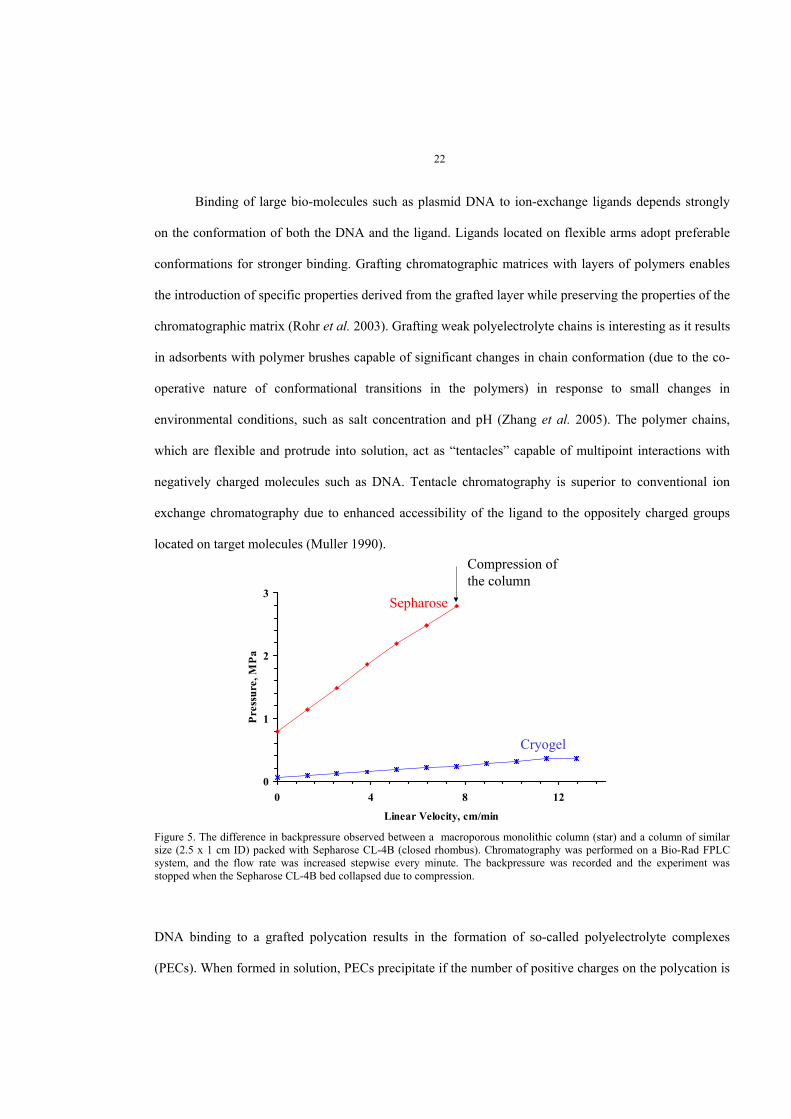
A significant problem associated with plasmid DNA chromatography in a packed-bed mode is
pronounced mass-transfer limitations. Plasmid DNA is characterized by its large size (> 200 nm) with a
helical length of about 370 nm, a diameter of double helix of 11.3 nm and a diffusion coefficient in the
order of 10-8 cm2/s, which is approximately an order of magnitude less than that of proteins (Diogo et al.
2005). Moreover, the high viscosity of the alkaline lysate generates high backpressures (between 15-60
MPa) in columns packed with beaded particles (Diogo et al. 2005). Monolithic chromatography has been
used for plasmid DNA purification, and mass-transfer resistance was shown to be significantly reduced
(Branovic et al. 2004; Urthaler et al. 2005). In columns packed with beaded particles, mass-transfer is
governed by diffusion whereas in monolithic columns mass-transfer is predominantly due to convective
transport. The macroporous monolithic cryogels used in this study (paper IV) were produced by radical
polymerization of a monomer, acrylamide, and cross-linker, N,N’-methylenebisacrylamide, at subzero
temperatures, as mentioned above. Large interconnected pores in monolithic cryogels allow for efficient
convective mass-transport of plasmid DNA. Moreover, only a small backpressure is generated when a
viscous solution is pumped through a macroporous monolithic cryogel column.
Viscous, particulate-containing solutions with a high content of contaminating substances such as
RNA, genomic DNA and protein need to be processed for the capture of the target molecule (plasmid
DNA), which is usually present in low amounts. Integration of a number of necessary unit operations,
such as clarification, plasmid capture and primary purification, into a single step will both improve the
final product recovery and the economy of the process. In addition, degradation of plasmid DNA is time-
dependent, hence fast processing of non-clarified cell lysate is desirable (Urthaler et al. 2005). Due to the
large pore size of the monolithic columns, it is possible to process particulate-containing solutions at high
flow rates without increases in backpressure or a reduction in binding capacity (papers III and IV) (Figure
5).
0
1
2
3
0 4 8 12
Linear Velocity, cm/min
Pres
sure
, MPa
Sepharose
Cryogel
Compression ofthe column
22
Binding of large bio-molecules such as plasmid DNA to ion-exchange ligands depends strongly
on the conformation of both the DNA and the ligand. Ligands located on flexible arms adopt preferable
conformations for stronger binding. Grafting chromatographic matrices with layers of polymers enables
the introduction of specific properties derived from the grafted layer while preserving the properties of the
chromatographic matrix (Rohr et al. 2003). Grafting weak polyelectrolyte chains is interesting as it results
in adsorbents with polymer brushes capable of significant changes in chain conformation (due to the co-
operative nature of conformational transitions in the polymers) in response to small changes in
environmental conditions, such as salt concentration and pH (Zhang et al. 2005). The polymer chains,
which are flexible and protrude into solution, act as “tentacles” capable of multipoint interactions with
negatively charged molecules such as DNA. Tentacle chromatography is superior to conventional ion
exchange chromatography due to enhanced accessibility of the ligand to the oppositely charged groups
located on target molecules (Muller 1990).
Figure 5. The difference in backpressure observed between a macroporous monolithic column (star) and a column of similarsize (2.5 x 1 cm ID) packed with Sepharose CL-4B (closed rhombus). Chromatography was performed on a Bio-Rad FPLCsystem, and the flow rate was increased stepwise every minute. The backpressure was recorded and the experiment wasstopped when the Sepharose CL-4B bed collapsed due to compression.
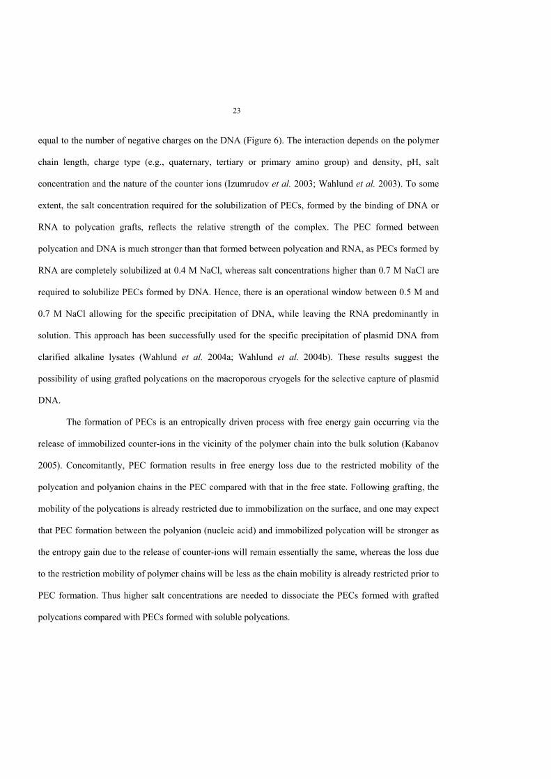
DNA binding to a grafted polycation results in the formation of so-called polyelectrolyte complexes
(PECs). When formed in solution, PECs precipitate if the number of positive charges on the polycation is
23
equal to the number of negative charges on the DNA (Figure 6). The interaction depends on the polymer
chain length, charge type (e.g., quaternary, tertiary or primary amino group) and density, pH, salt
concentration and the nature of the counter ions (Izumrudov et al. 2003; Wahlund et al. 2003). To some
extent, the salt concentration required for the solubilization of PECs, formed by the binding of DNA or
RNA to polycation grafts, reflects the relative strength of the complex. The PEC formed between
polycation and DNA is much stronger than that formed between polycation and RNA, as PECs formed by
RNA are completely solubilized at 0.4 M NaCl, whereas salt concentrations higher than 0.7 M NaCl are
required to solubilize PECs formed by DNA. Hence, there is an operational window between 0.5 M and
0.7 M NaCl allowing for the specific precipitation of DNA, while leaving the RNA predominantly in
solution. This approach has been successfully used for the specific precipitation of plasmid DNA from
clarified alkaline lysates (Wahlund et al. 2004a; Wahlund et al. 2004b). These results suggest the
possibility of using grafted polycations on the macroporous cryogels for the selective capture of plasmid
DNA.
The formation of PECs is an entropically driven process with free energy gain occurring via the
release of immobilized counter-ions in the vicinity of the polymer chain into the bulk solution (Kabanov
2005). Concomitantly, PEC formation results in free energy loss due to the restricted mobility of the
polycation and polyanion chains in the PEC compared with that in the free state. Following grafting, the
mobility of the polycations is already restricted due to immobilization on the surface, and one may expect
that PEC formation between the polyanion (nucleic acid) and immobilized polycation will be stronger as
the entropy gain due to the release of counter-ions will remain essentially the same, whereas the loss due
to the restriction mobility of polymer chains will be less as the chain mobility is already restricted prior to
PEC formation. Thus higher salt concentrations are needed to dissociate the PECs formed with grafted
polycations compared with PECs formed with soluble polycations.
0
0.2
0.4
0.6
0.8
1
0 0.2 0.4 0.6 0.8 1NaCl conc., M
A/A
o
-- --
+++ + +++----
0
0.2
0.4
0.6
0.8
1
0 1 2 3 4 5 6
(+/-) Ratio
A/A
o
+ + + + + + + +- - - - - - - -
+ + +- - - - - - - -
+ + + + + + + +- - - -
A) Effect of polymer/nucleic acids on formation of PECs
Figure 6. Polyelectrolyte complexes of nucleic acids with polycations.A. Schematic representation of the effect of molar ratio (+/-) on the formation of PECs between polymeric chains carryingquaternary amines and phosphate groups respectively (1); relative residual absorbance of RNA (open triangle), and genomicDNA (closed square) remaining in solution after precipitation with polyDMAEMA as a function of the charge ratio (+/-) at pH5 (2).B. Schematic representation of the effect of salt concentrations on the solubilization of PECs (1); the effect of saltconcentration on the solubilization of PECs between polyDMAEMA and genomic DNA (closed square) or RNA (opentriangle) (2). The relative residual absorbance of nucleic acids remaining in the solution after precipitation withpolyDMAEMA was expressed as (A/Ao).
In paper IV, polycations with tertiary and quaternary amine groups were introduced into the
macroporous cryogel matrix by graft polymerization of N,N,-dimethylaminoethyl methacrylate
(DMAEMA) and (2-(methacryloyloxy)ethyl)-trimethyl ammonium chloride (META) onto the pore
surface of the chromatographic matrix. In addition, partial quaternization of polyDMAEMA was used
(Figure 7).
25
Figure 7. Polycation grafting onto a macroporous polyacrylamide cryogel.
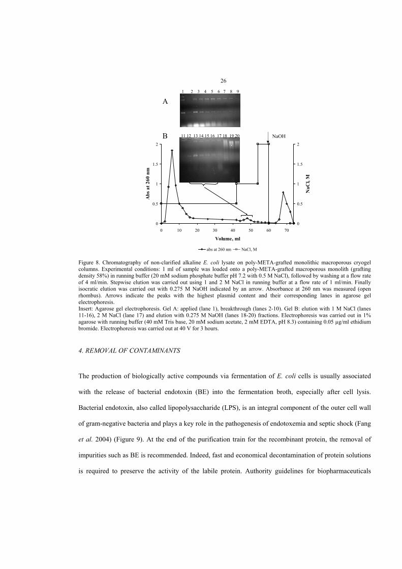
Plasmid DNA was efficiently captured by polyMETA-grafted, polyDMAEMA-grafted and
partially quaternized polyDMAEMA–monolithic macroporous cryogel columns directly from non-
clarified alkaline lysate; bound plasmid DNA was eluted with a NaCl gradient. When eluted with a
stepwise NaCl gradient from a polyMETA-grafted column, the plasmid DNA was free from RNA and
contamination with host proteins was negligible (Figure 8). A small percentage of RNA and protein was
co-eluted with plasmid DNA from both polyDMAEMA-grafted and partially quaternized
polyDMAEMA–grafted columns (paper IV).
C u ( I I I ) C u ( I I )O H
N H
N H 2N H N H 2
+
N CH C C
C H 3 O
O
n
CH 2
CH 2 N+
C H 3
C H 3
C H 3
CH 2
CH 2 N
C H 3
C H 3
CH 2 C C
C H 3 O
O
N H
N CH C C
C H 3 O
O
n
CH 2
CH 2 N
C H 3
C H 3
CH 2
CH 2 N+
C H 3
C H 3
CH 2 C C
C H 3 O
O C H 3
+ + ++ H 2 O
a ) A c t iv a t io n r e a c t io n
b ) G r a f t p o ly m e r iz a t io n
+
-
+
D M A E M A
M E T A
0
0.5
1
1.5
2
0 10 20 30 40 50 60 70
Volume, ml
Abs
at 2
60 n
m
0
0.5
1
1.5
2
NaC
l, M
abs at 260 nm NaCl, M
A
NaOH
1 2 3 4 5 6 7 8 910
11 12 13 14 15 16 17 18 19 20B
26
Figure 8. Chromatography of non-clarified alkaline E. coli lysate on poly-META-grafted monolithic macroporous cryogelcolumns. Experimental conditions: 1 ml of sample was loaded onto a poly-META-grafted macroporous monolith (graftingdensity 58%) in running buffer (20 mM sodium phosphate buffer pH 7.2 with 0.5 M NaCl), followed by washing at a flow rateof 4 ml/min. Stepwise elution was carried out using 1 and 2 M NaCl in running buffer at a flow rate of 1 ml/min. Finallyisocratic elution was carried out with 0.275 M NaOH indicated by an arrow. Absorbance at 260 nm was measured (openrhombus). Arrows indicate the peaks with the highest plasmid content and their corresponding lanes in agarose gelelectrophoresis.Insert: Agarose gel electrophoresis. Gel A: applied (lane 1), breakthrough (lanes 2-10). Gel B: elution with 1 M NaCl (lanes11-16), 2 M NaCl (lane 17) and elution with 0.275 M NaOH (lanes 18-20) fractions. Electrophoresis was carried out in 1%agarose with running buffer (40 mM Tris base, 20 mM sodium acetate, 2 mM EDTA, pH 8.3) containing 0.05 µg/ml ethidiumbromide. Electrophoresis was carried out at 40 V for 3 hours.
4. REMOVAL OF CONTAMINANTS
The production of biologically active compounds via fermentation of E. coli cells is usually associated
with the release of bacterial endotoxin (BE) into the fermentation broth, especially after cell lysis.
Bacterial endotoxin, also called lipopolysaccharide (LPS), is an integral component of the outer cell wall
of gram-negative bacteria and plays a key role in the pathogenesis of endotoxemia and septic shock (Fang
et al. 2004) (Figure 9). At the end of the purification train for the recombinant protein, the removal of
impurities such as BE is recommended. Indeed, fast and economical decontamination of protein solutions
is required to preserve the activity of the labile protein. Authority guidelines for biopharmaceuticals
27
require that the level of endotoxin for intravenous application is not more than 5 endotoxin units (EU) per
kg body weight per hour (Hirayama et al. 2002). One EU equals 100 pg of standard endotoxin.
Negative affinity chromatography has been employed for endotoxin removal. The biological
activity of BE is not of concern, whereas the biological activity of the product (protein) should be
preserved with 100% recovery (Anspach 2001). Selective binding between the immobilized ligand and
endotoxin, without affecting the target protein, is required. The affinity ligand should be able to bind BE,
which is present in very low concentrations, in the presence of protein of up to 6 orders of magnitude
higher concentrations (Anspach 2001; Petsch et al. 1998). Bacterial endotoxin removal from protein-
containing solutions also depends on the nature of the protein (Talmadge et al. 1989).
Polycationic ligands, such as polyethyleneimine (PEI) and poly(ε-lysine) (Sakata et al. 2002) have
been reported to have a high affinity to BE and have been used for BE capture. Highly branched PEI with
a ratio of primary, secondary and tertiary amines of 1:2:1, a molecular weight of 60 kDa, a pK > 9 for
primary amino groups and a pK > 10.5 for secondary amines, has been also used (Petsch et al. 1997;
Petsch et al. 1998).
Bacterial endotoxin has been removed from protein solutions using cellulose columns with
immobilized poly(ε-lysine) at physiological pH (Bemberis et al. 2005). Efficient removal of BE from a
bovine serum albumin (BSA) solution was reported using nylon flat-sheets with immobilized poly(L-
lysine) (Petsch et al. 1997). Several reports on the capture of BE by using matrices with immobilized PEI
showed efficient BE clearance (Anspach et al. 2000; Hirayama and Sakata 2002; Petsch et al. 1998).
The cyclic peptide antibiotic, polymyxin B, disorganizes the bacterial cell wall through its
interactions with lipid A. Polymyxin B immobilized on a solid matrix has also been used as an affinity
ligand for BE removal (Issekutz 1983; Liu et al. 1997). However, pronounced protein losses due to the
binding of negatively charged protein to polymyxin B have been reported. Karplus et al. showed a loss of
24% of bovine catalase with a 103 BE reduction (Karplus et al. 1987).
28
Lysozyme from hen egg white, is an enzyme with a molecular weight of 14.4 kDa and a pI of 11.2
which catalyses the hydrolysis of peptidoglycan in the cell wall. Lysozyme possesses a positive charge
under neutral conditions, while BE is negatively charged. Lysozyme binds BE through electrostatic forces
and with a concomitant hydrophobic interaction with the acyl-chain region (Brandenburg et al. 1998). BE
and lipid A bind to lysozyme with a 3:1 molar ratio (Brandenburg et al. 1998).
The appropriate selection of environmental conditions, such as pH and salt concentration, plays an
important role in BE removal from a protein-containing solution. The pH values below that of the
isoelectric point of a protein ensure best clearance and high recovery (Petsch et al. 1997). Basic proteins
possess a net positive charge at pH values close to 7 and act as carriers for BE, hence decreasing BE
clearance. The binding between the matrix with immobilized ligand and BE should be more efficient than
the binding between BE and basic proteins.
In paper III, macroporous monolithic cryogel columns with immobilized polyethyleneimine,
polymyxin B and lysozyme were employed for BE capture from BSA solution. Columns with
immobilized polymyxin B showed nearly 100% recovery of BSA and almost 100% BE capture at pH 7.2.
Bacterial endotoxin was quantitatively bound to the cryogel column with immobilized lysozyme at pH 4.7
with 100% BSA recovery, whereas at pH 7.2, 35% of the BE was in the breakthrough alongside the BSA.
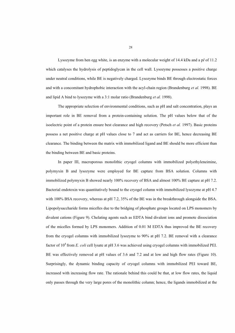
Lipopolysaccharide forms micelles due to the bridging of phosphate groups located on LPS monomers by
divalent cations (Figure 9). Chelating agents such as EDTA bind divalent ions and promote dissociation
of the micelles formed by LPS monomers. Addition of 0.01 M EDTA thus improved the BE recovery
from the cryogel columns with immobilized lysozyme to 90% at pH 7.2. BE removal with a clearance
factor of 104 from E. coli cell lysate at pH 3.6 was achieved using cryogel columns with immobilized PEI.
BE was effectively removed at pH values of 3.6 and 7.2 and at low and high flow rates (Figure 10).
Surprisingly, the dynamic binding capacity of cryogel columns with immobilized PEI toward BE,
increased with increasing flow rate. The rationale behind this could be that, at low flow rates, the liquid
only passes through the very large pores of the monolithic column; hence, the ligands immobilized at the
surface of smaller pores are essentially unavailable. At higher flow rates, the mobile phase may begin to
pass through the smaller pores and thus be exposed to a greater area of immobilized ligand.
Negative chromatography using macroporous monolithic cryogel columns proved to be an
efficient means of removing BE, both from protein solutions (e.g., target protein product), and from cell
lysates (e.g., waste waters). Adjusting conditions such as the ionic strength and pH of the applied solution
or adding a chelating agent eliminates retardation of the target protein on the column.
Figure 9. A: Chemical structure of bacterial endotoxin. B: Formation of endotoxin micelles.
n
O
PO
O NH2
O
PO
PO
NH2
O
O
O
O
OP
OH
O
O
O
NHO
OO
OO
OH
O NH OPO O
O
O O
OO
OH
OO
O
PO
OO
OO
OO
PO
O
OH
O- Antigen
n= 4-40
Coreoligosaccharide
Lipid A
Cell exterior
Cell interior
A
0
0.1
0.2
0 50 100
Volume, ml
BE, R
elat
ive
Units
0
100
200
Prot
ein
conc
., µg
/ml
2M NaCl
30
Figure 10. Capture of bacterial endotoxin (BE) from a BSA solution using macroporous monolith columns with immobilizedPEI. Experimental conditions: BSA solution containing BE was applied in 1 ml of the running buffer followed by washing andelution with 2 M NaCl in the running buffer at a flow rate of 1 ml/min. BSA was assayed using BCA (closed square, dashedline) and BE was assayed by measuring absorbance at 280 nm with subtraction of the BSA contribution to absorbance (openrhombus, straight line).
CONCLUSIONS
The individual studies presented in this thesis clearly demonstrate that macroporous monolithic cryogel
columns and composite cryogel matrices are materials with a large potential for the capture of
biologically active compounds from a variety of biotechnological feeds, including those which contain
particulate matter. The large interconnected pores typical of monolithic cryogels allow for minimal flow
resistance and efficient convective mass-transport of large biomolecules and bio-aggregates, such as
plasmid DNA and micelles formed by bacterial endotoxin. Further developments in this direction will
concentrate on the design and production of highly selective, affinity ligands capable of the exclusive
adsorption of the target biomolecule. Such ligands could be developed, for instance, using a triple-helix
approach for plasmid DNA capture. On the other hand, an area where the full potential of cryogels could
realize, may be the processing of even larger biological entities such as viruses, and microbial and even
31
mammalian cells. One may foresee the development of affinity chromatography methods for the
integrated purification of viruses and the selective isolation of particular cell lines from microbial
consortia or animal tissues. The challenge will be to isolate sub-populations of cells at different stages of
development (e.g., lag-phase, exponential growth or a stationary phase for microorganisms) or