Primary torsion of the greater omentum: Color Doppler sonography and CT correlated with surgery and pathology findings Hesham El Sheikh a, * , Nabil Abdulaziz b a Department of Diagnostic Radiology, Faculty of Medicine, Benha University, Cairo, Egypt b Department of General Surgery, Faculty of Medicine, Al Azhar University, Cairo, Egypt Received 30 September 2013; accepted 28 October 2013 Available online xxxx KEYWORDS Omental torsion; US; CT; Color Doppler; Surgical Abstract Primary torsion of the greater omentum is a rare cause of acute abdomen commonly diagnosed at surgery performed for appendicitis. We report nine cases of omental torsion who underwent surgery and correlate their preoperative color Doppler ultrasonography (US) and com- puted tomography (CT) findings with the surgical and pathological findings to assess the value of US and CT in the diagnosis of omental torsion. US findings of omental torsion correlated with the operative and pathological findings in seven patients and the diagnosis was missed in two patients suspected to have ruptured appendix. CT findings of omental torsion correlated with the operative and pathological findings in all five patients who did CT. US and CT scanning are useful for pre- operative diagnosis of omental torsion. Ó 2013 Production and hosting by Elsevier B.V. on behalf of Egyptian Society of Radiology and Nuclear Medicine. 1. Introduction Primary torsion of the greater omentum is a rare cause of acute abdomen that results when the greater omentum twists upon it- self along its long axis with resultant compromise of its vascular supply leading to hemorrhagic infarction and fat necrosis (1,2). The etiology of primary torsion of the omentum is not known but various predisposing and precipitating factors have been identified. Predisposing factors include anatomic malfor- mations of the omentum (bifid omentum or tongue-like projec- tions); accessory omentum; irregular distribution of omental fat; and anomalous veins that allow a fixed point for omental twisting (1,3). The identified precipitating factors include local trauma; over eating with resultant hyperperistalsis; sudden change in position; coughing and straining (1,3). The right side of the omentum is the most frequently involved portion, be- lieved to be due to its increased length and mobility (4,5). Patients with omental torsion may present with acute abdom- inal pain more commonly in the right lower quadrant (3,4,6). The pain is constant and non-radiating, gradually increasing in * Corresponding author. Tel.: +20 966598181340. E-mail address: [email protected](H. El Sheikh). Peer review under responsibility of Egyptian Society of Radiology and Nuclear Medicine. Production and hosting by Elsevier The Egyptian Journal of Radiology and Nuclear Medicine (2013) xxx, xxx–xxx Egyptian Society of Radiology and Nuclear Medicine The Egyptian Journal of Radiology and Nuclear Medicine www.elsevier.com/locate/ejrnm www.sciencedirect.com 0378-603X Ó 2013 Production and hosting by Elsevier B.V. on behalf of Egyptian Society of Radiology and Nuclear Medicine. http://dx.doi.org/10.1016/j.ejrnm.2013.10.006 ARTICLE IN PRESS Please cite this article in press as: El Sheikh H, Abdulaziz N, Primary torsion of the greater omentum: Color Doppler sonography and CT correlated with surgery and pathology findings, Egypt J Radiol Nucl Med (2013), http://dx.doi.org/10.1016/j.ejrnm.2013.10.006
Transcript
The Egyptian Journal of Radiology and Nuclear Medicine (2013) xxx, xxx–xxx
ARTICLE IN PRESS
Egyptian Society of Radiology and Nuclear Medicine
The Egyptian Journal of Radiology andNuclearMedicine
Peer review under responsibility of Egyptian Society of Radiology and
Nuclear Medicine.
Production and hosting by Elsevier
0378-603X � 2013 Production and hosting by Elsevier B.V. on behalf of Egyptian Society of Radiology and Nuclear Medicine.
http://dx.doi.org/10.1016/j.ejrnm.2013.10.006
Please cite this article in press as: El Sheikh H, Abdulaziz N, Primary torsion of the greater omentum: Color Doppler sonography acorrelated with surgery and pathology findings, Egypt J Radiol Nucl Med (2013), http://dx.doi.org/10.1016/j.ejrnm.2013.10.006
Hesham El Sheikha,*, Nabil Abdulaziz
b
a Department of Diagnostic Radiology, Faculty of Medicine, Benha University, Cairo, Egyptb Department of General Surgery, Faculty of Medicine, Al Azhar University, Cairo, Egypt
Received 30 September 2013; accepted 28 October 2013
Available online xxxx
KEYWORDS
Omental torsion;
US;
CT;
Color Doppler;
Surgical
Abstract Primary torsion of the greater omentum is a rare cause of acute abdomen commonly
diagnosed at surgery performed for appendicitis. We report nine cases of omental torsion who
underwent surgery and correlate their preoperative color Doppler ultrasonography (US) and com-
puted tomography (CT) findings with the surgical and pathological findings to assess the value of
US and CT in the diagnosis of omental torsion. US findings of omental torsion correlated with the
operative and pathological findings in seven patients and the diagnosis was missed in two patients
suspected to have ruptured appendix. CT findings of omental torsion correlated with the operative
and pathological findings in all five patients who did CT. US and CT scanning are useful for pre-
operative diagnosis of omental torsion.� 2013 Production and hosting by Elsevier B.V. on behalf of Egyptian Society of Radiology and Nuclear
Medicine.
1. Introduction
Primary torsion of the greater omentum is a rare cause of acuteabdomen that results when the greater omentum twists upon it-self along its long axis with resultant compromise of its vascularsupply leading to hemorrhagic infarction and fat necrosis (1,2).
The etiology of primary torsion of the omentum is notknown but various predisposing and precipitating factors have
been identified. Predisposing factors include anatomic malfor-mations of the omentum (bifid omentum or tongue-like projec-tions); accessory omentum; irregular distribution of omental
fat; and anomalous veins that allow a fixed point for omentaltwisting (1,3). The identified precipitating factors include localtrauma; over eating with resultant hyperperistalsis; suddenchange in position; coughing and straining (1,3). The right side
of the omentum is the most frequently involved portion, be-lieved to be due to its increased length and mobility (4,5).
Patients with omental torsion may present with acute abdom-
inal painmore commonly in the right lower quadrant (3,4,6). Thepain is constant and non-radiating, gradually increasing in
Fig. 1A Gray-scale US shows large hyperechoic mass next to
anterior abdominal wall in the right lower quadrant (arrows).
2 H. El Sheikh, N. Abdulaziz
ARTICLE IN PRESS
intensity. Physical examination usually shows evidence of localperitonitis in the right iliac fossa and a mass may be palpable(3). A mild leukocytosis may be found (3,7). The Most common
clinical diagnosis made in these patients is acute appendicitis.Other differential diagnoses include acute cholecystitis, torsionof an ovarian cyst and perforated peptic ulcer (4,6).
Primary torsion of the omentum is rarely diagnosed preop-
eratively and the literature found on this condition is limited tocase presentations and small case series (1–3,7–10). US and CTproved to provide typical and well-recognizable features in re-
cent studies (8–10).The current therapeutic management of choice is laparos-
copy proceeding to laparotomy identifying and removing the
infarcted section of omentum. The presence of free serosan-guineous fluid in the peritoneal cavity as a result of hemor-rhagic extravasion and normal appendix, gallbladder at
laparoscopy makes the diagnosis of omental torsion likely(3,8,11,12).
The purpose of our study is to report nine cases of primarytorsion of the greater omentum who underwent surgery and
correlate their color Doppler US and CT findings with the sur-gical and pathological findings to assess the value of US andCT in the diagnosis of omental torsion.
2. Subjects and methods
During the period between February 2008 and July 2012, 8297
patients with acute right abdominal pain referred to the radi-ology department; all underwent US and 487 patients under-went CT study. Surgery was performed for 4773 patients,
only 9 patients had primary torsion of the greater omentum.The remaining 4764 patients had different diagnoses (2162 pa-tients had acute appendicitis, 864 patients had calcular chole-
cystitis, 754 patients had ectopic pregnancy, 187 patients hadtorsion of an ovarian cyst, 192 patients had perforated duode-nal ulcer, and 605 patients had renal and ureteric stones). Allnine patients with primary torsion of the greater omentum
were included in the present study after an informed consentwas obtained and other patients were excluded. There were se-ven males and two females and their ages ranged from 18 to
35 years (mean age, 27 years). Eight patients presented withacute right lower abdominal pain with symptoms suggestiveof acute appendicitis and one patient presented with acute
right upper quadrant pain suggestive of acute cholecystitis. Se-ven patients presented with pain for more than 48 h and twopatients complained of pain for 5 days.
Please cite this article in press as: El Sheikh H, Abdulaziz N, Primary tcorrelated with surgery and pathology findings, Egypt J Radiol Nucl M
All nine patients were subjected to clinical examination,complete blood count, gray-scale and color Doppler US. CTstudy was done for 5 patients. Reviewing the data of the
remaining 4 patients who did not undergo CT study showedthat the diagnosis of omental torsion was missed on prelimin-ary US in two patients; their US suggested acute appendicitis
and two patients had US suspicion of torsion omentum butcontraindicated for CT study, one patient was pregnant at8 weeks gestation and other patient was obese.
US examination was performed first by an experiencedradiologist using a scanner Mindray DC-7 and a scanner Med-ison SONOACE SA 9900, both scanners with a 3–7 MHzcurved and 5–12 MHz linear-array transducers. The site, size,
echogenicity and form of the lesion were evaluated by grayscale US and lesion vascularity was evaluated by color Dopp-ler US.
CT study was performed for five patients using HITACHI8 slice scanner in a supine position with the head first. The pa-tients were instructed to continue quiet breathing for the dura-
tion of scan. The images were acquired from the diaphragmthrough the symphysis pubis with 5 mm slice thickness ob-tained at 5 mm intervals, 0.8 s scan time, a collimation of
2.5 · 8, 120 kV X-ray tube voltage, 250 MA current, 350 fieldof view (FOV) and scan type was volume scan. The duration ofscan was 25 s. CT study was performed first without IV con-trast for all five patients. CT findings of two patients suspected
the presence of a vascular pedicle for the mass forming omen-tal torsion, a finding required IV contrast injection. CT studywith IV contrast for the two patients was performed following
bolus injection of 50 ml of non-ionic, low osmolar contrastmedium, Iohexol (Omnipaque 300 mg I/ml-GE healthcare, Ire-land) through a 18 gage cannula placed into a superficial arm
vein. Only one patient received oral contrast for proper delin-eation of the mass from the related colon (Figs. 1C and 1D).Oral contrast given was Iohexol (Omnipaque 350 mg I/ml-
GE healthcare, Ireland) and 25 ml of contrast was mixed in1000 ml of water. The patient was instructed to consume900 ml of oral contrast 3 h before scanning and no specificinstruction concerning rate of ingestion was given (13). After
CT study, multiplanar reformation (MPR) was performed incoronal and sagittal planes that evolved as a routine supple-ment to axial images. Images were printed at abdominal soft
tissue window setting (level, 40 H; width, 250 H).US criteria for omental torsion were the identification of a
non-compressible hyperechoic mass in the right abdomen di-
rectly under the abdominal wall located exactly under the siteof maximum tenderness in conjunction with the absence of anyspecific findings of other acute abdominal conditions such asappendicitis or cholecystitis (14,15). CT diagnosis of omental
torsion was based on identifying abdominal mass of fat densitylocated directly under the abdominal wall and having concen-tric hyper-attenuated streaks that converge toward the center
of the torsion or a whirling mass of fatty and fibrous tissuearound a vascular pedicle or fatty mass with a whirling pattern(16–20).
At surgery, infarcted omental tissue and appendix were re-moved using successful laparoscopy in five patients and lapa-roscopy was extended to a mini-laparotomy in four patients
(3,8,11,12). All surgical specimens were sent for pathologicalexamination. Gray-scale and color Doppler US and CT find-ings were compared with the surgical and pathologicalfindings.
orsion of the greater omentum: Color Doppler sonography and CTed (2013), http://dx.doi.org/10.1016/j.ejrnm.2013.10.006
Fig. 1B Color Doppler US shows few vessels inside the
peripheral area of the mass reflecting tissue hyperemia (arrows).
Fig. 1C Coronal CT scan with IV and oral contrast shows large
abdominal mass next to anterior abdominal wall in the right lower
quadrant (arrows). The mass appears of fat density with concen-
tric linear strands.
Fig. 1D Axial CT scan with IV and oral contrast shows large
abdominal mass next to anterior abdominal wall in the right lower
quadrant (arrows). The mass appears of fat density with concen-
tric linear strands that converge toward the center of the mass.
Fig. 1E Operative picture demonstrating torted omentum sec-
tion hold by the surgeon with few twists (arrows).
Fig. 1F Photograph of bisected surgical specimen shows central
axis of omental torsion hold with an artery forceps (black arrow)
with dark brown tissues within the fatty specimen suggestive of
infracted or ischemic omentum (white arrows).
Primary torsion of the greater omentum: Color Doppler sonography and CT correlated with surgery and pathology findings 3
ARTICLE IN PRESS
3. Results
At surgery, all nine patients had primary omental torsion anddiagnosis was confirmed at pathological examination of the
surgical specimens. Physical examination showed that onlyone patient was febrile (38.5 �C) and abdominal masses werepalpable in five patients. Laboratory tests showed that one pa-
tient had leukocytosis (11 · 103/UL) and other patients had anormal blood count.
US findings suggested the diagnosis of omental torsion inseven patients (Figs. 1A and 2A) and the diagnosis was missed
in two patients (Fig. 3); their US suggested ruptured appendix.Omental mass diameters ranged from 3 to 8.5 cm. The masswas in the right lower quadrant in eight patients (Fig. 1),
and in the right upper quadrant in one patient (Fig. 2).Gray-scale US revealed hyperechoic mass in the right abdo-
men directly under the abdominal wall in all seven patients. In
four patients, US showed hypoechoic tubular structures withinthe hyperechoic mass (Fig. 2A). Color Doppler US showed ab-sence of vascualarity within these hypoechoic tubular struc-
tures (Fig. 2B). At surgery, these tubular structures werefound to be segments of greater omentum twisted on itself sev-eral times (Fig. 2C). Pathological examination of the twistedsegment revealed an area of intense infarction and necrosis
surrounded by edematous and congested omental tissue(Fig. 2D). In another two patients, color Doppler US showedfew vessels within the peripheral area of the hyperechoic mass
suggesting tissue hyperemia (Fig. 1B). Pathological examina-tion revealed areas of infarcted and necrotic omentum that
Please cite this article in press as: El Sheikh H, Abdulaziz N, Primary tcorrelated with surgery and pathology findings, Egypt J Radiol Nucl M
was surrounded by adipose tissue with vascular congestion
and edema and these findings were confirmed by microscopicexamination (Fig. 1G).
Five of seven patients with US suspicion of omental torsion
underwent CT study and their CT findings confirmed US sus-picion. CT findings correlated with the operative and patho-logical findings in all five patients. CT characteristics of
omental torsion included fatty mass with concentric hyper-attenuated streaks converged toward the center of the massin four patients (Fig. 1) and fatty mass with a whirling patternin one patient. After performing CT study without IV contrast,
orsion of the greater omentum: Color Doppler sonography and CTed (2013), http://dx.doi.org/10.1016/j.ejrnm.2013.10.006
Fig. 1G Histology displaying omental torsion characterized by
areas of hemorrhage, congested dilated blood spaces. No vascular
thrombosis.
Fig. 2A Gray-scale US shows well-defined hyperechoic mass
next to anterior abdominal wall in the right upper quadrant (white
arrows) with poorly defined hypoechoic areas (black short arrow).
Fig. 2B Color Doppler US shows absence of vascularity in
poorly defined linear hypoechoic area within the mass (short black
arrows) which corresponds to infarcted omentum surrounded by
hyperechoic adipose tissue.
Fig. 2C Operative picture shows torted omentum section with
few twists (arrows).
Fig. 2D Histology displaying omental torsion characterized by
areas of hemorrhage and fat necrosis (ischemic necrosis of
omentum). The blood vessels are dilated and congested with
numerous thrombosed vessels.
Fig. 3A Gray-scale US shows large poorly-defined hyperechoic
mass in right iliac fossa with poorly defined hypoechoic areas
(arrows). The mass was thought to be a ruptured appendix. At
surgery, the mass was large area of omental infraction secondary
to omental torsion in an operative surprise.
4 H. El Sheikh, N. Abdulaziz
ARTICLE IN PRESS
two patients received IV contrast for identification of vascularpedicle for torsion, a finding not confirmed on CT. One patientreceived oral contrast for delineation of the mass from the re-
lated colon but no more data could be obtained.At surgery, all patients had omental mass torted on a prox-
imal pedicle with few twists and signs suggestive of infracted or
ischemic omentum (dark brown tissues within the fatty speci-men better seen on bisected surgical specimen) (Figs. 1E, 1F,2C, 3B and 3C). A small amount of free serosanguineous fluid
was found in the peritoneal cavity (a finding was observed onUS and CT). Mesenteric lymph nodes were seen adjacent tothe mass forming omental torsion in three patients (their diam-eters ranged from 5–20 mm). Pathologic examination of the
Please cite this article in press as: El Sheikh H, Abdulaziz N, Primary tcorrelated with surgery and pathology findings, Egypt J Radiol Nucl M
lymph nodes revealed a nonspecific inflammatory process.The appendix was normal and removed from all patients and
pathological examination of the specimens showed no signifi-cant changes. The patients were discharged home 2–3 daysafter surgery without complications.
4. Discussion
Preoperative diagnosis of omental torsion via US or CT scanhas been reported in the literature in recent years (14,21). US
orsion of the greater omentum: Color Doppler sonography and CTed (2013), http://dx.doi.org/10.1016/j.ejrnm.2013.10.006
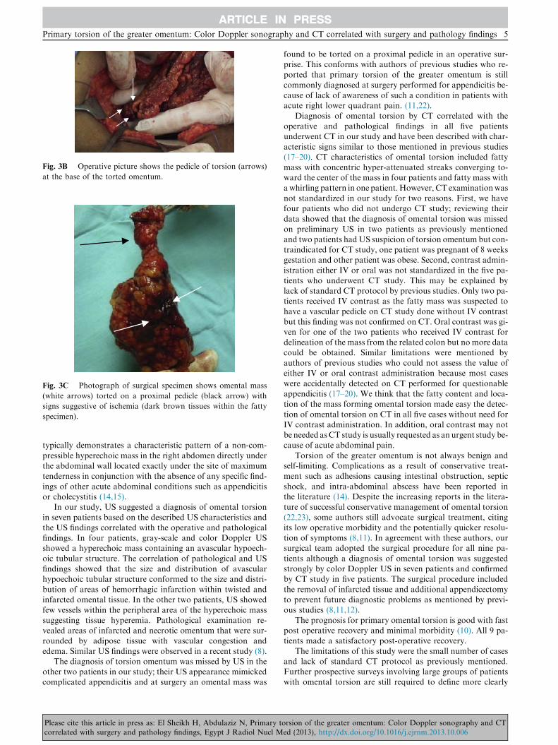
Fig. 3B Operative picture shows the pedicle of torsion (arrows)
at the base of the torted omentum.
Fig. 3C Photograph of surgical specimen shows omental mass
(white arrows) torted on a proximal pedicle (black arrow) with
signs suggestive of ischemia (dark brown tissues within the fatty
specimen).
Primary torsion of the greater omentum: Color Doppler sonography and CT correlated with surgery and pathology findings 5
ARTICLE IN PRESS
typically demonstrates a characteristic pattern of a non-com-
pressible hyperechoic mass in the right abdomen directly underthe abdominal wall located exactly under the site of maximumtenderness in conjunction with the absence of any specific find-
ings of other acute abdominal conditions such as appendicitisor cholecystitis (14,15).
In our study, US suggested a diagnosis of omental torsion
in seven patients based on the described US characteristics andthe US findings correlated with the operative and pathologicalfindings. In four patients, gray-scale and color Doppler USshowed a hyperechoic mass containing an avascular hypoech-
oic tubular structure. The correlation of pathological and USfindings showed that the size and distribution of avascularhypoechoic tubular structure conformed to the size and distri-
bution of areas of hemorrhagic infarction within twisted andinfarcted omental tissue. In the other two patients, US showedfew vessels within the peripheral area of the hyperechoic mass
suggesting tissue hyperemia. Pathological examination re-vealed areas of infarcted and necrotic omentum that were sur-rounded by adipose tissue with vascular congestion and
edema. Similar US findings were observed in a recent study (8).The diagnosis of torsion omentum was missed by US in the
other two patients in our study; their US appearance mimickedcomplicated appendicitis and at surgery an omental mass was
Please cite this article in press as: El Sheikh H, Abdulaziz N, Primary tcorrelated with surgery and pathology findings, Egypt J Radiol Nucl M
found to be torted on a proximal pedicle in an operative sur-prise. This conforms with authors of previous studies who re-ported that primary torsion of the greater omentum is still
commonly diagnosed at surgery performed for appendicitis be-cause of lack of awareness of such a condition in patients withacute right lower quadrant pain. (11,22).
Diagnosis of omental torsion by CT correlated with theoperative and pathological findings in all five patientsunderwent CT in our study and have been described with char-
acteristic signs similar to those mentioned in previous studies(17–20). CT characteristics of omental torsion included fattymass with concentric hyper-attenuated streaks converging to-ward the center of the mass in four patients and fatty mass with
awhirling pattern in one patient.However, CT examinationwasnot standardized in our study for two reasons. First, we havefour patients who did not undergo CT study; reviewing their
data showed that the diagnosis of omental torsion was missedon preliminary US in two patients as previously mentionedand two patients hadUS suspicion of torsion omentum but con-
traindicated for CT study, one patient was pregnant of 8 weeksgestation and other patient was obese. Second, contrast admin-istration either IV or oral was not standardized in the five pa-
tients who underwent CT study. This may be explained bylack of standard CT protocol by previous studies. Only two pa-tients received IV contrast as the fatty mass was suspected tohave a vascular pedicle on CT study done without IV contrast
but this finding was not confirmed on CT. Oral contrast was gi-ven for one of the two patients who received IV contrast fordelineation of the mass from the related colon but no more data
could be obtained. Similar limitations were mentioned byauthors of previous studies who could not assess the value ofeither IV or oral contrast administration because most cases
were accidentally detected on CT performed for questionableappendicitis (17–20). We think that the fatty content and loca-tion of the mass forming omental torsion made easy the detec-
tion of omental torsion on CT in all five cases without need forIV contrast administration. In addition, oral contrast may notbe needed asCT study is usually requested as an urgent study be-cause of acute abdominal pain.
Torsion of the greater omentum is not always benign andself-limiting. Complications as a result of conservative treat-ment such as adhesions causing intestinal obstruction, septic
shock, and intra-abdominal abscess have been reported inthe literature (14). Despite the increasing reports in the litera-ture of successful conservative management of omental torsion
(22,23), some authors still advocate surgical treatment, citingits low operative morbidity and the potentially quicker resolu-tion of symptoms (8,11). In agreement with these authors, oursurgical team adopted the surgical procedure for all nine pa-
tients although a diagnosis of omental torsion was suggestedstrongly by color Doppler US in seven patients and confirmedby CT study in five patients. The surgical procedure included
the removal of infarcted tissue and additional appendicectomyto prevent future diagnostic problems as mentioned by previ-ous studies (8,11,12).
The prognosis for primary omental torsion is good with fastpost operative recovery and minimal morbidity (10). All 9 pa-tients made a satisfactory post-operative recovery.
The limitations of this study were the small number of casesand lack of standard CT protocol as previously mentioned.Further prospective surveys involving large groups of patientswith omental torsion are still required to define more clearly
orsion of the greater omentum: Color Doppler sonography and CTed (2013), http://dx.doi.org/10.1016/j.ejrnm.2013.10.006