32
Prime Exposure Factors 1 By Professor Stelmark

Prime Exposure Factors 1
By Professor Stelmark

Properties of X-rays
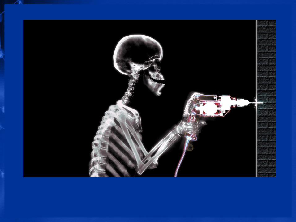
• Highly penetrative invisible rays• Electrically neutral• Polyenergetic• Liberate minute amounts of heat on passing through matter• Travel ordinarily in straight lines• Travel with the speed of light in vacuum• Ionize gasses• Cause fluorescence in certain crystals• Cannot be focused by lenses• Affect photographic film• Produce chemical and biological changes by ionization• Produce secondary and scatter radiation

Highly penetrative invisible rays
paper
Aluminum
Lead

Electrically neutral

Travel ordinarily in straight lines

Travel with the speed of light in vacuum
• 3 x 10⁸ m/s
• 3 x 10⁵ km/s •
• 186,000 miles/s

Affect photographic film

Produce secondary and scatter radiation

Methods of Image Identification
• Radiographic
• Photographic
• Electronic

Radiographic

Permanently generated in the image with the use of x-rays. Admissible in court

Photographic
Patient information, date, type of x-am.Admissible in court

Electronic
R upright
Patient information incorporated into the radiograph during the processing admissible in court . After
processing is NOT.
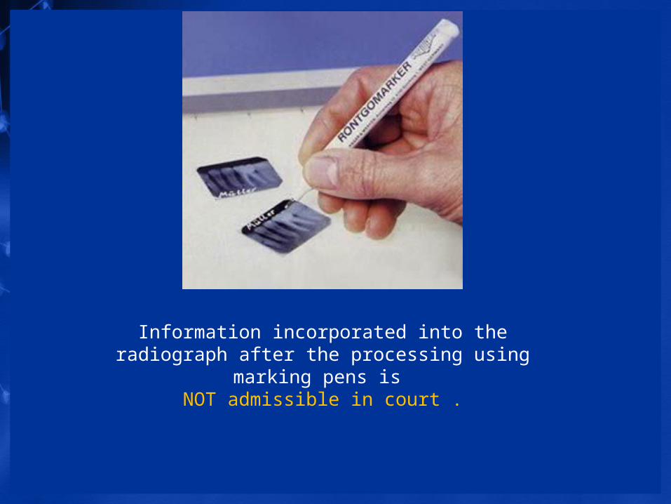
Information incorporated into the radiograph after the processing using marking pens is
NOT admissible in court .

Hostos Lab Radiograph Marking Practices
• Lead room letter markers A B C
• Lead anatomic side marker L or R
• Lead experiment exposure number marker 1 2 3 4

A
R
1

Important Radiological Science Terminology
• Photons• X-ray photons• X-ray beam quantity• X-ray beam quality• Radiation exposure• Exposure factors• Image receptor digital and analog• SID• SSD• X-ray beam field size• Irradiated area

Photons and x-ray photons
A photon is the smallest quantity of any type of electromagnetic energy, just as an atom is the smallest quantity of an element. A photon may be pictured as a small bundle of energy, sometimes called a quantum,that travels through space at the speed of light. We speak of x-ray photons, light photons, and other types of electromagnetic energy as photon radiation.
X-ray beam quantity
Refers to the total number of x-ray photons in a beam. Beam quantity is affected by mAs, kVp, and distance. The radiographer should associate quantity with radiation dose. All other factors remaining constant, an increase in quantity increases the radiation dose delivered to the patient. Beam quantity is directly proportional to mAs. Because mAs controls the number of electrons boiled off of the filament and available to produce x-rays, it is considered the primary factor controlling quantity. Doubling the mAs doubles the output. When adjustments in quantity are desired, mAs is the factor adjusted.

X-ray beam quality
Refers to the penetrating power of the x-ray beam. Penetration refers to those x-ray photons that are transmitted through the body and reach the image receptor. It is desirable for some of the x-ray photons to penetrate the anatomic area of interest, or no image would result. Photons that reach the image receptor create the dark shades of the image and areas where no photons reach result in the light or clear areas of the image. Both are needed to create the image. Beam quality is affected by kVp and is controlled mainly by adjusting kVp. As kVp increases, the beam's ability to penetrate matter also increases and vice versa. X-ray beams with high energy (from high kVp settings) are said to be high-quality or hard beams. X-ray beams with low energy (from low kVp settings) are said to be low-quality or soft beams.

Radiation exposure
The intensity of the x-ray beam of an x-ray imaging system is measured in roentgens (R) or milliroentgens (mR) and is called the x-ray quantity. Another term, radiation exposure, is often used instead of x-ray intensity or x-ray quantity. All have the same meaning and all are measured in roentgens.

Exposure factors
The primary exposure technique factors the radiographer selects on the control panel are milliamperage, time of exposure, and kilovoltage peak (kVp). Depending on the type of control panel, milliamperage and exposure time may be selected separately or combined as one factor, milliamperage/second (mAs). Regardless, it is important to understand how changing each separately or in combination affects the radiation reaching the IR and the radiographic image

Image receptor
Is any sensitive surface that receives the image forming x-rays from the patient and converts them into another form that can be visible
analog
CR computed radiography

DR digital

SID source to Image Receptor Distance
The distance between the source of the radiation and the IR

SOD source to Object Distance
Refers to the distance from the x-ray source (focal spot) to the object being radiographed.


X-ray beam field size
Refers to the dimension of an x-ray beam

Irradiated area
Refers to the object area being irradiated