and Laboratoire Associe INRA-ENV, Ecole Veterinaire de Lyon, BP83, 69280 Marcy l’Etoile,3 France,and Institute of Veterinary Virology, University of Bern, CH-3012 Bern, Switzerland4
Received 17 September 1997/Accepted 27 April 1998
We previously reported that infection of goats with caprine arthritis encephalitis virus (CAEV) tat2 proviralDNA or virus results in persistent infection, since the animals seroconverted and direct virus isolation fromcultures of blood-derived macrophages was positive. In this study we wanted to determine whether goatsinjected with CAEV tat2 proviral DNA or virus were protected against challenge with the pathogenic homol-ogous virus and to investigate whether CAEV tat2 was still pathogenic. All animals injected with CAEV tat2became infected as indicated by seroconversion and virus isolation. Challenge at 8 or 9 months postinfectiondemonstrated protection in four of four animals injected with CAEV tat2 but did not in three of threemock-inoculated challenged goats. Challenge virus was undetectable in the blood macrophages of protectedanimals during a period of 6 or 10 months postchallenge. In two of four protected animals, however, we wereable to detect the challenge wild-type virus by reverse transcriptase PCR on RNA directly extracted fromsynovial membrane cells surrounding the inoculation site. This result suggests that protection was achievedwithout complete sterilizing immunity. Animals injected with CAEV tat2 and mock challenged developedinflammatory lesions in the joints, although these lesions were not as severe as those in CAEV wild-type-injected goats. These results confirm the dispensable role of Tat in CAEV replication in vivo for the estab-lishment of infection and pathogenesis and demonstrate in another lentivirus infection model the efficacy oflive attenuated viruses to induce resistance to superinfection.
Like other lentiviral infections, caprine arthritis encephalitisvirus (CAEV) infection is characterized by viral persistencein the face of an immune response and the onset of slow andprogressive degenerative diseases (20). Synovitis, mastitis,pneumonia, and encephalitis characterize the course of diseasein CAEV infection (29). These inflammatory diseases are theresult of viral infection of cells of monocyte/macrophage lin-eage, which are the main target cells in vivo (8–10). Infectionof macrophages is a common feature of lentiviral infectionsand plays a central role in the development of associated dis-eases. The CAEV infection model thus provides a system toanalyze the pathogenesis of diseases associated with macro-phage infection and to develop vaccine strategies against mac-rophage-tropic viruses. The use of an infectious molecularclone of CAEV (34, 37) allowed us to investigate the role ofdifferent genes in the induction of infection and pathogenesis.We previously demonstrated the essential role of the vif genefor efficient CAEV replication in vitro and in vivo (13, 15),whereas the tat gene was shown to be dispensable in bothinstances (14). Goats inoculated with CAEV vif2 virus orproviral DNA were not protected against CAEV wild-type (wt)challenge, due to the reduced level of CAEV vif2 replication(15, 16). Similar results were obtained in the simian immuno-deficiency virus (SIV) macaque model, in which an inversecorrelation between the degree of virus attenuation and the
induction of protection was demonstrated (26, 42). In the ma-caque model, it is now clearly established that long-term pro-tection against systemic challenge can be achieved by systemicimmunization with live attenuated SIVmac239 Dnef or D3 (7,42) or SIVmac32H C8 (31). This approach was also shown tobe effective against mucosal challenge after either systemicSIVmac32H C8 (6) or mucosal simian/human immunodefi-ciency virus 89.6 (28) immunization. All these results wereobtained with lymphocyte-tropic viruses. Since macrophage-tropic strains of human immunodeficiency virus type 1 (HIV-1)seem to be the transmitted viruses responsible for initial infec-tion (36, 39, 44), they might be the initial targets to consider invaccine strategies. Indeed, in the SIV model, immunization ofmacaques with attenuated macrophage-tropic SIV/17E-CI re-sulted in protective immunity against heterologous challenge(4).
In a previous study, we reported that proviral DNA of theCAEV Cork molecular clone with deleted tat sequences(CAEV tat2) produced persistent infection in goats, with anantibody response showing kinetics of appearance and a reac-tivity pattern against viral proteins that were similar to those inCAEV wt-infected goats (14). The present study was designedto evaluate the capacity of this live attenuated virus to induceresistance to superinfection and to investigate the pathogenicproperties of CAEV tat2 compared to those of CAEV wt.Protection against challenge with a high dose (.250 100%animal infectious dose [AID100]) of homologous CAEV wt wasachieved in all goats inoculated with CAEV tat2 without com-plete sterilizing immunity. A control animal inoculated withCAEV tat2 and mock challenged developed mild inflamma-tory lesions in the joints, ruling out an essential role for the
CAEV tat gene in virus-induced pathogenesis. Although fur-ther attenuation of CAEV tat2 is required to obtain a non-pathogenic live attenuated vaccine strain, these experimentsdemonstrated the efficacy of this vaccine strategy in an addi-tional lentiviral animal model.
MATERIALS AND METHODS
Viruses. The infectious proviral DNA of the CAEV Cork strain was generatedby ligation of the 9- and 0.5-kb HindIII CAEV fragments (34, 37). The tat-deleted mutant, containing a 153-bp in-frame deletion between positions 5677and 5829, was previously described (14). Viral stocks were obtained by transfec-tion of CAEV wt or tat2 proviral DNA into primary goat synovial membranecells or by infection of goat synovial membrane cells with transfection superna-tants. These stocks contained 105 50% tissue culture infectious doses (TCID50)per ml. Preliminary in vivo virus titration with fivefold stock virus dilutionsrevealed that five of five goats that were given the last 2.5 3 1022 dilution by theintratracheal route became persistently infected (data not shown). Therefore, thestock virus is estimated to contain .250 AID100 per ml.
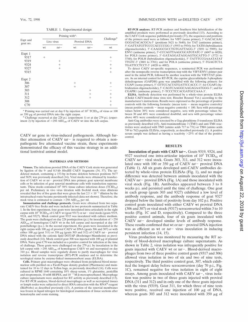
Immunization and challenge protocols. Goats were obtained from two sepa-rate CAEV-free flocks and were included in two protocols summarized in Table1. In the first experiment, four goats were inoculated intra-articularly in the rightcarpus with 105 TCID50 of CAEV wt (goat 9317) or tat2 viral stocks (goats 9319,9324, and 9327). Mock control goat 9315 was inoculated with culture medium.The goats were challenged on day 228 postinjection (p.i.) by inoculation of .250AID100 of homologous CAEV wt in the left carpus and necropsied at day 185postchallenge (p.c.). In the second experiment, five goats were inoculated in theright carpus with 100 mg of proviral CAEV wt DNA (goats 306 and 307) or witheither 100 mg (goat 311) or 350 mg (goats 303 and 312) of CAEV tat2 proviralDNA mixed with the cationic lipid DOTAP (Boehringer-Mannheim) as previ-ously described (14). Mock control goat 308 was injected with 100 mg of plasmidDNA. Naive goat C70 was included as a positive control for infection at the timeof challenge. These goats were challenged on day 279 p.i. by inoculation in theleft carpus with .250 AID100 of homologous CAEV wt and necropsied on day310 p.c. Blood samples were regularly drawn to purify macrophages for virusisolation and reverse transcriptase (RT)-PCR analyses and to determine theserological status by enzyme-linked immunosorbent assay (ELISA).
Cells. Primary goat macrophages were obtained from peripheral blood mono-nuclear cells purified on a Ficoll-Paque density gradient (Pharmacia) and main-tained for 10 to 12 days in Teflon bags, as previously described (14). Cells werecultured in RPMI 1640 containing 10% sheep serum, 1% glutamine, penicillinand streptomycin, 10 mM HEPES, and 1025 M b-mercaptoethanol. Macrophageculture supernatants were analyzed for RT activity as previously described (13).For RT-PCR analyses, blood-derived macrophages as well as necropsied tissuesor lymph nodes were subjected to direct RNA extraction with the RNAB reagent(BioProbe) as described previously (13). A portion of the synovial membraneswas frozen in liquid nitrogen for histological examination of tissue sections afterhematoxylin and eosin staining.
RT-PCR analyses. RT-PCR analyses and Southern blot hybridization of theamplified products were performed as previously described (13). According tothe CAEV Cork sequence published previously (37), the sequences and positionsof the primers used were as follows: for VIF5 (sense primer), 59-GACACAACGGGATACACGCA-39 (positions 5621 to 5640); for TAT (antisense primer),59-GATTATGTTCCCCACCCCGG-39 (5953 to 5934); for TATH (hybridizationoligonucleotide), 59-CAAGGCGCCTGTGATTAGG-39 (5891 to 5909); forENV1 (antisense primer), 59-CCCAGTTAAGCGCATGTATC-39 (6047 to 6028);for POLS (sense primer), 59-GATAGGATAGGAGTGCATTG-39 (3721 to3740); for POLH (hybridization oligonucleotide), 59-TATTTCCGAAATATATTTGTC-39 (3801 to 3781); and for POLA (antisense primer), 59-TGAGTCTATGATTCCTCCT-39 (4020 to 4002).
To detect CAEV tat-specific sequences, a seminested PCR was performedafter the nonspecific reverse transcription step with the VIF5/ENV1 primer pairused in the initial PCR, followed by another reaction with the VIF5/TAT prim-ers. As an internal control for RT-PCR, the caprine glyceraldehyde-3-phosphatedehydrogenase (GAPDH) gene was amplified with the following primers: forCGAP1 (sense primer), 59-GTTCCACTATGATTCCACCC-39; for CGAP2 (hy-bridization oligonucleotide), 59-CAGTCAAGGCAAGAGAATGGG-39; and forCGAPR1 (antisense primer), 59-TCCCTCCACGATGCCAAA-39.
ELISA. Antibody detection was performed by a whole-virus protein ELISA(Chekit CAEV/maedi-visna virus test; Hoechst Roussel Vet.) according to themanufacturer’s instructions. Results were expressed as the percentage of positivecontrols with the following formula: (mean tests 2 mean negative controls)/(mean positive controls 2 mean negative controls) 3 100. Sera with percentagevalues below 30% were considered negative, sera with percentage values be-tween 30 and 40% were considered doubtful, and sera with percentage valuesabove 40% were considered positive.
Anti-Gag antibodies were screened by a Gag-glutathione S-transferase ELISAas previously described (43). Anti-transmembrane 3 (TM3) and anti-TM4 reac-tivities were analyzed with TM3 (amino acids 717 to 731) or TM4 (amino acids749 to 762) peptide ELISAs, respectively, as described previously (1). A positiveserum sample was defined as having a reactivity .25% of that of the positivecontrol.
RESULTS
Inoculation of goats with CAEV tat2. Goats 9319, 9324, and9327 received one intra-articular injection of 105 TCID50 ofCAEV tat2 viral stock. Goats 303, 311, and 312 were inocu-lated once with 100 or 350 mg of CAEV tat2 proviral DNA(Table 1). All six goats developed anti-CAEV antibodies de-tected by whole-virus protein ELISAs (Fig. 1), and no majordifference was detected between animals inoculated with theCAEV tat2 proviral DNA (Fig. 1A) and those inoculated withviral stock (Fig. 1B). Antibodies appeared between 3 to 8weeks p.i. and persisted until the time of challenge. One goatin each group (goats 303 and 9327) exhibited a low level ofantibody response, and the antibody response of goat 9327dropped below the limit of positivity from day 102 p.i. Positivecontrol goats inoculated with either CAEV wt proviral DNA(306 and 307) or viral stock (9317) seroconverted within 3 to 10weeks (Fig. 1C and D, respectively). Compared to the threepositive control animals, four of six goats inoculated withCAEV tat2 developed similar levels of antibody response.These results confirmed that wt or tat2 proviral DNA injectionwas as efficient as wt or tat2 virus inoculation in inducingpersistent infection (14, 15).
Virus production was monitored by measuring the RT ac-tivity of blood-derived macrophage culture supernatants. Asshown in Table 2, virus isolation was infrequently positive forgoats injected with CAEV wt or tat2. Blood-derived macro-phages from two of three positive control goats (9317 and 306)allowed virus isolation in two of six and two of nine tests,respectively. The third positive control goat, 307, which exhib-ited the longest delay before seroconversion (day 70 p.i., Fig.1C), remained negative for virus isolation in eight of eightassays. Among goats inoculated with CAEV tat2, virus isola-tion was positive in two of three goats injected with proviralDNA (311 and 312) and in only one of the three goats injectedwith the virus (9319). Goat 311, for which three of nine testswere positive, received one injection of 100 mg of DNA,whereas goats 303 and 312 were inoculated with 350 mg of
DNA, suggesting that the inoculation dose did not affect thelevel of virus replication. Virus isolation remained negative forgoats 303, 9327, and 9324, although they developed low to highantibody responses, respectively (Fig. 1A and B). These resultsprobably reflect variation between animals in the ability tocontrol viral replication, together with the difficulty of isolatingvirus from a small percentage of infected blood monocytes (9).No significant difference was observed in the frequency ofisolation of viruses from animals injected with CAEV wt ortat2 proviral DNA or virus-injected animals. RT-PCR analysiswas performed on day 91 p.i. with RNA extracted from mac-rophage-produced viruses (Table 2, goats 306, 307, 303, 311,312, and 308) and showed that the deletion introduced into thetat gene was still detectable in tat2 virus particles (14), rulingout the possibility that a recombination event caused reversalto the wt phenotype, which would explain the lack of differencebetween goats injected with CAEV wt and those injected withtat2.
Antibody response analysis. In the CAEV infection model,a correlation was previously described between the severity ofdisease in infected animals and the titers of anti-Env antibod-ies (23), especially the anti-TM antibodies (27). Four immu-nodominant epitopes were delineated in the CAEV gp38 TM,and antibody reactivity against two of them, TM3 and TM4,was shown to be associated with the presence of clinical ar-thritis (1). To compare the ability of CAEV tat2 proviral DNAor virus infection to induce such antibody specificities with thatof CAEV wt infection, we tested the goat sera by ELISA withpeptides TM3 (amino acids 717 to 731) or TM4 (amino acids
749 to 762) as antigens. In parallel, an ELISA specific for therecombinant Gag fusion protein glutathione S-transferase-Gagwas used to detect anti-Gag antibodies. Sera were tested at day146 p.i. for the group injected with CAEV wt or tat2 proviralDNA and at day 221 p.i. for the group injected with CAEV wtor tat2 virus. We chose to analyze late sera, since it was re-ported that the kinetics of anti-TM antibody appearance werearound 12 to 32 weeks p.i. compared to 3 to 4 weeks p.i. foranti-Gag antibodies (1). Results are summarized in Table 3(left-hand side). Sera were considered positive for values.25% of the ELISA for the positive controls. All positivecontrol goats, 306, 307, and 9317, developed anti-Gag and anti-TM3 antibodies, whereas only goat 306 also produced anti-TM4 antibodies. In the groups injected with CAEV tat2, goat311 (tat2 proviral DNA) and goat 9319 (tat2 virus) developedall three antibody specificities. Goat 312 (tat2 proviral DNA)was weakly positive against Gag and TM4. Goat 303 (tat2 pro-viral DNA) and goats 9324 and 9327 (tat2 virus) were consid-ered unreactive in all three ELISAs. Western blot analysisagainst whole-virus proteins, however, demonstrated that day146 p.i. sera from goats 303 and 312 were weakly reactive againstmature Gag proteins p28, p18, and p14.5 (14). This discrep-ancy between ELISA and Western blot results has alreadybeen observed with sera from naturally infected goats (1).Noticeably, three of six animals inoculated with CAEV tat2developed anti-Gag and anti-TM antibodies after inoculation(goats 311, 312, and 9319) compared to three of three goatsinjected with CAEV wt, which developed both reactivities.
FIG. 1. Antibody response. Anti-CAEV antibodies to whole-virus proteins were measured by ELISA on different days (d) postinfection or p.c. Time of challengeis indicated by the arrows. (A) Goats inoculated with CAEV tat2 proviral DNA and mock-challenged (303 tat2/2) or wt-challenged goats (311, 312 tat2/wt). (B) Goatsinoculated with CAEV tat2 and mock-challenged (9319 tat2/2) or wt-challenged (9324, 9327 tat2/wt) goats. (C) Mock-inoculated and wt-challenged goat (308 2/wt)or naive and wt-challenged (C70 wt) goat and CAEV wt proviral DNA-positive control animals mock challenged (307 wt/2) or wt challenged (306 wt/wt). (D)Mock-inoculated challenged control goat (9315 2/wt) and positive control goat challenged with CAEV wt (9317 wt/wt).
Challenge of goats inoculated with CAEV tat2. Challengewas performed with a high dose of homologous CAEV wt(.250 AID100) injected in the joint, which is one of the maintargets of infection. Goats inoculated with CAEV tat2 or wtvirus were challenged on day 228 p.i., whereas goats injectedwith CAEV tat2 or wt proviral DNA were challenged on day279 p.i. A naive animal, goat C70, was included at that time asan additional positive control for infection (Table 1). Two of
three goats inoculated with CAEV wt, 306 and 9317, werechallenged with the homologous CAEV wt, whereas goat 307was mock challenged. Two goats inoculated with CAEV tat2,303 (tat2 proviral DNA) and 9319 (tat2 virus), were mockchallenged. Most additional results were obtained with goat9319, since goat 303 died accidentally at day 30 p.c. The fourother animals inoculated with CAEV tat2, 311 and 312 (tat2proviral DNA) and 9324 and 9327 (tat2 virus), were chal-
TABLE 2. Virus isolation and RT-PCR detection in blood-derived macrophages
Day
RT activity/RT-PCR detectiona in goat challenged as indicated
a Virus was isolated by RT activity measurement of 1 ml of macrophage culture supernatant at day 12, concentrated 1003/RT-PCR detection of RNA extracted fromblood-derived macrophages cultured for 12 days with the VIF5/ENV1 primer pair. 2, no fragment amplification; wt, detection of the 427-bp wt tat fragment; tat2,detection of the 274-bp tat-deleted fragment. Southern blots of the amplified fragments were hybridized with the 32P-labeled TATH oligonucleotide probe.
b Right intracarpal inoculation of CAEV wt or tat2 proviral DNA or virus.c Left intracarpal challenge with pathogenic CAEV.d Goat 303 died accidentally at day 30 p.c.
TABLE 3. Antibody specificitiesa after inoculation with CAEV tat2 or wt and pathogenic challenge
a Antibody reactivities were measured by ELISAs specific for a glutathione S-transferase-Gag fusion protein, the TM3 peptide (amino acids 717 to 731), and the TM4peptide (amino acids 749 to 762). Sera were diluted 1:100, 1:60, and 1:20, respectively, and values higher than 25% were considered positive. Samples analyzed weretaken at day 146 p.i. and day 152 p.c. for goats 308, 306, 307, 311, and 312 and at day 221 p.i. and day 84 p.c. for goats 9315, 9317, 9319, 9324, and 9327.
b Histopathological lesions observed in sections of synovial membranes were graded low (1), mild (11), or severe (111). ND, not determined.
VOL. 72, 1998 IMMUNIZATION WITH tat-DELETED CAEV 6799
lenged with CAEV wt. All control animals became infected asshown by the seroconversion curves (goats 308 and C70, Fig.1C and goat 9315, Fig. 1D). The antibody response level forgoat 9319 remained high (Fig. 1B), suggesting continuous stim-ulation by viral antigens. An increase in antibody response wasobserved in the four wt-challenged animals inoculated withCAEV tat2, goats 311 and 312 (Fig. 1A) and goats 9324 and9327 (Fig. 1B), as measured on day 84 p.c. Isolation of viruswas possible from blood-derived macrophages of positive con-trol goats (Table 2). Of the samples from the two animalschallenged and inoculated with CAEV wt, only those fromgoat 9317 allowed positive virus isolation by RT activity mea-surement and RT-PCR on RNA from blood-derived macro-phages (Table 2). Positive control mock-challenged goat 307developed a persistent low-level infection as revealed by thelate occasional RT-PCR virus detection on blood-derived mac-rophages (Table 2). Virus isolation was negative for all animalsimmunized with CAEV tat2, regardless of whether theywere mock challenged or challenged with CAEV wt (Table 2).In only one case (goat 9324 on day 15 p.c.) was the immu-nizing tat2 virus detected in blood-derived macrophages byRT-PCR with the VIF5/TAT primer pair (Table 2). This resultsuggests a transient reactivation of the CAEV tat2 by the wtchallenge virus. These observations indicate that prior im-munization with the attenuated CAEV tat2 induced protec-tion or superinfection resistance against homologous patho-genic challenge at the peripheral blood level.
Protein-specific ELISAs revealed that the two positive con-trol animals tested, 308 and 9315, developed both anti-Gag andanti-TM3 antibodies, but only goat 308 produced anti-TM4antibodies (Table 3, right-hand side). An increase of anti-Gag,anti-TM3, and anti-TM4 antibodies was observed in sera fromgoat 306, whereas goat 9317 demonstrated no change in anti-body specificity. The late development of anti-TM3 antibodieswas detected in goat 307 (Table 3, right-hand side). Whereas aslight decrease was observed in the anti-Gag, anti-TM3, andanti-TM4 reactivities of goat 9319, all these antibody specific-ities were strengthened in the four wt-challenged animals thatwere inoculated with CAEV tat2, without the appearance ofnew reactivities (Table 3, right-hand side), suggesting that the
humoral response was stimulated by the wt challenge virus.RT-PCR analyses performed on RNA extracted from differenttissues of positive control goats allowed the detection of chal-lenge virus (Table 4). Virus was also detected in the synovialmembranes and lymph nodes of goat 9317 as well as in thelymphoid tissues of goat 306 (Table 4). The infected status ofgoat 307 was confirmed by RT-PCR analyses on synovial mem-branes, mammary secretions, and lymphoid tissues (Table 4).Most of the necropsied tissues of goat 9319 were RT-PCRpositive with the POLS/POLA primer pair (Table 4), suggest-ing a continuous replication of CAEV tat2 in target tissues.RT-PCR analyses with the POLS/POLA primer pair on dif-ferent tissues of wt-challenged goats inoculated with CAEVtat2 gave positive reactions in the synovial membranes andmammary secretions of goats 311 and 312 and in the lymphnodes and bone marrow of goats 9324 and 9327 (Table 4).Since this reaction did not allow us to distinguish between thetat2 immunizing virus and the wt challenge virus, further RT-PCR analyses were performed with the VIF5/TAT primer pairto amplify the tat gene.
Viral RNA detection in necropsied tissues and histopathol-ogy. As CAEV wt challenge virus was not detected at theperipheral level, we next examined whether sequestration ofthe challenge virus occurred in protected wt-challenged goatsimmunized with tat2. Tissue samples were taken at necropsyand analyzed by RT-PCR for the presence of deleted or wt tatviral RNA. Figure 2 shows results obtained from cells frommammary secretions of female goats. wt tat RNA could beamplified from negative control challenged goats 9315 and C70as well as from positive control mock-challenged goat 307 orwt-challenged goat 9317. Only a deleted tat product was am-plified from mock-challenged goat 9319 or wt-challenged ani-mals 311 and 312 which had been inoculated with CAEV tat2.An RT-PCR specific for tat was negative for samples fromwt-challenged goats 9324 and 9327, which had been injectedwith CAEV tat2. No wt challenge virus was detected in fluidssecreted by animals immunized with tat2, confirming the re-sults obtained with blood-derived macrophages.
Figure 3 shows results obtained with RNA extracted fromsynovial membranes surrounding the inoculation sites. A wt tat
TABLE 4. Virus detection by RT-PCR analysis on necropsy tissues
Expt 2306 wt DNA wt 1 1 NT 2 1 2 1 2307 wt DNA Mock 1 2 1 2 2 2 2 2311 tat2 DNA wt 2 1 1 2 2 2 2 2312 tat2 DNA wt 1 1 1 2 2 2 2 2308 Mock wt 1 2 NT 2 2 2 2 2C70b wt 1 1 1 NT NT NT NT 1
a RT-PCR analysis on RNA directly extracted from tissues taken at sacrifice was performed with the POLS/POLA primer pair. Southern blots of the amplifiedfragments were hybridized with the 32P-labeled POLH oligonucleotide probe. 1, detection of the 299-bp pol fragment; 2, no detectable amplified products; NT, nottested. Tissue samples analyzed were from left or right synovial membranes (SM), mammary secretions (MS), left or right retromammary lymph node (RMLN), leftor right prescapular lymph node (PSLN), and bone marrow (BM).
b Goat C70 received no priming and was included at the time of challenge.
amplification product could be observed in all negative controlchallenged animals, in both the right and the left joints of goats9315 and C70 but in goat 308 in only the joint inoculated withCAEV wt. Among positive control animals, mock-challengedgoat 307 was negative for the tat-specific RT-PCR in bothjoints, whereas the pol-specific primer pair allowed viral RNAdetection in the left joint (Table 4). For the two other positivecontrol challenged animals, 306 and 9317, amplification of wttat was positive in synovial membranes from both joints. tat-deleted amplification products could be observed in three offour wt-challenged goats inoculated with CAEV tat2, in goats311 and 312 in the joint injected with CAEV tat2, and in bothjoints of goat 9327, although in this last case the tat2 amplifiedproduct exhibited a slightly different size. Both wt tat and tat2amplification products were detected in the wt-injected joint ofgoat 312 and in both synovial membranes of goat 9327. Sam-ples from goat 9324 were negative for tat-specific RT-PCRanalysis, whereas parallel amplification performed with pol-specific primers allowed viral RNA detection (Table 4). Thisdiscrepancy, also observed for goat 307, could be due to thelower efficiency of the VIF5/TAT primer pair compared to thatof POLA/POLS and/or to a lower viral load in these infectedanimals. We were able to detect the wt challenge virus at theinoculation site in two of four wt-challenged animals that hadbeen immunized with CAEV tat2; this suggests that control ofthe wt virus replication occurred in these animals without com-plete sterilizing immunity.
The pathogenic properties of the tat2 attenuated virus werethen analyzed. A portion of the necropsied synovial mem-branes of the challenged animals was frozen, and tissue sec-tions were examined for the presence of inflammatory lesions.Different tissue sections from each animal were examined in-dependently by two or three examiners, and representativepictures for each group are shown in Fig. 4. Compared to thesynovial membrane section of a naive animal (Fig. 4A), a tissuesection from the wt-challenged goat inoculated with CAEV wt(goat 9317) (Fig. 4B) showed the characteristics of severe vi-rus-induced inflammatory lesions with the high lymphocyticand monocytic infiltration that we and others have alreadydescribed with similar experimental infections (14, 38). CAEVtat2 injection resulted in lesions that were less severe thanthose resulting from the wt virus, as observed on a tissue sec-
tion from the mock-challenged goat injected with CAEV tat2virus (goat 9319) (Fig. 4C). Thickening of the synovial mem-brane as well as lymphocytic infiltration of the connective tis-sue was observed. Mild lesions were also observed in tissuesections of wt-challenged goat 9327, which was injected withCAEV tat2 (Fig. 4D); these lesions were less severe than thosein the positive control goat (Fig. 4B), indicating that wt-chal-lenged animals immunized with CAEV tat2 resisted the path-ogenicity of the challenge virus, since lesions observed in theseanimals were most probably due to replication of the immu-nizing virus itself. Most of the animals developed either onlyanti-TM3 antibodies (9315, 307, 312, and 9324) or only anti-TM3 and anti-TM4 antibodies (308, 306, 311, and 9319) andlesions that ranged from low to severe (Table 3). Goats 9317and 9327 developed severe and mild lesions, respectively, inthe absence of either of these antibody reactivities (Table 3). Inagreement with results previously obtained with a larger panelof sera (1), most sera in this study (80%) reacted with TM3peptide and Gag and correlated with an arthritic condition.
DISCUSSION
The infectivity of retroviral DNA has been proved in severalmodels, including SIV, bovine leukemia virus, CAEV, andfeline immunodeficiency virus (14, 15, 24, 35, 41). This methodprevents variation in infection efficacy due to the presenceof various proportions of defective viruses in individual viralstocks and allows the evaluation of the infectious and patho-genic properties of genetically modified retroviral genomes. Inthis study, we extended our previous results (14) and demon-strated the reliability and efficiency of this infection procedurecompared to those of infection with viral stocks obtained invitro by transfection of the same infectious CAEV molecularclone, either wt or tat2. Overall, time to seroconversion andvirus isolation were not significantly different between animalsinoculated with CAEV tat2 or wt proviral DNA and goatsinjected with CAEV tat2 or wt virus. A more detailed analysisof the antibody response, however, revealed that infection withCAEV tat2 was less efficient than injection with CAEV wt ininducing a high-level antibody response and stimulating theproduction of anti-Gag and anti-TM reactivities. The differ-ences detected in the antibody response in animals injectedwith CAEV tat2 compared to that in goats inoculated withCAEV wt could be due to an attenuated replication of CAEVtat2 in vivo. We observed that goats 303 and 9327, with the
FIG. 2. Challenge virus detection by RT-PCR on RNA extracted from cellsin mammary secretions. Amplification was performed with the VIF5/ENV1primer pair, and a Southern blot of the amplified products was hybridized withthe 32P-labeled TATH oligonucleotide probe. The CGAP1/CGAP2 primer pairwas used to amplify the caprine housekeeping gene GAPDH as an internalcontrol. Molecular size markers are indicated at the left.
FIG. 3. Challenge virus detection by RT-PCR on RNA extracted from right(a) or left (b) synovial membranes. Amplification was performed by seminestedPCR, first with the VIF5/ENV1 primer pair and then with primers VIF5/TAT,resulting in the production of a 333-bp fragment for the wt tat gene and a 180-bpfragment for the tat2 gene. Amplification of the GADPH control gene andSouthern blot hybridization were as described in the legend for Fig. 3.
VOL. 72, 1998 IMMUNIZATION WITH tat-DELETED CAEV 6801
lowest antibody response, had no detectable anti-Gag or an-ti-TM antibodies and were negative for virus isolation.Goats 312 and 9324, with a high antibody response, reactedweakly against Gag and TM epitopes before challenge andremained negative for virus isolation. Finally, goats 311 and9319, with the highest antibody level, reacted strongly againstthe Gag and TM epitopes, and virus could be isolated fromblood-derived macrophage cultures. Taking into account thevariation between the animals’ ability to control infection,these differences were not observed in the three positive con-
trol goats. In the CAEV infection model, there seems to be acorrelation between the intensity of the immune response andthe level of virus replication (15, 16); this correlation was alsoobserved in the case of SIVmac32H C8 infection (6) but not inthe SIVmac239 Dnef (7, 22) or equine infectious anemia virusDDU models (25).
The results demonstrate that infection with attenuatedCAEV tat2 via intracarpal inoculation can protect goats fromhomologous pathogenic challenge in the contralateral joint.Protection was defined as the absence of challenge virus de-
FIG. 4. Histopathology of synovial membranes from a naive goat (A), from positive control goat 9317, which was injected with CAEV wt (B), from mock-challengedgoat 9319, which was inoculated with CAEV tat2 (C), and from wt-challenged goat 9327, which was inoculated with CAEV tat2 (D). Synovial membrane samples weretaken at necropsy, and frozen sections were stained with hematoxylin and eosin. Magnification, 310.
tection in peripheral blood-derived macrophages during thepostchallenge period—185 days for the group injected withCAEV tat2 proviral DNA and 310 days for the group inocu-lated with CAEV tat2. No anamnestic antibody response wasdetected; however, an increase in anti-CAEV response wasobserved in goats 311 and 312, suggesting limited exposure toCAEV antigens. As in some vaccination studies with attenu-ated SIV (6, 28, 31), we found no correlation between the levelof antibody response and protection. Protection was achievedwithout complete sterilizing immunity, since RT-PCR analysesof different tissues obtained at necropsy allowed the detectionof the challenge wt virus in the inoculated joints of two of fourprotected animals. In one of these two goats, 9327, CAEV wtRNA was present in both carpal joints, showing that the chal-lenge virus may have diffused through the body before it wasbrought under control. The local immune response, eitherantibody or cell-mediated, may be responsible for this controlmechanism, since the synovial fluid and synovium of infectedgoats are rich in plasmocytes, activated CD41 and CD81 lym-phocytes, and macrophages (12, 21, 40).
In a previous study, we reported that immunization with thehighly attenuated CAEV vif2 failed to protect goats againsthomologous pathogenic challenge (15)—even when the goatswere challenged after a long period of immunization (about 8months p.i.)—whereas the slightly attenuated CAEV tat2 im-munization was protective. The fact that these two experimen-tal groups were inoculated and challenged with the same pro-tocol allows us to determine the impact of the degree ofattenuation of the vaccine viruses on their efficiency to induceprotection. In another lentiviral model, these results confirmthe inverse correlation established with live attenuated SIVsbetween the level of vaccine strain replication and the induc-tion of protection (26, 42). In addition to the replication effi-ciency of the vaccine strain, an important parameter of efficientprotection is duration of the vaccination. In the macaquemodel, protection was obtained with live attenuated SIVs, andresults demonstrate a clear trend toward increased protectionwith the time of vaccination (4, 7, 17, 31, 42). Most effectivevaccination assays against lentiviral infection have failed toclearly demonstrate whether protection was immunity medi-ated or based on superinfection resistance due to interference.Further analyses are necessary to evaluate the maturation ofthe immune response, described in the SIV and equine infec-tious anemia virus models (4, 5, 11), as well as to investigatethe induction of a cellular immune response to CAEV tat2immunization.
Examination of synovial membrane sections from a mock-challenged goat inoculated with CAEV tat2 revealed mildhistopathological changes compared to the severe inflamma-tory lesions observed in the joints of goats infected with CAEVwt. Since the CAEV tat gene is not strictly required to establishpersistent infection and the onset of clinical signs, the functionof Tat is still unknown. Several studies reported the correlationbetween the level of anti-Env (anti-SU and anti-TM) antibod-ies and the development of arthritic lesions in the CAEVinfection model (our results and references 1, 23, and 27),together with the high level of virus expression in tissue mac-rophages (45) and the massive infiltration of the arthritic sy-novium by B lymphocytes, plasmocytes, and activated CD41
and CD81 lymphocytes (2, 12, 21, 40). Recent reports sug-gested the role of differential activation of CAEV-reactiveT-helper subsets in virus expression control and disease out-come and associated the dominance of T-helper 2-like cellswith arthritis (3, 33, 40). Our results suggest a role for CAEVTat in the increase of the viral replication level, independentlyof its weak transactivation of the viral long terminal repeat (14,
18). As Tat of visna virus was reported to regulate the expres-sion of cellular genes involved in activation pathways (30), onehypothesis is that Tat of small ruminant lentiviruses activatesthe infected macrophages, resulting in increased viral expres-sion and thereby augmenting the reactivities of antibodies andactivated lymphocytes to viral antigens and infected cells in thesynovial tissue (2, 12, 21, 40). Nontranscriptional function ofHIV-1 Tat in virion infectivity and immune activation of HIV-1-infected cells by Tat were recently described (19, 32) andmay be a general feature of lentiviral Tat proteins.
ACKNOWLEDGMENTS
We thank J. M. Guibert, M. Vignoni (CNEVA, Sophia-Antipolis,France), E. Pardo, and P. Bolland (ENV, Lyon, France) for theirexcellent technical assistance and Sophie Dufour (IVV, Bern, Switzer-land) for her precious help. We thank our colleagues at INSERMU372 for their support and M. Guyader and K. E. Willett for criticalreading of the manuscript.
A. Harmache was the recipient of a doctoral fellowship from theFrench Agency against AIDS (ANRS). G. Bertoni was supported bygrant 31-41859.94 from the Swiss National Science Foundation. Thiswork was supported by INSERM and ANRS.
REFERENCES
1. Bertoni, G., M.-L. Zahno, R. Zanoni, H.-R. Vogt, E. Peterhans, G. Ruff, W. P.Cheevers, P. Sonigo, and G. Pancino. 1994. Antibody reactivity to the im-munodominant epitopes of the caprine arthritis encephalitis virus gp38transmembrane protein associates with the development of arthritis. J. Virol.68:7139–7147.
2. Cheevers, W. P., D. P. Knowles, T. C. McGuire, D. R. Cunningham, D. S.Adams, and J. R. Gorham. 1988. Chronic disease in goats orally infected withtwo isolates of caprine arthritis encephalitis virus. Lab. Investig. 58:510–517.
3. Cheevers, W. P., J. C. Beyer, and D. P. Knowles. 1997. Type 1 and type 2cytokine gene expression by viral gp135 surface protein-activated T lympho-cytes in caprine arthritis encephalitis lentivirus infection. J. Virol. 71:6259–6263.
4. Clements, J. E., R. C. Montelaro, M. C. Zink, A. M. Amedee, S. Miller, A. M.Trichel, B. Jagerski, D. Hauer, L. N. Martin, R. P. Bohm, and M. Murphey-Corb. 1995. Cross-protective immune responses induced in rhesus macaquesby immunization with attenuated macrophage-tropic simian immunodefi-ciency virus. J. Virol. 69:2737–2744.
5. Cole, K. S., J. L. Rowles, B. A. Jagerski, M. Murphey-Corb, T. Unangst, J. E.Clements, J. Robinson, M. S. Wyand, R. C. Desrosiers, and R. C. Montelaro.1997. Evolution of envelope-specific antibody responses in monkeys exper-imentally infected or immunized with simian immunodeficiency virus and itsassociation with the development of protective immunity. J. Virol. 71:5069–5079.
6. Cranage, M. P., A. M. Whatmore, S. A. Sharpe, N. Cook, N. Polyanskaya, S.Leech, J. D. Smith, E. W. Rud, M. J. Dennis, and G. A. Hall. 1997. Macaquesinfected with live attenuated SIVmac are protected against superinfectionvia the rectal mucosa. Virology 229:143–154.
7. Daniel, M. D., F. Kirchoff, S. C. Czajak, P. K. Sehgal, and R. C. Desrosiers.1992. Protective effects of a live attenuated SIV vaccine with a deletion in thenef gene. Science 258:1938–1941.
8. Gendelman, H. E., O. Narayan, S. Molineaux, J. E. Clements, and Z. Ghotbi.1985. Slow persistent replication of lentiviruses: role of macrophages andmacrophage precursors in bone marrow. Proc. Natl. Acad. Sci. USA 82:7086–7090.
9. Gendelman, H. E., O. Narayan, S. Kennedy-Stoskopf, P. G. E. Kennedy, Z.Ghotbi, J. E. Clements, J. Stanley, and G. Pezeshkpour. 1986. Tropism ofsheep lentiviruses for monocytes: susceptibility to infection and virus geneexpression increase during maturation of monocytes to macrophages. J. Vi-rol. 58:67–74.
10. Gorrell, M. D., M. R. Brandon, D. Sheffer, R. J. Adams, and O. Narayan.1992. Ovine lentivirus is macrophagetropic and does not replicate produc-tively in T lymphocytes. J. Virol. 66:2679–2688.
11. Hammond, S. A., S. J. Cook, D. L. Lichtenstein, C. J. Issel, and R. C.Montelaro. 1997. Maturation of the cellular and humoral responses to per-sistent infection in horses by equine infectious anemia virus is a complex andlengthy process. J. Virol. 71:3840–3852.
12. Harkiss, G. D., N. J. Watt, T. J. King, J. Williams, and J. Hopkins. 1991.Retroviral arthritis: phenotypic analysis of cells in the synovial fluid of sheepwith inflammatory synovitis associated with visna virus infection. Clin. Im-munol. Immunopathol. 60:106–117.
13. Harmache, A., M. Bouyac, G. Audoly, C. Hieblot, P. Peveri, R. Vigne, and M.Suzan. 1995. The vif gene is essential for replication of caprine arthritisencephalitis virus in goat synovial membrane cells and affects the late steps
VOL. 72, 1998 IMMUNIZATION WITH tat-DELETED CAEV 6803
of the virus replication cycle. J. Virol. 69:3247–3257.14. Harmache, A., C. Vitu, P. Russo, M. Bouyac, C. Hieblot, P. Peveri, R. Vigne,
and M. Suzan. 1995. The caprine arthritis encephalitis virus tat gene isdispensable for efficient viral replication in vitro and in vivo. J. Virol. 69:5445–5454.
15. Harmache, A., P. Russo, F. Guiguen, C. Vitu, M. Vignoni, M. Bouyac, C.Hieblot, M. Pepin, R. Vigne, and M. Suzan. 1996. Requirement of caprinearthritis encephalitis virus vif gene for in vivo replication. Virology 224:246–255.
16. Harmache, A., P. Russo, C. Vitu, F. Guiguen, J. F. Mornex, M. Pepin, R.Vigne, and M. Suzan. 1996. Replication in goats in vivo of caprine arthritisencephalitis virus deleted in vif or tat genes: possible use of these deletionmutants as live vaccines. AIDS Res. Hum. Retroviruses 12:409–411.
17. Heeney, J. L., L. Holtermann, P. TenHaaft, R. Dubbes, W. Koornstra, V.Teeuwsen, P. Bourquin, S. Norley, and H. Niphuis. 1994. Vaccine protectionand reduced virus load from heterologous macaque-propagated SIV chal-lenge. AIDS Res. Hum. Retroviruses 10(Suppl. 2):S117–S121.
18. Hess, J. L., J. M. Pyper, and J. E. Clements. 1986. Nucleotide sequence andtranscriptional activity of the caprine arthritis encephalitis virus long termi-nal repeat. J. Virol. 60:385–393.
19. Huang, L., A. Joshi, R. Willey, J. Orenstein, and K.-T. Jeang. 1994. Humanimmunodeficiency viruses regulated by alternative trans-activators: geneticevidence for a novel non-transcriptional function of Tat in virion infectivity.EMBO J. 13:2886–2896.
20. Joag, S. V., E. B. Stephens, and O. Narayan. 1996. Lentiviruses, p. 1977–1996. In B. N. Fields, D. M. Knipe, P. M. Howley, et al. (ed.), Virology.Lippincott-Raven Publishers, Philadelphia, Pa.
21. Kennedy-Stoskopf, S., C. Zink, and O. Narayan. 1989. Pathogenesis of ovinelentivirus-induced arthritis: phenotypic evaluation of T lymphocytes in syno-vial fluid, synovium and peripheral circulation. Clin. Immunol. Immuno-pathol. 52:323–330.
22. Kestler, H., III, D. J. Ringler, K. Mori, D. L. Panicali, P. K. Segal, M. D.Daniel, and R. C. Desrosiers. 1991. Importance of the nef gene for mainte-nance of high virus loads and for development of AIDS. Cell 65:651–662.
23. Knowles, D., Jr., W. Cheevers, T. McGuire, T. Stem, and P. Gorham. 1990.Severity of arthritis is predicted by antibody response to gp135 in chronicinfection with caprine arthritis encephalitis virus. J. Virol. 64:2396–2398.
24. Letvin, N., C. I. Lord, N. W. King, M. S. Wyand, K. V. Myrick, and W. A.Haseltine. 1991. Risks of handling HIV. Nature (London) 349:573.
25. Lichtenstein, D. L., K. E. Rushlow, R. F. Cook, M. L. Raabe, C. J. Swardson,G. J. Kociba, C. J. Issel, and R. C. Montelaro. 1995. Replication in vitro andin vivo of an equine infectious anemia virus mutant deficient in dUTPaseactivity. J. Virol. 69:2881–2888.
26. Lohman, B. L., M. B. McChesney, C. J. Miller, E. McGowan, S. M. Joye,K. K. A. Van Rompay, E. Reay, L. Antipa, N. C. Pedersen, and M. L.Marthas. 1994. A partially attenuated simian immunodeficiency virus in-duces host immunity that correlates with resistance to pathogenic viruschallenge. J. Virol. 68:7021–7029.
27. McGuire, T. C., D. P. Knowles, Jr., W. C. Davis, A. L. Brassfield, T. A. Stem,and W. P. Cheevers. 1992. Transmembrane protein oligomers of caprinearthritis encephalitis virus are immunodominant in goats with progressivearthritis. J. Virol. 66:3247–3250.
28. Miller, C. J., M. B. McChesney, X. Lu, P. J. Dailey, C. Chutkowski, D. Lu,P. Brosio, B. Roberts, and Y. Lu. 1997. Rhesus macaques previously infectedwith simian/human immunodeficiency virus are protected from vaginal chal-lenge with pathogenic SIVmac239. J. Virol. 71:1911–1921.
29. Narayan, O., and L. C. Cork. 1985. Lentiviral diseases of sheep and goats:chronic pneumonia, leukoencephalomyelitis and arthritis. Rev. Infect. Dis. 7:89–98.
30. Neuveut, C., R. Vigne, J. E. Clements, and J. Sire. 1993. The visna transcrip-tional activator Tat: effects on the viral LTR and on cellular genes. Virology197:236–244.
31. Norley, S., B. Beer, D. Binninger-Schinzel, C. Cosma, and R. Kurth. 1996.Protection from pathogenic SIVmac challenge following short-term infec-tion with a Nef-deficient attenuated virus. Virology 219:195–205.
32. Ott, M., S. Emiliani, C. Van Lint, G. Herbein, J. Lovett, N. Chirmule, T.McCloskey, S. Pahwa, and E. Verdin. 1997. Immune hyperactivation ofHIV-1 infected T cells mediated by Tat and the CD28 pathway. Science 275:1481–1485.
33. Perry, L. L., M. J. Wilkerson, G. A. Hullinger, and W. P. Cheevers. 1995.Depressed CD41 T lymphocyte response and enhanced antibody responseto viral antigen in chronic lentivirus-induced arthritis. J. Infect. Dis. 171:328–334.
34. Pyper, J. M., J. E. Clements, J. D. Strandberg, L. C. Cork, and D. E. Griffin.1986. Sequence homology between cloned caprine arthritis encephalitis virusand visna virus, two neurotropic lentiviruses. J. Virol. 58:665–670.
35. Rigby, M. A., M. J. Hosie, B. J. Willett, N. Mackay, M. McDonald, C.Cannon, T. Dunsford, O. Jarrett, and J. C. Neil. 1997. Comparative effi-ciency of feline immunodeficiency virus infection by DNA inoculation. AIDSRes. Hum. Retroviruses 13:405–412.
36. Roos, M. T. L., J. M. A. Lange, R. E. Y. de Goede, R. A. Coutinho, P. T. A.Schellekens, F. Miedema, and M. Tersmette. 1992. Viral phenotype andimmune response in primary human immunodeficiency virus type 1 infec-tion. J. Infect. Dis. 165:427–432.
37. Saltarelli, M., G. Querat, D. A. Konings, R. Vigne, and J. E. Clements. 1990.Nucleotide sequence and transcriptional analysis of molecular clones ofCAEV which generate infectious virus. Virology 179:347–364.
37a.Suzan, M., et al. Unpublished results.38. Turelli, P., F. Guiguen, J.-F. Mornex, R. Vigne, and G. Querat. 1997. dUTP-
ase-minus caprine arthritis-encephalitis virus is attenuated for pathogenesisand accumulates G-to-A substitutions. J. Virol. 71:4522–4530.
39. van’t Wout, A. B., N. A. Kootstra, G. A. Mulder-Kampinga, N. Albrecht-vanLent, H. J. Scherpbier, J. Veenstra, K. Boer, R. A. Coutinho, F. Miedema,and H. Schuitemaker. 1994. Macrophage-tropic variants initiate human im-munodeficiency virus type 1 infection after sexual, parenteral and verticaltransmission. J. Clin. Investig. 94:2060–2067.
40. Wilkerson, M. J., W. C. Davis, T. V. Baszler, and W. P. Cheevers. 1995.Immunopathology of chronic lentivirus-induced arthritis. Am. J. Pathol. 146:1433–1443.
41. Willems, L., D. Portetelle, P. Kerkhofs, G. Chen, A. Burny, M. Mammerickx,and R. Kettman. 1992. In vivo transfection of bovine leukemia virus intosheep. Virology 189:775–777.
42. Wyand, M. S., K. H. Manson, M. Garcia-Moll, D. Montefiori, and R. C.Desrosiers. 1996. Vaccine protection by a triple deletion mutant of simianimmunodeficiency virus. J. Virol. 70:3724–3733.
43. Zanoni, R. G., I. M. Nauta, U. Pauli, and E. Peterhans. 1991. Expression inEscherichia coli and sequencing of the coding region for the capsid protein ofDutch maedi-visna strain ZZV 1050: application of recombinant protein inenzyme-linked immunosorbent assay for the detection of caprine and ovinelentiviruses. J. Clin. Microbiol. 29:1290–1294.
44. Zhu, T., H. Mo, N. Wang, D. S. Nam, Y. Cao, R. A. Koup, and D. D. Ho. 1993.Genotypic and phenotypic characterization of HIV-1 in patients with pri-mary infection. Science 261:1179–1181.
45. Zink, M. C., J. A. Yager, and J. D. Myers. 1990. Pathogenesis of caprinearthritis encephalitis virus—cellular localization of viral transcripts in tissuesof infected goats. Am. J. Physiol. 136:843–854.