Principles of complex oncotherapy and surgical oncology. Methods of surgical reconstruction and oncological rehabilitation Director General of National Institute of Oncology, Budapest - Hungary Prof. Miklós Kásler MD, PhD, DSc, Dr.h.c., FRCS
Transcript
Principles of complex oncotherapy and surgical
oncology.
Methods of surgical reconstruction and
oncological rehabilitation
Director General of National Institute of Oncology, Budapest - Hungary
Prof. Miklós Kásler MD, PhD, DSc, Dr.h.c., FRCS
Top 10 cancers in Hungary (2010)
3
0 50 100 150
Brain
Larynx
Pancreas
Kidney, except renal pelvis
Stomach
Rectum
Bladder
Colon
Prostate
Bronchus & lung
ASR (W) rate per 100 000
Mortality Incidence
Male0 20 40 60 80 100 120 140
Cervix uteri
Stomach
Malignant melanoma of skin
Rectum
Ovary
Pancreas
Corpus uteri
Colon
Bronchus & lung
Breast
ASR (W) rate per 100 000
Mortality Incidence
Female
0 20 40 60 80 100 120
Malignant melanoma of skin
Kidney, except renal pelvis
Stomach
Pancreas
Rectum
Bladder
Prostate
Colon
Breast
Bronchus & lung
ASR (W) rate per 100 000
Mortality IncidenceMortality Incidence
Hungarian
Cancer
Registry
Treatment of cancerCancer treatment programmes
• „The main goals of a cancer treatment programme is to cure or considerably prolong the life of patients and to ensure the best possible quality of life to cancer survivors.
• The most effective and efficient treatment programmes are those that:
a) are provided in a sustained and equitable way;
b) are linked to early detection; and
c) adhere to evidence-based standards of care and a multidisciplinary approach.”
Edward A. Sausville; Dan L. Longo Principles of Cancer Treatment. In. Harrison’s Principles of
Internal Medicine 18th.
• „The goal of cancer treatment is first to eradicate the cancer.
• If this primary goal cannot be accomplished, the goal of cancer treatment shifts to palliation, the amelioration of symptoms, and preservation of quality of life while striving to extend life.
• One of the challenges of cancer treatment is to use the various treatment modalities alone and together in a fashion that maximizes the chances for patient benefit.
• Cancer treatments are divided into four main types: surgery, radiation therapy, chemotherapy (including hormonal therapy and molecularly targeted therapy), and biologic therapy (including immunotherapy and gene therapy).
• The modalities are often used in combination, and agents in one category can act by several mechanisms.
• Surgery and radiation therapy are considered local treatments, though their effects can influence the behavior of tumor at remote sites.
• Chemotherapy and biologic therapy are usually systemic treatments.
• Oncology, the study of tumors including treatment approaches, is a multidisciplinary effort with surgical-, radiotherapy-, and internal medicine–related areas of expertise.”
Principles of complex oncotherapy
Surgical Oncology
• „Over the past 4 decades cancer care has undergone a revolution.
• No longer is surgery the only treatment for most solid malignancies but adjuvant therapies with highly focussed radiotherapy, targeted molecular therapies and multimodal chemotherapy are the standard of care.
• These multimodal treatment regimes have had a great impact on cancer survival rates, as have improved diagnostics.
• Today, general surgeons can no longer work in isolation and must be part of a multidisciplinary team.
• The surgeon must be more than just a technician and must understand the contributions made by other disciplines
and how this may impact on the type and timing of surgery:
he/she must be a Surgical Oncologist. „
The Surgical Oncologist
• The oncologic surgeon is a physician of the
XXIst century, combining the modern theory of
diagnostical and therapeutic oncology and
the manual skills using high-technology.
• Surgical oncology is basic part of the
multidisciplinary tumor care, performed by the
Surgical Oncologist.
Why now?
• Increasing incidence of malignancies
• Biological hypothesis
• Population screening
• Modern evidence based oncology >> better overal survival (OS)
• Social improvements >> better OS >> need of better quality of life (eg. breast oncoplasty)
• Sofisticated extended- as well minimal-invasive operative techniques, according to multidisciplinary stage based but individual surgery
• High-technology (laparoscopy, DaVinci etc.)
• Industrial competition
Characteristics of a Surgical Oncologist
• Specialized- or superspecialized education and training (eg. breast oncoplastic surgeon)
• Special exams (UEMS/EBSQ surgical oncologist, breast surgeon)
• Specialized comprehensive centers
• Multi- and interdisciplinarity (core member of MDTs)
• Quality controll
• Clinical trials
• Education
The European Union of Medical Specialists (UEMS) and the European
Board of Surgery Qualification (EBSQ)
Principles of Surgical Oncology
DeVita,Hellman, and Rosenberg's Cancer: Principles and Practice of Oncology
Multidisciplinary team (MDT) decision making (eg. Multidisciplinary Breast Cancer Team)
The role of the oncological team in cancer patient management
1. Determination of the diagnostic algoritm
1.1. Evaluation of the available findings
1.2. Completion of the available findings
- necessary? (has any therapeutic impact?)
- for what purpose? (what information does it yield?)
- sensitivity and specificity of the examination of choice? (CT, MRI, PET CT, etc..)
- in what order, where ?
2. Determination of the therapeutic algoritm
- determination of the necessary therapeutic components(surgery, radiotherapy, medical therapy)
- type of the components (surgical, radiotherapeutic, drug)
- order of the components
- time of the delivery of the components
- site of the delivery of the components
3. Control of therapy: what, how, when, where ?
4. Rehabilitation – palliation20
Specialised Oncological Comprehensive Centers
• „Many studies have shown that increasing hospital volume for major cancer surgery also has positive impact on patient survival.
• In one study of 5,013 patients in the Surveillance, Epidemiology, and End Results registry of patients high hospital volume was linked with lower mortality for patients undergoing pancreatectomy (P = .004), esophagectomy (P <.001), liver resection (P = .04), and pelvic exenteration (P = .04).
• In patients undergoing esophagectomy, operative mortalitywas 17.3% in low-volume hospitals compared with 3.4% in high-volume hospitals.
• For patients undergoing pancreatectomy, the correspon-
ding rates were 12.9% vs. 5.8%.”
DeVita,Hellman, and Rosenberg's Cancer: Principles and Practice of Oncology 8th.,
Lippincott Williams&Wilkins
The National Institute of Oncology, Hungary
http://www.breastcentresnetwork.org
http://www.onkol.hu/en/Breast%20and%20Sarcoma
The main goals of Surgical Oncology
• To perform adequate radical surgery in a multidisciplinary context, according to the treatment plan of the MDT.
• Free jejunal graft for oesophageal reconstruction(microsurgery)
• Onco-vascular surgery
• Oncological bone surgey
Analysis of optimal application of surgical lasers and their
impact on tissues
C) Carbonized layer dotted
with vacuoles. Deeper, a few
necrotic zones can be
detected. Squamous
epithelium shows sporadic
intracellular oedema, 250x
- The features of laser destruction and wound healing under different parametersdifferent duration (0,1-0,2-0,5 sec. continuous, focused) given capacity (5-10-15-20-25 W), given angle of incidence (30o, 45o, 60o and 90o)
different capacity (5-10-15-20-25 W), given duration (0,1; 0,2; 0,5 sec.; continuous),
given angle of incidence (30o, 45o, 60o and 90o)
different angle of incidence (30o, 45o, 60o and 90o), given duration (0,1; 0,2; 0,5 sec.; continuous), given
capacity (5-10-15-20-25 W)
- The effect of the combined application of CO2 and Nd-YAG laser
on tissues and on wound healing
- The effect of surgical incisions with CO2 laserbeam,
electrocautery and scalpel on tissues and on wound healing
A) Typical picture of a laser
crater penetrating to the
mascular layer, 50x
Kásler M. et al: Orv. Hetil. 128. 1945-1946. 1987.
Gáspár L., ..., Kásler M., et al.: Bőrgyógy. Venerol. Szle. 67. 207-212. 1991.
Gáspár L., Kásler M.: Laserek az orvosi gyakorlatban. Budapest, Berlin, Heidelberg, New York, London, Paris,
Tokyo, Hong Kong, Barcelona, Springer-Verlag, 1993.
Kásler M. et al: Magy. Onkol. 52, 301-312, 2008.
The introduction of surgical lasers in Hungary (from 1980)
1.1 Patient group: 7977 patients had been operated between 1980 and 2007
Histological type
• benign 3640
• precancerous 298
• malignant 4108
7977
Localization
• skin 6137
• oral cavity 474
• larynx 848
• pharynx 518
1.2 Method:
– Anaesthesia
• Local anaesthesia (1%-os Lidocain):
- skin alterations
- benign and precancerous lesions in the oral cavity and mesopharynx
• Narcosis: malignant lesions of the oral cavity, pharynx and larynx
– Laser mode:
• coagulation: superficial layers of the epithelium
• vaporization: lesions infiltrating the epithelium
• excision: extended infiltrating lesions
– Laser protection:
• the patient and the operating staff (protective clothes)
• isolation with physiological saline
– histological control
– Control: 1-3-6 months, 1 year
33
Bánhidy F., Kásler M.: Gegenüberstellung der Ergebnisse von laserchirurgischer und mikrolaryngoskopischer Behandlung von Larynxpraekanzerosen.
In: Aktuelles in der Otorhinolaryngologie, 1983. Georg Thieme. Stuttgart, New York, (Editor: Mayer E., H. Zrunek M.)
Bánhidy F., Kásler M. et al: Die Möglichkeit der Anwendung des chirurgischen Lasers in der HNO. In: Aktuelles in der Otorhinolaryngologie, 1984. Georg
Thieme, Stuttgart-New York (Editor: Mayer E., Zrunek M.)
Bánhidy F., Kásler M. et al: Magy. Onkol. 38. 187-190, 1984.
Bánhidy F., Kásler M.,et al.: Über die CO2-Laser-Therapie von Stenosen von Larynx und Halstrachea. Wiss. Zeitschr. 33, 243, 1984.
Gáspár L., ..., Kásler M. et al: Bőrgyógy. Venerol. Szle. 67. 207-212. 1991.
Structure- and function preserving laser surgeries (423 oral cavity, 518 pharynx, 848 larynx - first in Hungary)
Lower lip
leucoplachia
B) After vaporization and coagulationA) Before surgery C) Healed surgical site
T2 oropharyngeal
A) Before surgery B) After surgery C) Healed surgical site
Papillomatosis
Before surgery After surgery Before CO2 surgery After surgery
Residual
laryngeal
tumour
34
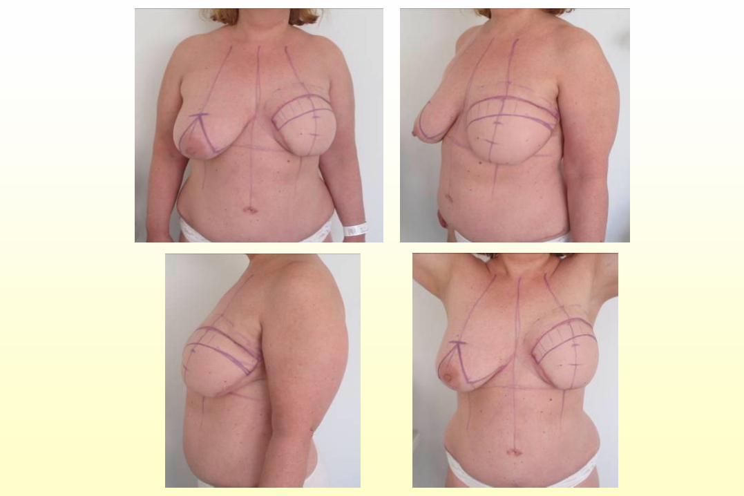
To perform adequate radical surgery
- to preserve maximal aesthetics
• Breast Conserving Surgery
• Oncoplastic breast conserving surgery
- volumen replacement, displacement
• Skin-, Areola-, Nipple Sparing Mastectomies
• Immediate / delayed postmastectomy breast
reconstructions
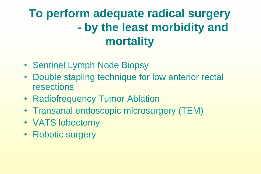
To perform adequate radical surgery
- by the least morbidity and
mortality
• Sentinel Lymph Node Biopsy
• Double stapling technique for low anterior rectal resections
• Radiofrequency Tumor Ablation
• Transanal endoscopic microsurgery (TEM)
• VATS lobectomy
• Robotic surgery
DaVinci Surgical Robotic System
To perform adequate radical surgery
- on the most cost effective way
• Cases detected by population screening, early
stage
• One day breast surgery
• Immediate one stage breast reconstruction
• Gastro-intestinal stanting
• Robotic Surgery
Quality of life in oncological surgery
• Able to reconstruct the missing parts of the
human body
• Surgical reconstruction of function
– Skin and musculo-sceletal, organ failure
• Reconstruction of aesthetic apperance
restoration or improvement
• Demand of immediate non delayed
reconstruction
PALLIATIVE CARE OF CANCER PATIENTS
Provides relief from pain and other distressing symptoms;
Affirms life and regards dying as a normal process;
Intends neither to hasten nor to postpone death;
Integrates the psychological and spiritual aspects of patient care;
Offers a support system to help patients live as actively as possible
until death;
Offers a support system to help the family cope during the patient’s
illness and in their own bereavement;
40
Principles of Surgical Oncology
DeVita,Hellman, and Rosenberg's Cancer: Principles and Practice of Oncology
8th., Lippincott Williams&Wilkins
Strategy of the reconstructive surgery
• Local flaps
- Taken from the direct vicinity
• Pedicle flaps
- Skin and myocutaneous flaps taken from the vicinity
- Skin flaps taken from more distant regions of the body
- Transposition flap – tube flap
• Free transplantation
- Full thickness skin graft
- Split thickness skin graft
- Transplantation of skin flap and myocutaneous flap
43
Left ingional exulcerated sof tissue sarcoma,
radical surgical resection and reconstruction
with TFL flap
FACIAL COSMETIC UNITS AND JUNCTION LINES
45
HOW TO USE AESTHETIC UNITS
Gary C. Burget (Plastic and Reconstructive Surgery, August 1985)
“Just as we can utilize skin lines, wrinkle lines and hair to cover our scars or to minimize theirunattractiveness and obviousness, we can choose, whenever possible, to make our defectsand our reconstructions fit topographic units.”
46
ESSENTIAL PARAMETERS OF TISSUE
DEFECT
Size
Depth
Contidion of the subcutaneous tissue (perichondrium,
periosteum, open cavity)
Free surgical margins
47
CLOSING TYPES OF FACIAL DEFECTS
Primary closing without tension and deformity
Skin grafting
Local flaps
Distant flaps composed of more hitology types (composite
flaps)
48
RECONSTRUCTION METHODS
• Free skin grafting:
in cases of superficial defects
• Flaps:
– If deep-seated tissues are to be covered
– If the wound base is of „poor quality”
– If the surface to be reconstructed is exposed to great mechanic
force
– If disturbing contour hiatus would develop
– If open cavity in body is to be closed
49
CLASSIFICATION OF FLAPS
ACCORDING TO THE TISSUES CONSTITUTING
THE FLAP:
• Skin
• Skin and fascia
• Skin and muscle
• Skin, muscle, bone
50
ACCORDING TO THE MOBILIZATION METHOD
OF THE FLAP:
– advancement flaps
– rotation flaps
– transposition flaps
– subcutaneously pedicled flaps
– interpolation flaps
– free flaps
– combined reconstruction
51
CLASSIFICATION OF FLAPS
TEMPORAL DEFECT – full thickness skin graft
DEFECT OF THE WING OF THE NOSE - full thickness skin graft
PREAURICULAR DEFECT
advancement flap
53
TRANSPOSITION FLAPS
54
DEFECT OF THE INNER PALPEBRA - transposition flap
55
MIDDLE FACE DEFECT - transposition flap
SUBCUTANEOUSLY PEDICLED FLAP
56
DEFECT OF THE EXTERNAL PALPEBRA - subcutaneously pedicled flap
LOWER LIP DEFECT
subcutaneously pedicled flap
UPPER LIP DEFECT
subcutaneously pedicled flap
INTERPOLATION FLAP
58
DEFECT OF THE WING OF THE NOSE - interpolation flap
59
FREE FLAP RECONSTRUCTION
radial forearm flap
60
FREE FLAP RECONSTRUCTION
anterolateral thigh flap
61
DOUBLE DEFECT OF THE WING OF THE NOSE - expander + interpolation flap
FOREHEAD DEFECT - COMBINED RECONSTRUCTION
63
COMBINED RECONSTRUCTION
64
Introduction of new techniques for reconstructive surgery in Hungary
Pectoralis major (PM) myocutaneous flap modified by the author – first time in Hungary
Localizations: - floor of the mouth, mandible, tongue, root of the tongue