7/23/13 Shark Lab Key crescentok.com/staff/jaskew/isr/botzo/shark/sharkkey.htm 1/8 Shark Lab Key Study this basic information about the spiny dogfish shark. Print this Shark Lab Report Guide . Pre-Lab Research Study this website . It provides several useful videos of large shark dissections. Study this lab key. Basic instructions for the dissection are noted in blue . Key structures of the shark are noted in red . dorsal surface click on picture for ventral surface External Anatomy Examine each of the following items on your shark. The streamlined body is divided into the head , trunk, and tail . Coloration is dark gray above and almost white below. The lateral line , made up of a series of tiny pores leading to nerve receptors, is sensitive to vibrations in the water. The Fins: The anterior dorsal fin is larger than the posterior dorsal fin . A spiny dogfish has two spines, one immediately in front of each dorsal fin. The spines carry a poison secreted by glands at their base. The caudal fin is divided into two lobes: a larger dorsal lobe and a smaller ventral lobe. This type of tail is known as a heterocercal tail . The paired pectoral fins act like wings to provide the lift needed to keep the shark from sinking. The paired pelvic fins , located on either side of the cloacal opening, are different in males and females.
Study this basic information about the spiny dogfish shark.
Print this Shark Lab Report Guide.
Pre-Lab Research
Study this website. It provides several useful videos of large shark dissections.
Study this lab key. Basic instructions for the dissection are noted in blue. Key structures of the shark are
noted in red.
dorsal surface
click on picture for ventral surface
External Anatomy
Examine each of the following items on your shark.
The streamlined body is divided into the head, trunk, and tail.
Coloration is dark gray above and almost white below.
The lateral line, made up of a series of tiny pores leading to nerve receptors, is sensitive to vibrations in
the water.
The Fins:
The anterior dorsal fin is larger than the posterior dorsal fin. A spiny dogfish has two spines, one
immediately in front of each dorsal fin. The spines carry a poison secreted by glands at their base.
The caudal fin is divided into two lobes: a larger dorsal lobe and a smaller ventral lobe. This type of tail is
known as a heterocercal tail.
The paired pectoral fins act like wings to provide the lift needed to keep the shark from sinking.The paired pelvic fins, located on either side of the cloacal opening, are different in males and females.
pigmented iris can be seen beneath the cornea with
the pupil at its center.
Upper and lower eyelids protect the eye. Just inside
the lower lid is a membrane that extends over the
surface of the eye to provide protection withoutclosing the eyes.
Spiracle openings are located above and behind each eye. The spiracle is an incurrent water passageway
leading into the mouth.
Most sharks have five external gill slits located behind the mouth and in front of the pectoral fins. Water
taken in by the mouth and spiracles is passed over the gills and forced out through the gill slits.
The Mouth:
Using both hands, carefully open the mouth to examine the teeth and tongue.
The triangular teeth are arranged in several rows beginning atthe outer edges of the upper and lower jaws. Behind thefunctional first row of teeth are additional rows folded
downward ready to replace any that are lost.The tongue is practically immovable and without muscles. It is
supported anteriorly and posteriorly by cartilage.The nares, external nostrils, are located on the ventral surface
of the rostrum anterior to the jaws. A nasal flap separates theincurrent from the excurrent opening. Water passes into and out
of the olfactory sac, permitting the shark to detect the odors ofthe water.
The patches of pores on the head in the areas of the eyes,snout, and nostrils are the openings of the ampullae ofLorenzini. These sense organs detect very small changes in
temperature, water pressure, and electrical fields.
Sexual Dimorphism
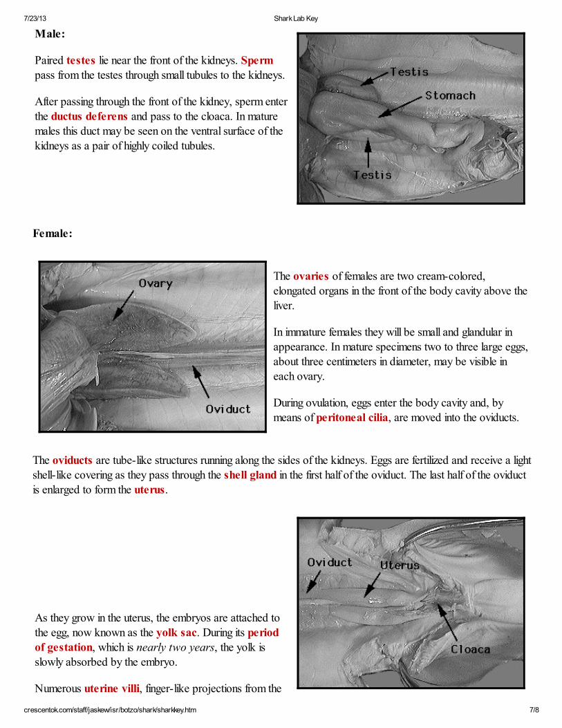
Males have stiff, grooved copulatory organs called claspers on the inner side of their pelvic fins. During
copulation, these are inserted through the cloaca into the oviduct of the female. Sperm travel from the cloaca ofthe male along the grooved dorsal surface of the clasper into the female.
The cloacal opening is located on the ventral surface between the pelvic fins. It receives the products of the
Turn the shark ventral side up and make an incision just in front of the cloacal opening. Extend the cut forward,all the way to the pectoral girdle (between the pectoral fins).
Digestive System:
A shiny membrane, the peritoneum,
lines the inside of the body wall. Theinternal organs are supported by a
membrane known as the mesentery.
The liver is the largest organ in the body.Its two main lobes, right and left, extend
from the pectoral girdle backward
through most of the body cavity. A third,
much shorter lobe (with the green gallbladder attached), is located between the
The liver is rich in oil, which stores energy for the shark and, because of its low specific gravity, provides alimited amount of buoyancy. (Sharks have no swim bladders.)
Remove the two main lobes of the liver to make the other internal organs easier to see.
The esophagus is the thick muscular tube connecting the oral cavity and pharynx with the stomach.
Open the stomach by cutting along its long axis. (Theremay be partially digested food that must be removed.)
The mucosa lines the stomach with longitudinal folds that
help churn and mix the food with digestive juices. Theback of the stomach narrows to a circular valve, the
pyloric sphincter, which regulates the passage of
partially digested food into the intestines.
Behind the stomach is the duodenum, the first section of
the small intestine. The bile duct from the gall bladder
enters the duodenum.
The pancreas is located at the connection of the
duodenum and the stomach. Secretions from the
pancreas enter the duodenum through the pancreaticduct.
The dark, triangular-shaped spleen is located near the
posterior end of the stomach.
The valvular intestine is the second, and much larger,
section of the small intestine. It is recognized by the rings
on its surface.
Open the valvular intestine as you did the stomach.
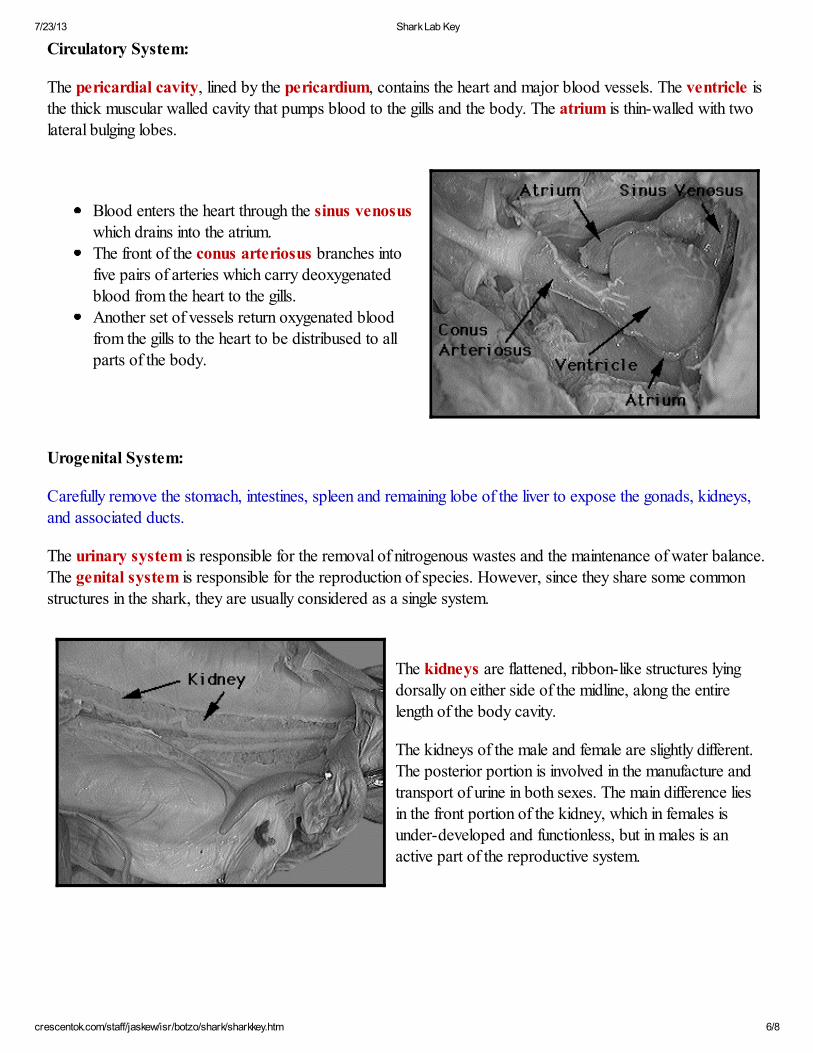
The pericardial cavity, lined by the pericardium, contains the heart and major blood vessels. The ventricle isthe thick muscular walled cavity that pumps blood to the gills and the body. The atrium is thin-walled with two
lateral bulging lobes.
Blood enters the heart through the sinus venosus
which drains into the atrium.The front of the conus arteriosus branches into
five pairs of arteries which carry deoxygenated
blood from the heart to the gills.
Another set of vessels return oxygenated bloodfrom the gills to the heart to be distribused to all
parts of the body.
Urogenital System:
Carefully remove the stomach, intestines, spleen and remaining lobe of the liver to expose the gonads, kidneys,
and associated ducts.
The urinary system is responsible for the removal of nitrogenous wastes and the maintenance of water balance.The genital system is responsible for the reproduction of species. However, since they share some common
structures in the shark, they are usually considered as a single system.
The kidneys are flattened, ribbon-like structures lying
dorsally on either side of the midline, along the entire
length of the body cavity.
The kidneys of the male and female are slightly different.The posterior portion is involved in the manufacture and
transport of urine in both sexes. The main difference lies
in the front portion of the kidney, which in females is
under-developed and functionless, but in males is an
uterine wall, make contact with the surface ot thedeveloping embryo and its yolk sac. It is believed that
these provide the embryo with water; all other nutrients
are supplied by the yolk.
The cloaca serves as the aperture through which the 23 to 29 centimeter long shark "pups" are born.
This type of development, where the young are born as miniature adults but have received hardly any nutrition
directly from the mother's uterus, is known as ovoviviparous.
Nervous System:
Remove the skin from the dorsal surface of the head and
shave off thin layers of the cartilage cranium until the brain
and cranial nerves are exposed.
There are two main parts of the nervous system:
central nervous system - the brain and spinal
cordperipheral nervous system - the sense organs,
cranial and spinal nerves, and their branches.
Parts of the shark brain:
Forebrain:The two cerebral hemispheres are rounded lobes at the front of the brain. The first portion of the
cerebrum is known as the olfactory lobes, responsible for the sense of smell.
The second portion of the forebrain consists of the epithalamus, pineal body, thalamus,
hypothalamus and pituitary body.
Midbrain: The optic lobes are prominent bulges of the brain responsible for sight.
Hindbrain:
The cerebellum is an oval-shaped portion that partly overlaps the optic lobes.The medulla oblongata is the elongated region at the back of the brain that narrows into the spinal