J. Novakofski, M. S. Brewer, N. Mateus-Pinilla, J. Killefer and R. H. McCusker Prion biology relevant to bovine spongiform encephalopathy 2005, 83:1455-1476. J ANIM SCI http://www.journalofanimalscience.org/content/83/6/1455 the World Wide Web at: The online version of this article, along with updated information and services, is located on www.asas.org at Acquisitions Dept on December 12, 2013 www.journalofanimalscience.org Downloaded from at Acquisitions Dept on December 12, 2013 www.journalofanimalscience.org Downloaded from
Transcript
J. Novakofski, M. S. Brewer, N. Mateus-Pinilla, J. Killefer and R. H. McCuskerPrion biology relevant to bovine spongiform encephalopathy
2005, 83:1455-1476.J ANIM SCI
http://www.journalofanimalscience.org/content/83/6/1455the World Wide Web at:
The online version of this article, along with updated information and services, is located on
www.asas.org
at Acquisitions Dept on December 12, 2013www.journalofanimalscience.orgDownloaded from at Acquisitions Dept on December 12, 2013www.journalofanimalscience.orgDownloaded from
Prion biology relevant to bovine spongiform encephalopathy1
J. Novakofski*2, M. S. Brewer†, N. Mateus-Pinilla‡, J. Killefer*, and R. H. McCusker*
Departments of *Animal Sciences and †Food Science and Human Nutrition,University of Illinois at Urbana–Champaign 61801-4737; and ‡Illinois Natural History Survey,
Center for Wildlife and Plant Ecology, Champaign, IL 61820
ABSTRACT: Bovine spongiform encephalopathy(BSE) and chronic wasting disease (CWD) of deer andelk are a threat to agriculture and natural resources,as well as a human health concern. Both diseases aretransmissible spongiform encephalopathies (TSE), orprion diseases, caused by autocatalytic conversion ofendogenously encoded prion protein (PrP) to an abnor-mal, neurotoxic conformation designated PrPsc. Mostmammalian species are susceptible to TSE, which, de-spite a range of species-linked names, is caused by asingle highly conserved protein, with no apparent nor-mal function. In the simplest sense, TSE transmissioncan occur because PrPsc is resistant to both endogenousand environmental proteinases, although many detailsremain unclear. Questions about the transmission ofTSE are central to practical issues such as livestocktesting, access to international livestock markets, andwildlife management strategies, as well as intangibleissues such as consumer confidence in the safety of themeat supply. The majority of BSE cases seem to havebeen transmitted by feed containing meat and bone
2005 American Society of Animal Science. All rights reserved. J. Anim. Sci. 2005. 83:1455–1476
Introduction
Bovine spongiform encephalopathy (BSE) is one ofseveral transmissible spongiform encephalopathies(TSE), or prion diseases, which are progressive neuro-logical disorders thought to be caused by conversion ofendogenous, host-encoded prion protein (PrP) to anabnormal conformation designated PrPsc (Prusiner,1982; Legname et al., 2004). Prion diseases may begenetic, sporadic, or transmitted, although in all cases,progression of the disease is associated with accumula-
1The authors acknowledge the generous support of Cargill, Incor-porated, Minneapolis, MN, in publication of this review.
2Correspondence: 1503 South Maryland Dr. (phone: 217-333-6181;e-mail: [email protected]).
Received November 22, 2004.Accepted March 7, 2005.
1455
meal from infected animals. In the United Kingdom,there was a dramatic decrease in BSE cases after neuraltissue and, later, all ruminant tissues were bannedfrom ruminant feed. However, probably because ofheightened awareness and widespread testing, there isgrowing evidence that new variants of BSE are arising“spontaneously,” suggesting ongoing surveillance willcontinue to find infected animals. Interspecies trans-mission is inefficient and depends on exposure, se-quence homology, TSE donor strain, genetic polymor-phism of the host, and architecture of the visceralnerves if exposure is by an oral route. Considering thelow probability of interspecies transmission, the lowefficiency of oral transmission, and the low prion levelsin nonnervous tissues, consumption of conventional an-imal products represents minimal risk. However, detec-tion of rare events is challenging, and TSE literatureis characterized by subsequently unsupported claimsof species barriers or absolute tissue safety. This reviewpresents an overview of TSE and summarizes recentresearch on pathogenesis and transmission.
tion of abnormal PrPsc conformer in the brain and sub-sequent neurodegeneration (Prusiner, 2004). Both thedisease process and disease transmission are relatedto stability of the PrPsc conformer, which is markedlyresistant to biological or chemical degradation. The ab-breviation PrPsc refers to scrapie, the prototypical priondisease, whereas an alternative, PrPres, refers to pro-teinase resistance. The mechanisms of prion diseasetransmission are of great interest because spontaneousoccurrences of prion disease are rare and most cases indomestic and wild animals result from transmission.Moreover, based on DNA analyses, the AA sequence ofPrP is well conserved in mammalian species; therefore,most, if not all, mammalian species are susceptible toprion diseases (Table 1) and the disease may be trans-mitted between species. Most prion diseases may betransmitted to laboratory animals, and substantial evi-dence indicates that a variant form of human Creutz-feldt-Jakob Disease (vCJD; sporadic CJD is the proto-
at Acquisitions Dept on December 12, 2013www.journalofanimalscience.orgDownloaded from
aLab rodents include gerbils, guinea pigs, hamsters, mice, and rats.bRepassage means secondary transmission after adaptation in an intermediate species.cOther primates may include chimpanzees, macaque monkeys, marmosets, prosimians, squirrel monkeys.
typical TSE of humans) was caused by interspeciestransmission following the BSE outbreak in the UnitedKingdom. The geographic distributions of BSE andchronic wasting disease (CWD; TSE of cervids) havecontinued to increase in recent years, despite wide-spread control efforts, indicating the importance of bet-ter understanding these diseases. The purpose of thisarticle is to review characteristics of prion diseases andtransmission as well as to examine recent findings thatsuggest it may be necessary to revise common percep-tions about the disease.
Basis of Prion Disease
Transmission of TSE seem to result from three un-usual characteristics of PrPsc, although many specificsare unclear. First, PrPsc is autocatalytic in that PrPsc
promotes conversion of additional PrP molecules intothe PrPsc conformation (Raymond et al., 2000; Prusiner,2004). Second, PrPsc is resistant to proteases (intracel-lar, intestinal, and environmental) that would normallydestroy the protein (Telling et al. 1996; Riesner 2003).This results in accumulation of undegraded PrPsc invivo and persistence ex vivo. Third, accumulated PrPsc
results in neuronal damage and spongiform changes inthe brain (Unterberger et al., 2005).
The prnp gene is present in most, if not all, wild-typemammals and is highly conserved across species (vanRheede, et al., 2003). Endogenous PrP is encoded by asingle exon of the prnp gene, which codes for a 256-to 264-AA precursor of approximately 28 kDa that isprocessed by cleavage of a 22- to 24-AA signal peptideyielding a mature protein of 231 to 253 AA (Riesner,2003; Van Rheede, et al., 2003). Species differences inPrP size are primarily a function of differences in thenumber of octapeptide repeat regions.
Normal PrP is a glycoprotein primarily found onnerve and immune cell membranes, although the pro-tein is found at lower concentrations on many cell types.The PrP protein has O- and N-linked glycosylation sitesin its precursor form (Riesner, 2003) and is found as amixture of nonglycosylated, monoglycosylated and dig-lycosylated forms. Cell surface attachment occurs viaa glycosyl phosphatidyl inositol anchor (Riesner, 2003).Membrane bound PrP also associates with heparan sul-fates (Caughey et al., 1994) and laminin (Graner et al.,2000). In neurons, PrP is associated with both the cellbody and synaptic vesicles (Collinge et al. 1994; Saleset al., 1998).
The PrP is a metal-binding protein having at leastone site that binds Cu2+, with a Kd of 10−14 M (Jacksonet al., 2001). Other transition metal ions (Zn, Mn, andNi) bind with lower affinity. Metal binding alters PrPbiochemical characteristics, including protease resis-tance (Brown, et al. 1997; Lehmann, 2002). The PrPmutations that have additional copies of the metal-binding octapeptide repeat have properties similar toPrPsc (Lehmann and Harris, 1996). Copper bindingstimulates PrP endocytosis (Pauly and Harris, 1998).In addition to extracellular function, intracellular PrPmay modify properties of intracellular proteins, includ-ing antiapoptotic Bcl-2 (Kurschner and Morgan, 1995),several heat shock proteins (Edenhofer et al., 1996;Zanata et al., 2002), neuronal synapsin, and the growthfactor signal adapter Grb2 (Spielhaupter and Schatzl,2001). Knockout mice without PrP often seem to benormal, suggesting that normal PrP function may beredundant with an unidentified protein. Neurologicalabnormalities in some strains of PrP knockout miceresult from an incomplete construct, which upregulatesthe adjacent dpl gene (Aguzzi, 2003). The dpl gene prod-
at Acquisitions Dept on December 12, 2013www.journalofanimalscience.orgDownloaded from
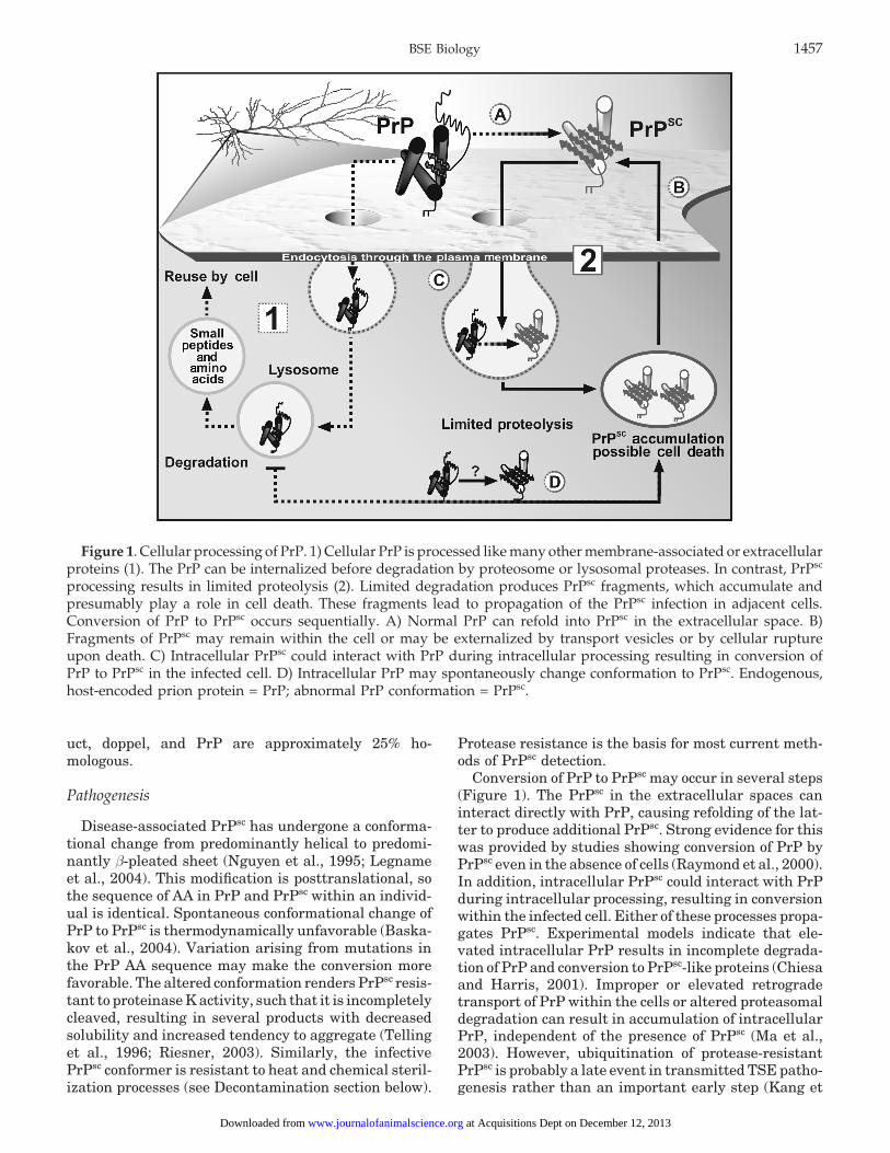
Figure 1. Cellular processing of PrP. 1) Cellular PrP is processed like many other membrane-associated or extracellularproteins (1). The PrP can be internalized before degradation by proteosome or lysosomal proteases. In contrast, PrPsc
processing results in limited proteolysis (2). Limited degradation produces PrPsc fragments, which accumulate andpresumably play a role in cell death. These fragments lead to propagation of the PrPsc infection in adjacent cells.Conversion of PrP to PrPsc occurs sequentially. A) Normal PrP can refold into PrPsc in the extracellular space. B)Fragments of PrPsc may remain within the cell or may be externalized by transport vesicles or by cellular ruptureupon death. C) Intracellular PrPsc could interact with PrP during intracellular processing resulting in conversion ofPrP to PrPsc in the infected cell. D) Intracellular PrP may spontaneously change conformation to PrPsc. Endogenous,host-encoded prion protein = PrP; abnormal PrP conformation = PrPsc.
uct, doppel, and PrP are approximately 25% ho-mologous.
Pathogenesis
Disease-associated PrPsc has undergone a conforma-tional change from predominantly helical to predomi-nantly β-pleated sheet (Nguyen et al., 1995; Legnameet al., 2004). This modification is posttranslational, sothe sequence of AA in PrP and PrPsc within an individ-ual is identical. Spontaneous conformational change ofPrP to PrPsc is thermodynamically unfavorable (Baska-kov et al., 2004). Variation arising from mutations inthe PrP AA sequence may make the conversion morefavorable. The altered conformation renders PrPsc resis-tant to proteinase K activity, such that it is incompletelycleaved, resulting in several products with decreasedsolubility and increased tendency to aggregate (Tellinget al., 1996; Riesner, 2003). Similarly, the infectivePrPsc conformer is resistant to heat and chemical steril-ization processes (see Decontamination section below).
Protease resistance is the basis for most current meth-ods of PrPsc detection.
Conversion of PrP to PrPsc may occur in several steps(Figure 1). The PrPsc in the extracellular spaces caninteract directly with PrP, causing refolding of the lat-ter to produce additional PrPsc. Strong evidence for thiswas provided by studies showing conversion of PrP byPrPsc even in the absence of cells (Raymond et al., 2000).In addition, intracellular PrPsc could interact with PrPduring intracellular processing, resulting in conversionwithin the infected cell. Either of these processes propa-gates PrPsc. Experimental models indicate that ele-vated intracellular PrP results in incomplete degrada-tion of PrP and conversion to PrPsc-like proteins (Chiesaand Harris, 2001). Improper or elevated retrogradetransport of PrP within the cells or altered proteasomaldegradation can result in accumulation of intracellularPrP, independent of the presence of PrPsc (Ma et al.,2003). However, ubiquitination of protease-resistantPrPsc is probably a late event in transmitted TSE patho-genesis rather than an important early step (Kang et
at Acquisitions Dept on December 12, 2013www.journalofanimalscience.orgDownloaded from
al., 2004). Extrapolating from these findings, it is possi-ble to speculate that sporadic TSE may arise by a combi-nation of these intracellular events.
The neurotoxic mechanism of PrPsc remains unclear,with different lines of evidence supporting some combi-nation of PrPsc toxicity and alteration in normal PrPfunction. The presence of both PrPsc and synthesis ofPrP are required for pathogenesis. Inoculation of a doseof PrPsc that is infectious in normal mice does not causedisease in PrP knockout mice (Prusiner et al., 1993),indicating that endogenous PrP conversion to PrPsc isnecessary to cause pathology. Loss of normal PrP func-tion also is not the cause of pathology because PrPknockout mice do not develop disease. Crosslinking ofPrP with antibodies in the absence of any PrPsc resultsin rapid onset of neuronal disease (Solforosi et al.,2004), suggesting that disruption of normal PrP func-tion or accumulation alone may result in a neurotoxicprocess.
Clinical signs of TSE are primarily neurological: be-havior changes, impaired coordination, muscle spasms,and repetitive movements. Body wasting in spite ofnormal food intake is common, although obesity andabnormal glucose metabolism are observed in somestrain/host combinations (Ye and Carp, 1995; Wads-worth et al., 2003). Neurological deterioration is pro-gressive after clinical symptoms appear. Specific combi-nations of symptoms, referred to as TSE phenotype,reflect different pathogenic TSE strains. The TSE typi-cally have long incubation periods of months (rodents,cats), years (cattle, sheep, deer), or decades (man), soclinical signs usually are evident in older animals. Be-cause the sequence of PrP protein is highly conservedand most of the PrPsc is eventually derived from con-verted host-encoded PrP, infection does not evoke asubstantial immune response or inflammatory reactionin host animals (Aguzzi, 2003; Prusiner, 2004); how-ever, chronic inflammatory conditions may modify nat-ural PrPsc transmission (Heikenwalder et al., 2005). AllTSE cause spongiform changes in the brain associatedwith some degree of amyloid plaque development. Amy-loid plaques consist of PrPsc aggregates, which may bedetected histologically as diffuse, condensed, or floridplaques in infected brain tissue. Specific combinationsof clinical symptoms reflect the specific regions in thebrain where PrPsc accumulates, the characteristic pat-tern of damage and the nature of plaque deposition(Brown et al., 1994; DeArmond et al., 1997).
Animals may develop PrPsc-based disease 1) by inges-tion of PrPsc; 2) by peripheral exposure to PrPsc, mostcommonly by an iatrogenic route (surgery, cadavericgrowth hormone injection, corneal transplantation); 3)by hereditary transmission as an autosomal, dominanttrait; or 4) sporadically by unknown origin (Wadsworthet al., 2003; Prusiner, 2004).
Oral infection by ingestion involves transfer of PrPsc
from the digestive tract to the spleen or lymphoreticularsystem, and then to the peripheral nervous system andeventually to the brain. Infectivity is established in
peripheral lymphoid organs before infective PrPsc isfound in the central nervous system (CNS), indicatingthat peripheral conversion of PrP to PrPsc is necessarystep for infection. There seem to be two potential routesof neuro-invasion by PrPsc via lymphoid tissue: one pathinvolving the spleen, and the other path involving vis-ceral lymph nodes (Kimberlin and Walker, 1989). Fol-lowing ingestion, PrPsc may be degraded by digestiveenzymes, leaving a pathogenic fragment similar to thatof a proteinase K-resistant PrPsc fragment. The PrPsc
fragment may be cotransported across the intestinalepithelial with ferritin (Mishra et al., 2004). The PrPsc
or PrPsc fragments are transported from the intestineto secondary lymphoid organs by intestinal dendriticcells, which are specialized to acquire antigen from pe-ripheral tissues (Huang and MacPherson, 2004). Den-dritic cells from the intestine present PrPsc to T and Blymphocytes within lymphoid tissues such as Peyer’spatches of the intestine or follicular dendritic cells(FDC) of the spleen, thymus, and tonsils (Mabbott andBruce, 2003; Prinz et al., 2003). In the lymphoid tissues,PrPsc molecules accumulate following conversion fromthe PrP conformation. Accumulation of PrPsc and pro-gression to the CNS nervous system requires B-cellcytokines, although expression of PrP on the B-cells isnot necessary for transmission (Klein et al., 1997; 1998).The spleen is a rich source of B-cells, and B-cell cytok-ines enable splenic FDC cells to mature (Prinz et al.,2003; Aguzzi and Miele, 2004). Splenic FDC may serveas the source of PrP for peripheral PrPsc formation asthey express a high level of PrP (Aguzzi, 2003). Subse-quent transfer of PrPsc from FDC to sympathetic nervesdepends on the distance between the two structures(Prinz et al., 2003). Chronic lymphocytic inflammation,which upregulates cyotkines, enables PrPsc accumula-tion in otherwise PrPsc free tissues (Heikenwalder etal., 2005).
After oral inoculation, PrPsc can be detected in a por-tion of the follicles of Peyer’s patches in the distal ileumof cattle for much of the disease progression (Terryet al., 2003). Detection of PrPsc in lymphoid tissue ofexperimentally infected animals is possible before clini-cal signs are observed. In the CNS, detectable PrPsc isfirst seen in the spinal cord, then the brain stem, andthen in higher brain areas. Levels of PrPsc in the brainincrease exponentially following infection before the de-velopment of clinical symptoms. By the onset of clinicalsymptoms, PrPsc titers in spleen and lymph tissues haveeither plateaued or decreased, although in the laterstages of disease, PrPsc levels are increased in Peyer’spatches. (Kimberlin and Walker, 1988; Anderson et al.,1996). Oral infectivity demonstrates that PrPsc is resis-tant to stomach acids, mammalian digestive proteases,and in the case of ruminants, bacterial proteases.
Neural invasion by PrPsc does not require exposureto lymphoid tissue. Peripheral exposure, for examplethrough damaged skin (Taylor et al., 1996), the tongue(Bartz et al., 2003), or vascular system (Kimberlin andWalker, 1989), results in direct PrPsc exposure of the
at Acquisitions Dept on December 12, 2013www.journalofanimalscience.orgDownloaded from
nervous system with subsequent retrograde transportto the CNS and rapid infection. Infection seems to beparticularly rapid and effective by peripheral exposureof heavily innervated tissue such as the tongue (Bartzet al., 2003). There is also retrograde transmission bynerves as PrPsc is found in sensory nerves and neuro-muscular junction in the tongue of intracerebrally (i.c.)inoculated animals (Bartz et al., 2002; Mulcahy et al.,2004). Iatrogenic infection by medical procedures re-sults from central or peripheral neural inoculation. Itis worth noting that there have been more than twiceas many iatrogenic CJD cases as vCJD cases. Iatrogenicprion disease transmission has been documented fromexposure to neuro-surgical instruments, from injectionof cadaver-derived growth hormone or gonadotrophin,and from corneal or dura tissue transplants (Brown etal., 2000; Weissmann et al., 2002).
Another route of peripheral infection is maternaltransmission to the fetus or newborn. The traditionalview is that scrapie is maternally transmitted to thefetus; however, it is clear that separating fetal (vertical)transmission from postnatal lateral transmission is dif-ficult (Ridley and Baker, 1995). Transmission of PrPsc
in utero across the maternal-fetal barrier requires aplacental architecture permitting relocation of infectivePrPsc and genetic susceptibility of the fetus.
Genetics of Prion Disease
Specific TSE phenotypes result from the combinedeffects of pathogenic mutations, genetic polymorphismsof prnp, and the specific strain of infectious PrPsc. Spo-radic (spontaneous) and hereditary forms of prion dis-ease share a common pathogenesis except that the ini-tiating events are unknown in the first case and prede-termined by a pathogenic mutation in the second.“Sporadic” refers to development of prion disease withno familial history or apparent exogenous infection. Thespontaneous conversion of PrP to PrPsc is thermody-namically unfavorable, as indicated by the low inci-dence of sporadic CJD (Baskakov et al., 2004). Variationin the PrP AA sequence may make the conversion moreor less favorable, thereby altering susceptibility to priondisease. There are 86 reported mutations or polymor-phisms of the prnp sequence, with at least 25 that in-crease risk of spontaneous PrPsc formation, predisposethe individual to exogenous infection, or both (Heatonet al., 2003; Wadsworth et al., 2003). Susceptibility maybe reflected by minimal infectious dose, incubation timefollowing exposure, or relative statistical risk in a popu-lation.
A common protein polymorphism of human PrP, me-thionine or valine at residue 129 (129M/V), seems tobe predictive of CJD susceptibility (Figure 2). Heterozy-gosity at 129 protects individuals from CJD, whereas129V homozygotes are strongly overrepresented in indi-viduals with iatogenic or sporadic CJD (Collinge et al.,1991; Palmer et al., 1991). All vCJD patients have the129M homozygous genotype (Collinge et al., 1996). Be-
sides susceptibility, PrP polymorphisms may result indifferent symptoms. In sporadic CJD, homozygous129M individuals developed no amyloid plaque,whereas homozygous 129V individuals developed ex-tensive plaques (Pickering-Brown et al., 1995).
Approximately 10 to 15% of human prion diseasesare familial, meaning the individual has inherited aprnp mutation that confers a high susceptibility. Allfamilies identified with inherited forms of prion dis-eases have either point mutations or insertions in theprnp gene (Collinge and Palmer, 1994; Prusiner, 2004).Mutation D178N is found in fatal familial insomniaand familial CJD (Gambetti et al., 2003). The clinicaland pathological differences between familial CJD andfatal familial insomnia, which have the same D178Nmutation, are the result of the129M/V polymorphism(Goldfarb et al., 1992). Mutations responsible for Gerst-mann-Straussler-Scheinker syndrome include P102L,A117V, F198S D202N, Q212P, and Q217R (Piccardo etal., 2001).
PrPsc Strains: Conformation and Glycoslyation
Besides the AA sequence variation that contributesto phenotypic variation of TSE, a single PrP sequencecan change into multiple PrPsc conformers, each produc-ing a distinct pathologic phenotype (Telling et al., 1996;Safar et al., 2000). Strain differences result from varia-tion of the α helix to β sheet conversion within PrPsc,as well as variation in degree of PrP glycoslyation. Ingeneral, transmitted TSE strains retain their pheno-typic fingerprint, so the existence of different strainsof PrPsc is an important key to understanding the riskfor interspecies transmission.
At least 20 strains of scrapie have been differentiatedby their incubation period, clinical symptoms, and brainlesion pattern (Bruce et al., 1994, 2003). Differentstrains of TSE can be serially passaged in inbred micewith no change in the TSE strain characteristics, dem-onstrating that strain differences may be independentof PrP amino acid sequence in the host (Bruce, 2003).Characteristics of both the hyper and drowsy hamster-adapted scrapie PrPsc strains also can be reproducedin a cell-free conversion system, indicating the impor-tance of PrPsc conformation (Bessen et al., 1995). Differ-ent strains of PrPsc generated from the same PrP pre-cursor are cleaved at distinct sites by proteinase K,demonstrating differences in tertiary structure of PrPsc.Variation in glycosylation patterns of PrPsc may playa role in TSE phenotype (DeArmond et al., 1997; Hillet al., 1998), and because of the different cleavage sites,the size distribution of glycosylated proteinase K diges-tion fragments provides another signature of straintype (Wadsworth et al., 2003). The BSE PrPsc containshigh levels of diglycosylated and low levels of monogly-cosylated protein. Some scrapie strains contain primar-ily monoglycoslyated protein, whereas the glycosylationprofile in other strains is more similar, but not identical,to BSE (Baron et al., 2004). Human prion disease also
at Acquisitions Dept on December 12, 2013www.journalofanimalscience.orgDownloaded from
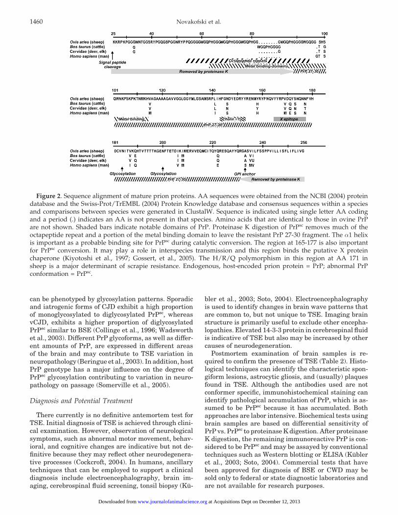
Figure 2. Sequence alignment of mature prion proteins. AA sequences were obtained from the NCBI (2004) proteindatabase and the Swiss-Prot/TrEMBL (2004) Protein Knowledge database and consensus sequences within a speciesand comparisons between species were generated in ClustalW. Sequence is indicated using single letter AA codingand a period (.) indicates an AA is not present in that species. Amino acids that are identical to those in ovine PrPare not shown. Shaded bars indicate notable domains of PrP. Proteinase K digestion of PrPsc removes much of theoctapeptide repeat and a portion of the metal binding domain to leave the resistant PrP 27-30 fragment. The α1 helixis important as a probable binding site for PrPsc during catalytic conversion. The region at 165-177 is also importantfor PrPsc conversion. It may play a role in interspecies transmission and this region binds the putative X proteinchaperone (Kiyotoshi et al., 1997; Gossert, et al., 2005). The H/R/Q polymorphism in this region at AA 171 insheep is a major determinant of scrapie resistance. Endogenous, host-encoded prion protein = PrP; abnormal PrPconformation = PrPsc.
can be phenotyped by glycosylation patterns. Sporadicand iatrogenic forms of CJD exhibit a high proportionof monoglycosylated to diglycosylated PrPsc, whereasvCJD, exhibits a higher proportion of diglycosylatedPrPsc similar to BSE (Collinge et al., 1996; Wadsworthet al., 2003). Different PrP glycoforms, as well as differ-ent amounts of PrP, are expressed in different areasof the brain and may contribute to TSE variation inneuropathology (Beringue et al., 2003). In addition, hostPrP genotype has a major influence on the degree ofPrPsc glycosylation contributing to variation in neuro-pathology on passage (Somerville et al., 2005).
Diagnosis and Potential Treatment
There currently is no definitive antemortem test forTSE. Initial diagnosis of TSE is achieved through clini-cal examination. However, observation of neurologicalsymptoms, such as abnormal motor movement, behav-ioral, and cognitive changes are indicative but not de-finitive because they may reflect other neurodegenera-tive processes (Cockcroft, 2004). In humans, ancillarytechniques that can be employed to support a clinicaldiagnosis include electroencephalography, brain im-aging, cerebrospinal fluid screening, tonsil biopsy (Ku-
bler et al., 2003; Soto, 2004). Electroencephalographyis used to identify changes in brain wave patterns thatare common to, but not unique to TSE. Imaging brainstructure is primarily useful to exclude other encepha-lopathies. Elevated 14-3-3 protein in cerebrospinal fluidis indicative of TSE but also may be increased by othercauses of neurodegeneration.
Postmortem examination of brain samples is re-quired to confirm the presence of TSE (Table 2). Histo-logical techniques can identify the characteristic spon-giform lesions, astrocytic gliosis, and (usually) plaquesfound in TSE. Although the antibodies used are notconformer specific, immunohistochemical staining canidentify pathological accumulation of PrP, which is as-sumed to be PrPsc because it has accumulated. Bothapproaches are labor intensive. Biochemical tests usingbrain samples are based on differential sensitivity ofPrP vs. PrPsc to proteinase K digestion. After proteinaseK digestion, the remaining immunoreactive PrP is con-sidered to be PrPsc and may be assayed by conventionaltechniques such as Western blotting or ELISA (Kubleret al., 2003; Soto, 2004). Commercial tests that havebeen approved for diagnosis of BSE or CWD may besold only to federal or state diagnostic laboratories andare not available for research purposes.
at Acquisitions Dept on December 12, 2013www.journalofanimalscience.orgDownloaded from
Immunohistochemistry Microscopic examination of brain sections All Gold standard for TSE detection.stained with antibodies for PrP protein.Antibodies are not conformation-specific Completion time: 2 to 3 d. Lowbut PrP does not accumulate except sensitivity to postmortem autolysis.under pathological conditions.
Detects PrPsc deposits in brain beforehistopathological identification ofprion-induced vacuoles.
Bioassay Suspect brain sample injected into the All Completion time: several months.brain or periphery of test animal(usually mouse). Infectivity Lowest detection limits of all tests.diagnosed by clinical signs ofinfection and incubation period. Sensitivity depends mouse strainTest results confirmed by other used.methods.
Histopathology Microscopic examination of brain sections All Completion time: 2 to 3 d. Properfor changes indicative of spongiform fixation required to preventencephalopathy, vacuoles, or presence postmortem autolysis.of prion-associated fibrils.
Western blotting Sample is homogenized, treated with All Visualization techniques vary fromproteinase K, which degrades normal chromogenic precipitate to radioPrP, electrophoretically separated isotopes.with SDS-PAGE, blotted onto amembrane, and probed with anantibody to detect the protease-resistant fragments (PrPsc).
Rapid methods (company)
Prionics-Check Western Test Western blot of protease-treated samples Cattle European 0.5-g obex sample required.(Prionics AG, Schlieren, with PrP antibody Union, Dilution accuracy: 1:10.c
Switzerland) United States Detection limit: 5.0 to 20 pmol.Detection via alkaline phosphatase- Completion time: 6 to 8 h.coupled secondary antibody with achemiluminescent substrate, a Validated for sheep and goatsreduced molecular weight comparedwith native PrP, and glycosylation-dependent band pattern of resistant PrP
Prionics-Check LIA Enzyme-linked immunosorbant assay Cattle European 0.5-g obex sample required.(Prionics AG) (ELISA) Union, Dilution accuracy: 1:10 to 1:30.
United StatesUses monoclonal antibodies and Detection limit: 1.0 to 5.0 pmol.chemiluminescent detection. Completion time: 4 h.
Bio-Rad test ELISA sandwich with colorimetric Cattle European 0.35-g obex sample.(Bio-Rad, Hercules, CA) substrate. Union, Dilution accuracy: 1:300.
Deer United States, Detection limit: 0.5 to 2.0 pmol.Sample degradation of normal protein Japan Completion time: 6 h.with proteinase K. Elk
Detection with two specific monoclonalantibodies for protease resistant PrP(colorimetric).
Enfer test ELISA Cattle European Dilution accuracy: 1:10 to 1:30.(Abbott Labs, Abbott Park, One-step sample preparation and Union, Detection limit: 1.0 to 10 pmol.IL) proteinase K treatment, polyclonal United States, Completion time: 4 h
antibodies, chemiluminescence Japandetection.
Continued
at Acquisitions Dept on December 12, 2013www.journalofanimalscience.orgDownloaded from
CDI test Conformation-dependent immunoassay. Cattle European Dilution accuracy: 1:30.(Impro Biotechnology, South) Union, Detection limit: 0.5 to 5.0 pmol.San Francisco, CA) Denatures sample to expose buried United States, Completion time: 8 h.
epitope in PrPsc. Uses Japan Does not involve proteinase K.two radio-labeled antibody fragmentsto capture and detect PrP.
Herd-check Immunoassay after affinity based Cattle, United States Completion time: 4 to 5 h(IDEXX Laboratories, separation of PrPsc in brain deer Does not involve proteinase K.Westbrook, ME) homogenate.
aSee Table 1 for abbreviations. Endogenous, host-encoded prion protein = PrP; abnormal PrP conformation = PrPsc.bTests approved by the European Commission for BSE detection are based on immunodetection of the pathological PrPsc conformer.bDilution accuracy is a reflection of test accuracy with known positive samples.
Animal bioassay remains the most sensitive methodfor detection of PrPsc (Soto, 2004). Although time con-suming and labor intensive, it is necessary to detectvery low levels of infectivity. Use of animals overex-pressing PrP has shortened bioassay time, whereas useof animals transgenic for another species of prion iscentral to interspecies transmission research. Cell cul-ture bioassays with various cell lines, including com-mon fibroblasts, may be used to detect PrPsc that is notmeasurable with biochemical techniques (Weissman etal., 2002; Vorberg et al., 2004).
A variety of substances have been investigated toprevent transmission or arrest progress of TSE. Inhibi-tory compounds could interfere with PrP conforma-tional change, PrPsc aggregate formation, or PrPsc pro-tease resistance. In addition, primary therapeutic con-siderations are toxicity and ability to cross the bloodbrain barrier. Early interest focused on compounds sim-ilar to Congo red, which is used as a histological stainfor PrPsc. Compounds showing activity in vitro includeCongo red (Caughey and Race, 1992; Rudyk et al.,2000), porphyrins (Caughey et al., 1998), lysosomotrop-ics or protease inhibitors (Doh-ura et al., 2000), pyri-dine-based compounds (Perrier et al., 2000), branchedpolyamines (Supattapone et al., 2001), and phenothi-azines or bis-acridines (May et al., 2003). Several com-pounds have shown promise in vivo including tetracy-cline (Forloni et al., 2002) and quinolines (Murakami-Kubo et al., 2004). Quinacrine (an acridine) and chlopro-mazine depress PrPsc aggregation, but they are ineffec-tive in treating animal models (Barret et al., 2003).
Transmissible Spongiform Encephalopathies
Scrapie
Scrapie was first reported in 1732, and it is the proto-typical spongiform encephalopathy found in ovines andcaprines. Scrapie refers to the pruritus-induced rubbingand scratching (scraping) that results in wool loss. Itis an acquired infection with an incubation period of 2to 5 yr and an average age of onset of clinical symptoms
of 2.5 yr (Schreuder, 1994). Typically, no more than5% of the animals in a herd show clinical symptomsof infection.
Scrapie epidemics are self-sustaining because of hori-zontal transmission (animal-to-animal) and apparentenvironmental persistence. Contaminated pastureshave caused numerous outbreaks of scrapie (Chatelainet al., 1983; Redman et al., 2002). In Iceland, eradica-tion attempts failed when scrapie-free sheep were re-stocked onto pastures that had been kept free of sheepfor 3 yr (Sigurdarson, 1991) indicating the stability ofPrPsc even in the soil. Infectious material buried in thesoil loses only approximately 50% of its infectivity in 3yr (Brown and Gajdusek, 1991). There is limited evi-dence for specific mechanisms of environmental trans-mission, although it clearly occurs. Likely possibilitiesinclude saliva or feces (Miller et al., 2004), urine(Shaked et al., 2001), or decay residue of infectioustissues such as placenta (Table 3). Because PrPsc isstable in the soil, animals could become infected byconsumption of soil or inhalation of contaminated dust.
Scrapie PrPsc is recoverable from lymphatic tissue,tonsils, spleen, and thymus, as well as nervous tissue.Accumulation of PrPsc in placental tissue depends onsusceptible fetal genotype and stage of gestation (Tuoet al., 2001, 2002; Andreoletti et al., 2002); however,the presence or absence of PrPsc in placentomes of fe-tuses does not correlate with time to postnatal infection(Andreoletti et al., 2002). This suggests that infectiondoes not occur in utero but is postnatal. Infection mayoccur from direct postnatal contact with placental tis-sue or from pasture contaminated with placental tissue.Scrapie has been experimentally transmitted (by i.c.inoculation) to a number of animals, including ham-sters, mice, mink, ferrets, goats, monkeys, and cattle.Oral infection has been successfully demonstrated inmice (Chandler, 1963), sheep, goats (Pattison et al.,1972), and squirrel monkeys (Gibbs et al., 1980).
Variation in the coding region of the prnp gene, pre-viously known as the scrapie gene Sip, influences sus-ceptibility to scrapie. The major AA polymorphisms areA136V, H154R, and H171Q,R (Tranulis, 2002). Ani-
at Acquisitions Dept on December 12, 2013www.journalofanimalscience.orgDownloaded from
aDEFRA (2004b). Based on scrapie-infected sheep and goats.bTonsil and distal ileum classified by USDA (2004a).cBlood moved from DEFRA infectivity category; Ironside and Head (2003).dFeces moved from DEFRA infectivity category; Miller et al. (2004).eUterus moved from DEFRA infectivity category; Tuo et al. (2002).
mals with prnp genotypes, A136R154Q171/ARQ, ARQ/VRQ, and VRQ/VRQ seem to be the most susceptibleto scrapie. Heterozygous AHQ or ARR animals are par-tially susceptible, whereas homozygous ARR sheep areclinically resistant (Hunter, 2003; Baylis and Gold-mann, 2004). Sheep with a R171Q polymorphism areresistant, but not immune, to scrapie (Goldmann etal., 1994).
Scrapie is a reportable disease in the United States,and the Animal and Plant Health Inspection Servicehas had a voluntary scrapie-free flock certification pro-gram since October 1992. The national prevalence ofscrapie is 0.2% in the United States, with approxi-mately 84% of cases occurring in the Suffolk or Hamp-shire breeds (USDA, 2004d).
Bovine Spongiform Encephalopathy
First observed as a progressive neural degenerativedisease in the United Kingdom in 1984, BSE was spe-cifically diagnosed in 1987 (DEFRA, 2000). By June1990, there were some 14,000 confirmed cases out of anestimated population of 10 million cattle in the UnitedKingdom. Since 1986, nearly 200,000 cases of BSE havebeen identified in the United Kingdom (DEFRA, 2004a).The geographic distribution of BSE has increased to 24countries, including the United States (Office Interna-tional des Epizooties, 2004).
Symptoms of BSE include abnormal gait and stum-bling, hyperresponsiveness to stimuli, tremors, loss ofBW (>75% of cases), aggression, licking or rubbing, anddecreased milk yield (roughly 50% of cases; Cockcroft,2004). Unlike scrapie, however, pruritus (itching orscratching) is not a common symptom (Wilesmith etal., 1988). The median age for clinical signs of naturalBSE infection is 42 mo, with an incubation period of 2to 8 yr. Animals as young as 20 mo have tested positivefor BSE (DEFRA, 2004a). The anatomical locations andseverity of brain lesions are similar in most cases ofBSE. Vacuolar changes are prominent in the nucleus ofthe solitary tract (hindbrain, glossopharyngeal nerves),nucleus of the spinal tract of V (hindbrain, trigeminalnerves), vestibular nuclear complex (medulla; the obex
is the portion of medulla or brainstem just distal tothe cerebellum), central gray matter, rostral colliculus(mesencephalon; orientation reflex), and hypothalamus(Simmons et al., 1996).
Bovine spongiform encephalopathy is considered a“common source” epidemic, meaning that animals con-tract the disease from a common element, most likelyeating feed containing rendered meat and bone meal(MBM) contaminated with bovine brain and spinal cordfrom infected cattle. Development of infection is relatedto dose and tissue PrPsc content. Semen, chemicals,autosomal inheritance, biologics, and pharmaceuticalshave been ruled out as the common source (Wilesmithet al., 1988, 1991, 1992).
There is little evidence for vertical transmission ofBSE from dam to fetus (Foster et al., 1999; Wrathallet al., 2002); however, calves born in close contact withBSE-infected cows may be infected within days aftercalving (Hoinville et al., 1995; Donnelly et al., 1997).Calves and mice fed placenta from confirmed BSE casesremained healthy (Middleton and Barlow, 1993; Hoin-ville et al., 1995), suggesting that maternal transfer ispostnatal in cattle, although the route remains unclear.No PrPsc has been demonstrated in bovine colostrum,although PrPsc infectivity has been demonstrated inhuman colostrum (Tamai et al., 1992).
In the United Kingdom epidemic, BSE occurred pre-dominantly in Friesian and Friesian crossbred cattle,although this is likely to be a function of managementpractices rather than genetics. There are two sequencesof the bovine prnp gene, one with five repeats of anoctapeptide metal binding sequence and one with six(Goldmann et al., 1991), but this genotype differencedoes not influence the occurrence of BSE (Hunter et al.,1994). An analysis of 370 cattle in Scotland indicatedsimilar frequencies of the two genotypes betweenhealthy cattle and animals with BSE; however, thenumber of octapeptide repeats may influence rate ofprogression or age at onset (Castilla et al., 2004; Croeset al., 2004). Besides the octapeptide repeat, there is a23-bp insertion/deletion polymorphism in the noncod-ing prnp promoter region that is associated with BSEsusceptibility (Sander et al., 2004). Other members of
at Acquisitions Dept on December 12, 2013www.journalofanimalscience.orgDownloaded from
the Bovidae family, including captive nyala, eland,kudu, gemsbok, and oryx, also are susceptible to BSEor very similar diseases (Kirkwood and Cunningham,1994).
Specified bovine offals, which included brain, spinalcord, spleen, thymus, tonsils, and intestines, werebanned from use in ruminant feed in the United King-dom in 1989. The BSE epidemic peaked in 1992 atalmost 1,000 cases per week. Subsequently, the numberof positive animals decreased by 1993, which is consis-tent with a 2- to 8-yr incubation in cattle. A large partof the decrease is attributed to the ban on feeding rumi-nant-derived MBM to other ruminants (Anderson etal., 1996); however, BSE cases continued to occur at alower rate, which was attributed to cross-contamina-tion of ruminant feed. This led to complete exclusion ofMBM from all farm animal feeds in 1996. The UnitedKingdom stopped exporting MBM in 1996. Currently,there are approximately 5 to 10 BSE-positive cases/wk being confirmed in the United Kingdom (DEFRA,2004a). Of these cases, more than 95% were born afterthe initiation of the ban on feeding ruminant-derivedMBM. The small number of BSE cases continuing tooccur in animals born more than 15 yr after the feedban suggests additional routes of infection, such as hori-zontal transmission or environmental reservoirs.
There are several hypotheses concerning the originsof BSE. One theory is that BSE existed at very low,and therefore undetected, levels in the cattle populationbefore 1988, as a rare sporadic disease, and that itsincrease in incidence in the United Kingdom resultedfrom coincidental changes in the industry or in theenvironment. Given the delay between exposure anddevelopment of clinical symptoms, British cattle werelikely exposed to a TSE that resulted in disease in 1981or 1982. This exposure corresponded to cessation ofsolvent extraction of fat from MBM in most renderingplants (Wilesmith et al., 1991, 1992; Figure 3). Downercattle or cattle with “staggers” may have been infectedwith BSE before its recognition as a transmissible dis-ease. A second theory suggests that BSE initially ap-peared as a new TSE in the 1980s, which was spreadby the contemporary industry practices of low-tempera-ture rendering and inclusion of bovine-derived MBMin bovine feed. A third theory suggests that scrapiereached infective levels through recycling in MBM ulti-mately adapting to the strain currently recognized asBSE. A fourth theory suggests that a new strain ofscrapie that was more readily transmitted to cattlearose in sheep (DEFRA, 2004b). In fact, sheep-passagedBSE has strain characteristics similar to the originalBSE, and sheep infected with BSE are not clinicallydistinct from sheep infected with scrapie (Hill et al.,1998).
Atypical Strains of BSE. Unlike scrapie, most casesof BSE seem to be a single strain based on three charac-teristics: 1) a typical BSE case has a high concentrationof lesions in the brain stem with unremarkable olfactorybulb involvement; 2) a typical BSE case has an overre-
Figure 3. Meat and bone meal production from ren-dered animal by-products.
presentation of high-molecular-mass/diglycosylatedPrPsc and a lower proportion of monoglycoslyated glyco-forms; and 3) a typical BSE case develops very littleamyloid plaque in damaged tissues.
Recently, three cases of BSE in France (Biacabe etal., 2004) and two cases in Italy (Casalone et al., 2004)have been described that were atypical based on stan-dard strain typing using Western blotting of proteinaseK-digested brain stem samples. The atypical cases fromItaly also had a distribution of brain lesions differentthan expected from BSE. The French atypical BSEcases occurred in cattle that were older than 24 mo andthat did not have clinical symptoms when tested. Inthese cattle, PrPsc was characterized by an unglycosyl-ated proteinase fragment with a greater molecularweight than typical BSE, a characteristic similar to thePrPsc from cattle experimentally infected with scrapie.In contrast, the two Italian atypical cases were charac-terized by lower-molecular-weight unglycosylatedPrPsc, similar to sporadic CJD. Moreover, mouse pas-sages of BSE PrPsc suggest that two distinct BSEstrains may exist in British cattle as well (Lloyd et al.,2004). These observations strongly suggest that BSEof cattle exists in multiple sporadic strains, as do TSEin other species. Therefore, increased surveillance islikely to detect additional BSE strains and additionalcases in spite of rigorous preventative measure.
at Acquisitions Dept on December 12, 2013www.journalofanimalscience.orgDownloaded from
The first recognition of CWD occurred in 1967 incaptive mule deer and hybrid mule deer × white-taileddeer at a Colorado research facility (Williams andYoung, 1980). By 1985, it had been identified as a TSEin free-ranging cervids, including mule deer, white taildeer, and elk in Colorado and Wyoming. Currently,CWD is found in 12 states and two Canadian provincesin both farmed-raised and wild cervid populations.
When clinical signs of CWD infection appear, cervidspresent behavioral changes (loss of fear to humans),progressive weight loss, excessive salivation, polydip-sia, polyuria, ataxia, weakness, inability to stand, dehy-dration, dull hair coat, drooping head and ears, andemaciation (Williams and Young, 1980; Spraker et al.,1997). The age at onset of clinical symptoms in elkranges from 2 to 8 yr, and the duration of symptomsranges from 5 to 12 mo before death (Miller et al., 1998).The youngest naturally infected mule deer reported wasapproximately 17 mo old, whereas the youngest elkwas approximately 24 mo old (Miller et al., 2000; Ball,2002). Based on dentition, white tail deer may be in-fected as young as 5 to 7 mo of age (Illinois NaturalHistory Survey, unpublished data). Amino acid poly-morphisms of cervid prnp occur at Q95H, G96S, A116G,M132L and S138N (O’Rourke et al., 1999, 2004; John-son et al., 2003). No genotype seems to be resistant,although Q95G96A116S138 deer are less likely to haveCWD, whereas elk homozygous for M132 are overrepre-sented among CWD-positive animals.
In CWD, microscopic lesions and pathologicalchanges are limited to the CNS, characterized by neu-ronal degeneration and spongiform encephalopathywith no inflammatory response, intracytoplasmic vacu-oles in neurons, and astrocytic hypertrophy and hyper-plasia with occurrence of amyloidal plaques (Williamsand Young, 1993). Abnormal PrPsc has been demon-strated by immunohistochemistry in the brain, palatinetonsils, lymph nodes, along the small and large intes-tine, Peyer’s patches and spleen of affected deer (Si-gurdson et al., 1999). Accumulation in the alimentarytract and lymphoid tissue precedes accumulation inbrainstem (Williams and Miller, 2002).
The origin of CWD is undetermined. Like BSE, it hasbeen suggested that CWD originated with the transmis-sion of scrapie from domestic sheep to cervids. Scrapieinoculation results in a TSE of elk indistinguishablefrom CWD (Hamir et al., 2004b); however, countriessuch as the United Kingdom, with more prevalent ratesof scrapie, have not reported CWD. Another theory sug-gests that it is the result of a spontaneous naturallyoccurring TSE of cervids (Williams and Young, 1980;Miller and Williams, 2004).
Like scrapie, CWD epidemics are self-sustaining inboth captive and free-ranging cervid populations. It isunlikely that the primary route of CWD transmissionoccurs by consumption of infected tissue, although arti-ficial feeding stations for cervids could exacerbate the
problem because of environmental contamination. Epi-demics in captive deer and elk provide strong evidenceof horizontal transmission similar to scrapie epidemics(Miller and Williams, 2003; 2004). It is likely that trans-mission occurs by both direct and indirect contact. Con-taminated pastures have resulted in CWD in captiveherds similar to outbreaks of scrapie. Early involve-ment of lymphoid tissue of the alimentary tract is con-sistent with reported horizontal transmission from sa-liva or feces in contaminated pens (Miller et al., 2004).Placental transmission remains a possible route for ver-tical transmission of CWD in deer (Miller and Williams,2003); however, the specific mechanisms for transmis-sion, including source of PrPsc, reservoirs, route of infec-tion, and infective dose, remain unknown.
Sources of Transmissible PrPsc
The degree to which PrPsc in different tissues cantransmit infection is critical to understanding risk tothe livestock industry and human population becausePrPsc is not destroyed by cooking or proteinases andmay be infectious if consumed (Taylor, 1999). Manynonneuronal tissues, including abomasum, adrenalgland, heart, intestine, kidney, lung, lymph nodes,mammary gland, parotid gland, skeletal muscle, ton-sils, spleen, and uterus, express PrP; however, PrPsc
accumulation is not a direct function of PrP expression,nor is it tissue-specific (Table 3). In animals with clini-cal TSE symptoms, levels of PrPsc are greatest in theCNS but rarely are found in peripheral tissues otherthan lymph nodes, spleen, and tonsil (Horiuchi et al.,1995; Wadsworth et al., 2001).
Infectious dose usually is expressed in terms of LD50,the amount of agent required to infect or to kill 50% ofthe population exposed, or in terms of infectiousunits, the amount required to kill one animal from agroup. One picogram of PrPsc contains approximately100 infectious units, or 10 LD50 (Brown et al., 2001).An oral LD50 of brain tissue is in the milligram range(Bolton et al., 1991). The infectivity of PrPsc-containingtissue varies depending on the route of exposure andthe PrPsc content, which increases with disease progres-sion, peaking when clinical symptoms appear (Kimber-lin and Walker, 1988; Anderson et al., 1996). Intraperi-toneal injection is approximately 104 times more effi-cient, and i.c. inoculation is approximately 109 timesmore efficient than oral ingestion (Prusiner, 2004). In-travenous infection requires a five- to sevenfold greaterdose compared with an i.c. inoculation (Brown et al.,2001).
Meat and Bone Meal
Although there are different hypotheses concerningthe origins of BSE, clearly the BSE infective agentspread through the cattle population via ruminant feed-ing of rendered ruminant MBM. Meat and bone meal isthe rendered product from mammal tissues, including
at Acquisitions Dept on December 12, 2013www.journalofanimalscience.orgDownloaded from
bone, exclusive of any added blood, hair, hoof, horn,hide, manure, stomach, and ruminal contents. It mustcontain a minimum of 4.0% P, and the Ca level mustnot exceed 2.2 times the P level. (AAFCO, 2004; CFIA,2004). Rendering methods are not capable of fully inac-tivating PrPsc (Taylor et al., 1997), and it is plausiblethat changes in the rendering industry concurrent witha rare event of scrapie adaptation to a bovine host re-sulted in the BSE epidemic. Specifically, a shift frombatch rendering to continuous processing, eliminationof solvent extraction with steam stripping, or a reduc-tion in rendering temperature may have resulted ininfectious MBM (Taylor et al., 1995b; Anderson et al.,1996). Before 1970, high-temperature batch rendering(of sheep carcasses) was the predominant practice inthe United Kingdom. In the mid-1970s, rendering tem-perature was lowered, and then, during the late 1970sand early 1980s, solvent extraction, typically appliedto “greaves” after the primary cooking process, wasabandoned on a large scale by the United Kingdom’srendering industry (Wilesmith et al., 1992; Taylor etal., 1997; Figure 3). This may have resulted in lowerinactivation of the infectious PrPsc, allowing it to con-centrate through recycling of infected animals in MBM(Phillips et al., 2000; Horn et al., 2001).
The U.S. Food Safety and Inspection Service (FSIS)has amended meat inspection regulations to designatethe brain, skull, eyes, trigeminal ganglia, spinal cord,vertebral column (excluding the vertebrae of the tail,the transverse processes of the thoracic and lumbarvertebrae, and the wings of the sacrum), and dorsalroot ganglia of cattle 30 mo of age and older, and thetonsils and distal ileum of the small intestine of allcattle, as specified risk materials, which are inedibleand prohibited for human food (USDA, 2004a). Specificmethods for disposition are not prescribed by FSIS,although processors must outline methods in theirHACCP plans. In addition, protein derived from mam-malian tissues, with some exceptions, such as blood,milk, or any product composed entirely of porcine orequine protein, must be excluded from ruminant feed(Code of Federal Regulations, 2000).
Stunning
It has been shown that muscle foods may becomecontaminated with CNS tissue or other high-risk tissueduring the slaughter or processing of cattle. Contamina-tion can result from stunning with pneumatic or car-tridge-fired penetrating bolt devices, advanced meat-recovery systems (mechanically deboned meat), inad-vertent inclusion of nervous tissue during fabrication,or from contaminated equipment during fabrication(Kelley et al., 2000; Daly et al., 2002; USDA, 2004c).Air injection stunners fragment CNS tissues, which canenter the circulatory system as emboli and lodge in thecapillaries of peripheral tissues, such as lung and heart,which are heavily vascularized (Schmidt et al., 1999;Horlacher et al., 2002). Experiments suggest captive-
bolt stunning could distribute CNS material into lymphnodes, spleen, liver, kidney, and muscle (Daly et al.,2002). Stunning also has been shown to increase CNScontent of jugular blood samples (Love et al., 2000).Central nervous system tissue has been detected at lowlevels (�g/g) on subprimal cuts, in ground meat, and intissue derived from advanced meat-recovery systems inU.S. commercial plants (Schmidt et al., 2001). Nervoustissue also has been detected in whole muscle cuts fromcattle harvested in European Union commercial facili-ties (Prendergast et al., 2004). Emboli containing CNStissue were detected in just under 1% of slaughteredcattle (Lucker et al., 2002). Therefore, FSIS prohibitedthe use of penetrating captive-bolt stunning devicesthat inject air into the cranial cavity (USDA, 2004b).
The sawdust from carcass splitting clearly has thepotential to distribute CNS tissue onto both equipmentand carcasses (Schmidt et al., 2001; Lucker, et al.,2002). Distribution of CNS tissue onto operators andequipment also may occur via other routes (Daly et al.,2002). Removal of cervical vertebrae and spinal canalfrom chuck bones before advanced meat-recovery-sys-tem processing decreases contamination with CNS tis-sue (Schmidt et al., 2001). Commercial kits that detectCNS tissue at levels of 1% or more in meat products(Hughson et al., 2003) will not be useful for detectionbecause research suggests levels present in meat fromcommercial processing plants are in the range of 0.001%(Schmidt et al., 2001).
Muscle Tissue
It is generally thought that transmission of priondisease by muscle foods is unlikely (Table 3). Attemptsat experimental transmission by feeding muscle fromBSE-infected animals have failed (European Commis-sion 2002a,b); however, ground meat products carrysome risk from inadvertent nervous tissue contamina-tion during slaughter or processing. The use of mechani-cal deboning or advanced meat recovery systems to re-cover tissue from bones containing nervous tissue rep-resents the greatest risk of transmission.
Like most tissues, muscle contains PrP at roughly 2to 10% of the concentration in brain (Moudjou et al.,2001). The PrP is likely produced by the muscle cellsrather than by other cellular components because cul-tured myoblasts and myotubes express PrP (Brown etal., 1998). The presence of PrPsc in muscle is unusual.Muscle containing PrPsc has been reported from i.c.-infected transgenic mice that overexpress prnp, frominfected outbred wild-type mice (Bosque et al., 2002),in orally infected Syrian hamsters (Thomzig et al., 2003;2004), and in a subset of CJD patients (Glatzel et al.,2003; Kovacs et al., 2004). Accumulation of PrPsc alsohas been reported in the skeletal muscle of tongue fromhamsters following either intralingual or i.c. inocula-tion with transmissible mink encephalopathy (Mulcahyet al., 2004). Several muscles (psoas major, supraspina-lis, semimembranous) from genetically susceptible
at Acquisitions Dept on December 12, 2013www.journalofanimalscience.orgDownloaded from
scrapie-infected sheep have tested positive for PrPsc
(Andreoletti et al., 2004). The PrPsc was located in mus-cle spindles, part of the neural reflex mechanism inmuscle. Immunodetectable PrPsc has not been found inmuscle from cattle, sheep, elk, or raccoons experimen-tally infected with scrapie, CWD, or mink encephalopa-thy by other groups (Hamir et al., 2004a). Furthermore,in small trials, PrPsc has not been demonstrated inmuscle from BSE-infected cattle (European Commis-sion, 2002b). Other investigators have failed to observePrPsc in muscle from either CJD or vCJD patients (Headet al., 2004).
The issue of possible transmission from muscle foodsis a critical one and needs additional investigation todefine actual risk. Pathogenesis of PrPsc must be estab-lished specifically for a host species, PrPsc strain, andinfection route because extrapolation of results fromanimal models and across interspecies combinations isnot necessarily predictive of transmission rates (Thom-zig et al., 2003). To date, no infectivity has been detectedin skeletal muscle of food animals using bioassays, thecurrent gold standard for infectivity detection, althoughthis does not rule out detection of rare events in thefuture.
Tallow and Gelatin
Edible tallow is rendered only from the tissue ofhealthy bovine animals (Codex Alimentarius, 1998);however, because tallow might be rendered from in-fected animals without clinical symptoms, there is rea-son for concern. The risk seems low because epidemio-logical studies have failed to connect tallow consump-tion with BSE occurrence (Wilesmith et al., 1988).Moreover, rendering of BSE-spiked material producedtallow that was not infectious, although the MBM pro-duced from the same material was highly infectious(Taylor et al., 1995b). Continued vigilance is warrantedin light of a report that inactivation of PrPsc by autoclav-ing is less effective in the presence of lipid (Appel etal., 2001).
Two-thirds of the gelatin produced worldwide comesfrom cattle by-products (Schrieber and Seybold, 1993).The European Commission evaluated whether changesin temperature, pH, and fat removal during the manu-facturing process produce “safe” gelatin. Both acid andalkaline processing methods to produce gelatin reduceBSE infectivity by 102- to 103-fold (Grobben et al., 2003);however, the European Commission (2002a) concludedthat the process could not be modified and still producea functional product. Because questions remain, theFDA (1997) issued Guidance for Industry. The Sourcingand Processing of Gelatin to Reduce the Potential RiskPosed by Bovine Spongiform Encephalopathy (BSE) inFDA-Regulated Products for Human Use to ensure thesafety of gelatin. The FDA stated that there is no scien-tific basis for banning the use of gelatin in consumerproducts intended for oral consumption or cosmetic use,even if the gelatin is derived from countries reporting
cases of BSE. However, products regulated by the FDAthat are used for human injection, for use in or on theeye, or implantation should not be manufactured fromgelatin produced using cattle from BSE-positivecountries.
Milk
Because infective PrPsc is found in the lymphoreticu-lar system and lymphocytes are found in milk, it ispossible that milk may be infective. High concentra-tions of PrP have been found in the secretory vesiclesof epithelial cells in the digestive tract, suggesting thisalso may occur in mammary gland epithelial cells (Four-nier et al., 1998). Normal PrP is found in the mammarygland (Horiuchi et al., 1995). Taylor et al. (1995a) dem-onstrated that mice receiving milk from BSE-infectedcows (orally, i.c., or intraperitoneally) showed no signsof neurological disease, spongiform encephalopathy, orother specific pathology after 2 yr. Colostrum, whichcontains different proteins from milk, also might beconsidered an increased risk. Human colostrum of aCJD patient was able to transmit PrPsc to mice (Tamaiet al., 1992), although the presence of PrPsc in bovinecolostrum has not been reported. Milk, colostrum, andmammary tissue are not currently considered to be in-fective (DEFRA, 2004b; Table 3). Nonetheless, this con-clusion is based on relatively low sensitivity assays, soinvestigations began in United Kingdom in 2000 to testmilk and milk components for the presence of PrPsc
using more sensitive methods (SEAC, 2002).
Blood
Experiments to demonstrate the infectivity in bloodfrom animals with prion disease have produced mixedresults. Not all infected animals exhibit detectable lev-els of PrPsc in the blood nor does blood from infectedanimals consistently transmit disease (Ironside andHead, 2003). Transmissible PrPsc has been demon-strated in blood and blood components from infectedmice, hamsters, guinea pigs, chimpanzees, and mon-keys (Brown et al., 1998; 2001). Experiments examiningBSE infectivity from blood indicate a strong likelihoodthat blood or blood components from some, but not all,donor animals are infectious. Hunter et al. (2002) foundthat sheep-adapted BSE was transmitted to host sheepby transfusion of whole blood in 17% of exposed ani-mals, whereas scrapie transmission by the same routewas approximately 19%. Llewelyn et al. (2004) foundthat one of 48 people became infected after receiving atransfusion from asymptomatic individuals who subse-quently died with vCJD. The recipient developed vCJD6.5 yr after transfusion and 3 yr after the donor devel-oped symptoms. Infectivity of blood components in-creases with disease progression (Cervenakova et al.,2003). Transmission in animal models shows that thegreatest infectivity is associated with blood cells and,in lesser amounts, with plasma fractions (Brown et al.,
at Acquisitions Dept on December 12, 2013www.journalofanimalscience.orgDownloaded from
1998; Cervenakova et al., 2003). Both PrP and PrPsc
bind plasminogen (Fischer et al., 2000; Maissen et al.,2001). The potential transmission of prion disease byblood is clear, although the actual risk to humans underroutine medical circumstances is low (Table 3). TheFDA (2002) has issued a Guidance for Industry. RevisedPreventive Measures to Reduce the Possible Risk ofTransmission of Creutzfeldt-Jakob Disease (CJD) andVariant Creutzfeldt-Jakob Disease (vCJD) by Blood andBlood Products, which was intended to decrease therisk of prion disease transmission by blood transfusion.
Urine
A protease-resistant form of PrP has been found inurine of animals and humans infected with PrPsc
(Shaked et al., 2001). The protein found in urine, whichseems to differ from brain-derived PrPsc, was entitledUPrPsc by the authors. The UPrPsc was found in theurine of hamsters inoculated with scrapie, cattle withBSE, and humans with CJD but was not found in theurine of uninfected hamsters, cattle, or humans. TheUPrPsc was found in the urine as early as 17 d afteri.c. inoculation of hamsters with scrapie PrPsc and longbefore clinical or pathological changes occurred. Clini-cal signs appeared at d 105; however, in contrast to theoriginal brain-derived PrPsc, the UPrPsc did not causeovert clinical infection, although inoculated hamstersdid have UPrPsc in their urine, implying that UPrPsc
was transmitted and replicated within the new host.
Interspecies Transmission
There is overwhelming evidence that TSE can betransmitted between species. This occurs primarily be-cause there is a very high homology across all mamma-lian PrP protein sequences (Figure 4). Glycosylationsites, disulfide bonds, and trans-membrane regions areperfectly conserved in 26 mammalian species, spanningall placental orders, whereas sequences responsible forsecondary structure, processing, and membrane attach-ment are well conserved (Krakauer et al., 1998; VanRheede et al., 2003). The conservation of PrP amongmammals implies that the protein has a useful role,although its physiological function is unclear (Oesch etal., 1991).
Interspecies infection is generally less efficient thanintraspecies transmission, which is reflected by ahigher LD50 or delay in onset of symptoms in the firstpassage from a donor species to a new host species.Transmission of BSE to mice requires 1,000 times moreinfectivity than transmission to cattle (Bradley, 1999).These differences are referred to as a “species barrier.”However, subsequent passages within the new host spe-cies are usually more efficient, indicating PrPsc adapta-tion. Species barriers are dependent on several factors,including PrP protein sequence, and the physiology ofthe digestive tract, immune, and nervous systems. Thespecies barrier is most protective if the delay in onset
of symptoms approaches the normal life expectancy ofthe host species. The likelihood of interspecies trans-mission may be very low or result in subclinical disease,but it is unlikely there are absolute barriers betweenmammalian species. There may be a substantial speciesbarrier, but it is important to recognize that the numberof documented species barriers is small, whereas thedemonstrated examples of interspecies transmissionare clearly increasing.
Bovine to Livestock Transmission
Experimental transmission of BSE to a variety ofanimals, including pigs, sheep, and goats, has beendemonstrated (Table 1). Exposing pigs to BSE brainhomogenate by intracranial, i.v., and i.p. routes re-sulted in TSE in approximately 1 to 3 yr, whereas short-term feeding of infected MBM did not result in disease(Wells et al., 2003). Not all exposed pigs developed clini-cal disease, although preclinical histopathology sug-gested this was a function of a long incubation periodrather than complete resistance. Sheep succumb to ex-perimental BSE challenge in approximately 500 d (Fos-ter et al., 1993). This is similar to the time required forintraspecies scrapie transmission between sheep whereno PrP sequence difference exists. Ovine PrP differsfrom bovine PrP at several AA besides known polymor-phic sites, which suggests that susceptibility is not en-tirely controlled by overall sequence similarity betweendonor and recipient PrP. Sheep with the R171Q poly-morphism are resistant but not immune to BSE (Jeffreyet al., 2001; Houston et al., 2003). Moreover, human PrPcontains R171, indicating that this particular residue isnot an important locus for resistance.
Cervid to Livestock Transmission
Circumstantial evidence as well as experimental ex-posure data (Hamir et al., 2001) indicate the likelihoodof transmitting CWD to cattle or humans is low. Straintyping indicates that PrPsc from BSE-infected cattleand CWD-infected deer or elk are different (Stack etal., 2004); however, interspecies transmission has beenexperimentally demonstrated by CWD mule deer braintissue inoculation into cattle (Hamir et al, 2001). Simi-larly, CWD can be transmitted to racoons and ferrets.Moreover, ferret-passaged CWD demonstrated an in-creased host range and pathogenicity. Ferret-adaptedCWD PrPsc was subsequently transmissible to ham-sters and became more pathogenic with a shorter incu-bation period as the number of passages increased inferrets (Bartz et al., 1998). A comparison of in vitro PrPconversion to PrPsc suggests the likelihood of cervid-to-bovine transmission may be similar to that of bovine-to-human transmission (Raymond et al., 2000). Conver-sion of PrP to PrPsc by cervid PrPsc is most efficient forcervid PrP, moderate for ovine, and least efficient forhumans or bovine PrP (Raymond et al., 2000). In thiscase, the number of cattle tested with CWD may be
at Acquisitions Dept on December 12, 2013www.journalofanimalscience.orgDownloaded from
Figure 4. Phylogeny of mature prion protein AA sequence. Amino acid sequences were obtained from the NCBI(2004) protein database and the Swiss-Prot/TrEMBL (2004) Protein Knowledge database; comparisons were generatedin ClustalW and phylogenetic relationships visualized by PHYLIP (2004) using a bootstrap of 500. Branch points andhorizontal distances reflect amino acid dissimilarity. The break in link to Gallus gallus was added to size the tree toprintable space, as the genetic distance is more than 10 times any of the mammalian differences; fewer than half theAA are conserved between sheep and chickens. The sheep, cattle, and human sequences included as 1 and 2 reflectpolymorphisms within each species.
too small to be conclusive. Interspecies transmissionof BSE occurs between bovine and human with PrPsequences that are 88% identical, whereas bovine, por-cine, and cervid PrP sequences are 94% identical (Fig-ure 2). The greater sequence similarity of cervid to live-stock species, which may share pasture space and ecolo-gies, compared with the similarity between humansand bovines, suggests the potential for CWD transmis-sion livestock species. The threat that CWD poses wasformalized in a declaration by the U.S. Secretary ofAgriculture that “the possibility [that] CWD from deerand elk could cause disease in humans or in domesticlivestock … is an emergency that threatens the live-stock industry of this country” (Federal Register, 2001).
Livestock to Human Transmission
Transmission of BSE to humans as vCJD may neverbe proven unequivocally. It is clear from TSE strainanalyses that BSE can be transmitted to humans, caus-ing vCJD, which is distinct from sporadic CJD (Collingeet al., 1996; Bruce et al., 1997; Hill et al., 1997). Of the144 cases of vCJD reported, all the individuals thathave been genotyped were homozygous 129M, whichalso is known to increase the risk for CJD. Evidencefrom transgenic mice expressing human PrP (129V and/or 129M) suggests that BSE may also be transmittedto humans but manifest as sporadic CJD (Asante et al.,2002; Wadsworth et al., 2004). Transmission of BSE to
at Acquisitions Dept on December 12, 2013www.journalofanimalscience.orgDownloaded from
humans must be very inefficient considering the num-ber of vCJD cases relative to the number of exposures.It also is likely that scrapie transmission to man isextremely inefficient because scrapie is common insheep, whereas CJD, the most similar human TSE,occurs in roughly one in one million individuals.
Decontamination of PrPsc
One of the most striking characteristics of PrPsc isits resistance to inactivation by common cleaning andsterilization techniques (Brown et al., 1982; Taylor,1999). Contamination with PrPsc has two components;PrPsc in organic material, and PrPsc that is tightlybound to surfaces such as stainless steel and plastic.Cleaning with conventional detergents removes organicmaterial, and any associated PrPsc, which would other-wise inactivate or dilute sterilizing agents. Therefore,cleaning before disinfection can result in greater decon-tamination, even though cleaning agents do not inacti-vate remaining PrPsc. Inactivation of PrPsc has not beenaccomplished by treatment with detergents, formalde-hyde, hydrogen peroxide, ethanol, ether, or acetone (Ru-tala and Weber, 2001).
Decontamination of PrPsc requires a one log reductiongreater than the LD50. For example, if the concentrationof PrPsc in infectious brain is 106 i.c. LD50 per gram,a 107, or seven-log reduction of infectivity would berequired for safe disinfection. Treatment with chlorineat 1,000 to 5,000 ppm (full strength commercial bleach)for 30 min produces less than a four-log reduction ininfectivity (Brown et al., 1982). Longer exposure pro-duces minimal improvement in decontamination,whereas higher levels of chlorine (10,000 ppm) resultin greater than a four-log reduction (Kimberlin et al.,1983; Rutala and Weber, 2001). Standard steam steril-ization (121°C for 15 min) results in less than a three-log reduction. To effect a greater than three-log reduc-tion requires autoclaving at 121°C or higher for morethan 30 min (Kimberlin et al., 1983; Taguchi et al.,1991). Treatment with sodium hydroxide (1 N) for 15to 30 min is effective at decontamination (Rutala andWeber, 2001). High concentrations of phenol or guani-dine thiocyanate also inactivate PrPsc (Ernst and Race,1993; Manuelidis, 1997). If conventional cleaning beforedisinfection can produce a four-log reduction, subse-quent sterilization must produce an additional three-log reduction to produce minimally adequate decontam-ination of high-risk material.
Metal Surfaces
Binding of PrPsc to surgical instruments is believedto be a major source of iatrogenic transmission of CJD(WHO, 1999). The PrPsc binds to surfaces (metal andplastic) without losing infectivity (Weissmann et al.,2002). Instruments that have undergone multiple cy-cles of cleaning and sterilization have demonstrablyresulted in cases of iatrogenic CJD (Weissmann et al.,
2002). Stainless-steel-bound PrPsc may be more infec-tive than soluble PrPsc. Stainless-steel wire (0.5 × 5mm) exposed to PrPsc retained 105 LD50 infective unitsand required only 5 min of i.c. exposure to cause infec-tion (Flechsig et al., 2001). Metal-bound PrPsc seemsparticularly resistant to decontamination. Using a bio-assay, wire contaminated with PrPsc remained infec-tious after cleaning with sodium hydroxide and auto-claving, conditions that would have inactivated PrPsc
in tissue (Yan et al., 2004).
Environmental Persistence
It is apparent that PrPsc is extremely resistant todegradation and that environmental reservoirs capableof transmission exist; however, because laboratory de-tection of infectious PrPsc at low levels is very difficult,there is little additional experimental informationabout environmental persistence of infectious PrPsc.The first published report of PrPsc persistence is thatof Brown and Gajdusek (1991), showing that scrapie-infected fluid, mixed with soil, packed into perforatedPetri dishes, and embedded within soil-containing pots,retained infectivity after being buried for 3 yr. Anec-dotal evidence suggests that environmental decontami-nation is exceedingly difficult. Facilities that have beenexposed to scrapie remain infectious for long periods,and there are no published reports describing success-ful decontamination of a facility infected with CWD.Miller et al. (2004) recently demonstrated CWD infec-tion from pens contaminated by carcasses or fecal mate-rial. Published risk assessments for environmentaltransmission of scrapie and BSE, either within or be-tween species, are based almost exclusively on assump-tions because little experimental data are available(Ridley and Baker, 1999; Cummins et al., 2002).
Implications
Most mammalian species are susceptible to transmis-sible spongiform encephalopathies caused by aberrantprions. Transmission between animals and species oc-curs via exposure to infected tissue or residue. In allcases, infectivity is concentrated in nervous tissue,which has been banned from the food and feed supply.Interspecies transmission is rare, but like intraspeciestransmission, it is more likely to occur between geneti-cally susceptible individuals. Although bovine spongi-form encephalopathy may have arisen from interspe-cies transmission of scrapie, increasing evidence sug-gests that it continues to arise spontaneously, andcontinued surveillance will eventually detect thesecases as well. Similarly, strain phenotype is largelymaintained with passage through different species,suggesting that many of the multiple strains of trans-missible spongiform encephalopathies in domestic ani-mals (>20 scrapie, >3 bovine spongiform encephalopa-thy) probably arose independently. Together, these ob-servations indicate that transmissible spongiform
at Acquisitions Dept on December 12, 2013www.journalofanimalscience.orgDownloaded from
encephalopathies cannot be eradicated. Consideringthe low probability of interspecies transmission and thelow prion levels in nonnervous tissues, consumptionof conventional animals products represents minimumrisk to humans.
Literature Cited
AAFFCO. 2004. Official Publication. Assoc. Am. Feed Control Offi-cials, Oxford, IN.
Aguzzi, A. 2003. Prions and the immune system: A journey throughgut, spleen, and nerves. Adv. Immunol. 81:123–171.
Aguzzi, A., and G. Miele. 2004. Recent advances in prion biology.Curr. Opin. Neurol. 17:337–342.
Anderson, R. M., C. A. Donnelly, N. M. Ferguson, M. E. J. Woolhouse,C. J. Whatt, H. J. Udy, S. MaWhinney, S. P. Dunstan, T. R. E.Southwood, J. W. Wilesmith, J. B. M. Ryan, L. J. Hoinville, J.E. Hillerton, A. R. Austin, and G. A. H. Wells. 1996. Transmissiondynamics and epidemiology of BSE in British cattle. Nature382:779–788.
Andreoletti, O., C. Lacroux, A. Chabert, L. Monnereau, G. Tabouret,F. Lantier, P. Berthon, F. Eychenne, S. Lafond-Benestad, J. M.Elsen, and F. Schelcher. 2002. PrP(Sc) accumulation in placentasof ewes exposed to natural scrapie: Influence of foetal PrP geno-type and effect on ewe-to-lamb transmission. J. Gen. Virol. (Pt.10) 83:2607–2616.
Andreoletti, O., S. Simon, C. Lacroux, N. Morel, G. Tabouret, A.Chabert, S. Lugan, F. Corbiere P. Ferre, G. Foucras, H. Laude,F. Eychenne, J. Grassi, and F. Schelcher. 2004. PrPSc accumula-tion in myocytes from sheep incubating natural scrapie. Nat.Med. 10:591–593.
Appel, T., M. Wolff, F. von Rheinbaben, M. Heinzel, and D. Riesner.2001. Heat stability of prion rods and recombinant prion proteinin water, lipid and lipid-water mixtures. J. Gen. Virol. (Pt 2)82:465–473.
Asante, E. A., J. M. Linehan, M. Desbruslais, S. Joiner, I. Gowland,A. L. Wood, J. Welch, A. F. Hill, S. E. Lloyd, J. D. Wadsworth,and J. Collinge. 2002. BSE prions propagate as either variantCJD-like or sporadic CJD-like prion strains in transgenic miceexpressing human prion protein. EMBO J. 21:6358–6366.
Ball, K. 2002. Chronic wasting disease in a Rocky Mountain elk. Can.Vet. J. 43:880–882.
Baron, T. G., and A. G. Biacabe, 2001. Molecular analysis of theabnormal prion protein during co-infection of mice with a bovineencephalopathy and a scrapie agent. J. Virol. 75:107–114.
Barret, A., F. Tagliavini, G. Forloni, C. Bate, M. Salmona, L. Colombo,A. De Luigi, L. Limido, S. Suardi, G. Rossi, F. Auvre, K. T. Adjou,N. Sales, A. Williams, C. Lasmezas, and J. P. Deslys. 2003.Evaluation of quinacrine treatment for prion diseases. J. Virol.77:8462–8469.
Bartz, J. C., A. E. Kincaid, and R. A. Bessen. 2002. Retrograde trans-port of transmissible mink encephalopathy within descendingmotor tracts. J. Virol. 76:5759–5768.
Bartz, J. C., A. E. Kincaid, and R. A. Bessen. 2003. Rapid prionneuroinvasion following tongue infection. J. Virol. 77:583–591.
Bartz, J. C., R. F. March, D. I. McKenzie, and J. M. Aiken. 1998. Thehost range of chronic wasting disease is altered on passage inferrets. J. Virol. 251:297–301.
Baskakov, I. V., G. Legname, Z. Gryczynski, and S. B. Prusiner. 2004.The peculiar nature of unfolding of the human prion protein.Protein Sci. 13:586–595.
Beringue, V., G. Mallinson, M. Kaisar, M. Tayebi, Z. Sattar, G. Jack-son, D. Anstee, J. Collinge and S. Hawke. 2003. Regional hetero-geneity of cellular prion protein isoforms in the mouse brain.Brain 126:2065–2073.
Bessen, R. A., D. A. Kocisko, G. J. Raymond, S. Nandan, P. T. Lans-bury, and B. Caughey. 1995. Non-genetic propagation of strain-specific properties of scrapie prion protein. Nature 375:698–700.
Biacabe, A. G., J. L. Laplanche, S. Ryder, and T. G. Baron. 2004.Distinct molecular phenotypes in bovine prion diseases. EMBORep. 5(1):110–115.
Bolton, D. C., S. J. Seligman, G. Bablanian, D. Windsor, L. J. Scala,K. S. Kim, C. M. Chen, R. J. Kascsak, and P. E. Bendheim. 1991.Molecular location of a species-specific epitope on the hamsterscrapie agent protein. J. Virol. 65:3667–3675.
Bosque, P. J., C. Ryou, G. Telling, D. Peretz, G. Legname, S. J.DeArmond, and S. B. Prusiner. 2002. Prions in skeletal muscle.Proc. Natl. Acad. Sci. USA 99:3812–3817.
Bradley, R. 1999. BSE transmission studies with particular referenceto blood. Dev. Biol. Stand. 99:35–40.
Brown, P., and D. C. Gajdusek. 1991. Survival of scrapie virus after3 years’ interment. Lancet 337:269–270.
Brown, P., R. G. Rohwer, E. M. Green, and D. C. Gajdusek. 1982.Effect of chemicals, heat, and histopathologic processing on high-infectivity hamster-adapted scrapie virus. J. Infect. Dis.145:683–687.
Brown, P., C. J. Gibbs, Jr., P. Rodgers-Johnson, D. M. Asher, M. P.Sulima, A. Bacote, L. G. Goldfarb, and D. C. Gajdusek. 1994.Human spongiform encephalopathy: The National Institutes ofHealth series of 300 cases of experimentally transmitted disease.Ann. Neurol. 35:513–529.
Brown, D. R., K. Qin, J. W. Herms, A. Madlung, J. Manson, R. Strome,P. E. Fraser, T. Kruck, A. von Bohlen, W. Schulz-Schaeffer, A.Giese, D. Westaway, and H. Kretzschmar. 1997. The cellularprion protein binds copper in vivo. Nature 390:684–687.
Brown, P., R. G. Rohwer, B. C. Dunstan, C. MacAuley, D. C. Gajdusek,and W. N. Drohan. 1998. The distribution of infectivity in bloodcomponents and plasma derivatives in experimental models oftransmissible spongiform encephalopathy. Transfusion38:810–816.
Brown, P., M. Preece, J. P. Brandel, T. Sato, L. McShane, I. Zerr, A.Fletcher, R. G. Will, M. Pocchiari, N. R. Cashman, J. H. d’Aig-naux, L. Cervenakova, J. Fradkin, L. B. Schonberger, and S. J.Collins. 2000. Iatrogenic Creutzfeldt-Jakob disease at the mil-lennium. Neurology 55:1075–1081.
Brown P., L. Cervenakova, and H. Diringer. 2001. Blood infectivityand the prospects for a diagnostic screening test in Creutzfeldt-Jakob disease. J. Lab. Clin. Med. 137:5–13.
Bruce, M. E. 2003. TSE strain variation. Br. Med. Bull. 66:99–108.Bruce, M., A. Chree, I. McConnell, J. Foster, G. Pearson, and H.
Fraser. 1994. Transmission of bovine spongiform encephalopa-thy and scrapie to mice: Strain variation and the species barrier.Philos. Trans. R. Soc. Lond. (B. Biol. Sci.) 343:405–411.
Bruce, M. E., R. G. Will, J. W. Ironside, I. McConnell, D. Drummond,A. Suttie, L. McCardle, A. Chree, J. Hope, C. Birkett, S. Cousens,H. Fraser, and C. J. Bostock. 1997. Transmissions to mice indi-cate that ‘new variant’ CJD is caused by the BSE agent. Nature389:498–501.
Casalone, C., G. Zanusso, P. Acutis, S. Ferrari, L. Capucci, F. Taglia-vini, S. Monaco, and M. Caramelli. 2004. Identification of asecond bovine amyloidotic spongiform encephalopathy: Molecu-lar similarities with sporadic Creutzfeldt-Jakob disease. Proc.Natl. Acad. Sci. USA 101:3065–3070.
Castilla, J., A. Gutierrez-Adan, A. Brun, B. Pintado, B. Parra, M. A.Ramirez, F. J. Salguero, F. Diaz San Segundo, A. Rabano, M.J. Cano, and J. M. Torres. 2004. Different behavior toward bovinespongiform encephalopathy infection of bovine prion proteintransgenic mice with one extra repeat octapeptide insert muta-tion. J. Neurosci. 24:2156–2164.
Caughey, B., K. Brown, G. J. Raymond, G. E. Katzenstein, and W.Thresher. 1994. Binding of the protease-sensitive form of PrP(prion protein) to sulfated glycosaminoglycan and congo red. J.Virol. 68:2135–2141.
Caughey, B., and R. E. Race. 1992. Potent inhibition of scrapie-associ-ated PrP accumulation by congo red. J. Neurochem. 59:768–771.
Caughey, W. S., L. D. Raymond, M. Horiuchi, and B. Caughey. 1998.Inhibition of protease-resistant prion protein formation by por-phyrins and phthalocyanines. Proc. Natl. Acad. Sci. USA95:12117–12122.
at Acquisitions Dept on December 12, 2013www.journalofanimalscience.orgDownloaded from
Cervenakova., L., O. Yakovleva, C. McKenzie, S. Kolchinsky, L.McShane, W. N. Drohan, and P. Brown. 2003. Similar levels ofinfectivity in the blood of mice infected with human-derivedvCJD and GSS strains of transmissible spongiform encephalopa-thy. Transfusion 43:1687–1694.
CFIA. 2004. Approved feed ingredients Schedule IV, Part 1, 5.1.7.Canadian Food Inspection Agency. Available: http://www.inspection.gc.ca/english/anima/feebet/feebete.shtml. AccessedNov. 4, 2004.
Chandler, R. L. 1963. Experimental scrapie in the mouse. Res. Vet.Sci. 4:269–275.
Chatelain, J., N. Delasnerie-Laupretre, F. Cathala, and P. Brown.1983. Scrapie in France: Some possible predisposing factors inthe naturally-acquired disease of sheep. Vet. Microbiol. 8:511–515.
Chiesa, R., and D. A. Harris. 2001. Prion diseases: What is the neuro-toxic molecule? Neurobiol. Dis. 8:743–763.
Cockcroft, P. D. 2004. The similarity of the physical sign frequenciesof bovine spongiform encephalopathy and selected differentialdiagnoses. Vet. J. 167:175–180.
Code of Federal Regulations. 2000. Substances Prohibited from Usein Animal Food or Feed. Title 21, Vol. 6, Parts 589, 523–526.U.S. Government Printing Office, Washington, DC.
Codex Alimentarius. 1998. Food Labelling Complete Texts. CurrentOfficial Standards. Codex Standard 211, Part 2.4.1. FAO, WHO,United Nations, Geneva, Switzerland.
Collinge, J., and M. S. Palmer. 1994. Molecular genetics of humanprion diseases. Phil. Trans. R. Soc. Lond. Biol. Sci. 343:371–378.
Collinge, J., M. S. Palmer, and A. J. Dryden. 1991. Genetic predisposi-tion to iatrogenic Creutzfeldt-Jakob disease. Lancet 337:1441–1442.
Collinge, J., M. A. Whittington, K. S. Sidle, C. J. Smith, M. S. Palmer,A. R. Clarke, and J. G. Jefferys. 1994. Prion protein is necessaryfor normal synaptic function. Nature 370:295–297.
Collinge, J., K. C. Sidle, J. Meads, J. Ironside, A. F. Hill. 1996. Molecu-lar analysis of prion strain variation and the aetiology of ‘newvariant’ CJD. Nature 383:685–690.
Croes, E. A., J. Theuns, J. J. Houwing-Duistermaat, B. Dermaut, K.Sleegers, G. Roks, M. Van Den Broeck, B. Van Harten, J. C.Van Swieten, M. Cruts, C. Van Broeckhoven, and C. M. VanDuijn. 2004. Octapeptide repeat insertions in the prion proteingene and early onset dementia. J. Neurol. Neurosurg. Psychiatr.75:1166–1170.
Cummins, E. J., P. M. Grace, D. J. Fry, K. P. McDonnell, S. F. Colgan,and S. M. Ward. 2002. Quantitative exposure assessment forthe combustion of meat and bone meal derived from specifiedrisk material in the context of BSE in Ireland. J. Agric. Saf.Health 8:365–383.
Daly, D. J., D. M. Prendergast, J. J. Sheridan, I. S. Blair, and D. A.McDowell. 2002. Use of a marker organism to model the spreadof central nervous system tissue in cattle and the abattoir envi-ronment during commercial stunning and carcass dressing.Appl. Environ. Microbiol. 68:791–798.
DeArmond, S. J., H. Sanchez, F. Yehiely, Y. Qiu, A. Ninchak-Casey,V. Daggett, A. P. Camerino, J. Cayetano, M. Rogers, D. Groth,M. Torchia, P. Tremblay, M. R. Scott, F. E. Cohen, and S. B.Prusiner. 1997. Selective neuronal targeting in prion disease.Neuron 19:1337–1348.
DEFRA. 2000. The BSE inquiry: The Report. House of Commonspapers 1999–2000. Her Majesty’s Stationary Office, London.Available: http://www.bseinquiry.gov.uk/report/index.html. Ac-cessed Nov. 4, 2004.
DEFRA. 2004a. Animal Health and Welfare, Bovine Spongiform En-cephalopathy of BSE. U.K. Dept. Environ. Food and Rural Af-fairs. Available: http://www.defra.gov.uk/animalh/bse/index.html. Accessed Nov. 4, 2004.
DEFRA. 2004b. Animal Health and Welfare, Bovine Spongiform En-cephalopathy, Science and Research, Epidemiology of BSE. U.K.Dept. Environ. Food and Rural Affairs. Available: http://www.defra.gov.uk/animalh/bse/science-research/epidem.html. Ac-cessed Nov. 4, 2004.
DEFRA. 2004c. Animal Health and Welfare, Bovine Spongiform En-cephalopathy, Science and Research, Transmission of BSE. U.K.Dept. Environ. Food and Rural Affairs. Available: http://www.defra.gov.uk/animalh/bse/science-research/transmis.html. Ac-cessed Nov. 4, 2004.
Doh-ura, K., T. Iwaki, and B. Caughey. 2000. Lysosomotropic agentsand cysteine protease inhibitors inhibit scrapie-associated prionprotein accumulation. J. Virol. 74:4894–4897.
Donnelly, C. A., N. M. Ferguson, A. C. Ghani, J. W. Wilesmith, R.M. Anderson. 1997. Analysis of dam-calf pairs of BSE cases:Confirmation of a maternal risk enhancement. Proc. R. Soc.Lond. (B. Biol. Sci.) 264:1647–1656.
Edenhofer, F., R. Rieger, M. Famulok, W. Wendler, S. Weiss, E. L.Winnacker. 1996. Prion protein PrPc interacts with molecularchaperones of the Hsp60 family. J. Virol. 70:4724–4728.
Ernst, D. R., and R. E. Race. 1993. Comparative analysis of scrapieagent inactivation methods. J. Virol. Methods 41:193–201.
European Commission. 2002a. Opinion on TSE infectivity distribu-tion in ruminant tissues. EC Scientific Steering Committee.Available: http://www.europa.eu.int/comm/food/fs/sc/ssc/outcome_en.html. Accessed Nov. 4, 2004.
European Commission. 2002b. Prions in Muscle. Statement Adoptedby the Scientific Steering Committee. 4–5 April. EC Health andConsumer Protection Directorate. Available: www.europa.eu.int/comm/food/fs/sc/ssc/out254_en.pdf. Accessed Nov. 4, 2004.
FDA. 1997. Guidance for Industry. The sourcing and processing ofgelatin to reduce the potential risk posed by bovine spongiformencephalopathy (BSE) in FDA-regulated products for humanuse. Available: http://www.fda.gov/opacom/morechoices/industry/guidance/gelguide.htm. Accessed Nov. 4, 2004.
FDA. 2002. Guidance for Industry. Revised Preventive Measures toReduce the Possible Risk of Transmission of Creutzfeldt-JakobDisease (CJD) and Variant Creutzfeldt-Jakob Disease (vCJD) byBlood and Blood Products. Available: http://www.fda.gov/cber/gdlns/cjdvcjd.pdf. Accessed Nov. 4, 2004.
Federal Register. 2001. Declaration of Emergency Because of ChronicWasting Disease. Docket No. 01-019-1. Fed. Regis. 66:49342–49343.
Fischer, M. B., C. Roeckl, P. Parizek, H. P. Schwarz, and A. Aguzzi.2000. Binding of disease-associated prion protein to plasmino-gen. Nature 408:479–483.
Flechsig, E., I. Hegyi, M. Enari, P. Schwarz, J. Collinge, and C.Weissmann. 2001. Transmission of scrapie by steel-surface-bound prions. Mol. Med. 7:679–684.
Forloni, G., S. Iussich, T. Awan, L. Colombo, N. Angeretti, L. Girola,I. Bertani, G. Poli, M. Caramelli, M. Grazia Bruzzone, L. Farina,L. Limido, G. Rossi, G. Giaccone, J. W. Ironside, O. Bugiani, M.Salmona, F. Tagliavini. 2002. Tetracyclines affect prion infecti-vity. Proc. Natl. Acad. Sci. USA 99:10849–10854.
Foster J. D., J. Hope, and H. Fraser. 1993. Transmission of bovinespongiform encephalopathy to sheep and goats. Vet. Rec.133:339–441.
Foster, J., W. McKelvey, H. Fraser, A. Chong, A. Ross, D. Parnham,W. Goldmann, and N. Hunter. 1999. Experimentally inducedbovine spongiform encephalopathy did not transmit via goatembryos. J. Gen. Virol. (Pt. 2) 80:517–524.
Fournier, J. G., F. Escaig-Haye, T. Billette de Villemeur, O. Robain,C. I. Lasmezas, J. P. Deslys, D. Dormont, and P. Brown. 1998.Distribution and submicroscopic immunogold localization of cel-lular prion protein (PrPc) in extracerebral tissues. Cell TissueRes. 292:77–84.
Gambetti, P., P. Parchi, and S. G. Chen. 2003. Hereditary Creutzfeldt-Jakob disease and fatal familial insomnia. Clin. Lab. Med.23:43–64.
Gibbs, Jr., C. J., H. L. Amyx, A. Bacoate, C. I. Masters, and D. C.Gajdusek. 1980. Oral transmission of Kuru, Creutzfeldt-JakobDisease, and Scrapie to non-human primates. J. Infect. Dis.142:205–208.
Goldfarb, L. G., R. B. Petersen, M. Tabaton, P. Brown, A. C. LeBlanc,P. Montagna, P. Cortelli, J. Julien, C. Vital C, and W. W. Pendel-bury. 1992. Fatal familial insomnia and familial Creutzfeldt-
at Acquisitions Dept on December 12, 2013www.journalofanimalscience.orgDownloaded from
Jakob disease: Disease phenotype determined by a DNA poly-morphism. Science 258:806–808.
Goldmann, W., N. Hunter, G. Benson, J. D. Foster, and J. Hope.1991. Different forms of the bovine PrP gene have five or sixcopies of a short, G-C-rich element within the protein-codingexon. J. Gen. Virol. (Pt. 1) 72:201–204.
Goldmann, W., N. Hunter, G. Smith, J. Foster, and I. Hope. 1994.PrP genotype and agent effects in scrapie: change in allelic inter-action with different isolates of agent in sheep, a natural hostof scrapie. J. Gen. Virol. 75(Pt 5):989–995.
Gossert, A. D., S. Bonjour, D. A. Lysek, F. Fiorito and K. Wuthrich.2005. Prion protein NMR structures of elk and of mouse/elkhybrids. Proc. Natl. Acad. Sci. USA 18:102:646–650.
Graner, E., A. F. Mercadante, S. M. Zanata, O. V. Forlenza, A. L.Cabral, S. S. Veiga, M. A. Juliano, R. Roesler, R. Walz, A. Minetti,I. Izquierdo, V. R. Martins, R. R. Brentani. 2000. Cellular prionprotein binds laminin and mediates neuritogenesis. Brain Res.Mol. Brain Res. 76:85–92.
Grobben, A. H., P. J. Steele, R. A. Somerville, D. M. Taylor. 2003.Inactivation of the bovine spongiform encephalopathy (BSE)agent by the acid and alkaline processes for manufacturing ofbone gelatine. Biotechnol. Appl. Biochem. 39:329–338.
Hamir, A. N., R. C. Cutlip, J. M. Miller, E. S. Williams, M. J. Stack, M.W. Miller, K. I. O’Rourke, and M. J. Chaplin. 2001. Preliminaryfindings on the experimental transmission of chronic wastingdisease agent of mule deer to cattle. J. Vet. Diagn. Invest.13:91–96.
Hamir, A. N., J. M. Miller and R. C. Cutlip. 2004a. Failure to detectprion protein (PrPres) by immunohistochemistry in striatedmuscle tissues of animals experimentally inoculated with agentsof transmissible spongiform encephalopathy. Vet. Pathol.41:78–81.
Hamir, A. N., J. M. Miller, R. C. Cutlip, R. A. Kunkle, A. L. Jenny,M. J. Stack, M. J. Chaplin, and J. A. Richt. 2004b. Transmissionof sheep scrapie to elk (Cervus elaphus nelsoni) by intracerebralinoculation: Final outcome of the experiment. J. Vet. Diagn.Invest. 16:316–321.
Head, M. W., D. Ritchie, N. Smith, V. McLoughlin, W. Nailon, S.Samad, S. Masson, M. Bishop, L. McCardle, J. W. Ironside.2004. Peripheral tissue involvement in sporadic, iatrogenic, andvariant Creutzfeldt-Jakob disease: An immunohistochemical,quantitative, and biochemical study. Am. J. Pathol. 164:143–153.
Heaton, M. P., K. A. Leymaster, B. A. Freking, D. A. Hawk, T. P.Smith, J. W. Keele, W. M. Snelling, J. M. Fox, C. G. Chitko-McKown, and W. W. Laegreid. 2003. Prion gene sequence varia-tion within diverse groups of U.S. sheep, beef cattle, and deer.Mamm. Genome 14:765–777.
Heikenwalder, M., N. Zeller, H. Seeger, M. Prinz, P. C. Klohn, P.Schwarz, N. H. Ruddle, C. Weissmann and A. Aguzzi. 2005.Chronic lymphocytic inflammation specifies the organ tropismof prions. Science 307:1107–1110.
Huang, F. P., and G. G. MacPherson. 2004. Dendritic cells and oraltransmission of prion diseases. Adv. Drug. Deliv. Rev. 56:901–913.
Hill, A. F., M. Desbruslais, S. Joiner, K. S. Sidle, I. Gowland, J.Collinge, L. J. Doey, and P. Lantos. 1997. The same prion straincauses vCJD and BSE. Nature 389:448–450, 526.
Hill, A. F., K. C. Sidle, S. Joiner, P. Keyes, T. C. Martin, M. Dawson,and J. Collinge. 1998. Molecular screening of sheep for bovinespongiform encephalopathy. Neurosci. Lett. 255:159–162.
Hoinville, L. J., J. W. Wilesmith and M. S. Richards. 1995. An investi-gation of risk factors for cases of bovine spongiform encephalopa-thy born after the introduction of the “feed ban.” Vet. Rec.136:312–318.
Horiuchi, M., N. Yamazaki, T. Ikeda, N. Ishiguro, and M. Shinagawa.1995. A cellular form of prion protein (PrPC) exists in many non-neuronal tissues of sheep. J. Gen. Virol. (Pt. 10) 76:2583–2587.
Horlacher. S., E. Lucker, E. Eigenbrodt, and S. Wenisch. 2002. Brainemboli in the lungs of cattle. Berl. Munch. Tierarztl. Wochenschr.115:1–5.
Horn, G., M. Bobrow, M. Bruce, M. Goedert, A. McLean, and J. Web-ster. 2001. Review of the origin of BSE. Review prepared for theUK Dept. Environ., Food and Rural Affairs. Available: http://www.defra.gov.uk/animalh/bse/publications/bseorigin.pdf. Ac-cessed Nov. 4, 2004.
Houston, F., W. Goldmann, A. Chong, M. Jeffrey, L. Gonzalez, J.Foster, D. Parnham, and N. Hunter. 2003. Prion diseases: BSEin sheep bred for resistance to infection. Nature 423:498.
Hughson, E., P. Reece, M. J. Dennis, and S. Oehlschlager. 2003.Comparative evaluation of the performance of two commercialkits for the detection of central nervous system tissue in meat.Food Addit. Contam. 20:1034–1043.
Hunter, N. 2003. Scrapie and experimental BSE in sheep. Br. Med.Bull. 66:171–183.
Hunter, N., W. Goldmann, E. Marshall, and G. O’Neill. 2000. Sheepand goats: natural and experimental TSEs and factors influenc-ing incidence of disease. Arch. Virol. Suppl. 16:181–188.
Hunter, N., J. Foster, A. Chong, S. McCutcheon, D. Parnham, S.Eaton, C. MacKenzie and F. Houston. 2002. Transmission ofprion diseases by blood transfusion. J. Gen. Virol. 8311:2897–2905.
Ironside. J. W., and M. W. Head. 2003. Variant Creutzfeldt-Jakobdisease and its transmission by blood. J. Thromb. Haemost.1:1479–1486.
Jackson, G. S., I. Murray, L. L. Hosszu, N. Gibbs, J. P. Waltho, A.R. Clarke, and J. Collinge. 2001. Location and properties ofmetal-binding sites on the human prion protein. Proc. Natl.Acad. Sci. USA 98:8531–8535.
Jeffrey, M., S. Ryder, S. Martin, S. A. Hawkins, L. Terry, C. Berthelin-Baker, and S. J. Bellworthy. 2001. Oral inoculation of sheepwith the agent of bovine spongiform encephalopathy (BSE). 1.Onset and distribution of disease-specific PrP accumulation inbrain and viscera. J. Comp. Pathol. 124:280–289.
Johnson, C., J. Johnson, M. Clayton, D. McKenzie and J. Aiken. 2003.Prion protein gene heterogeneity in free-ranging white-taileddeer within the chronic wasting disease affected region of Wis-consin. J. Wildlife Dis. 39:576–581.
Kang, S. C., D. R. Brown, M. Whiteman, R. Li, T. Pan, G. Perry,T. Wisniewski, M. S. Sy, B. S. Wong. 2004. Prion protein isubiquitinated after developing protease resistance in the brainsof scrapie-infected mice. J. Pathol. 203:603–608.
Kelley, L. C., S. Hafner, P. C. McCaskey, M. T. Sutton, and K. A.Langheinrich. 2000. An evaluation of methods for the detectionof spinal cord in product derived from advanced meat recoverysystems. J. Food Prot. 63:1107–1112.
Kimberlin, R. H., and C. A. Walker. 1988. Scrapie. Ciba FoundationSymposium 135, 37–88.
Kimberlin, R. H., and C. A. Walker. 1989. The role of the spleen inthe neuroinvasion of scrapie in mice. Virus Res. 12:201–211.
Kimberlin, R. H., C. A. Walker, G. C. Millson, D. M. Taylor, P. A.Robertson, A. H. Tomlinson, A. G. Dickinson. 1983. Disinfectionstudies with two strains of mouse-passaged scrapie agent. Guide-lines for Creutzfeldt-Jakob and related agents. J. Neurol. Sci.59:355–369.
Kirkwood, J. K., and A. A. Cunningham. 1994. Epidemiological obser-vations on spongiform encephalopathies in captive wild animalsin the British Isles. Vet. Rec. 135:296–303.
Kiyotoshi, K., L. Zulianello, M. Scott, C. M. Cooper, A. C. Wallace,T. L. James, F. E. Cohen and S. B. Prusiner. 1997. Evidence forprotein X binding to a discontinuous epitope on the cellular prionprotein during scrapie prion propagation. Proc. Natl. Acad. Sci.USA 94:10069–10074.
Klein, M. A., R. Frigg, E. Flechsig, A. J. Raeber, U. Kalinke, H.Bluethmann, F. Bootz, M. Suter, R. M. Zinkernagel, and A.Aguzzi. 1997. A crucial role for B cells in neuroinvasive scrapie.Nature 390:687–690.
Klein, M. A., R. Frigg, A. J. Raeber, E. Flechsig, I. Hegyi, R. M.Zinkernagel, C. Weissmann, and A. Aguzzi. 1998. PrP expressionin B lymphocytes is not required for prion neuroinvasion. Nat.Med. 4:1429–1433.
at Acquisitions Dept on December 12, 2013www.journalofanimalscience.orgDownloaded from
Kovacs, G. G., E. Lindeck-Pozza, L. Chimelli, A. Q. Araujo, A. A.Gabbai, T. Strobel, M. Glatzel, A. Aguzzi, and H. Budka. 2004.Creutzfeldt-Jakob disease and inclusion body myositis: Abun-dant disease-associated prion protein in muscle. Ann. Neurol.55:121–125.
Krakauer, D. C., P. M. Zanotto, and M. Pagel. 1998. Prion’s progress:Patterns and rates of molecular evolution in relation to spongi-form disease. J. Mol. Evol. 47:133–145.
Kubler, E., B. Oesch, and A. J. Raeber. 2003. Diagnosis of priondiseases. Br. Med. Bull. 66:267–279.
Kurschner, C., and J. L. Morgan. 1995. The cellular prion protein(PrP) selectively binds to Bcl-2 in the yeast two-hybrid system.Brain Res. Mol. Brain Res. 30:165–168.
Legname, G., I. V. Baskakov, H. O. Nguyen, D. Riesner, F. E. Cohen,S. J. DeArmond and S. B. Prusiner. 2004. Synthetic mammalianprions. Science 305:673–676.
Lehmann, S. 2002. Metal ions and prion diseases. Curr. Opin. Chem.Biol. 6:187–192.
Lehmann, S., and D. A. Harris. 1996. Two mutant prion proteinsexpressed in cultured cells acquire biochemical properties remi-niscent of the scrapie isoform. Proc. Natl. Acad. Sci. USA93:5610–5614.
Llewelyn, C. A., P. E. Hewitt, R. S. G. Knight, K. Amar, S. Cousens,J. Mackenzie, and R. G. Will. 2004. Possible transmission ofvariant Creutzfeldt-Jakob disease by blood transfusion. Lancet363:417–421.
Lloyd, S. E., J. M. Linehan, M. Desbruslais, S. Joiner, J. Buckell, S.Brandner, J. D. Wadsworth and J. Collinge. 2004 J. Character-ization of two distinct prion strains derived from bovine spongi-form encephalopathy transmissions to inbred mice. J. Gen. Virol.85:2471–2478.
Love, S., C. R. Helps, S. Williams, A. Shand, J. L. McKinstry, S. N.Brown, D. A. Harbour, and M. M. Anil. 2000. Methods for detec-tion of haematogenous dissemination of brain tissue after stun-ning of cattle with captive bolt guns. J. Neurosci. Methods99:53–58.
Lucker, E., B. Schlottermuller, and A. Martin. 2002. Studies on con-tamination of beef with tissues of the central nervous system(CNS) as pertaining to slaughtering technology and human BSE-exposure risk. Berl. Munch. Tierarztl. Wochenschr. 115:118–121.
Ma, J., R. Wollmann, and S. Lindquist. 2002. Neurotoxicity and neu-rodegeneration when PrP accumulates in the cytosol. Science298:1781–1785.
Mabbott, N. A., and M. E. Bruce. 2003. Prion disease: Bridging thespleen-nerve gap. Nat. Med. 9:1463–1464.
Maissen, M., C. Roeckl, M. Glatzel, W. Goldmann, and A. Aguzzi.2001. Plasminogen binds to disease-associated prion protein ofmultiple species. Lancet 357:2026–2028.
Manuelidis, L. 1997. Decontamination of Creutzfeldt-Jakob diseaseand other transmissible agents. J. Neurovirol. 3:62–65.
May, B. C., A. T. Fafarman, S. B. Hong, M. Rogers, L. W. Deady, S.B. Prusiner, and F. E. Cohen. 2003. Potent inhibition of scrapieprion replication in cultured cells by bis-acridines. Proc. Natl.Acad. Sci. USA 100:3416–3421.
Middleton, D. J., and R. M. Barlow. 1993. Failure to transmit bovinespongiform encephalopathy to mice by feeding them with ex-traneural tissues of affected cattle. Vet. Rec. 132:545–547.
Miller, M. W., M. A. Wild, and E. S. Williams. 1998. Epidemiologyof chronic wasting disease in captive Rocky Mountain elk. J.Wildlife Dis. 34:532–538.
Miller, M. W., and E. S. Williams. 2003. Prion disease: Horizontalprion transmission in muledeer. Nature 425:35–36.
Miller, M. W., and E. S. Williams. 2004. Chronic wasting disease ofcervids. Curr. Top. Microbiol. Immunol. 284:193–214.
Miller, M. W., E. S. Williams, C. W. McCarty, T. R. Spraker, T. J.Kreeger, C. T. Larsen, and E. T. Thorne. 2000. Epizootiology ofchronic wasting disease in free-ranging cervids in Colorado andWyoming. J. Wildlife Dis. 36:676–690.
Miller, M., E. S. Williams, N. T. Hobbs, and L. L. Wolfe. 2004. Environ-mental sources of prion transmission in mule deer. Emerg. In-fect. Dis. 10:1003–1006.
Mishra, R. S., S. Basu, Y. Gu, X. Luo, W. Q. Zou, R. Mishra, R. Li, S.G. Chen, P. Gambetti, H. Fujioka and N. Singh. 2004. Protease-resistant human prion protein and ferritin are cotransportedacross Caco-2 epithelial cells: Implications for species barrier inprion uptake from the intestine. J. Neurosci. 24:11280–11290.
Moudjou, M., Y. Frobert, J. Grassi, and C. La Bonnardiere. 2001.Cellular prion protein status in sheep: Tissue-specific biochemi-cal signatures. J. Gen. Virol. (Pt. 8) 82:2017–2024.
Mulcahy, E. R., J. C. Bartz, A. E. Kincaid, and R. A. Bessen. 2004.Prion infection of skeletal muscle cells and papillae in the tongue.J. Virol. 78:6792–6798.
Murakami-Kubo, I., K. Doh-Ura, K. Ishikawa, S. Kawatake, K. Sa-saki, J. Kira, S. Ohta, and T. Iwaki. 2004. Quinoline derivativesare therapeutic candidates for transmissible spongiform enceph-alopathies. J. Virol. 78:1281–1288.
NCBI. 2004. Protein sequence database. National Center for Biotech-nology Information. Available: http://www.ncbi.nlm.nih.gov/.Accessed Nov. 4, 2004.
Nguyen, J., M. A. Baldwin, F. E. Cohen, and S. B. Prusiner. 1995.Prion protein peptides induce alpha-helix to beta-sheet confor-mational transitions. Biochemistry 34:4186–4192.
Office International des Epizooties. 2004. Number of reported casesof bovine spongiform encephalopathy (BSE) in farmed cattleworldwide. World Organisation for Animal Health (OIE). Avail-able: http://www.oie.int/eng/info/en_esbmonde.htm. AccessedNov. 4, 2004.
O’Rourke, K. I., T. E. Besser, M. W. Miller, T. F. Cline, T. R. Spraker,A. L. Jenny, M. A. Wild, G. L. Zebarth and E. S. Williams. 1999.PrP genotypes of captive and free-ranging Rocky Mountain elk(Cervus elaphus nelsoni) with chronic wasting disease. J. Gen.Virol. 80:2765–2769.
O’Rourke, K. I., T. R. Spraker, L. K. Hamburg, T. E. Besser, K. A.Brayton, and D. P. Knowles. 2004. Polymorphisms in the prionprecursor functional gene but not the pseudogene are associatedwith susceptibility to chronic wasting disease in white-taileddeer. J. Gen. Virol. 85(Pt 5):1339–1346.
Oesch, B., D. Westaway, and S. B. Prusiner. 1991. Prion proteingenes: evolutionary and functional aspects. Curr. Top. Microbiol.Immunol. 172:109–124.
Palmer, M. S., A. J . Dryden, J. T. Hughes, and J. Collinge. 1991.Homozygous prion protein genotype predisposes to sporadicCreutzfeldt-Jakob disease. Nature 352:340–342.
Pattison, I. H., M. N. Hoare, J. N. Jebbett, and W. A. Watson. 1972.Further observations on the production of scrapie in sheep byoral dosing with foetal membranes from scrapie-infected sheep.Br. Vet. J. 130:1xv–1xvii.
Pauly, P. C., and D. A. Harris. 1998. Copper stimulates endocytosisof the prion protein. J. Biol. Chem. 273:33107–33110.
Perrier, V., A. C. Wallace, K. Kaneko, J. Safar, S. B. Prusiner, andF. E. Cohen. 2000. Mimicking dominant negative inhibition ofprion replication through structure-based drug design. Proc.Natl. Acad. Sci. USA. 97:6073–6078.
Phillips, N. A., J. Bridgeman, and M. Ferguson-Smith. 2000. Reportof the inquiry into BSE and variant CJD. Vol. 1: Findings andconclusions. The Stationary Office, London. Available: http://www.bseinquiry.gov.uk/. Accessed Nov. 4, 2004.
PHYLIP. 2004. The Phylogeny Inference Package. Univ. of Washing-ton and Joseph Felsenstein. Available: http://evolution.gs.was-hington.edu/phylip.html. Accessed Nov. 4, 2004.
Piccardo, P., J. J. Liepnieks, A. William, S. R. Dlouhy, M. R. Farlow,K. Young, D. Nochlin, T. D. Bird, R. R. Nixon, M. J. Ball, C.DeCarli, O. Bugiani, F. Tagliavini, M. D. Benson, and B. Ghetti.2001. Prion proteins with different conformations accumulatein Gerstmann-Straussler-Scheinker disease caused by A117Vand F198S mutations. Am. J. Pathol. 158:2201–2207.
Pickering-Brown, S. M., D. M. A. Mann, F. Owen, J. W. Ironside, R.de Silva, D. A. Roberts, D. J. Balderson, and P. N. Cooper. 1995.Allelic variations in apolipoprotein E and prion protein genotyperelated to plaque formation and age of onset in sporadic Creutz-feldt-Jakob disease. Neurosci. Lett. 187:127–129.
at Acquisitions Dept on December 12, 2013www.journalofanimalscience.orgDownloaded from
Prendergast, D. M., J. J. Sheridan, D. J. Daly, D. A. McDowell, andI. S. Blair. 2004. Dissemination of central nervous system tissueduring the slaughter of cattle in three Irish abattoirs. Vet. Rec.154:21–24.
Prinz, M., M. Heikenwalder, T. Junt, P. Schwarz, M. Glatzel, F. L.Heppner, Y. X. Fu, M. Lipp, A. Aguzzi. 2003. Positioning offollicular dendritic cells within the spleen controls prion neuroin-vasion. Nature 425:957–962.
Prusiner, S. B. 1982. Novel proteinaceous infectious particles causescrapie. Science 216:136–144.
Prusiner, S. B. 2004. Prion Biology and Diseases. 2nd ed. Cold SpringHarbor Monograph Series, Cold Spring Harbor LaboratoryPress, Cold Spring Harbor, NY.
Prusiner, S. B., D. Groth, A. Serban, R. Koehler, D. Foster, M. Torchia,D. Burton, S. L. Yang, and S. J. DeArmond. 1993. Ablationof the prion protein (PrP) gene in mice prevents scrapie andfacilitates production of anti-PrP antibodies. Proc. Natl. Acad.Sci. USA 90:10608–10612.
Raymond. G. J., A. Bossers, L. D. Raymond, K. I. O’-Rourke, L. E.McHolland, P. K. Bryant, III, M. W. Miller, E. S. Williams, M.Smits, and B. Caughey. 2000. Evidence of a molecular barrierlimiting susceptibility of humans, cattle and sheep to chronicwasting disease. EMBO J. 19:4425–4430.
Redman, C. A., P. G. Coen, L. Matthews, R. M. Lewis, W. S. Dingwall,J. D. Foster, M. E. Chase-Topping, N. Hunter, and M. E. Wool-house. 2002. Comparative epidemiology of scrapie outbreaks inindividual sheep flocks. Epidemiol. Infect. 128:513–521.
Ridley, R. M., and H. F. Baker. 1999. The myth of maternal transmis-sion of spongiform encephalopathy. Br. Med. J.311(7012):1071–1075.
Riesner, D. 2003. Biochemistry and structure of PrP(C) and PrP(Sc).Br. Med. Bull. 66:21–33.
Rudyk, H., S. Vasiljevic, R. M. Hennion, C. R. Birkett, J. Hope, andI. H. Gilbert. 2000. Screening Congo Red and its analogues fortheir ability to prevent the formation of PrP-res in scrapie-in-fected cells. J. Gen. Virol. 81:1155–1164.
Rutala, W. A., and D. J. Weber. 2001. Creutzfeldt-Jakob disease:Recommendations for disinfection and sterilization. Clin. Infect.Dis. 32:1348–1356.
Safar, J., F. E. Cohen, and S. B. Prusiner. 2000. Quantitative traitsof prion strains are enciphered in the conformation of the prionprotein. Arch. Virol. Suppl. 2000:227–235.
Sales, N., K. Rodolfo, R. Hassig, B. Faucheux, L. Di Giamberardino,and M. L. Moya. 1998. Cellular prion protein localization inrodent and primate brain. Eur. J. Neurosci. 10:2464–2471.
Sander, P., H. Hamann, I. Pfeiffer, W. Wemheuer, B. Brenig, M. H.Groschup, U. Ziegler, O. Distl, and T. Leeb. 2004. Analysis ofsequence variability of the bovine prion protein gene (PRNP) inGerman cattle breeds. Neurogenetics 5:19–25.
Schmidt, G. R., K. L. Hossner, R. S. Yemm, and D. H. Gould. 1999.Potential for disruption of central nervous system tissue in beefcattle by different types of captive bolt stunners. J. Food Prot.62:390–393.
Schmidt, G. R., R. S. Yemm, K. D. Childs, J. P. O’Callaghan, and K.L. Hossner. 2001. The detection of central nervous system tissueon beef carcasses and in comminuted beef. J. Food Prot.64:2047–2052.
Schreuder, B. E. C. 1994. General aspects of spongiform encephalopa-thies and hypotheses on the agents. Vet. Q. 15:167–174.
Schrieber, R., and U. Seybold. 1993. Gelatine production, the sixsteps to maximum safety. Dev. Biol. Stand. 80:195–198.
SEAC. 2002. Determination of abnormal prion protein in the milkof cattle experimentally infected with the BSE agent. Summary:Paper No: SEAC/INF/75/22. Spongiform Encephalopathy Advi-sory Committee. Available: http://www.seac.gov.uk/papers/milkSEAC75.pdf. Accessed Nov. 4, 2004.
Shaked, G. M., Y. Shaked, Z. Kariv-Inbal, M. Halimi, I. Avraham,and R. Gabizon. 2001. A protease-resistant prion protein isoformis present in urine of animals and humans affected with priondiseases. J. Biol. Chem. 276:31479–31482.
Sigurdson, C. J., E. S. Williams, M. S. Miller, T. R. Spraker, K. I.O’-Rourke, and E. A. Hoover. 1999. Oral transmission and earlylymphoid tropism of chronic wasting disease PrPres in muledeer fawns (Odocoileus hemionus). J. Gen. Virol. 80:2757–2764.
Simmons, M. M., P. Harris, M. Jeffrey, S. C. Meek, I. W. Blamire,G. A. Wells. 1996. BSE in Great Britain: Consistency of theneurohistopathological findings in two random annual samplesof clinically suspect cases. Vet. Rec. 138:175–177.
Solforosi, L., J. R. Criado, D. B. McGavern, S. Wirz, M. Sanchez-Alavez, S. Sugama, L. A. DeGiorgio, B. T. Volpe, E. Wiseman,G. Abalos, E. Masliah, D. Gilden, M. B. Oldstone, B. Conti,and R. A. Williamson. 2004. Cross-linking cellular prion proteintriggers neuronal apoptosis in vivo. Science 303:1514–1516.
Somerville, R. A., S. Hamilton and K. Fernie. 2005. Transmissiblespongiform encephalopathy strain, PrP genotype and brain re-gion all affect the degree of glycosylation of PrPSc. J. Gen. Virol.(Pt. 1) 86:241–246.
Soto, C. 2004. Diagnosing prion diseases: needs, challenges and hopes.Nat. Rev. Microbiol. 2:809–819.
Spielhaupter, C., and H. M. Schatzl. 2001. PrPC directly interactswith proteins involved in signaling pathways. J. Biol. Chem.276:44604–44612.
Spraker, T. R., M. W. Miller, E. S. Williams, D. M. Getzy, W. J.Adrian, G. G. Schoonveld, R. A. Spowart, K. I. O’Rourke, J. M.Miller, and P. A. Merz. 1997. Spongiform encephalopathy infree-ranging mule deer (Odocoileus hemionus), white-tailed deer(Odocoileus virginianus) and Rocky Mountain elk (Cervus ela-phus nelsoni) in north central Colorado. J. Wildlife Dis. 33:1–6.
Stack, M. J., A. Balachandran, M. Chaplin, L. Davis, S. Czub andB. Miller. 2004. The first Canadian indigenous case of bovinespongiform encephalopathy (BSE) has molecular characteristicsfor prion protein that are similar to those of BSE in the UnitedKingdom but differ from those of chronic wasting disease incaptive elk and deer. Can. Vet. J. 45:825–830.
Swiss-Prot/TrEMBL. 2004. Protein Knowledge Base. The Swiss Insti-tute of Bioinformatics and the European Bioinformatics Insti-tute. Available: http://us.expasy.org/. Accessed Nov. 4, 2004.
Supattapone, S., H. Wille, L. Uyechi, J. Safar, P. Tremblay, F. C.Szoka, F. E. Cohen, S. B. Prusiner, and M. R. Scott. 2001.Branched polyamines cure prion-infected neuroblastoma cells.J. Virol. 75:3453–3461.
Taguchi, F., Y. Tamai, K. Uchida, R. Kitajima, H. Kojima, T. Kawa-guchi, Y. Ohtani, and S. Miura. 1991. Proposal for a procedurefor complete inactivation of the Creutzfeldt-Jakob disease agent.Arch. Virol. 119:297–301.
Tamai Y., H. Kojima, R. Kitajima, F. Taguchi, Y. Ohtani, T. Kawa-guchi, S. Miura, M. Sato, and Y. Ishihara. 1992. Demonstrationof the transmissible agent in tissue from a pregnant womanwith Creutzfeldt-Jakob disease. N. Engl. J. Med. 327:649.
Taylor, D. M., C. E. Ferguson, C. J. Bostock, and M. Dawson. 1995a.Absence of disease in mice receiving milk from cows with bovinespongiform encephalopathy. Vet. Rec. 136:592–596.
Taylor, D. M., and K. Fernie. 1996. Exposure to autoclaving or sodium-hydroxide extends the dose-response curve of the 263K strainof scrapie agent in hamsters. J. Gen. Virol. 77:811–813.
Taylor, D. M., S. L. Woodgate, and M. J. Atkinson. 1995b. Inactivationof the bovine spongiform encephalopathy agent by renderingprocedures. Vet. Rec. 136:605–610.
Taylor, D. M., I. McConnell, and H. Fraser. 1996. Scrapie infectioncan be established readily through skin scarification in immuno-competent but not immunodeficient mice. J. Gen. Virol.77:1595–1599.
Taylor, D. M., S. L. Woodgate, A. J. Fleetwood, and R. J. Cawthorne.1997. Effect of rendering procedures on the scrapie agent. Vet.Rec. 141:643–649.
Telling, G. C., P. Parchi, S. J. DeArmond, P. Cortelli, P. Montagna,R. Gabizon, J. Mastrianni, E. Lugaresi, P. Gambetti, and S. B.Prusiner. 1996. Evidence for the conformation of the pathologicisoform of the prion protein enciphering and propagating priondiversity. Science 274:2079–2082.
at Acquisitions Dept on December 12, 2013www.journalofanimalscience.orgDownloaded from
Terry, L. A., S. Marsh, S. J. Ryder, S. A. Hawkins, G. A. Wells, andY. I. Spencer. 2003. Detection of disease-specific PrP in the distalileum of cattle exposed orally to the agent of bovine spongiformencephalopathy. Vet. Rec. 152:387–392.
Thomzig, A., C. Kratzel, G. Lenz, D. Kruger, and M. Beekes. 2003.Widespread PrPSc accumulation in muscles of hamsters orallyinfected with scrapie. EMBO Rep. 4:530–533.
Thomzig, A., W. Schulz-Schaeffer, C. Kratzel, J. Mai, and M. Beekes.2004. Preclinical deposition of pathological prion protein PrP(Sc)in muscles of hamsters orally exposed to scrapie. J. Clin. Invest.113:1465–1472.
Tranulis, M. A. 2002. Influence of the prion protein gene, Prnp, onscrapie susceptibility in sheep. APMIS 110:33–43.
Tuo, W., K. I. O’Rourke, D. Zhuang, W. P. Cheevers, T. R. Spraker, andD. P. Knowles. 2002. Pregnancy status and fetal prion geneticsdetermine PrPSc accumulation in placentomes of scrapie-in-fected sheep. Proc. Natl. Acad. Sci. USA 99:6310–6315.
Tuo, W., D. Zhuang, D. P. Knowles, W. P. Cheevers, M. S. Sy, andK. I. O’Rourke. 2001. PrP-C and PrP-Sc at the fetal-maternalinterface. J. Biol. 276:18229–18234.
Unterberger, U., T. Voigtlander, and H. Budka. 2005. Pathogenesisof prion diseases. Acta Neuropathol. 109:32–48.
USDA. 2004a. Food Safety and Inspection Service. Prohibition of theuse of specified risk materials for human food and requirementsfor disposition of non-ambulatory disabled cattle. Fed. Regis.69:1861–1874.
USDA. 2004b. Food Safety and Inspection Service. Prohibition of theUse of Certain Stunning Devices Used to Immobilize CattleDuring Slaughter. Fed. Regis. 69:1885–1891.
USDA. 2004c. Food Safety and Inspection Service. Meat Produced byAdvanced Meat/Bone Separation Machinery and Meat Recovery(AMR) Systems. Fed. Regis. 69:1874–1885.
van Rheede, T., N. M. Smolenaars, O. Madsen, W. W. de Jong. 2003.Molecular evolution of the mammalian prion protein. Mol. Biol.Evol. 20:111–121.
Vorberg, I., A. Raines, B. Story and S. A. Priola. 2004. Susceptibilityof common fibroblast cell lines to transmissible spongiform en-cephalopathy agents. J. Infect. Dis. 189:431–439.
Wadsworth, J. D., E. A. Asante, M. Desbruslais, J. M. Linehan, S.Joiner, I. Gowland, J. Welch, L. Stone, S. E. Lloyd, A. F. Hill,S. Brandner, and J. Collinge. 2004. Human prion protein withvaline 129 prevents expression of variant CJD phenotype. Sci-ence 306:1793–1796.
Wadsworth, J. D., A. F. Hill, J. A. Beck, and J. Collinge. 2003. Molecu-lar and clinical classification of human prion disease. Br. Med.Bull. 66:241–254.
Wadsworth, J. D., S. Joiner, A. F. Hill, T. A. Campbell, M. Desbruslais,P. J. Luthert, and J. Collinge. 2001. Tissue distribution of prote-ase resistant prion protein in variant Creutzfeldt-Jakob diseaseusing a highly sensitive immunoblotting assay. Lancet358:171–180.
Weissmann, C., M. Enari, P. C. Klohn, D. Rossi, and E. Flechsig. 2002.Transmission of prions. J. Infect. Dis. 186(Suppl. 2):S157–S165.
Wells G. A., S. A. Hawkins, A. R. Austin, S. J. Ryder, S. H. Done, R.B. Green, I. Dexter, M. Dawson and R. H. Kimberlin. 2003.Studies of the transmissibility of the agent of bovine spongiformencephalopathy to pigs. J. Gen. Virol. (Pt. 4) 84:1021–1031.
Wilesmith, J. W., J. B. M. Ryan, and M. J. Atkinson. 1991. Bovinespongiform encephalopathy: Epidemiological studies on the ori-gin. Vet. Rec. 128:199–203.
Wilesmith, J. W., J. B. M. Ryan, and W. D. Hueston. 1992. Bovinespongiform encephalopathy: Epidemiological features 1985–1990. Vet. Rec. 130:90–94.
Wilesmith, J. W., G. A. Wells, M. P. Cranwell, and J. B. Ryan. 1988.Bovine spongiform encephalopathy: Epidemiological studies.Vet. Rec. 123:638–644.
Williams, E. S., and M. W. Miller. 2002. Chronic wasting disease indeer and elk in North America. Rev. Sci. Tech. 21:305–316.
Williams, E. S., and S. Young. 1992. Spongiform encephalopathiesin Cervidae. Rev. Sci. Tech. 11:551–567.
Williams, E. S., and S. Young. 1993. Neuropathology of chronic wast-ing disease of mule deer (Odocoileus hemionus) and elk (Cervuselaphus nelsoni). Vet. Pathol. 30:36–45.
WHO. 1999. WHO Infection Control Guidelines for TransmissibleSpongiform Encephalopathies. Report of a WHO Consultation,Geneva, Switzerland. Available: http://www.who.int/csr/resources/publications/bse/en/whocdscsraph2003.pdf. AccessedNov. 4, 2004.
Wrathall, A. E., K. F. Brown, A. R. Sayers, G. A. Wells, M. M. Sim-mons, S. S. Farrelly, P. Bellerby, J. Squirrell, Y. I. Spencer, M.Wells, M. J. Stack, B. Bastiman, D. Pullar, J. Scatcherd, L.Heasman, J. Parker, D. A. Hannam, D. W. Helliwell, A. Chree,and H. Fraser. 2002. Studies of embryo transfer from cattleclinically affected by bovine spongiform encephalopathy (BSE).Vet. Rec. 150:365–378.
Yan, Z. X., L. Stitz, P. Heeg, E. Pfaff, and K. Roth. 2004. Infectivityof prion protein bound to stainless steel wires: a model for testingdecontamination procedures for transmissible spongiform en-cephalopathies. Infect. Control Hosp. Epidemiol. 25:280–283.
Ye, X., and R. I. Carp. 1995. The pathological changes in peripheralorgans of scrapie-infected animals. Histol. Histopathol.10:995–1021.
Zanata, S. M., M. H. Lopes, A. F. Mercadante, G. N. Hajj, L. B.Chiarini, R. Nomizo, A. R. Freitas, A. L. Cabral, K. S. Lee, M.A. Juliano, E. de Oliveira, S. G. Jachieri, A. Burlingame, L.Huang, R. Linden, R. R. Brentani, and V. R. Martins. 2002.Stress-inducible protein 1 is a cell surface ligand for cellularprion that triggers neuroprotection. EMBO J. 21:3307–3316.
at Acquisitions Dept on December 12, 2013www.journalofanimalscience.orgDownloaded from