Prior Authorization Review Panel MCO Policy Submission A separate copy of this form must accompany each policy submitted for review. Policies submitted without this form will not be considered for review. Plan: Aetna Better Health Submission Date: 07/01/2018 Policy Number: 0270 Effective Date: Revision Date: 05/08/2018 Policy Name: Proton Beam and Neutron Beam Radiotherapy Type of Submission – Check all that apply: New Policy Revised Policy* Annual Review – No Revisions *All revisions to the policy must be highlighted using track changes throughout the document. Please provide any clarifying information for the policy below: CPB 0270 Proton Beam and Neutron Beam Radiotherapy This CPB has been revised to state that proton beam radiotherapy is considered experimental and investigational for atypical meningioma, hemangioblastoma, liposarcoma, and sacral chordoma. Name of Authorized Individual (Please type or print): Dr. Bernard Lewin, M.D. Signature of Authorized Individual:
Policy Name: Proton Beam and Neutron Beam Radiotherapy
Type of Submission – Check all that apply: New Policy Revised Policy* Annual Review – No Revisions
*All revisions to the policy must be highlighted using track changes throughout the document. Please provide any clarifying information for the policy below:
CPB 0270 Proton Beam and Neutron Beam Radiotherapy This CPB has been revised to state that proton beam radiotherapy is considered experimental and
investigational for atypical meningioma, hemangioblastoma, liposarcoma, and sacral chordoma.
Name of Authorized Individual (Please type or print):
Dr. Bernard Lewin, M.D.
Signature of Authorized Individual:
Proton Beam and Neutron Beam Radiotherapy - Medical Clinical Policy Bulletins | Aetna Page 1 of 107
(https://www.aetna.com/)
Proton Beam and Neutron Beam Radiotherapy
Policy History
Last Review 03/08/2018 Effective: 07/16/1998 Next Review: 03/14/2019
Review History
Definitions
Additional Information
Clinical Policy Bulletin Notes
Number: 0270
Policy
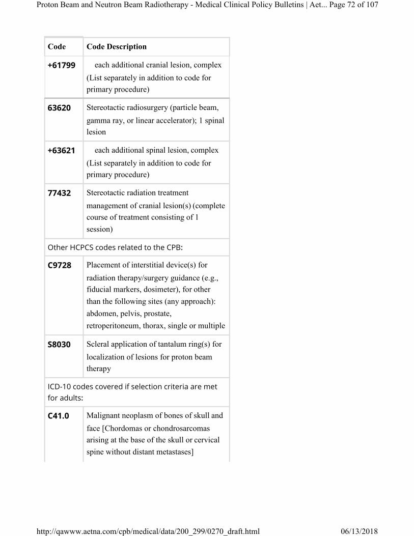
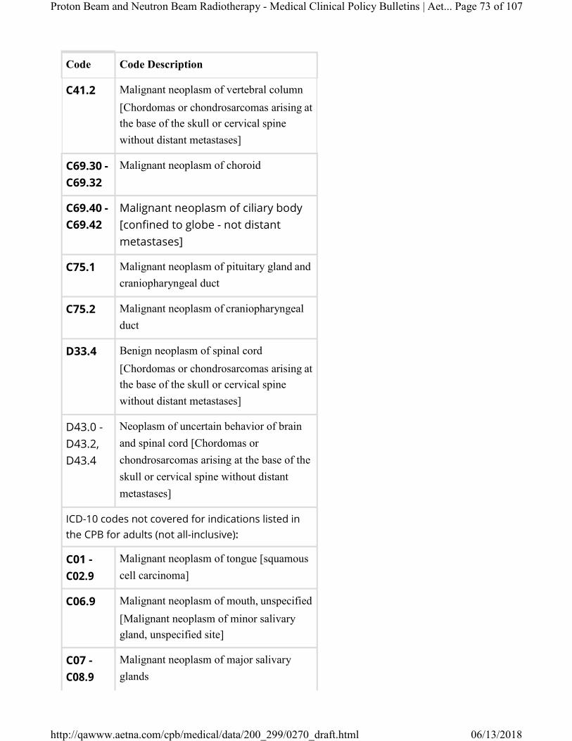
I. Aetna considers proton beam radiotherapy (PBRT) medically necessary in any of the following radiosensitive tumors:
A. Chordomas or chondrosarcomas arising at the base of the skull or cervical spine without distant metastases; or
B. Malignancies in children (21 years of age and younger); or
C. Uveal melanomas confined to the globe (i.e., not distant metastases) (the uvea is comprised of the iris, ciliary body, and choroid [the vascular middle coat of the eye]).
II. Aetna considers proton beam radiotherapy for treatment of prostate cancer not medically necessary for individuals with localized prostate cancer because it has not
Proton Beam and Neutron Beam Radiotherapy - Medical Clinical Policy Bulletins | Aetna Page 2 of 107
been proven to be more effective than other radiotherapy modalities for this indication. Proton beam therapy for metastatic prostate cancer is considered experimental and investigational.
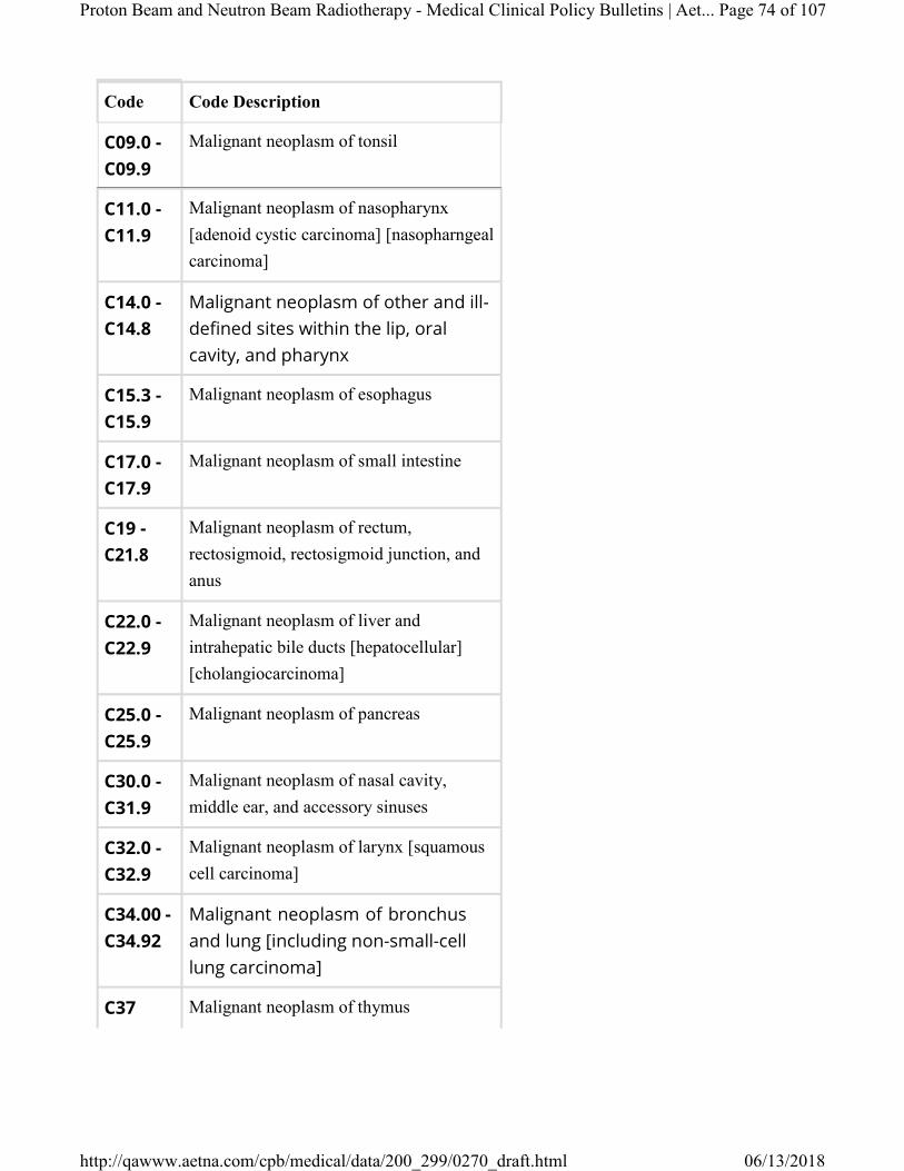
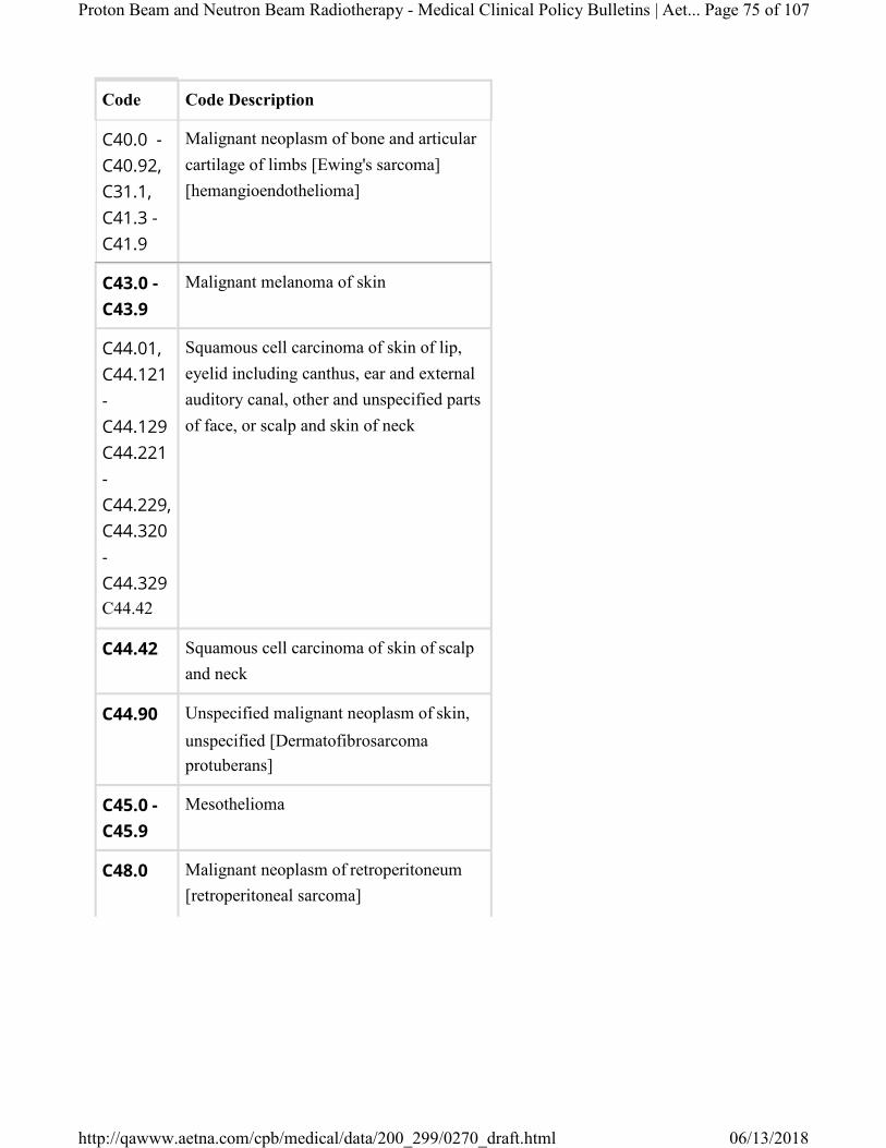
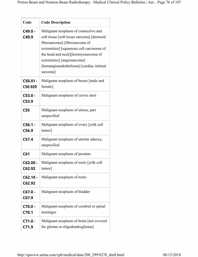
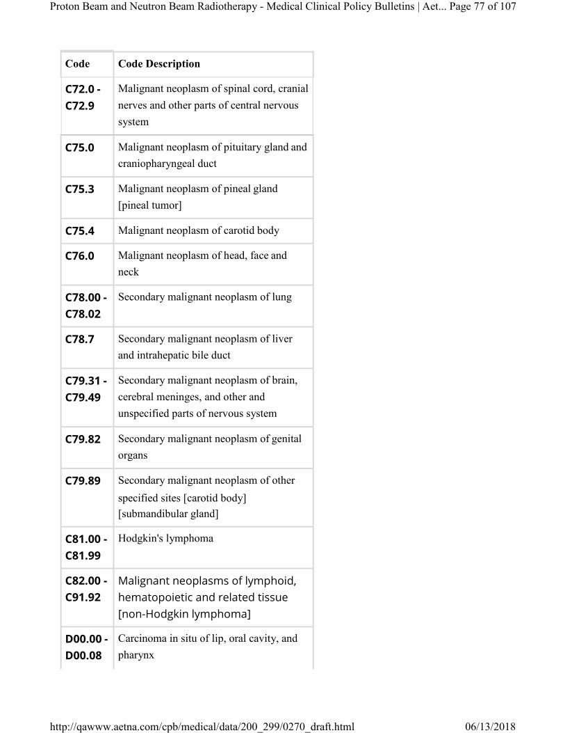
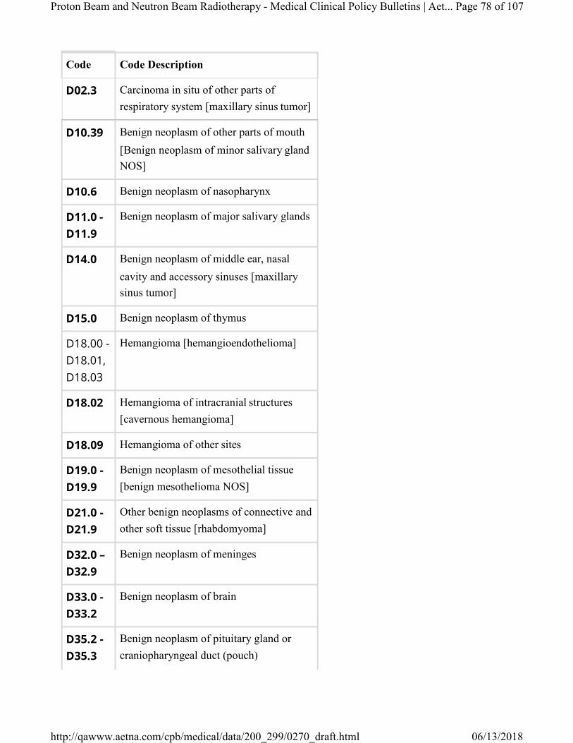
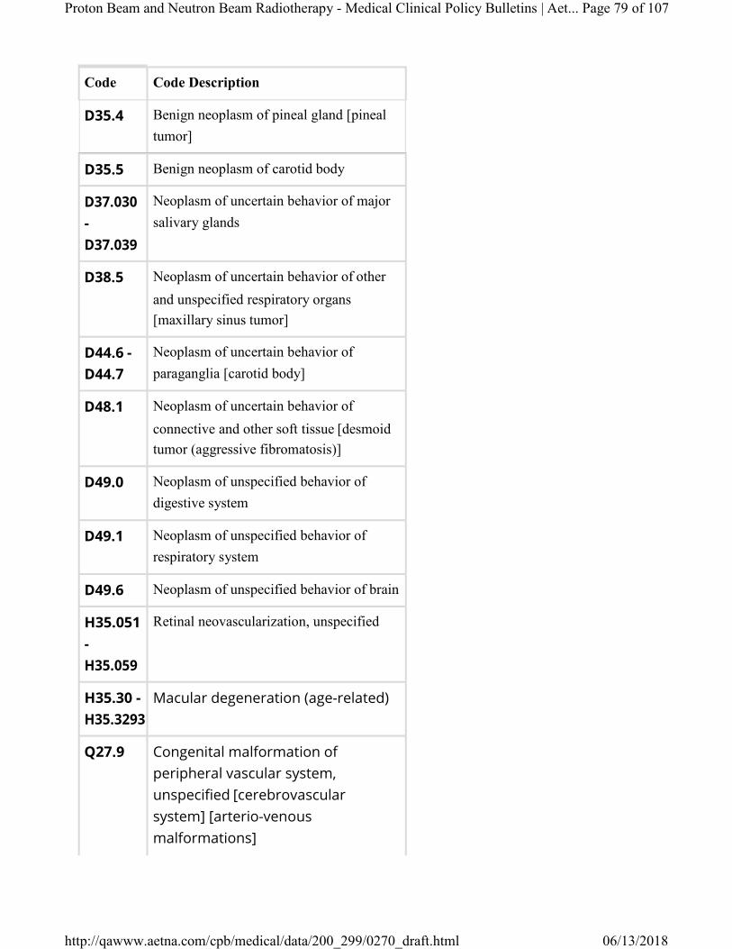
III. Aetna considers proton beam radiotherapy experimental and investigational for all other indications, including the following indications in adults (over age 21) (not an all-inclusive list) because its effectiveness for these indications has not been established:
▪ Adenoid cystic carcinoma ▪ Age-related macular degeneration ▪ Angiosarcoma ▪ Atypical meningioma ▪ Bladder cancer ▪ Brain tumors ▪ Breast cancer ▪ Cardiac intimal sarcoma ▪ Carotid body tumor ▪ Cavernous hemangioma ▪ Cervical cancer ▪ Cholangiocarcinoma ▪ Choroidal hemangioma ▪ Dermatofibrosarcoma protuberans ▪ Desmoid fibromatosis ▪ Desmoid tumor (aggressive fibromatosis) ▪ Ependymoma ▪ Esophageal cancer ▪ Ewing's sarcoma ▪ Fibrosarcoma of the extremities ▪ Gangliomas ▪ Glioma ▪ Head and neck cancer (including
Proton Beam and Neutron Beam Radiotherapy - Medical Clinical Policy Bulletins | Aetna Page 4 of 107
▪ Thymoma ▪ Tonsillar cancer ▪ Uterine cancer ▪ Vestibular schwannoma ▪ Yolk cell tumor
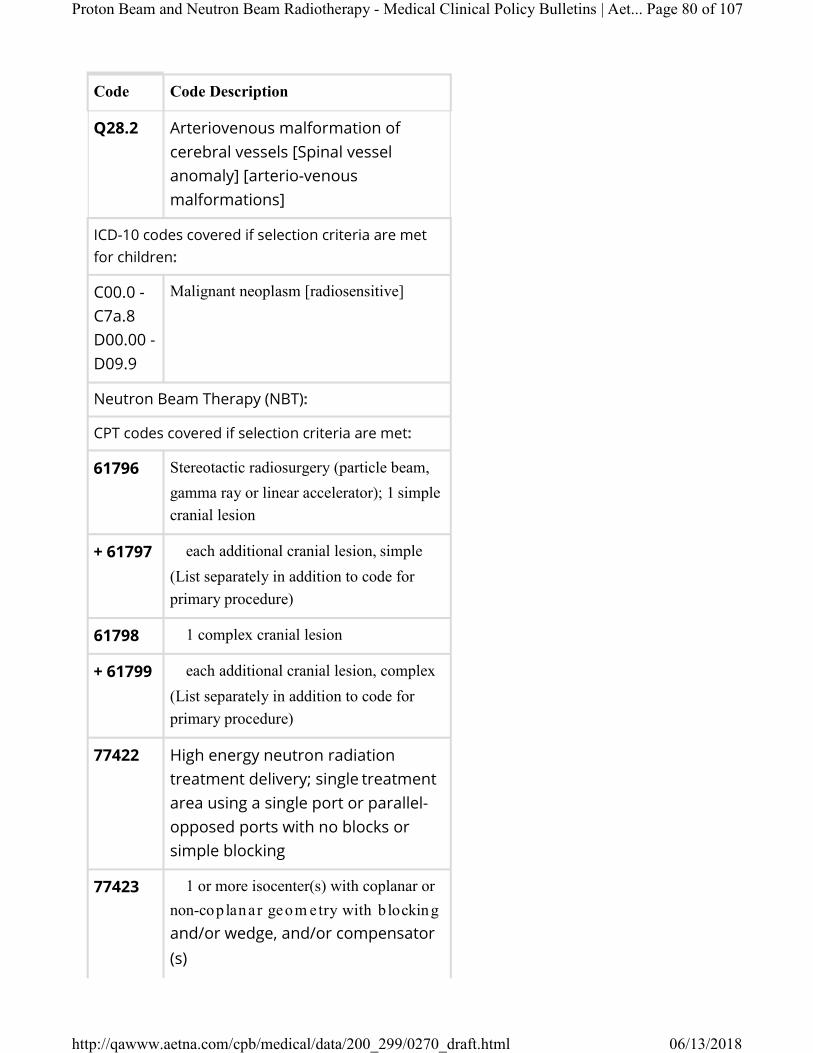
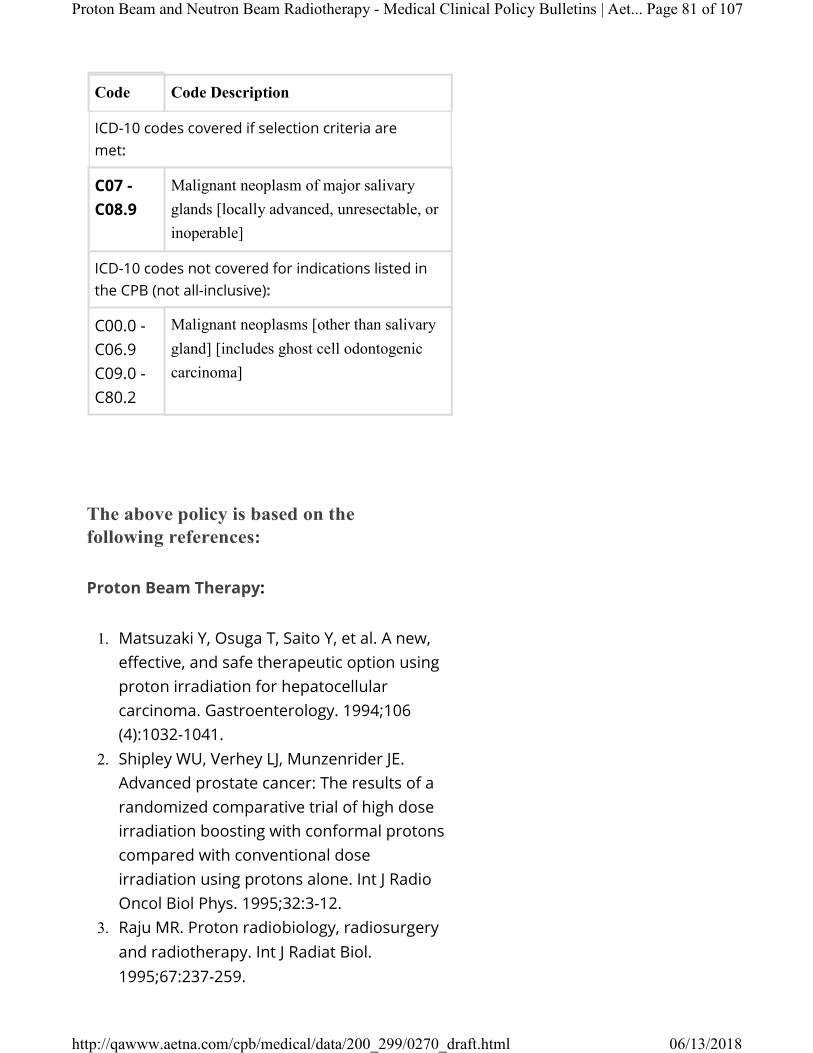
IV. Aetna considers neutron beam therapy medically necessary for the treatment of any of the following salivary gland tumors:
▪ Inoperable tumor; or ▪ Locally advanced tumor especially in
persons with gross residual disease; or ▪ Unresectable tumor.
V. Aetna considers neutron beam therapy experimental and investigational for all other indications including malignancies listed below (not an all inclusive list) because its effectiveness for these indications has not been established:
▪ Colon cancer ▪ Dermatofibrosarcoma protuberans ▪ Ghost cell odontogenic carcinoma ▪ Glioma ▪ Kidney cancer ▪ Laryngeal cancer ▪ Lung cancer ▪ Pancreatic cancer ▪ Prostate cancer ▪ Rectal cancer ▪ Soft tissue sarcoma.
Proton Beam and Neutron Beam Radiotherapy - Medical Clinical Policy Bulletins | Aetna Page 5 of 107
Background
Proton Beam Therapy
Proton beam radiation therapy (PBRT) is a type of external beam radiation therapy (EBRT) that utilizes protons (positively charged subatomic particles) that are precisely targeted to a specific tissue mass. Proton beams have the ability to penetrate deep into tissues to reach tumors, while delivering less radiation to superficial tissues such as the skin. This may make PBRT more effective for inoperable tumors or for those individuals in which damage to healthy tissue would pose an unacceptable risk.
Proton beams have less scatter than other sources of energy such as gamma rays, x-rays, or electrons. Because of this feature, proton beam radiotherapy (PBRT) has been used to escalate radiation dose to diseased tissues while minimizing damage to adjacent normal tissues. Proton beams have been used in stereotactic radiosurgery of intracranial lesions; the gamma knife and linear accelerator have also been used in stereotactic radiosurgery. Proton beam radiotherapy has been shown to be particularly useful in treating radiosensitive tumors that are located next to vital structures, where complete surgical excision or administration of adequate doses of conventional radiation is difficult or impossible. Examples include uveal melanomas, chordomas and chondrosarcomas at the base of the skull, and inoperable arterio-venous malformations. There is inadequate data on the application of PBRT for the treatment of nonuveal melanoma.
Proton Beam and Neutron Beam Radiotherapy - Medical Clinical Policy Bulletins | Aetna Page 6 of 107
beam therapy by the Veteran's Health Administration (2015) reached the following conclusions: "For the cancer sites and types reviewed by the ESP report, there are no reliable data from long-term randomized trials on survival, quality of life, or functional capacity of patients who underwent PBT compared with any other modality. The authors could not fully assess the overall net health benefit of proton beam therapy versus its comparators because comparative observational studies did not consistently report many outcomes of greatest interest. Existing comparative studies have numerous methodological deficiencies that limited confidence in the findings, and the findings may have limited applicability across all US proton beam facilities. Although numerous randomized controlled trials are underway that carry the promise of improved toxicity measurement, the report noted it is unclear whether these will fully address gaps in evidence on such outcomes as recurrence, ability to deliver planned chemotherapy and radiation regimens, functional capacity, overall severe late toxicity, and secondary malignancies. Although the report found comparative studies in giant cell tumors, head and neck cancer, uveal hemangiomas, and meningiomas, they provided insufficient evidence for drawing conclusions. There is insufficient evidence to draw conclusions about the comparative effects of PBT versus other radiation modalities among patients with recurrent tumors or how the comparative effects of proton and photon beam therapies differ according to variation in tumor motion."
Proton Beam and Neutron Beam Radiotherapy - Medical Clinical Policy Bulletins | Aetna Page 7 of 107
Technologies in Health (CADTH, 2016) found: "Two systematic reviews of clinical evidence, two systematic reviews of economic evidence, and one primary economic evaluation were identified regarding the clinical and cost-effectiveness of proton beam therapy compared to photon radiotherapy for the treatment of cancer patients. There was limited comparative evidence, with insufficient evidence available for many indications, comparators, and outcomes. Comparable benefits and harms were demonstrated by most studies for the majority of outcomes in prostate, esophageal, lung, and breast cancer, as well as medulloblastoma, and pediatric brain tumours. An increased risk of patient harms was observed for some outcomes in breast, esophageal, prostate and lung cancer. As well, reduced survival was reported for spinal cord gliomas. Reduced harms were reported for some outcomes in patients with medulloblastoma, as well as lung, esophageal, and prostate cancer, and pediatric retinoblastoma. There was insufficient evidence to draw conclusions for recurrent liver and brain cancers, meningioma, head and neck cancers, uveal hemangioma, and for the outcome of secondary malignancies. The identified economic evidence is likely not generalizable to the Canadian context, and may not reflect accurate and up-to-date cost and benefit estimates. Most evaluations reported that PBT was not cost- effective; however, the technology was more likely to be cost-effective in pediatric populations, and under specific circumstances in younger adults, and patients with more advanced disease (e.g., high risk head and neck, lung cancer, and breast cancer patients). Overall, current comparative evidence does not suggest
Proton Beam and Neutron Beam Radiotherapy - Medical Clinical Policy Bulletins | Aetna Page 8 of 107
that PBT is superior to photon therapy from a clinical or cost perspective for the majority of indications. There are concerns regarding the quantity, quality and generalizability of the available evidence. Current ongoing studies and future investigation into differences in hard clinical endpoints and long-term outcomes may resolve some of this uncertainty."
A report by the Canadian Agency for Drugs and Technologies in Health (CADTH, 2017) found: "A review of the clinical evidence from nine systematic reviews found that PBT, alone or in combination with photon radiotherapy, is comparable to other types of radiotherapy for most types of cancer. Exceptions include meningioma and subgroups of malignant meningioma, and poorly differentiated tumours of prostate cancer in adults, for which greater benefits were found with PBT; some intramedullary spinal cord glioma in both children and adults, for which lower benefits were found with PBT; and eye cancer in adults, for which both greater benefits and lower benefits, depending on the specific type of eye cancer, were found with PBT. The clinical evidence also found that the safety of PBT, alone or in combination with photon radiotherapy, varies by the type of cancer it is used to treat, compared with other types of radiotherapy. It was found to be associated with greater harms in breast cancer and prostate cancer in adults; lower harms in retinoblastoma in children and medulloblastoma in adults; and both greater harms and lower harms in adults depending on the specific type of the following cancers:
Proton Beam and Neutron Beam Radiotherapy - Medical Clinical Policy Bulletins | Aetna Page 9 of 107
esophageal cancer, optic nerve sheath meningioma, and lung cancer."
Regarding cost-effectiveness analyses of PBRT, the VATAP assessment found that the availability of studies titled by their authors as “economic evaluations” is misleading (Flynn, 2010). The assessment stated that such studies require cost and efficacy data about both the intervention and its alternatives (costs and consequences of alternative interventions), hence should be conducted only after efficacy data from randomized controlled trials are available. The assessment noted that, in the case of proton therapy, the economic studies are premature, really should be labeled simple cost rather than cost-effectiveness analyses, and their conclusions based on unwarranted efficacy assumptions. Cost data have been carefully collected and reported, but these are only one element of decision making about investment in proton therapy. The VATAP assessment concluded that there are no indications for which proton therapy has been shown unequivocally to be effective, or more effective than its alternatives. The VATAP assessment also concluded that no research published subsequent to the searches conducted for available systematic reviews has changed the conclusions of those reviews.
Regarding research implications, the VATAP assessment concluded that, in order to obtain the next generation of data, explicit decisions need to be made about which malignancies are amenable to/should require randomized trials (e.g., prostate cancer is sufficiently common) and which malignancies are sufficiently rare or difficult to treat with surgery or conventional radiotherapy (e.g., ocular tumors, tumors of the optical nerve, spinal cord, or central nervous system) that observational studies with larger cohorts than studies to date are the best approach (Flynn, 2010).
Proton Beam and Neutron Beam Radiotherapy - Medical Clinical Policy Bulletins | Aet... Page 10 of 107
The VATAP assessment also concluded that future studies should strongly consider valid and reliable embedded collection of cost data in order to inform better quality economic evaluation than currently available.
An assessment prepared for the Agency for Healthcare Research and Quality (Trikalinos, et al., 2009) found that a large number of scientific papers on charged particle radiotherapy for the treatment of cancer currently exist. However, these studies do not document the circumstances in contemporary treatment strategies in which radiotherapy with charged particles is superior to other modalities. Comparative studies in general, and randomized trials in particular (when feasible), are needed to document the theoretical advantages of charged particle radiotherapy to specific clinical situations. The assessment noted that most eligible studies were noncomparative in nature and had small sample sizes. The report stated that it is likely that focused systematic reviews will not be able to provide a definitive answer on the effectiveness and safety of charged particle beam radiotherapies compared with alternative interventions. This is simply because of the relative lack of comparative studies in general, and randomized trials in particular. The report stated that comparative studies (preferably randomized) are likely necessary to provide meaningful answers on the relative safety and effectiveness of particle beam therapy versus other treatment options in the context of current clinical practice. This is especially true for the treatment of common cancers. The report stated that, especially for many common cancers, such as breast, prostate, lung, and pancreatic cancers,
Proton Beam and Neutron Beam Radiotherapy - Medical Clinical Policy Bulletins | Aet... Page 11 of 107
it is essential that the theorized advantages of particle beam therapy versus contemporary alternative interventions are proven in controlled clinical trials, along with concomitant economic evaluations.
An assessment by the Riete Italiana Health Technology Assessment (RIHTA, 2011) concluded: "The lack of comparative studies comparing HT [hadron therapy] with other currently available treatments (other RT techniques and/or chemotherapy) does not allow drawing firm conclusions about the effects of HT in cancer treatment. In specific circumstances, clinical studies suggested improvements in safety and effectiveness by using HT instead of traditional RT for some types of tumours (in particular uveal melanoma, skull and neck chordomas, and NSCLC). Nonetheless, there is uncertainty regarding these estimates, due to methodological and design flaws of available studies. Therefore presently available evidence is not sufficient to support routine clinical use of HT. "
An assessment of proton therapy by the Ludwig Boltzmann Institute (Wild, et al., 2013) concluded: "The evidence basis for an added benefit is only very moderate. Since surrogate endpoints were primarily measured and no/hardly any prospectively comparative trial results with up-to-date photon therapy are available, there is no confirmed knowledge of whether the promise of theoretical advantages can be translated into patient-relevant advantages (longer survival, quality of life through fewer side effects)."
Proton Beam and Neutron Beam Radiotherapy - Medical Clinical Policy Bulletins | Aet... Page 12 of 107
The American Society of Radiation Oncology (ASTRO, 2013) has stated: "At the present time, ASTRO believes the comparative efficacy evidence of proton beam therapy with other prostate cancer treatments is still being developed, and thus the role of proton beam therapy for localized prostate cancer within the current availability of treatment options remains unclear."
The emerging technology committee of the American Society of Radiation Oncology (ASTRO) concluded that current evidence provides a limited indication for proton beam therapy (Allen, et al., 2012). The ASTRO report concluded that current data do not provide sufficient evidence to recommend proton beam therapy in lung cancer, head and neck cancer, gastrointestinal malignancies, and pediatric non-CNS malignancies. The ASTRO report stated that, in hepatocellular carcinoma and prostate cancer, there is evidence for the efficacy of proton beam therapy but no suggestion that it is superior to photon based approaches. In pediatric central nervous system (CNS) malignancies, proton beam therapy appears superior to photon approaches but more data is needed. The report found that, in large ocular melanomas and chordomas, there is evidence for a benefit of proton beam therapy over photon approaches. The ASTRO report stated that more robust prospective clinical trials are needed to determine the appropriate clinical setting for proton beam therapy.
A systematic evidence review (Lodge et al, 2007) compared the efficacy and cost-effectiveness of PBRT and other types of hadron therapy (neutron and heavy and light ion therapy) with photon therapy. The authors concluded that, overall, the introduction or extension of PBRT and other types of hadron therapy as a major treatment modality into standard clinical care is not supported by the current evidence base. The authors stated, however, that the efficacy of PBRT appears
Proton Beam and Neutron Beam Radiotherapy - Medical Clinical Policy Bulletins | Aet... Page 13 of 107
superior to that of photon therapy for some ocular and skull base tumours. The authors found that, for prostate cancer, the efficacy of PBRT seems comparable to photon therapy. The authors stated that no definitive conclusions can be drawn for the other cancer types. The authors also noted that they found little evidence on the relative cost-effectiveness of PBRT and other types of hadron therapy compared to photon therapy or with other cancer treatments. Other systematic evidence reviews of PBRT have reached similar conclusions (Lance, 2010; Brada et al, 2009; Efstathiou et al, 2009; ICER, 2008; Wilt et al, 2008; Brada et al, 2007; Olsen et al, 2007).
The only randomized controlled clinical trial comparing PBRT to conventional radiotherapy for prostate cancer published to date found no advantage of PBRT in overall survival (OS), disease-specific survival, or total recurrence-free survival (Shipley, 1995). A total of 202 patients with stage T3-T4 prostate cancer were randomly assigned to a standard dose of conventional radiotherapy plus a 25.2 Gy equivalent PBRT boost or to a standard dose of conventional radiotherapy with a 16.8 Gy boost of conventional radiotherapy. After a median follow-up of 61 months, there were no significant differences between the 2 groups in OS, disease-specific survival, total recurrence-free survival, or local control. Local control was better with the proton beam boost only among the subgroup of patients with poorly differentiated carcinoma. Patients receiving the proton beam boost had increased rates of late radiation sequelae.
Loma Linda University’s experience with PBRT of prostate cancer was reported in an article published in 1999 by Rossi et al. These investigators reported the results of an uncontrolled study of PBRT treatment of 319 patients with biopsy-proven early-stage prostate
Proton Beam and Neutron Beam Radiotherapy - Medical Clinical Policy Bulletins | Aet... Page 14 of 107
cancer, with no patient having an initial PSA of greater than 15. Because the study was uncontrolled, one is unable to determine whether the results of PBRT are superior to conventional forms of radiation therapy. In addition, the definitions of success and failure used in this study are not comparable to those used in other recent studies of prostate cancer treatments. In the study by Rossi et al, patients were considered to have an adequate response if their PSA level fell below 1.0; most other recent studies define an adequate response as PSA level below 0.5. In the study by Rossi et al, patients were considered treatment failures if they had 3 consecutive rises of PSA of 10 % or more, measured at 6-month intervals. In other words, for a patient to be considered a treatment failure, it would take at least 18 months, and patients would have to have 3 consecutive rises in PSA, each greater than 10 %. By contrast, other reported studies of prostate cancer radiotherapy have defined failure as any PSA elevation over a target PSA nadir. Finally, Rossi et al defined clinical disease free survival as having "no symptoms and no evidence of disease upon physical examination or radionuclide scans". These are very gross tests to determine success, and one would expect these tests to be negative in a high number of patients who harbored occult disease.
Of the 319 patients included in the study by Rossi et al, only 288 patients (91 %) who had achieved a nadir (any nadir) or who had been followed for at least 24 months were included in the analysis. This would indicate that 31 (9 %) of the patients originally included in the study either had persistently rising PSA levels without a nadir despite treatment, had dropped out of the study, or had not been followed for a sufficient length of time for some unspecified reason. Only 187 patients (59 % of the original 319 patients) achieved a PSA nadir of 0.5 or less, 66 (21 %) achieved a PSA nadir of 0.51 to 1.0, and 35 (11 %) achieved a PSA nadir of 1.0 and above. Thus,
Proton Beam and Neutron Beam Radiotherapy - Medical Clinical Policy Bulletins | Aet... Page 15 of 107
only 59 % of patients would be considered to have had an adequate response by the measure most commonly used in other recent prostate cancer treatment studies. In addition, because of the peculiar way the results are reported, there is no way of knowing how many patients' PSA nadirs were maintained.
In a randomized, prospective, sham-controlled, double- masked study (n = 37), Ciulla et al (2002) examined the effect of PBRT on subfoveal choroidal neovascular membranes associated with age-related macular degeneration. These investigators concluded that with the acceptance of photodynamic therapy, future studies will require more complex design and larger sample size to determine whether radiation can play either a primary or adjunctive role in treating these lesions.
In a phase II clinical study (n = 30), Kawashima and colleagues (2005) assessed the safety and effectiveness PBRT for patients with hepatocellular carcinoma (HCC). Eligibility criteria for this study were: solitary HCC; no indication for surgery or local ablation therapy; no ascites; age of 20 years or older; Zubrod performance status of 0 to 2; no serious co-morbidities other than liver cirrhosis; written informed consent. Proton beam radiotherapy was administered in doses of 76 cobalt gray equivalent in 20 fractions for 5 weeks. No patients received transarterial chemoembolization or local ablation in combination with PBRT. All patients had liver cirrhosis, the degree of which was Child-Pugh class A in 20, and class B in 10 patients. Acute reactions of PBRT were well-tolerated, and PBRT was completed as planned in all patients. Four patients died of hepatic insufficiency without tumor recurrence at 6 to 9 months; 3 of these 4 patients had pre-treatment indocyanine green retention rate at 15 minutes of more than 50 %. After a median follow-up period of 31 months (range of 16 to 54 months), only 1 patient experienced recurrence
Proton Beam and Neutron Beam Radiotherapy - Medical Clinical Policy Bulletins | Aet... Page 16 of 107
of the primary tumor, and 2-year actuarial local progression-free rate was 96 %. Actuarial overall survival rate at 2 years was 66 %. These investigators concluded that PBRT showed excellent control of the primary tumor, with minimal acute toxicity. They stated that further study is warranted to scrutinize adequate patient selection in order to maximize survival benefit of this promising modality.
In a phase II prospective trial, Bush et al (2011) evaluated the safety and effectiveness of PBRT for HCC. Patients with cirrhosis who had radiological features or biopsy-proven HCC were included in the study. Patients without cirrhosis and patients with extra- hepatic metastasis were excluded. The mean age was 62.7 years. The mean tumor size was 5.5 cm. Eleven patients had multiple tumors, and 46 % were within the Milan criteria. Patients received 63 Gy delivered over a 3-week period with PBRT. A total of 76 patients were treated and followed prospectively. Acute toxicity was minimal; all patients completed the full course of treatment. Radiation-induced liver disease was evaluated using liver enzyme, bilirubin, and albumin levels; no significant change supervened 6 months post- treatment. Median progression-free survival for the entire group was 36 months, with a 60 % 3-year progression-free survival rate for patients within the Milan criteria. Eighteen patients subsequently underwent liver transplantation; 6 (33 %) explants showed pathological complete response and 7 (39 %) showed only microscopic residual. The authors concluded that PBRT was found to be a safe and effective local-regional therapy for inoperable HCC. They noted that a randomized controlled trial to compare its efficacy to a standard therapy has been initiated.
Proton Beam and Neutron Beam Radiotherapy - Medical Clinical Policy Bulletins | Aet... Page 17 of 107
Olfactory neuroblastoma (ONB) is a rare disease, and a standard treatment strategy has not been established. Radiation therapy for ONB is challenging because of the proximity of ONB to critical organs. Nishimura et al (2007) analyzed the feasibility and effectiveness of PBRT for ONB. A retrospective review was performed on 14 patients who underwent PBRT for ONB as definitive treatment. The total dose of PBRT was 65 cobalt Gray equivalents (Gy(E)), with 2.5-Gy(E) once- daily fractionations. The median follow-up period for surviving patients was 40 months. One patient died from disseminated disease. There were 2 persistent diseases, 1 of which was successfully salvaged with surgery. The 5-year overall survival rate was 93 %, the 5-year local progression-free survival rate was 84 %, and the 5-year relapse-free survival rate was 71 %. Liquorrhea was observed in 1 patient with Kadish's stage C disease (widely destroying the skull base). Most patients experienced grade 1 to 2 dermatitis in the acute phase. No other adverse events of grade 3 or greater were observed according to the RTOG/EORTC acute and late morbidity scoring system. The authors concluded that these preliminary findings of PBRT for ONB achieved excellent local control and survival outcomes without serious adverse effects. They stated that PBRT is considered a safe and effective modality that warrants further study.
Proton beam radiotherapy represents a special case for children for several reasons (Wilson et al, 2005; Hall, 2006; Merchant, 2009). It has been shown in dosimetric planning studies to have a potential advantage over conventional photon therapy because of the ability to confine the high-dose treatment area to the tumor volume and minimize the radiation dose to the surrounding tissue. This especially important in children, as children are more sensitive to radiation- induced cancer than adults. An increased risk of second
Proton Beam and Neutron Beam Radiotherapy - Medical Clinical Policy Bulletins | Aet... Page 18 of 107
cancers in long-term survivors is more important in children than older adults. In addition to second malignant neoplasms, late effects of radiation to normal tissue can include developmental delay. Also, radiation scattered from the treatment volume is more important in the small body of the child. Finally, the question of genetic susceptibility arises because many childhood cancers involve a germline mutation.
An assessment of proton beam radiotherapy by the Veterans Health Administration Technology Assessment Program (VATAP) (Flynn, 2010) found that available English-language reviews for proton therapy generally concur on the state of the literature as consisting primarily of observational studies from which conclusions about the relative effectiveness of proton therapy versus alternatives cannot validly be made. The assessment reported that available reviews reflect the state of the literature in that they attempt to cover so much territory (multiple poor-prognosis inoperable tumors in both children and adults) that the reviews themselves are cumbersome to read, not well organized, and provide only diffuse or equivocal conclusions by individual diagnoses. "In other words, the literature reflects the early clinical investigation status of proton therapy, where observational studies are framed in terms of potential benefits, reasoning from pathophysiology, dose-finding, refinement of treatment protocols, and baseline safety of the entire approach" (Flynn, 2010). The assessment noted that only prostate cancer is represented by randomized controlled clinical trials, and in that case two small ones primarily concerned with refinement of protocol/dose escalation.
An assessment of the comparative effectiveness and value of management options in low-risk prostate cancer by the Institute for Clinical and Economic Review (ICER) (Ollendorf et al, 2008) found that the evidence
Proton Beam and Neutron Beam Radiotherapy - Medical Clinical Policy Bulletins | Aet... Page 19 of 107
on the comparative effectiveness and harms of proton beam therapy is limited to relatively small, highly selective case series of short duration, making any judgments about its relative benefit or inferiority to other options premature. The uncertainty regarding PBRT is accentuated because this technology involves delivery of a novel form of radiation, and there remain important questions about the full spectrum of possible effects. ICER rated PBRT's comparative clinical effectiveness as "insufficient", indicating that there is not enough evidence to allow a reasonable judgment of the likely balance of harms and benefits of PBRT in comparison to radical prostatectomy or other management options. ICER judged the comparative value of PBRT to be low compared to other options. The ICER reported explained, that, while ICER does not always provide a comparative value rating for technologies with insufficient evidence on comparative clinical effectiveness, the decision was made to rate the comparative value of PBRT as “low” relative to radical prostatectomy, based on current levels of reimbursement that are more than 3-fo ld h igh e r fo r PBRT.
A BCBS TEC assessment found insufficient evidence for PBRT in the treatment of non-smallcell lung cancer. In addition, the American Society for Radiation Oncology (ASTRO) guidelines (Allen et al, 2012) found insufficient evidence for PBRT in lung cancer.
The Blue Cross and Blue Shield Association Medical Advisory Panel (BCBSA, 2010) concluded that proton beam radiation therapy for treatment of non-small-cell lung cancer at any stage or for recurrent non-small-cell lung cancer does not meet the Technology Evaluation Center criteria. The TEC assessment stated that, overall, evidence is insufficient to permit conclusions
Proton Beam and Neutron Beam Radiotherapy - Medical Clinical Policy Bulletins | Aet... Page 20 of 107
about the results of proton beam therapy for any stage of non-small-cell lung cancer. The report found that all proton beam therapy studies are case series; there are no studies directly comparing proton beam therapy and stereotactic body radiotherapy. Among study quality concerns, no study mentioned using an independent assessor of patient reported adverse events, adverse events were generally poorly reported, and details were lacking on several aspects of proton beam therapy treatment regimens. The proton beam therapy studies had similar patient ages, but there was great variability in percent within stage Ia, sex ratio, and percent medically inoperable. There is a high degree of treatment heterogeneity among the proton beam therapy studies, particularly with respect to planning volume, total dose, number of fractions, and number of beams. Survival results are highly variable. It is unclear if the heterogeneity of results can be explained by differences in patient and treatment characteristics. Indirect comparisons between proton beam therapy and stereotactic body radiotherapy, comparing separate sets of single-arm studies on proton beam therapy and stereotactic body radiotherapy, may be distorted by confounding. In the absence of randomized, controlled trials, the comparative effectiveness of proton beam therapy and stereotactic body radiotherapy is uncertain.
Bassim et al (2010) reviewed the literature on radiation therapy for the treatment of vestibular schwannoma (VS). PubMed searches for English language articles on radiation treatment of VS published from January 2002 to July 2007 were
Proton Beam and Neutron Beam Radiotherapy - Medical Clinical Policy Bulletins | Aet... Page 21 of 107
conducted. Studies presenting outcomes were selected, yielding 56 articles (58 studies) in journals of neurosurgery (30), oncology (18), otolaryngology (6), and other (2). Data included type of study, number of subjects, demographics, follow-up times, type of radiation, tumor size, tumor control definition, control rates, facial nerve function measure and outcome, type of hearing and vestibular testing and outcomes, and complications. Descriptive statistics were performed. Studies (72.9 %) were retrospective reviews with stated sample sizes ranging from 5 to 829. Gamma-knife (49.2 %), linear accelerator (35.6 %), and proton beam (6.8 %) were used with various doses. Average follow-up was less than 5 years in 79.6 % of studies, and 67.4 % included patients at less than or equal to 1 year. Tumor size was reported as diameter (23.7 %), volume (49.2 %), both (11.9 %), other (3.4 %), or not reported (11.9 %). Definition of tumor control varied: less than or equal to 2 mm growth (22.0 %), no visible/measurable change (16.9 %), required surgery (10.2 %), other (17.0 %), and not clearly specified (33.9 %). Facial nerve outcome was reported as House-Brackmann (64.4 %), normal/abnormal (11.9 %), other (1.7 %), or was not reported (22 %). The authors concluded that the lack of uniform reporting criteria for tumor control, facial function and hearing preservation, and variability in follow-up times make it difficult to compare studies of radiation treatment for VS. They recommended consideration of reporting guidelines such as those used in otology for reporting VS resection results.
Proton Beam and Neutron Beam Radiotherapy - Medical Clinical Policy Bulletins | Aet... Page 22 of 107
Mizumoto et al (2010) evaluated the efficacy and safety of PBRT for locoregionally advanced esophageal cancer. The subjects were 51 patients with esophageal cancer who were treated between 1985 and 2005 using proton beams with or without X-rays. All but 1 had squamous cell carcinoma. Of the 51 patients, 33 received combinations of X-rays (median of 46 Gy) and protons (median of 36 GyE) as a boost. The median total dose of combined X-rays and proton radiation for these 33 patients was 80 GyE (range of 70 to 90 GyE). The other 18 patients received PBRT alone (median of 79 GyE, range of 62 to 98 GyE). Treatment interruption due to radiation-induced esophagitis or hematologic toxicity was not required for any patient. The overall 5-year actuarial survival rate for the 51 patients was 21.1 % and the median survival time was 20.5 months (95 % confidence interval [CI]: 10.9 to 30.2). Of the 51 patients, 40 (78 %) showed a complete response within 4 months after completing treatment and 7 (14 %) showed a partial response, giving a response rate of 92 % (47/51). The 5-year local control rate for all 51 patients was 38.0 % and the median local control time was 25.5 months (95 % CI: 14.6 to 36.3). The authors concluded that these findings suggested that PBRT is an effective treatment for patients with locally advanced esophageal cancer. Moreover, they stated that further studies are needed to determine the optimal total dose, fractionation schedules, and best combination of PBRT with chemotherapy. Furthermore, the National Comprehensive Cancer Network (NCCN) guideline on esophageal cancer (2011) does not mention the use of PBRT as a therapeutic option for this condition.
Mizumoto et al (2011) evaluated the safety and effectiveness of hyper-fractionated concomitant boost proton beam therapy (PBT) for patients with esophageal cancer. The study participants were 19 patients with esophageal cancer who were treated with
Proton Beam and Neutron Beam Radiotherapy - Medical Clinical Policy Bulletins | Aet... Page 23 of 107
hyperfractionated photon therapy and PBT between 1990 and 2007. The median total dose was 78 GyE (range of 70 to 83 GyE) over a median treatment period of 48 days (range of 38 to 53 days). Ten of the 19 patients were at clinical T Stage 3 or 4. There were no cases in which treatment interruption was required because of radiation-induced esophagitis or hematologic toxicity. The overall 1- and 5-year actuarial survival rates for all 19 patients were 79.0 % and 42.8 %, respectively, and the median survival time was 31.5 months (95 % limits: 16.7 to 46.3 months). Of the 19 patients, 17 (89 %) showed a complete response within 4 months after completing treatment and 2 (11 %) showed a partial response, giving a response rate of 100 % (19/19). The 1- and 5-year local control rates for all 19 patients were 93.8 % and 84.4 %, respectively. Only 1 patient had late esophageal toxicity of Grade 3 at 6 months after hyperfractionated PBT. There were no other non-hematologic toxicities, including no cases of radiation pneumonia or cardiac failure of Grade 3 or higher. The authors concluded that these findings suggested that hyperfractionated PBT is safe and effective for patients with esophageal cancer. They stated that further studies are needed to establish the appropriate role and treatment schedule for use of PBT for esophageal cancer.
In a phase I clinical study, Hong et al (2011) evaluated the safety of 1 week of chemo-radiation with proton beam therapy and capecitabine followed by early surgery on 15 patients with localized resectable, pancreatic ductal adenocarcinoma of the head. Patients received radiation with proton beam. In dose level 1, patients received 3 GyE × 10 (week 1, Monday to Friday; week 2, Monday to Friday). Patients in dose levels 2 to 4 received 5 GyE × 5 in progressively shortened schedules: level 2 (week 1, Monday, Wednesday, and Friday; week 2, Tuesday and
Proton Beam and Neutron Beam Radiotherapy - Medical Clinical Policy Bulletins | Aet... Page 24 of 107
Thursday), level 3 (week 1, Monday, Tuesday, Thursday, and Friday; week 2, Monday), level 4 (week 1, Monday through Friday). Capecitabine was given as 825 mg/m(2) b.i.d. Weeks 1 and 2 Monday through Friday for a total of 10 days in all dose levels. Surgery was performed 4 to 6 weeks after completion of chemotherapy for dose levels 1 to 3 and then after 1 to 3 weeks for dose Level 4. Three patients were treated at dose levels 1 to 3 and 6 patients at dose level 4, which was selected as the MTD. No dose limiting toxicities were observed. Grade 3 toxicity was noted in 4 patients (pain in 1; stent obstruction or infection in 3). Eleven patients underwent resection. Reasons for no resection were metastatic disease (3 patients) and unresectable tumor (1 patient). Mean post-surgical length of stay was 6 days (range of 5 to 10 days). No unexpected 30-day post-operative complications, including leak or obstruction, were found. The authors concluded that pre-operative chemo-radiation with 1 week of PBRT and capecitabine followed by early surgery is feasible. A phase II study is underway.
UpToDate reviews on "Management of locally advanced and borderline resectable exocrine pancreatic cancer" (Ryan and Mamon, 2012) and "Surgery in the treatment of exocrine pancreatic cancer and prognosis" (Fernandez-del Castillo et al, 2012) do not mention the use of proton beam therapy. Furthermore, the NCCN's clinical practice guideline on "Pancreatic adenocarcinoma" (2017) does not mention the use of proton beam.
Available peer-reviewed published evidence does not support the use of PBRT for squamous cell carcinomas of the head and neck. There is a lack of clinical outcome studies comparing PBRT to stereotactic radiosurgery or other photon- based methods. What few comparative studies
Proton Beam and Neutron Beam Radiotherapy - Medical Clinical Policy Bulletins | Aet... Page 25 of 107
exist are limited to dosimetric planning studies and not studies of clinical outcomes. Current guidelines from the National Cancer Institute (PDQ) include no recommendation for use of PBRT for squamous cell carcinoma of the head and neck. NCCN guidelines on head and neck cancer (NCCN, 2018) state that "[p]roton therapy can be considered when normal tissue constraints cannot be met by photon-based therapy" (emphasis added). A report from the American Society for Therapeutic and Radiation Oncology (ASTRO) (2012) concludes that there is insufficient evidence to support the use of proton beam therapy for head and neck cancers, and conclude that “current data do not provide sufficient evidence to recommend PBT in ... head and neck cancer… “. An AHRQ comparative effectiveness review (2010) on radiotherapy for head and neck cancer reached the following conclusions regarding proton beam therapy versus other radiotherapy treatments for head and neck cancer: “The strength of evidence is insufficient as there were no studies comparing proton beam therapy to any other radiotherapy modality. Therefore, no conclusions can be reached regarding the comparative effectiveness of proton beam therapy for any of the four key questions.” An updated AHRQ Comparative Effectiveness Review of radiotherapy treatments for head and neck cancer (Ratko, et al., 2014) concluded: "We did not identify any evidence for PBT."
An assessment by the Australian & New Zealand Horizon Scanning Network (2013) on "Proton beam therapy for the treatment of neoplams involving (or adjacent to) cranial structures" stated: "To date, treatment-planning studies
Proton Beam and Neutron Beam Radiotherapy - Medical Clinical Policy Bulletins | Aet... Page 26 of 107
have concluded that proton beam therapy results in substantial dose-sparing to adjacent critical structures which may translate to lower toxicity and thus increased survival. Unfortunately, clinical studies have not consistently proven that proton beam therapy is significantly better compared to conventional photon therapy. The prevalence of proton radiation-induced side effects in the studies included in this assessment appears to be within the range expected for conventional photon therapy, with some studies inferring that proton therapy is substantially safer. However the lack of consistency across studies and the lack of direct comparative studies severely limit the conclusiveness of these results. Meanwhile, most included studies reported local tumour control rates which are similar to conventional photon radiotherapy as well. . . . In conclusion, the evidence for proton beam therapy in neoplasms involving, or adjacent to, cranial structures remains inconclusive. Further studies are required to determine if proton therapy is indeed substantially better compared to conventional radiotherapy, as inferred by numerous treatment-planning studies."
An UpToDate review on "Clinical presentation and management of thymoma and thymic carcinoma" (Salgia, 2012) does not mention the use of proton beam therapy.
Guidelines on soft tissue sarcoma from the National Comprehensive Cancer Network (2012) indicate a potential role for proton therapy in retroperitoneal soft tissue sarcomas in persons who did not receive preoperative radiotherapy. The guidelines state: "Postoperative RT using newer techniques such as
Proton Beam and Neutron Beam Radiotherapy - Medical Clinical Policy Bulletins | Aet... Page 27 of 107
intensity-modulated radiation therapy (IMRT), 3D conformal proton therapy, and intensity modulated proton therapy (IMPT) may allow tumor target coverage and acceptable clinical outcomes within normal tissue dose constraints to adjacent organs at risk in some patients with retroperitoneal STS who did not receive pre-operative radiotherapy. Multicenter randomized controlled trials are needed to address the toxicities and therapeutic benefits of adjuvant RT techniques in patients with retroperitoneal STS."
An UpToDate review on "Malignant salivary gland tumors: Treatment of recurrent and metastatic disease" (Laurie, 2012) stated that "The most common malignant salivary gland tumors include mucoepidermoid carcinoma, adenoid cystic carcinoma, polymorphous low grade adenocarcinoma, carcinoma ex pleomorphic adenoma, acinic cell carcinoma, and adenocarcinoma not otherwise specified". However, it does not mention the use of PBRT as a therapeutic option.
UpToDate reviews on “Treatment of early (stage I and II) head and neck cancer: The larynx” (Koch and Machtay, 2012) and “Treatment of locoregionally advanced (stage III and IV) head and neck cancer: The larynx and hypopharynx” (Brockstein et al, 2012) do not mention the use of NBT.
Given concerns of excess malignancies following adjuvant radiation for seminoma, Efstathiou et al (2012) evaluated photon beam therapy and PBRT treatment plans to assess dose distributions to organs at risk and model rates of second cancers. A total of 10 stage I seminoma patients who were treated with conventional para-aortic AP-PA photon radiation to 25.5 Gy at Massachusetts General Hospital had PBRT plans generated (AP-PA, PA alone). Dose differences to
Proton Beam and Neutron Beam Radiotherapy - Medical Clinical Policy Bulletins | Aet... Page 28 of 107
critical organs were examined. Risks of second primary malignancies were calculated. Proton beam radiotherapy plans were superior to photons in limiting dose to organs at risk; PBRT decreased dose by 46 % (8.2 Gy) and 64 % (10.2 Gy) to the stomach and large bowel, respectively (p < 0.01). Notably, PBRT was found to avert 300 excess second cancers among 10,000 men treated at a median age of 39 and surviving to 75 (p < 0.01). The authors concluded that in this study, the use of protons provided a favorable dose distribution with an ability to limit unnecessary exposure to critical normal structures in the treatment of early-stage seminoma. It is expected that this will translate into decreased acute toxicity and reduced risk of second cancers, for which prospective studies are warranted. Furthermore, UpToDate reviews on “Treatment of stage I seminoma” (Beard, 2012) and “Treatment of stage II seminoma” (Beard and Oh, 2012) do not mention the use of PBRT.
Proton beam radiotherapy has been used as therapeutic option for choroidal hemangiomas. However, available evidence on its effectiveness for this indication is mainly in the form of retrospective reviews with small sample size and a lack of comparison to standard therapies. Furthermore, a review on “Choroidal hemangioma” (Finger, 2013) from the Eye Cancer Network’s website does not mention PBRT as a therapeutic option. Thus, PBRT is not an established treatment for patients with choroidal hemangiomas.
In a retrospective study, Hocht et al (2006) compared the results of therapy in patients with uveal hemangioma treated with photon or proton irradiation at a single center. From 1993 to 2002, a total of 44 patients were treated. Until 1998 radiotherapy was given with 6 MV photons in standard fractionation of 2.0 Gy 5 times per week. In 1998 PBRT became available and was used
Proton Beam and Neutron Beam Radiotherapy - Medical Clinical Policy Bulletins | Aet... Page 29 of 107
since then. A dose of 20 to 22.5 Cobalt Gray Equivalent (CGE; CGE = proton Gy x relative biological effectiveness 1.1) 68 MeV protons was given on 4 consecutive days. Progressive symptoms or deterioration of vision were the indications for therapy. Of the 44 patients treated, 36 had circumscribed choroidal hemangiomas (CCH) and 8 had diffuse choroidal hemangiomas (DCH) and Sturge-Weber syndrome. Of the patients, 19 were treated with photons with a total dose in the range of 16 to 30 Gy. A total of 25 patients were treated with PBRT. All patients with DCH but 1 were treated with photons. Stabilization of visual acuity was achieved in 93.2 % of all patients. Tumor thickness decreased in 95.4 % and retinal detachment resolved in 92.9 %. Late effects, although generally mild or moderate, were frequently detected. In all, 40.9 % showed radiation-induced optic neuropathy, maximum Grade I. Retinopathy was found in 29.5 % of cases, but only 1 patient experienced more than Grade II severity. Retinopathy and radiation-induced optic neuropathy were reversible in some of the patients and in some resolved completely. No differences could be detected between patients with CCH treated with protons and photons; treatment was less effective in DCH patients (75 %). The authors concluded that radiotherapy is effective in treating choroidal hemangiomas with respect to visual acuity and tumor thickness; but a benefit of PBRT could not be detected.
In a retrospective review, Levy-Gabriel et al (2009) evaluated the long-term effectiveness and outcome of low-dose PBRT in the treatment of symptomatic CCH. A total of 71 patients with symptomatic CCH were treated by PBRT between September 1994 and October 2002 using a total dose of 20 CGE. The median follow- up was 52 months (range of 8 to 133 months). Retinal re-attachment was obtained in all cases. Tumor thickness decreased in all cases and a completely flat
Proton Beam and Neutron Beam Radiotherapy - Medical Clinical Policy Bulletins | Aet... Page 30 of 107
scar was obtained in 65 patients (91.5 %). Visual acuity was improved by 2 lines or more in 37 of the 71 patients (52 %), and in 30 of the 40 patients (75 %) treated within 6 months after onset of the first symptoms. The main radiation complications detected during follow-up were cataract (28 %) and radiation-induced maculopathy (8 %). None of the 71 patients developed eyelid sequelae or neovascular glaucoma. The authors concluded that PBRT with a total dose of 20 CGE appeared to be a valid treatment for CCH, inducing definitive retinal re-attachment and decreasing tumor thickness. However, delayed radiation-induced maculopathy may occur. A successful functional outcome is dependent on a short interval between onset of the first symptoms and initiation of therapy.
In a retrospective chart review, Chan et al (2010) described the clinical outcomes of patients (n = 19) with CCH and DCH treated by PBRT using a non-surgical light-field technique. Choroidal hemangiomas were treated with PBRT using a light-field technique and doses ranging from 15 to 30 CGE in 4 fractions. Patients with at least 6 months' follow-up were included in the study. Tumor regression, visual acuity, absorption of sub-retinal fluid, and treatment-associated complications were evaluated by clinical examination and ultrasonography. Visual acuity improved or remained stable in 14 of 18 eyes (78 %). Sub-retinal fluid was initially present in 16 of 19 eyes (84 %), and completely resolved in all 16 eyes. Tumor height, as measured by B-scan ultrasonography, decreased in 18 of 19 eyes and remained stable in 1 of 19, as of the last examination. Complications of radiation developed in 9 of 19 eyes (47 %) with the total applied dose ranging from 15 to 30 CGE for these 9 eyes. The authors concluded that PBRT using a light-field technique without surgical tumor localization is an effective treatment option in managing both CCH and DCH
Proton Beam and Neutron Beam Radiotherapy - Medical Clinical Policy Bulletins | Aet... Page 31 of 107
associated with Sturge-Weber syndrome. A total proton dose as low as 15 CGE applied in 4 fractions appeared to be sufficient to reduce tumor size, promote absorption of sub-retinal fluid, and improve or stabilize vision in most patients.
Published studies of proton beam therapy for Hodgkin lymphoma are limited to dosimetric planning studies; there is a lack of published clinical outcome studies of proton beam therapy demonstrating improvements over photon therapy modalities. Guidelines on Hodgkin Lymphoma from the National Comprehensive Cancer Network (NCCN, 2018) state, in under the section Principles of Radiation Therapy, “Treatment with protons, electrons or photons may all be appropriate depending upon the clinical circumstances.” Guidelines on radiation therapy for Hodgkin lymphoma from the International Lymphoma Radiation Oncology Group (2014) state: “The role of proton therapy has not yet been defined, and it is not widely available.” American College of Radiology Appropriateness Criteria (2010) for adult Hodgkin lymphoma have no recommendation for proton beam therapy in Hodgkin lymphoma. European Society for Medical Oncology guidelines on Hodgkin disease (Eichenauer, et al., 2014) have no recommendation for proton beam therapy. Other international Hodgkin disease guidelines (British Committee for Standards in Haematology, 2014; BC Cancer Agency, 2013; Alberta Health Services, 2013) have no recommendation for proton beam radiation therapy. Guidelines on proton beam therapy from Alberta Health Services (2013) do not recommend proton beam therapy for lymphomas in adults “due to an insufficient evidence base.”
A technology assessment of proton beam therapy for the Washington State Health Care Authority (2014) found no comparative studies of proton beam therapy for
Proton Beam and Neutron Beam Radiotherapy - Medical Clinical Policy Bulletins | Aet... Page 32 of 107
lymphomas that met inclusion criteria for the systematic evidence review. The assessment concluded that the evidence for proton beam therapy for lymphomas was “insufficient” based on no evidence, and reported that their review of guidelines and coverage policies on proton beam found lymphoma was not recommended or not covered.
Meyer et al (2012) noted that chemotherapy plus radiation treatment is effective in controlling stage IA or IIA non-bulky Hodgkin's lymphoma in 90 % of patients but is associated with late treatment-related deaths. Chemotherapy alone may improve survival because it is associated with fewer late deaths. These researchers randomly assigned 405 patients with previously untreated stage IA or IIA non-bulky Hodgkin's lymphoma to treatment with doxorubicin, bleomycin, vinblastine, and dacarbazine (ABVD) alone or to treatment with subtotal nodal radiation therapy, with or without ABVD therapy. Patients in the ABVD-only group, those with a favorable risk profile as well as those with an unfavorable risk-profile, received 4 to 6 cycles of ABVD. Among patients assigned to subtotal nodal radiation therapy, those who had a favorable risk-profile received subtotal nodal radiation therapy alone and those with an unfavorable risk-profile received 2 cycles of ABVD plus subtotal nodal radiation therapy. The primary end-point was 12-year OS. The median length of follow-up was 11.3 years. At 12 years, the rate of OS was 94 % among those receiving ABVD alone, as compared with 87 % among those receiving subtotal nodal radiation therapy (hazard ratio [HR] for death with ABVD alone, 0.50; 95 % CI: 0.25 to 0.99; p = 0.04); the rates of freedom from disease progression were 87 % and 92 % in the 2 groups, respectively (HR for disease progression, 1.91; 95 % CI: 0.99 to 3.69; p = 0.05); and the rates of event-free survival were 85 % and 80 %, respectively (HR for event, 0.88; 95 % CI: 0.54 to 1.43;
Proton Beam and Neutron Beam Radiotherapy - Medical Clinical Policy Bulletins | Aet... Page 33 of 107
p = 0.60). Among the patients randomly assigned to ABVD alone, 6 patients died from Hodgkin's lymphoma or an early treatment complication and 6 died from another cause; among those receiving radiation therapy, 4 deaths were related to Hodgkin's lymphoma or early toxic effects from the treatment and 20 were related to another cause. The authors concluded that among patients with Hodgkin's lymphoma, ABVD therapy alone, as compared with treatment that included subtotal nodal radiation therapy, was associated with a higher rate of OS owing to a lower rate of death from other causes. This study did not address the use of PBT for the treatment of Hodgkin lymphoma; in fact it argued against the combination use of chemo- and radiation- therapy.
The Alberta Health Services, Cancer Care’s clinical practice guideline on “Proton beam radiation therapy” (2013) noted that “Members of the working group do not currently recommend that patients with prostate cancer, non-small cell lung cancer, or most lymphomas be referred for proton beam radiotherapy, due to an insufficient evidence base”.
The European Society for Medical Oncology’s guidelines on biliary cancers (Eckel et al, 2011) made no recommendation regarding the use of PBT in the treatment of cholangiocarcinoma. Furthermore, NCCN guidelines on “Hepatobiliary cancers” (Version 2.2013) made no recommendation for use PBT in cholangiocarcinoma.
A systematic evidence review of proton beam therapy prepared for the Washington State Healthcare Authority (2014) reviewed studies comparing proton beam therapy to photon therapies. The investigators identified two poor-quality retrospective comparative cohort studies of primary PBT for brain, spinal, and paraspinal tumors.
Proton Beam and Neutron Beam Radiotherapy - Medical Clinical Policy Bulletins | Aet... Page 34 of 107
One was an evaluation of proton beam therapy versus photon therapy in 40 adults who received surgical and radiation treatment of medulloblastoma at MD Anderson Cancer Center (citing Brown, et al., 2013). No statistical differences between radiation modalities were seen in Kaplan-Meier assessment of either overall or progression-free survival at two years. A numeric difference was seen in the rate of local or regional failure (5% for PBT vs. 14% for photon), but this was not assessed statistically. The second study involved 32 patients treated for intramedullary gliomas at Massachusetts General Hospital (citing Kahn, et al., 2011) with either proton beam therapy (n=10) or IMRT (n=22). While explicit comparisons were made between groups, the proton beam therapy population was primarily pediatric (mean age 14 years), while the IMRT population was adult (mean age 44 years). Patients in both groups were followed for a median of 24 months. While the crude mortality rate was lower in the proton beam therapy group (20% vs. 32% for IMRT), in multivariate analyses controlling for age, tumor pathology, and treatment modality, proton beam therapy was associated with significantly increased mortality risk (Hazard Ratio 40.0, p = 0.02). The rate of brain metastasis was numerically higher in the proton beam therapy group (10% vs. 5% for IMRT), but this was not statistically tested. Rates of local or regional recurrence did not differ between groups.
NCCN guidelines on central nervous system cancers (2018) state that, "[t]o reduce toxicity from craniospinal radiation in adults, consider the use of intensity- modulated radiation therapy or protons if available." International guidelines on CNS malignancies (ESMO, 2010; Alberta Cancer Care, 2012; Cancer Council Australia, 2009) have no recommendation for proton beam therapy. An ASTRO Technology Review of proton beam therapy (2012) stated that, for CNS
Proton Beam and Neutron Beam Radiotherapy - Medical Clinical Policy Bulletins | Aet... Page 35 of 107
malignancies other than skull base and cervical spine chordomas and chondrosarcomas, “the potential benefit of proton beam therapy remains theoretical and deserving of further study.”
Dermatofibrosarcoma protuberans is an uncommon tumor that arises in the skin. The tumor is firm and often flesh-colored although it can be reddish, bluish, or purplish. The tumor is often found on the chest or shoulders, but it can be found on other parts of the body. Dermatofibrosarcoma protuberans may cause no symptoms, and the initial size of the tumor tends to be around 1 to 5 centimeters. This tumor has a low potential to spread to other tissues (metastasize). Treatment often involves surgery to remove the tumor, such as by Mohs’ micrographic surgery. Moreover, NCCN’s clinical practice guideline on “Dermatofibrosarcoma protuberans” (Version 1.2014) does not mention proton or neutron beam therapy as a therapeutic option.
Plastaras et al (2014) stated that the dose distributions that can be achieved with protons are usually superior to those of conventional photon external-beam radiation. There are special cases where proton therapy may offer a substantial potential benefit compared to photon treatments where toxicity concerns dominate. Re- irradiation may theoretically be made safer with proton therapy due to lower cumulative lifetime doses to sensitive tissues, such as the spinal cord. Proton therapy has been used in a limited number of patients with rectal, pancreatic, esophageal, and lung cancers. Chordomas and soft tissue sarcomas require particularly high radiation doses, posing a dditional challenges for re- irradiation. Lymphoma is another special case where proton therapy may be advantageous. Late toxicities from even relatively low radiation doses, including cardiac complications and second cancers, are of
Proton Beam and Neutron Beam Radiotherapy - Medical Clinical Policy Bulletins | Aet... Page 36 of 107
concern in lymphoma patients with high cure rates and long life expectancies. Proton therapy has begun to be used for consolidation after chemotherapy in patients with Hodgkin and non-Hodgkin lymphoma. Breast cancer is another emerging area of proton therapy development and use. Proton therapy may offer advantages compared to other techniques in the setting of breast boosts, accelerated partial breast irradiation, and post-mastectomy radiotherapy. In these settings, proton therapy may decrease toxicity associated with breast radiotherapy. The authors concluded that as techniques are refined in proton therapy, one may be able to improve the therapeutic ratio by maintaining the benefits of radiotherapy while better minimizing the risks.
Mendenhall et al (2014) reported 5-year clinical outcomes of 3 prospective trials of image-guided proton therapy for prostate cancer. A total of 211 prostate cancer patients (89 low-risk, 82 intermediate-risk, and 40 high-risk) were treated in institutional review board- approved trials of 78 cobalt gray equivalent (CGE) in 39 fractions for low-risk disease, 78 to 82 CGE for intermediate-risk disease, and 78 CGE with concomitant docetaxel therapy followed by androgen deprivation therapy for high-risk disease. Toxicities were graded according to Common Terminology Criteria for Adverse Events (CTCAE), version 3.0. Median follow-up was 5.2 years. Five-year rates of biochemical and clinical freedom from disease progression were 99 %, 99 %, and 76 % in low-, intermediate-, and high-risk patients, respectively. Actuarial 5-year rates of late CTCAE, version 3.0 (or version 4.0) grade 3 gastrointestinal and urologic toxicity were 1.0 % (0.5 %) and 5.4 % (1.0 %), respectively. Median pre-treatment scores and International Prostate Symptom Scores at greater than 4 years post-treatment were 8 and 7, 6 and 6, and 9 and 8, respectively, among the low-, intermediate-, and high-
Proton Beam and Neutron Beam Radiotherapy - Medical Clinical Policy Bulletins | Aet... Page 37 of 107
risk patients. There were no significant changes between median pre-treatment summary scores and Expanded Prostate Cancer Index Composite scores at greater than 4 years for bowel, urinary irritation and/or obstructive, and urinary continence. The authors concluded that 5-year clinical outcomes with image- guided proton therapy included extremely high efficacy, minimal physician-assessed toxicity, and excellent patient-reported outcomes. Moreover, they stated that further follow-up and a larger patient experience are needed to confirm these favorable outcomes.
Furthermore, NCCN’s clinical practice guideline on “Prostate cancer” (Version 1.2015) states that “An ongoing prospective randomized trial is accruing patients and comparing prostate proton therapy to prostate IMRT. The NCCN panel believes that there is no clear evidence supporting a benefit or decrement to proton therapy over IMRT for either treatment efficacy or long-term toxicity”.
Amsbaugh (2012) reported acute toxicities and preliminary outcomes for pediatric patients with ependymomas of the spine treated with proton beam therapy at the MD Anderson Cancer Center. A total of 8 pediatric patients received proton beam irradiation between October 2006 and September 2010 for spinal ependymomas. Toxicity data were collected weekly during radiation therapy and all follow-up visits. Toxicities were graded according to the Common Terminology Criteria for Adverse Events version 3.0. All patients had surgical resection of the tumor before irradiation (7 subtotal resection and 1 gross total resection). Six patients had World Health Organization Grade I ependymomas, and 2 had World Health Organization Grade II ependymomas. Patients had up to 3 surgical interventions before radiation therapy (range of 1 to 3; median, 1). Three patients received proton
Proton Beam and Neutron Beam Radiotherapy - Medical Clinical Policy Bulletins | Aet... Page 38 of 107
therapy after recurrence and 5 as part of their primary management. The entire vertebral body was treated in all but 2 patients. The mean radiation dose was 51.1 cobalt gray equivalents (range of 45 to 54 cobalt gray equivalents). With a mean follow-up of 26 months from the radiation therapy start date (range of 7 to 51 months), local control, event-free survival, and overall survival rates were all 100 %. The most common toxicities during treatment were Grade 1 or 2 erythema (75 %) and Grade 1 fatigue (38 %). No patients had a Grade 3 or higher adverse event. Proton therapy dramatically reduced dose to all normal tissues anterior to the vertebral bodies in comparison to photon therapy. The authors concluded that preliminary outcomes showed the expected control rates with favorable acute toxicity profiles. They noted that proton beam therapy offers a powerful treatment option in the pediatric population, where adverse events related to radiation exposure are of concern. Moreover, they stated that extended follow-up will be required to assess for late recurrences and long-term adverse effects.
The American College of Radiology’s “Appropriateness Criteria® retreatment of recurrent head and neck cancer after prior definitive radiation” (McDonald et al, 2014) stated that “Newer conformal radiation modalities, including stereotactic body radiation therapy and proton therapy, may be appropriate in select cases. Additional data are needed to determine which patient subsets will most likely benefit from these modalities”.
In a review on “Promise and pitfalls of heavy-particle therapy”, Mitin and Zietman (2014) stated that “Particle therapy [including proton beam], on a relatively thin
Proton Beam and Neutron Beam Radiotherapy - Medical Clinical Policy Bulletins | Aet... Page 39 of 107
evidence base, has established itself as the standard of care for these rare malignancies [chordoma and chondrosarcoma]”.
An UpToDate review on “Ependymoma” (Kieran, 2014) does not mention proton beam as a therapeutic option.
Greenberger et al (2014) reported their experience with pediatric patients treated with PBT. A total of 32 pediatric patients with low-grade gliomas of the brain or spinal cord were treated with PBT from 1995 to 2007; 16 patients received at least 1 regimen of chemotherapy before definitive radiotherapy (RT). The median radiation dose was 52.2 GyRBE (48.6 to 54 GyRBE). The median age at treatment was 11.0 years (range of 2.7 to 21.5 years), with a median follow-up time of 7.6 years (range of 3.2 to 18.2 years). The 6-year and 8-year rates of progression-free survival were 89.7 % and 82.8 %, respectively, with an 8-year overall survival of 100 %. For the subset of patients who received serial neurocognitive testing, there were no significant declines in Full-Scale Intelligence Quotient (p = 0.80), with a median neurocognitive testing interval of 4.5 years (range of 1.2 to 8.1 years) from baseline to follow-up, but subgroup analysis indicated some significant decline in neurocognitive outcomes for young children (less than 7 years) and those with significant dose to the left temporal lobe/hippocampus. The incidence of endocrinopathy correlated with a mean dose of greater than or equal to 40 GyRBE to the hypothalamus, pituitary, or optic chiasm. Stabilization or improvement of visual acuity was achieved in 83.3 % of patients at risk for radiation-induced injury to the optic pathways. The authors concluded that this report of late effects in children with low-grade gliomas after PBT is encouraging. Proton beam therapy appears to be associated with good clinical outcome, especially when the tumor location allows for increased sparing of the
Proton Beam and Neutron Beam Radiotherapy - Medical Clinical Policy Bulletins | Aet... Page 40 of 107
left temporal lobe, hippocampus, and hypothalamic- pituitary axis. The authors also stated that larger cohorts are likely needed to enable accurate assessment of the incidence of moyamoya disease after PBT.
In a review on “Promise and pitfalls of heavy-particle therapy”, Mitin and Zietman (2014) stated that “In others, the benefits are likely to be small or non-existent such as with skin cancer; and proton beam therapy should not be considered”.
Li et al (2011) stated that the papillary tumor of the pineal region (PTPR) is a distinct entity that is particularly rare in the pediatric population. The authors documented the youngest reported patient with this clinicopathological entity to date. These researchers described the case of PTPR in a 15-month old boy. Initially thought to be a tectal glioma, the tumor was later identified as a pineal region tumor after demonstrating growth on routine imaging. Diagnosis of PTPR was established by histopathological evaluation of biopsy samples, which revealed papillary, cystic, and solid tumor components. The patient's post-operative course was complicated by tumor growth despite several debulking procedures and chemotherapy, as well as persistent hydrocephalus requiring 2 endoscopic third ventriculostomies and eventual ventriculo-peritoneal shunt placement. After a 15-month follow-up period, the patient has received proton-beam therapy (PBT) and has a stable tumor size. The PTPR is a recently described tumor of the CNS that must be included in the differential diagnosis of pineal region masses. The biological behavior, prognosis, and appropriate treatment of PTPR have yet to be fully defined.
An UpToDate review on “Pineal gland masses” (Moschovi and Chrousos, 2014) does not mention PBT as a therapeutic option.
Proton Beam and Neutron Beam Radiotherapy - Medical Clinical Policy Bulletins | Aet... Page 41 of 107
Clivio et al (2013) evaluated intensity modulated proton therapy (IMPT) in patients with cervical cancer in terms of coverage, conformity, and DVH parameters correlated with recommendations from magnetic resonance imaging (MRI)-guided brachytherapy. A total of 11 patients with histologically proven cervical cancer underwent primary chemo-radiation for the pelvic lymph nodes, the uterus, the cervix, and the parametric region, with a symmetric margin of 1 cm. The prescription was for 50.4 Gy, with 1.8 Gy per fraction. The prescribed dose to the parametria was 2.12 Gy up to 59.36 Gy in 28 fractions as a simultaneous boost. For several reasons, the patients were unable to undergo brachytherapy. As an alternative, IMPT was planned with 5 fractions of 6 Gy to the cervix, including the macroscopic tumor with an MRI-guided target definition, with an isotropic margin of 5 mm for PTV definition. Groupe-Europeen de Curietherapie and European society for Radiotherapy and Oncology (GEC-ESTRO) criteria were used for DVH evaluation. Reference comparison plans were optimized for volumetric modulated rapid arc (VMAT) therapy with the RapidArc (RA). The dose to the high- risk volume was calculated with α/β = 10 with 89.6 Gy. For IMPT, the clinical target volume showed a mean dose of 38.2 ± 5.0 Gy (35.0 ± 1.8 Gy for RA). The D98% was 31.9 ± 2.6 Gy (RA: 30.8 ± 1.0 Gy). With regard to the organs at risk, the 2Gy Equivalent Dose (EQD2) (α/β = 3) to 2 cm(3) of the rectal wall, sigmoid wall, and bladder wall was 62.2 ± 6.4 Gy, 57.8 ± 6.1 Gy, and 80.6 ± 8.7 Gy (for RA: 75.3 ± 6.1 Gy, 66.9 ± 6.9 Gy, and 89.0 ± 7.2 Gy, respectively). For the IMPT boost plans in combination with external beam radiation therapy, all DVH parameters correlated with less than 5 % risk for grades 2 to 4 late gastro-intestinal and genitourinary toxicity. The authors concluded that in patients who are not eligible for brachytherapy, IMPT as a boost technique additionally to external beam radiation therapy provides good target coverage and conformity
Proton Beam and Neutron Beam Radiotherapy - Medical Clinical Policy Bulletins | Aet... Page 42 of 107
and superior DVH parameters, compared with recommendations to MRI-guided brachytherapy. They stated that for selected patients, IMPT might be a valid alternative to brachytherapy and also superior to reference VMAT plans. (These preliminary findings from a small study [n = 11] need to be validated by well- designed studies).
UpToDate reviews on “Approach to adjuvant treatment of endometrial cancer” (Plaxe and Mundt, 2014) and “Treatment of recurrent or metastatic endometrial cancer” (Campos and Cohn, 2014) do not mention proton beam therapy as a therapeutic option.
Moreover, the National Comprehensive Cancer Network’s clinical practice guideline on “Uterine cancer” (Version 1.2015) does not list proton beam therapy as a therapeutic option.
An UpToDate review on “Management of anaplastic oligodendroglial tumors” (van den Bent, 2014) does not mention proton beam therapy as a therapeutic option.
Romesser et al (2016) stated that re-irradiation therapy (re-RT) is the only potentially curative treatment option for patients with locally recurrent head and neck cancer (HNC). Given the significant morbidity with head and neck re-RT, interest in proton beam radiation therapy (PBRT) has increased. These investigators reported the first multi-institutional clinical experience using curative-intent PBRT for re-RT in recurrent HNC. A retrospective analysis of ongoing prospective data registries from 2 hybrid community practice and academic proton centers was conducted. Patients with recurrent HNC who underwent at least 1 prior course of definitive-intent external beam radiation therapy (RT) were included. Acute and late toxicities were assessed with the National Cancer Institute Common
Proton Beam and Neutron Beam Radiotherapy - Medical Clinical Policy Bulletins | Aet... Page 43 of 107
Terminology Criteria for Adverse Events version 4.0 and the Radiation Therapy Oncology Group late radiation morbidity scoring system, respectively. The cumulative incidence of loco-regional failure was calculated with death as a competing risk. The actuarial 12-month freedom-from-distant metastasis and overall survival (OS) rates were calculated with the Kaplan-Meier method. A total of 92 consecutive patients were treated with curative-intent re-RT with PBRT between 2011 and 2014. Median follow-up among surviving patients was 13.3 months and among all patients was 10.4 months. The median time between last RT and PBRT was 34.4 months. There were 76 patients with 1 prior RT course and 16 with 2 or more courses. The median PBRT dose was 60.6 Gy (relative biological effectiveness, [RBE]); 85 % of patients underwent prior HNC RT for an oropharynx primary, and 39 % underwent salvage surgery before re-RT. The cumulative incidence of loco-regional failure at 12 months, with death as a competing risk, was 25.1 %. The actuarial 12-month freedom-from-distant metastasis and OS rates were 84.0 % and 65.2 %, respectively. Acute toxicities of grade 3 or greater included mucositis (9.9 %), dysphagia (9.1 %), esophagitis (9.1 %), and dermatitis (3.3 %). There was 1 death during PBRT due to disease progression. Grade 3 or greater late skin and dysphagia toxicities were noted in 6 patients (8.7 %) and 4 patients (7.1 %), respectively; 2 patients had grade 5 toxicity due to treatment-related bleeding. The authors concluded that proton beam re-RT of the head and neck can provide effective tumor control with acceptable acute and late toxicity profiles likely because of the decreased dose to the surrounding normal, albeit previously irradiated, tissue, although longer follow-up is needed to confirm these findings.
Proton Beam and Neutron Beam Radiotherapy - Medical Clinical Policy Bulletins | Aet... Page 44 of 107
McDonald et al (2016) reported the clinical outcomes of head and neck re-RT with proton therapy. From 2004 to 2014, a total of 61 patients received curative-intent proton re-irradiation, primarily for disease involving skull base structures, at a median of 23 months from the most recent previous course of radiation. Most had squamous cell (52.5 %) or adenoid cystic (16.4 %) carcinoma. Salvage surgery before re-irradiation was undertaken in 47.5 %. Gross residual disease was present in 70.5 %. For patients with microscopic residual disease, the median dose of re-irradiation was 66 Gy (relative biological effectiveness), and for gross disease was 70.2 Gy (relative biological effectiveness). Concurrent chemotherapy was given in 27.9 %. The median follow-up time was 15.2 months and was 28.7 months for patients remaining alive. The 2-year OS estimate was 32.7 %, and the median OS was 16.5 months. The 2-year cumulative incidence of local failure with death as a competing risk was 19.7 %; regional nodal failure, 3.3 %; and distant metastases, 38.3 %. On multi-variable analysis, Karnofsky performance status less than or equal to 70 %, the presence of a gastrostomy tube before re-irradiation, and an increasing number of previous courses of radiation therapy were associated with a greater hazard ratio for death. A cutaneous primary tumor, gross residual disease, increasing g ross tumor volume, and a lower radiation dose were associated with a greater hazard ratio for local failure. Grade greater than or equal to 3 toxicities were seen in 14.7 % acutely and 24.6 % in the late setting, including 3 treatment-related deaths. The authors concluded that re-irradiation with proton therapy, with or without chemotherapy, provided reasonable loco-regional disease control, toxicity profiles, and survival outcomes for an advanced-stage and heavily pre-treated population. Moreover, they stated that additional data are needed to identify which patients are most likely to benefit from aggressive
Proton Beam and Neutron Beam Radiotherapy - Medical Clinical Policy Bulletins | Aet... Page 45 of 107
efforts to achieve local disease control and to evaluate the potential benefit of proton therapy relative to other modalities of re-irradiation.
Desmoid Fibromatosis:
UpToDate reviews on “Desmoid tumors: Epidemiology, risk factors, molecular pathogenesis, clinical presentation, diagnosis, and local therapy” (Ravi et al, 2016) and “Desmoid tumors: Systemic therapy” (Ravi and Patel, 2016) do not mention PBT as a therapeutic option.
Hemangioendothelioma:
Hemangioendothelioma refers to a group of vascular neoplasms that may be considered benign as well as malignant, depending on the specific group member's activity. The primary treatment of intra-cranial hemangioendothelioma is surgical excision. Although some have advocated adjuvant radiotherapy or chemotherapy, there is insufficient evidence on the use of PBT for hemangioendotheliomas. The “Consensus-derived practice standards plan for complicated Kaposiform hemangioendothelioma” (Drolet et al, 2013) had no recommendation for PBT.
An UpToDate review on “Tufted angioma, kaposiform hemangioendothelioma, and the Kasabach-Merritt phenomenon” (Adams and Frieden, 2016) does not mention PBT as a therapeutic option.
Mesothelioma:
Cao and colleagues (2014) noted that IMPT is commonly delivered via the spot-scanning technique. To “scan” the target volume, the proton beam is controlled by varying its energy to penetrate the patient's
Proton Beam and Neutron Beam Radiotherapy - Medical Clinical Policy Bulletins | Aet... Page 46 of 107
body at different depths. Although scanning the proton beamlets or spots with the same energy can be as fast as 10 to 20 m s(-1), changing from one proton energy to another requires approximately 2 additional seconds. The total IMPT delivery time thus depends mainly on the number of proton energies used in a treatment. Current treatment planning systems typically use all proton energies that are required for the proton beam to penetrate in a range from the distal edge to the proximal edge of the target. The optimal selection of proton energies has not been well-studied. In this study, these researchers sought to determine the feasibility of optimizing and reducing t he number of proton energies in IMPT planning. They proposed an iterative mixed- integer programming optimization method to select a subset of all available proton energies while satisfying dosimetric criteria. They applied their proposed method to 6 patient datasets: 4 cases of prostate cancer, 1 case of lung cancer, and 1 case of mesothelioma. The numbers of energies were reduced by 14.3 % to 18.9% for the prostate cancer cases, 11.0 % for the lung cancer cases and 26.5 % for the mesothelioma case. The results indicated that the number of proton energies used in conventionally designed IMPT plans can be reduced without degrading dosimetric performance. The IMPT delivery efficiency could be improved by energy layer optimization leading to increased throughput for a busy proton center in which a delivery system with slow energy switch is employed. This was a small (n = 6; 1 case of mesothelioma) feasibility study; it did not provide any data regarding the effectiveness of PBRT for the treatment of mesothelioma.
In a case-series study, Pan et al (2015) described their experience implementing IMPT for lung-intact malignant pleural mesothelioma (MPM), including patient selection, treatment planning, dose verification, and process optimization. A total of 7 patients with
Proton Beam and Neutron Beam Radiotherapy - Medical Clinical Policy Bulletins | Aet... Page 47 of 107
epithelioid MPM were reviewed; 6 underwent pleurectomy, whereas 1 had biopsy alone. Four patients received IMPT and 3 received intensity modulated radiation therapy. Treatment plans for the other modality were created for dosimetric comparisons. Quality assurance processes included dose verification and robustness analysis. Image-guided set-up was performed with the first isocenter, and couch shifts were applied to reposition to the second isocenter. Treatment with IMPT was well-tolerated and completed without breaks. IMPT plans were designed with 2 isocenters, 4 beams, and ≤64 energy layers per beam. Dose verification processes were completed in 3 hours. Total daily treatment time was approximately 45 minutes (20 minutes for set-up and 25 minutes for delivery). IMPT produced lower mean doses to the contralateral lung, heart, esophagus, liver, and ipsilateral kidney, with increased contralateral lung sparing when mediastinal boost was required for nodal disease. The authors concluded that their initial experience showed that IMPT was feasible for routine care of patients with lung-intact MPM. This was a small (n = 4) feasibility study; it did not provide any data regarding the effectiveness of PBRT for the treatment of mesothelioma.
Retroperitoneal/Pelvic Sarcoma:
Kelly et al (2015) compared outcomes of patients with retroperitoneal or pelvic sarcoma treated with peri-operative (RT versus those treated without peri-operative RT. Prospectively maintained databases were reviewed to retrospectively compare patients with primary retroperitoneal or pelvic sarcoma treated during 2003 to 2011. Multivariate Cox regression models were used to assess associations with the primary end-points: local recurrence-free survival (LRFS) and disease- specific survival. At 1 institution, 172 patients were treated with surgery alone, whereas at another institution
Proton Beam and Neutron Beam Radiotherapy - Medical Clinical Policy Bulletins | Aet... Page 48 of 107
32 patients were treated with surgery and peri-operative PBRT or IMRT with or without intra-operative RT. The groups were similar in age, tumor size, grade, and margin status (all p > 0.08). The RT group had a higher percentage of pelvic tumors (p = 0.03) and a different distribution of histologies (p = 0.04). Peri-operative morbidity was higher in the RT group (44 % versus 16 % of patients; p = 0.004). After a median follow-up of 39 months, 5-year LRFS was 91 % (95 % CI: 79 % to 100 %) in the RT group and 65 % (57 % to 74 %) in the surgery-only group (p = 0.02). On multivariate analysis, RT was associated with better LRFS (HR, 0.26; p = 0.03); 5-year disease-specific survival was 93 % (95 % CI: 82 % to 100 %) in the RT group and 85 % (78 % to 92 %) in the surgery-only group (p = 0.3). The authors concluded that the addition of advanced-modality RT to surgery for primary retroperitoneal or pelvic sarcoma was associated with improved LRFS, although this did not translate into significantly better disease-specific survival. They stated that this treatment strategy warrants further investigation in a randomized trial.
Salivary Gland Tumors:
An UpToDate review on “Salivary gland tumors: Treatment of locoregional disease” (Lydiatt and Quivey, 2016) mentions neutrons and carbon ions, but not protons, for the treatment of salivary gland tumors.
Choroidal Melanoma:
In a retrospective review, Patel and colleagues (2016) reported visual outcomes in patients undergoing PBRT of tumors located within 1 disc diameter of the fovea. Patients with choroidal melanoma involving the fovea treated with proton beam therapy between 1975 and 2009 were included in this analysis. A total of 351
Proton Beam and Neutron Beam Radiotherapy - Medical Clinical Policy Bulletins | Aet... Page 49 of 107
patients with choroidal melanomas located 1 disc diameter (DD) or less from the fovea and more than 1 DD away from the optic nerve were included in this study. In a subgroup of 203 of the patients with small and medium choroidal melanomas, the effect of a reduced dose of radiation, 50 Gy (relative biological effectiveness [RBE]) versus 70 Gy (RBE), on visual outcomes was analyzed. The Kaplan-Meier method and Cox regression analysis were performed to calculate cumulative rates of vision loss and to assess risk factors for vision loss, respectively. Visual acuity (VA) and radiation complications, which included radiation maculopathy, papillopathy, retinal detachment, and rubeosis, were assessed. This study had a mean follow-up time of 68.7 months. More than 1/3 of patients (35.5 %) retained 20/200 or better vision 5 years after PBRT. For those patients with a baseline VA of 20/40 or better, 16.2 % of patients retained this level of vision 5 years after PBRT. Tumor height less than 5 mm and baseline VA 20/40 or better were associated significantly with a better visual outcome (p < 0.001). More than 2/3 (70.4 %) of patients receiving 50 Gy (RBE) and nearly half (45.1 %) of patients receiving 70 Gy (RBE) retained 20/200 or better vision 5 years after treatment, but this difference was not significant. Approximately 20 % of patients with these smaller macular tumors retained 20/40 vision or better 5 years after irradiation. The authors concluded that the results of this retrospective analysis demonstrated that despite receiving a full dose of radiation to the fovea, many patients with choroidal melanoma with foveal involvement maintain useful vision. A radiation dose reduction from 70 to 50 Gy (RBE) did not appear to increase the proportion of
Proton Beam and Neutron Beam Radiotherapy - Medical Clinical Policy Bulletins | Aet... Page 50 of 107
patients who retain usable vision. The major drawbacks of this study were: (i) its retrospective design, and (ii) despite decreasing the risk of coincidental maculopathy and papillopathy by choosing tumors more than 1 DD away from the optic nerve, there is still a definite overlap in the incidence of maculopathy and papillopathy occurring in these patients. These researchers were unable to distinguish whether the cause of vision loss in individual patient was the result of radiation effects on the macula or the optic nerve. The authors stated that further elucidation of factors that distinguish the small minority of patients able to retain excellent vision is needed for future improvement of visual outcomes.
Desmoid Tumor (Aggressive Fibromatosis):
UpToDate reviews on “Desmoid tumors: Epidemiology, risk factors, molecular pathogenesis, clinical presentation, diagnosis, and local therapy” (Ravi et al, 2016) and “Desmoid tumors: Systemic therapy” (Ravi and Patel, 2016) do not mention PBT as a therapeutic option.
Multiple Myeloma:
UpToDate reviews on “Overview of the management of multiple myeloma” (Rajkumar, 2016a) and “Treatment of relapsed or refractory multiple myeloma” (Rajkumar, 2016b) do not mention proton beam therapy as a therapeutic option.
Furthermore, National Comprehensive Cancer Network’s clinical practice guideline on “Multiple myeloma” (Version 3.2017) does not mention proton beam therapy as a therapeutic option.
Proton Beam and Neutron Beam Radiotherapy - Medical Clinical Policy Bulletins | Aet... Page 51 of 107
Thymic Tumor:
Vogel et al (2016) stated that radiation is an important modality in treatment of thymic tumors. However, toxicity may reduce its overall benefit. These researchers hypothesized that double-scattering proton beam therapy (DS-PT) can achieve excellent local control with limited toxicity in patients with thymic malignancies. Patients with thymoma or thymic carcinoma treated with DS-PT between 2011 and 2015 were prospectively analyzed for toxicity and patterns of failure on an IRB-approved study. A total of 27 consecutive patients were evaluated. Patients were a median of 56 years and had thymoma (85 %). They were treated with definitive (22 %), salvage (15 %) or adjuvant (63 %) DS-PT to a median of 61.2/1.8 Gy [CGE]. No patient experienced grade greater than or equal to 3 toxicity. Acute grade 2 toxicities included dermatitis (37 %), fatigue (11 %), esophagitis (7 %), and pneumonitis (4 %). Late grade 2 toxicity was limited to 1 patient with chronic dyspnea. At a median follow-up of 2 years, 100 % local control was achieved; 3-year regional control, distant control, and overall survival (OS) rates were 96 % (95 % confidence interval [CI]: 76 to 99 %), 74 % (95 % CI: 41 to 90 %), and 94 % (95 % CI: 63 to 99 %), respectively. The authors concluded that this was the first cohort and prospective series of proton therapy to treat thymic tumors, demonstrating low rates of early toxicity and excellent initial outcomes.
Parikh et al (2016) evaluated the dosimetric differences between proton beam therapy (PBT) and intensity modulated radiation therapy (IMRT) for resected thymoma. These investigators simultaneously reported their early clinical experience with PBT in this cohort. These researchers identified 4 patients with thymoma or thymic carcinoma treated at their center from 2012 to 2014 who completed adjuvant PBT to a median dose of
Proton Beam and Neutron Beam Radiotherapy - Medical Clinical Policy Bulletins | Aet... Page 52 of 107
57.0 cobalt Gy equivalents (CGE; range of 50.4 to 66.6 CGE) after definitive resection. Adjuvant radiation was indicated for positive (n = 3) or close margin (n = 1). Median age was 45 (range of 32 to 70) years. Stages included II (n = 2), III (n = 1), and IVA (n = 1). Analogous IMRT plans were generated for each patient for comparison, and pre-set dosimetric end-points were evaluated. Early toxicities were assessed according to retrospective chart review. Compared with IMRT, PBT was associated with lower mean doses to the lung (4.6 versus 8.1 Gy; p = 0.02), esophagus (5.4 versus 20.6 Gy; p = 0.003), and heart (6.0 versus 10.4 Gy; p = 0.007). Percentages of lung, esophagus, and heart receiving radiation were consistently lower in the PBT plans over a wide range of radiation doses. There was no difference in mean breast dose (2.68 versus 3.01 Gy; p = 0.37). Of the 4 patients treated with PBT, 3 patients experienced grade 1 radiation dermatitis, and 1 patient experienced grade 2 dermatitis, which resolved after treatment. With a median follow-up of 5.5 months, there were no additional grade greater than or equal to 2 acute or sub-acute toxicities, including radiation pneumonitis. The authors concluded that PBT is clinically well-tolerated after surgical resection of thymoma, and was associated with a significant reduction in dose to critical structures without compromising coverage of the target volume. Moreover, they stated that prospective evaluation and longer follow-up is needed to assess clinical outcomes and late toxicities.
Furthermore, National Comprehensive Cancer Network’s clinical practice guideline on “Thymomas and thymic carcinomas” (Version 3.2016) does not mention proton beam radiotherapy as a therapeutic option.
Proton Beam and Neutron Beam Radiotherapy - Medical Clinical Policy Bulletins | Aet... Page 53 of 107
Yolk Cell Tumor:
Park et al (2015) performed dosimetric comparisons between proton beam therapy and intensity modulated radiotherapy (IMRT) of intra- cranial germ cell tumors (ICGCTs) arising in various locations of the brain. IMRT, passively scattered proton therapy (PSPT), and spot scanning proton therapy (SSPT) plans were performed for 4 different target volumes: (i) the whole ventricle (WV), (ii) pineal gland (PG), (iii) supra-sellar (SS), and (iv) basal ganglia (BG); 5 consecutive clinical cases were selected from the patients treated between 2011 and 2014 for each target volume. A total 20 cases from the 17 patients were included in the analyses with 3 overlap cases which were used in plan comparison both for the whole ventricle and boost targets. The conformity index, homogeneity index, gradient index, plan quality index (PQI), and doses applied to the normal substructures of the brain were calculated for each treatment plan. The PQI was significantly superior for PSPT and SSPT than IMRT for ICGCTs in all locations (median; WV: 2.89 and 2.37 versus 4.06, PG: 3.38 and 2.70 versus 4.39, SS: 3.92 and 2.49 versus 4.46, BG: 3.01 and 2.49 versus 4.45). PSPT and SSPT significantly reduced the mean dose, and the 10 and 15 Gy dose volumes applied to the normal brain compared with IMRT (p ≤ 0.05). PSPT and SSPT saved significantly greater volumes of the temporal lobes and hippocampi (p < 0.05) in the SS and PG targets than IMRT. For tumors arising in the BG, PSPT and SSPT also saved greater volumes of the contralateral temporal lobes. The authors concluded that PSPT and SSPT provided superior target volume coverage and saved more normal
Proton Beam and Neutron Beam Radiotherapy - Medical Clinical Policy Bulletins | Aet... Page 54 of 107
tissue compared with IMRT for ICGCTs in various locations. Moreover, they stated that future studies should examine if the extent of normal tissue saved has clinical benefits in children with ICGCTs.
Furthermore, an UpToDate review on “Diagnosis and treatment of relapsed and refractory testicular germ cell tumors” (Gilliga n a n d Ka n to ff, 2016) d o e s n o t mention proton beam radiotherapy as a therapeutic option.
Atypical Meningioma:
An UpToDate review on “Management of atypical and malignant (WHO grade II and III) meningioma” (Shih and Park. 2018) states that “Complete surgical resection is generally difficult to achieve for malignant and atypical meningiomas, and partially resected tumors have a high rate of local recurrence as well as increased disease-specific mortality. Adjuvant radiation therapy (RT) is a standard component of initial therapy in patients with malignant meningiomas and subtotally resected atypical meningiomas in an effort to improve local control. For patients with atypical meningioma who undergo apparent gross total resection, the role of adjuvant radiation is less clear … Newer RT techniques that have been used in small series include stereotactic radiosurgery, hypofractionated stereotactic RT, and heavy particle irradiation (proton beam, carbon ion). No single approach has been shown to be superior to others, but they all reflect advances in conformal techniques”.
Proton Beam and Neutron Beam Radiotherapy - Medical Clinical Policy Bulletins | Aet... Page 55 of 107
National Comprehensive Cancer Network’s clinical practice guideline on “Soft tissue sarcoma” (Version 1.2018) does not mention proton beam therapy as a therapeutic option.
Sacral Chordoma:
Takahashi et al (2010) noted that sacral chordomas constitute more than 50 % of all chordomas and have a slower local growth than other bone malignant tumors. Although complete radical resection produces a longer local control and disease-free survival (DFS) at the initial visit, chordomas are already often too large for complete resection to be possible. Particle radiotherapy consisting of proton and carbon-ion is a promising new modality that has an inherent anti-tumor effect against many types of malignancies. However, the application of particle radiotherapy for tumors adjacent to the gastro-intestinal tract like sacral chordoma is restricted because the tolerance dose of the intestine is extremely low. A novel 2-step treatment was developed with surgical spacer placement and subsequent proton radiotherapy to administer particle radiotherapy with curative intent. This report presented a case of a patient with a huge sacral chordoma treated by this method. The authors concluded that this new strategy may potentially be an innovative and standard therapy for unresectable sacral chordoma in the near future.
Mima et al (2014) retrospectively evaluated the effectiveness and toxicity of particle therapy using carbon ions or protons for primary sacral chordomas. These investigators evaluated 23
Proton Beam and Neutron Beam Radiotherapy - Medical Clinical Policy Bulletins | Aet... Page 56 of 107
patients with primary sacral chordoma treated with carbon ion therapy (CIT) or proton therapy (PT) between July 2005 and June 2011 at the Hyogo Ion Beam Medical Center, Hyogo, Japan. The median patient age was 72 years. 14 patients were treated with 70.4 Gy equivalents (GyE) in 16 fractions and 9 were treated with 70.4 GyE in 32 fractions. CIT was used for 16 patients, and PT was used for 7 patients. The median follow-up period was 38 months. At 3 years, local control (LC), overall survival (OS) and progression-free survival (PFS) for all patients were 94 %, 83 % and 68 %, respectively. The log- rank test revealed that male sex was significantly related to better PFS (p = 0.029). No other factors, including dose fractionation and ion type, were significant for LC, OS or PFS. In 9 patients, greater than or equal to Grade 3 acute dermatitis was observed, and greater than or equal to Grade 3 late toxicities were observed in 9 patients. The 32-fraction protocol reduced severe toxicities in both the acute and late phases compared with the 16-fraction protocol. The authors concluded that particle therapy for patients with sacral chordoma showed favorable LC and OS. Severe toxicities were successfully reduced by modifying the dose fractionation and treatment planning in the later treatment era. They stated that this therapeutic modality should be considered useful and safe. This study had several drawbacks. This study was retrospective, and the statistical power was low. The number of patients was small (23 patients; and only 7 patients received proton beam therapy). The follow-up period was relatively short (median of 38 months). The authors noted that they continued recruiting and observing patients and reported these follow-up data.
Proton Beam and Neutron Beam Radiotherapy - Medical Clinical Policy Bulletins | Aet... Page 57 of 107
They stated that no conclusions about the clinical merit of PT or CIT can be drawn from this study; dosimetric comparisons with IMRT would be needed; the recruitment of greater numbers of patients with a longer follow-up period is needed.
Yu and colleagues (2016) noted that sacral chordomas represent 50 % of all chordomas, a rare neoplasm of notochordal remnants. Current NCCN guidelines recommend surgical resection with or without adjuvant radiotherapy or definitive radiation for unresectable cases. Recent advances in radiation for chordomas include conformal photon and proton beam radiation. These investigators examined the incidence, treatment, and survival outcomes to observe any trends in response to improvements in surgical and radiation techniques over a near 40-year time period. A total of 345 microscopically confirmed cases of sacral chordoma were identified between 1974 and 2011 from the surveillance, epidemiology, and end results program of the National Cancer Institute. Cases were divided into three cohorts by calendar year, 1974 to 1989, 1990 to 1999, and 2000 to 2011, as well as into 2 groups by age less than or equal to 65 versus greater than 65 to investigate trends over time and age via Chi- square analysis. Kaplan-Meier analyses were performed to determine effects of treatment on survival. Multi-variate Cox regression analysis was performed to determine predictors of OS. Five-year OS for the entire cohort was 60.0 %; OS correlated significantly with treatment modality, with 44 % surviving at 5 years with no treatment, 52 % with radiation alone, 82 % surgery alone, and 78 % surgery and radiation (p < 0.001). Age
Proton Beam and Neutron Beam Radiotherapy - Medical Clinical Policy Bulletins | Aet... Page 58 of 107
greater than 65 was significantly associated with non-surgical management with radiation alone or no treatment (p < 0.001). Relatively, fewer patients received radiation between 2000 and 2011 compared to prior time periods (p = 0.03) versus surgery, for which rates which did not vary significantly over time (p = 0.55). However, 5-year OS was not significantly different by time period. Age group and treatment modality were predictive for OS on multivariate analysis (p < 0.001). The authors concluded that surgery remains an important component in the treatment of sacral chordomas in current practice. Fewer patients were treated with radiation more recently despite advances in photon and proton beam radiation; OS remains unchanged. They stated that additional analyses of margin status, radiation modality, and local control in current practice are needed.
Demizu and associates (2017) conducted a retrospective, nationwide multi-center study to evaluate the clinical outcomes of PBT for bone sarcomas of the skull base and spine in Japan. Eligibility criteria included histologically proven bone sarcomas of the skull base or spine; (no metastases; greater than or equal to 20 years of age; and no prior treatment with radiotherapy. Of the 103 patients treated between January 2004 and January 2012, these researchers retrospectively analyzed data from 96 patients who were followed-up for greater than 6 months or had died within 6 months. A total of 72 patients (75.0 %) had chordoma, 20 patients (20.8 %) had chondrosarcoma, and 4 patients (7.2 %) had osteosarcoma. The most frequent tumor locations included the skull base in 68 patients (70.8 %) and the sacral spine in 13
Proton Beam and Neutron Beam Radiotherapy - Medical Clinical Policy Bulletins | Aet... Page 59 of 107
patients (13.5 %). Patients received a median total dose of 70.0 Gy (relative biological effectiveness). The median follow-up was 52.6 (range of 6.3 to 131.9) months. The 5-year OS, progression-free survival (PFS), and local control rates were 75.3 %, 49.6 %, and 71.1 %, respectively. Performance status was a significant factor for OS and PFS, while sex was a significant factor for local control. Acute grade-3 and late toxicities of greater than or equal to grade-3 were observed in 9 patients (9.4 %) each (late grade-4 toxicities [n = 3 patients; 3.1 %]). No treatment-related deaths occurred. The authors concluded that PBT was safe and effective for the treatment of bone sarcomas of the skull base and spine in Japan; however, larger prospective studies with a longer follow-up are needed to validate these findings. This study had several drawbacks. The foremost were its retrospective design and the relatively low impact of the statistical analyses. The follow‐up period was relatively short (median of 52.6 months) and the major histological subtypes (chordoma and CS) in this study were slow growing with the potential for recurrence 5 years post‐PBT.
Furthermore, an UpToDate review on “Spinal cord tumors” (Welch et al, 2018) states that “Newer radiation therapy techniques, including stereotactic radiosurgery and charged particle irradiation (e.g., protons, carbon ions), have been used to target the bone lesion while reducing the radiation exposure to the surrounding nerve roots and the cauda equina. Even for patients with unresectable spine and sacral chordomas, high-dose definitive radiation therapy using
Proton Beam and Neutron Beam Radiotherapy - Medical Clinical Policy Bulletins | Aet... Page 60 of 107
advanced techniques may achieve durable local control and disease-free survival in a subset of patients”.
Neutron Beam Therapy:
Most radiation therapies utilize photons -lightweight particles that damage cancerous cells. Neutron beam therapy (NBT) uses neutrons, which are much heavier than photons and appear to be more effective in destroying very dense tumors. Compared to roentgen ray (X-ray), neutrons are characterized by several properties: (i) reduced oxygen enhancement factor, (ii) less or no repair of sub-lethal or potentially lethal cell damage, and (iii) less variation of sensitivity through cell cycle.
Neutron beam radiation therapy (NBRT) is a specialized type of EBRT that uses high energy neutrons (neutral charged subatomic particles). The neutrons are targeted toward tissue masses that are characterized by lower tumor oxygen levels and a slower cell cycle, since neutrons require less oxygen and are less dependent on the cell’s position in the cell division cycle. Neutrons impact with approximately 20 to 100 times more energy than conventional photon radiation and may be more damaging to surrounding tissues. For that reason, the radiation is delivered utilizing a sophisticated planning and delivery system.
Neutron beam therapy entails the use of a particle accelerator; protons from the accelerator are deflected by a magnet to a target which creates the neutron beam. Neutron bean therapy has been employed mainly for the treatment of the salivary gland cancers. It has also been used to treat other malignancies such as soft tissue sarcoma (STS) as well as lung, pancreatic, colon, kidney
Proton Beam and Neutron Beam Radiotherapy - Medical Clinical Policy Bulletins | Aet... Page 61 of 107
and prostate cancers. Nevertheless, NBT has not gained wide acceptance because of the clinical difficulty in generating neutron particles. It should be noted that NBT is different from boron neutron capture therapy (BNCT), which is a radiotherapy based on the preferential targeting of tumor cells with non-radioactive isotope (10)B and subsequent activation with thermal neutrons to produce a highly localized radiation, and is often used to treat brain tumors. In BNCT, the patient is given a drink containing boron, which is taken up by tumor cells. The tumor is then irradiated with a neutron beam, causing the boron to split into two highly energetic particles (helium and lithium) that destroy the cancerous cells while largely sparing adjacent healthy cells.
Salivary Gland Cancer:
In the treatment of patients with salivary gland cancer, primary radiation including NBT may play a role in certain histological types or non-operative patients (Day, 2004). Neutron beam therapy has been most extensively used either for an incompletely excised primary tumor or for recurrent disease.
In a randomized clinical study, Laramore and associates (1993) compared the effectiveness of fast neutron radiotherapy versus conventional photon and/or electron radiotherapy for unresectable, malignant salivary gland tumors. Eligibility criteria included either inoperable primary or recurrent major or minor salivary gland tumors. Patients were stratified by surgical status (primary versus recurrent), tumor size (less than or greater than 5 cm), and histology (squamous or malignant mixed versus other). After a total of 32 patients were entered into this study, it appeared that the group receiving fast neutron radiotherapy had a significantly improved local/regional control rate and
Proton Beam and Neutron Beam Radiotherapy - Medical Clinical Policy Bulletins | Aet... Page 62 of 107
also a borderline improvement in survival and the study was stopped earlier than planned for ethical reasons. Twenty-five patients were study-eligible and analyzable. Ten-year follow-up data for this study was presented. On an actuarial basis, there was a statistically-significant improvement in local/regional control for the neutron radiotherapy group (56 % versus 25 %, p = 0.009), but there was no statistically significant improvement in OS (15 % versus 25 %). Patterns of failure were analyzed and it was demonstrated that distant metastases account for the majority of failures on the neutron radiotherapy arm and local/regional failures account for the majority of failures on the photon/electron radiotherapy arm. Long-term, treatment-related morbidity was analyzed and while the incidence of morbidity graded "severe" was greater on the neutron arm, there was no significant difference in "life-threatening" complications. These investigators concluded that fast neutron radiotherapy appeared to be the treatment-of-choice for patients with inoperable primary or recurrent malignant salivary gland tumors.
Prott et al (2000) reported their findings of fast neutron therapy in 72 patients with adenoid cystic carcinoma (ACC) of the salivary glands. The median age was 54 years; and the median follow-up was 50 months. This study showed that 39.1 % of the patients achieved a complete remission and 48.6 % achieved partial remission. The survival probability was 86 % after 1 year, 73 % after 2 years and 53 % after 5 years. The recurrence-free survival was 83 % after 1 year, 71 % after 2 years and 45 % after 5 years. These investigators concluded that NBT appeared to have been an effective treatment in these selected patients.
Proton Beam and Neutron Beam Radiotherapy - Medical Clinical Policy Bulletins | Aet... Page 63 of 107
Huber and colleagues (2001) compared retrospectively radiotherapy with neutrons, photons, and a photon/neutron mixed beam in patients (n = 75) with advanced ACC of the head and neck. Local control, survival, distant failure, and complications were analyzed. Follow-up ranged from 1 to 160 months (median 51 months), and the surviving patients had a minimum follow-up of 3 years at the time of analysis. The actuarial 5-year local control was 75 % for neutrons, and 32 % for both mixed beam and photons (p = 0.015, log-rank). This advantage for neutrons in local control was not transferred to significant differences in survival (p > 0.1). In multi-variate analysis post-operative radiotherapy (p = 0.003) and small tumor size (p = 0.01) were associated with high local control, while primary therapy (p = 0.006) and negative lymph nodes (p = 0.01) were associated with longer survival. While acute toxicity was similar in all 3 radiotherapy groups, severe late grade 3 and 4 toxicity tended to be more prevalent (p > 0.1) with neutrons (19 %) than with mixed beam (10 %) and photons (4 %). These researchers concluded that fast neutron radiotherapy provides higher local control rates than a mixed beam and photons in advanced, recurrent or not completely resected ACC of the major and minor salivary glands. Neutron radiotherapy can be recommended in patients with bad prognosis with gross/macroscopic residual disease (R2 resection), with unresectable tumors, or inoperable tumors.
Douglas et al (2003) evaluated the effectiveness of fast neutron radiotherapy for the treatment of salivary gland neoplasms. Of the 279 patients, 263 had evidence of gross residual disease at the time of treatment, while16 had no evidence of gross residual disease; 141 had tumors of a major salivary gland, and 138 had tumors of minor salivary glands. The median follow-up period was 36 months (range of 1 to 142 months). The main
Proton Beam and Neutron Beam Radiotherapy - Medical Clinical Policy Bulletins | Aet... Page 64 of 107
outcome measures were local-regional control, cause- specific survival, and freedom from metastasis. The 6-year actuarial cause-specific survival rate was 67 %. Multi-variate analysis revealed that low group stage (I -II) disease, minor salivary sites, lack of skull base invasion, and primary disease were associated with a statistically significant improvement in cause-specific survival. The 6-year actuarial local-regional control rate was 59 %. Multi-variate analysis revealed size 4 cm or smaller, lack of base of skull invasion, prior surgical resection, and no previous radiotherapy to have a statistically significant improved local-regional control. Patients without evidence of gross residual disease had a 100 % 6-year actuarial local-regional control. The 6- year actuarial freedom from metastasis rate was 64 %. Factors associated with decreased development of systemic metastases included negative lymph nodes at the time of treatment and lack of base of skull involvement. The 6-year actuarial rate of development of grade 3 or 4 long-term toxicity (using the Radiation Therapy Oncology Group and European Organization for Research on the Treatment of Cancer criteria) was 10 %. No patient experienced grade 5 toxic effects. The authors concluded that NBT is an effective treatment for patients with salivary gland neoplasms who have gross residual disease and achieves excellent local-regional control in patients without evidence of gross disease.
Other Types of Cancer:
Russell et al (1994) evaluated the effectiveness of fast neutron radiation therapy in treatment of locally advanced carcinomas of the prostate (n = 178). Median follow-up was 68 months (range of 40 to 86 months). The 5-year actuarial clinical local-regional failure rate was significantly better for neutron-treated patients than photon-treated patients (11 % versus 32 %). When findings of routine post-treatment prostate biopsies were
Proton Beam and Neutron Beam Radiotherapy - Medical Clinical Policy Bulletins | Aet... Page 65 of 107
incorporated, the resulting "histological" local-regional tumor failure rates were 13 % for the neutron-treated group versus 32 % for the photon-treated group (p = 0.01). Moreover, actuarial survival and cause-specific survival rates were statistically indistinguishable for the 2 patient cohorts, with 32 % of the neutron-treated patient deaths and 41 % of the photon-treated patient deaths caused by prostate cancer. Prostate specific antigen values were elevated in 17 % of neutron-treated patients and 45 % of photon-treated patients at 5 years (p < 0.001). Severe late complications of treatment were higher for the neutron-treated patients (11 % versus 3 %), and were inversely correlated with the degree of neutron beam shaping available at the participating institutions. The authors concluded that high energy fast neutron radiotherapy is safe and effective when adequate beam delivery systems and collimation are available, and it is significantly superior to external beam photon radiotherapy in the local-regional treatment of large prostate tumors.
In a review on the use of fast neutron radiation for the treatment of prostatic adenocarcinomas, Lindsley et al (1998) stated that the Radiation Therapy Oncology Group performed a multi-institutional randomized trial comparing mixed beam (neutron plus photon) irradiation to conventional photon irradiation for the treatment of locally advanced prostate cancer. A subsequent randomized trial by the Neutron Therapy Collaborative Working Group compared pure neutron irradiation to standard photon irradiation. Both studies reported significant improvement in loco-regional control with neutron irradiation compared to conventional photon irradiation in the treatment of locally advanced prostate carcinoma. To date, only the mixed beam study has demonstrated a significant survival benefit. Future analysis of the larger Neutron Therapy Collaborative
Proton Beam and Neutron Beam Radiotherapy - Medical Clinical Policy Bulletins | Aet... Page 66 of 107
Working Group trial at the 10-year follow-up should confirm whether or not improved loco-regional control translates into a survival advantage.
Lindsley et al (1996) noted that a phase III clinical study comparing NBT to photon radiotherapy for inoperable regional non-small cell lung cancer showed no overall improvement in survival. However, a statistically significant improvement in survival was observed in the subset of patients with squamous cell histology. Engenhart-Cabillic and colleagues (1998) discussed the use of NBT in the management of locally advanced non-resectable primary or recurrent rectal cancer. They noted that the value of radiation therapy in managing such patients is being appreciated, although up to 40 % of the treated patients have no symptomatic response. The authors also stated that over 350 patients were entered in studies comparing NBT alone and mixed- beam treatments. At present, no therapeutic gain for long-lasting survival has been achieved. However, local control and pain improvement seems to be better with NBT than with photon therapy. There is insufficient evidence regarding the effectiveness of NBT for rectal and lung cancers.
Strander et al (2003) stated that there is some evidence that adjuvant radiation therapy in combination with conservative surgery improves the local control rate in the treatment of STS of extremities and trunk in patients with negative, marginal or minimal microscopic positive surgical margins. A local control rate of 90 % has been achieved. Improvement is obtained with radiation therapy added in the case of intralesional surgery, but the local control rate is somewhat lower. More studies are needed on this issue. For STS in other anatomical sites, retroperitoneum, head and neck, breast and uterus, there is only weak indication of a benefit for the local control rate, with the use of adjuvant radiation therapy.
Proton Beam and Neutron Beam Radiotherapy - Medical Clinical Policy Bulletins | Aet... Page 67 of 107
There is still insufficient data to establish that pre-operative radiotherapy is favorable compared to post-operative radiotherapy for local control in patients presenting primarily with large tumors. One small study has shown a possible survival benefit for pre-operative radiotherapy. There is fairly good evidence to suggest that the pre-operative setting results in more wound complications. There is no randomized study comparing external beam radiotherapy and brachytherapy. The data suggested that external beam radiotherapy and low-dose rate brachytherapy result in comparable local control for high-grade tumors. Some patients with low-grade STS benefit from external beam radiotherapy in terms of local control. Brachytherapy with low-dose rate for low- grade tumors seems to be of no benefit, but data are sparse. The available data are inconclusive concerning the effect of intra-operative high-dose rate radiotherapy for retroperitoneal STS. Further studies are needed. Neutron radiotherapy might be beneficial for patients with low-grade and intermediate-grade tumors considered inoperable and for those operated with intralesional margins. More severe adverse effects for NBT have been reported.
Murray (2004) noted that the commonest STS of the upper extremity are the epithelioid sarcoma, synovial cell sarcoma, and malignant fibrous histiocytoma. Limb salvage surgery is the treatment of choice for STS to preserve upper extremity function. Following wide tumor resection, adjuvant therapies such as chemotherapy, external beam radiation therapy, and brachytherapy may lessen local recurrence rates, but their effect on overall survival remains unclear.
A review by Hassen-Khodja and Lance (2003) stated that the efficacy of NBT is well-established only for the treatment of inoperable or unresectable salivary gland tumors, regardless of their degree of malignancy or stage
Proton Beam and Neutron Beam Radiotherapy - Medical Clinical Policy Bulletins | Aet... Page 68 of 107
of progression, and for the treatment of large residual tumors after surgical resection. The authors also examined the data on the effectiveness of for NBT in the treatment of malignant prostate tumors, STS and central nervous system tumors. However, these data are insufficient to rule on its therapeutic efficacy.
An assessment of the evidence for neutron beam radiotherapy prepared by the Australia and New Zealand Horizon Scanning Network (Purins et al, 2007) found that NBT is a promising technology. The assessment cautioned, however, that "[t]he studies identified in this prioritising summary were not of high quality and, as such, the conclusions must be taken as preliminary in nature."
In a phase I study, Kankaanranta and colleagues (2011) examined the safety of BNCT in the treatment of malignant gliomas that progress after surgery and conventional external beam radiation therapy. Adult patients who had histologically confirmed malignant glioma that had progressed after surgery and external beam radiotherapy were included in this study, provided that greater than 6 months had elapsed from the last date of radiation therapy. The first 10 patients received a fixed dose, 290 mg/kg, of l-boronophenylalanine-fructose (l-BPA-F) as a 2-hour infusion before neutron irradiation, and the remaining patients were treated with escalating doses of l-BPA-F, either 350 mg/kg, 400 mg/kg, or 450 mg/kg, using 3 patients on each dose level. Adverse effects were assessed using National Cancer Institute Common Toxicity Criteria version 2.0. A total of 22 patients entered the study. Twenty subjects had glioblastoma, and 2 patients had anaplastic astrocytoma, and the median cumulative dose of prior external beam radiotherapy was 59.4 Gy. The maximally tolerated l-BPA-F dose was reached at the 450 mg/kg level, where 4 of 6 patients treated had a
Proton Beam and Neutron Beam Radiotherapy - Medical Clinical Policy Bulletins | Aet... Page 69 of 107
grade 3 adverse event. Patients who were given more than 290 mg/kg of l-BPA-F received a higher estimated average planning target volume dose than those who received 290 mg/kg (median of 36 versus 31 Gy [W, i.e., a weighted dose]; p = 0.018). The median survival time following BNCT was 7 months. The authors concluded that BNCT administered with an l-BPA-F dose of up to 400 mg/kg as a 2-hour infusion is feasible in the treatment of malignant gliomas that recur after conventional radiation therapy.
Ghost Cell Odontogenic Carcinoma:
Martos-Fernandez et al (2014) noted that ghost cell odontogenic carcinoma is a rare condition characterized by ameloblastic-like islands of epithelial cells with aberrant keratinitation in the form of ghost cell with varying amounts of dysplastic dentina. These investigators reported a case of a 70-year old woman with a rapid onset of painful swelling right maxillary tumor. Magnetic resonance showed a huge tumor dependent on the right half of the right hard palate with invasion of the pterygoid process and focally to the second branch of the trigeminal. Radiological stage was T4N0. The patient underwent a right subtotal maxillectomy with clear margins. Adjuvant radiotherapy was given. The patient was free of residual or recurrent disease 12 months after surgery. The tumor was 3.9 cm in diameter. It was spongy and whitish gray. Microscopically the tumor was arranged in nets and trabeculae, occasionally forming palisade. Tumoral cells had clear cytoplasm with vesicular nuclei. There was atipia and mitosi with vascular and peri-neural invasion. The excised tumor was diagnosed as a GCOC. The authors concluded that ghost cell carcinoma is a rare odontogenic carcinoma. Its course is unpredictable, ranging from locally invasive tumors of slow growth to highly aggressive and infiltrative ones.
Proton Beam and Neutron Beam Radiotherapy - Medical Clinical Policy Bulletins | Aet... Page 70 of 107
Wide surgical excision with clean margins is the treatment of choice although its combination with post-operative radiation therapy, with or without chemotherapy, remains controversial. This was a single- case study, and it’s unclear whether NBT was used as adjuvant radiotherapy. Moreover the authors stated that post-operative radiation therapy, with or without chemotherapy, remains controversial.
Li et al (2014) stated that the diagnosis of ameloblastic carcinoma is often difficult and the optimal treatment methods remain controversial. The current study retrospectively investigated the optimal diagnosis and treatment methods of 12 ameloblastic carcinoma patients at the West China Hospital of Stomatology, Sichuan University (Chengdu, China), and 20 patients selected from the PubMed database, were reviewed. The clinical features, diagnosis and outcome of the different treatments were evaluated. Ameloblastic carcinoma occurred in 12 out of a total of 538 ameloblastoma patients; the majority were of the primary type. Of the 538 ameloblastoma patients, 294 were males, 244 were females with a male to female ratio of 1.2:1. The predilection age is 20 to 30 years, which accounts for 40 % of the total. In total, 461 cases were in the mandible and 77 were located in the maxilla. The cure rate of the primary type and the recurrence rate of the secondary type tumors were higher in the patients from the West China Hospital of Stomatology compared with those reported in the literature. In particular, a case with a long-term survival of 30 years was presented, which was considered to be relatively rare. The evolution of the clinical course has experienced 3 stages: Ameloblastoma (1978) followed by metastatic ameloblastoma (2000) and finally ameloblastic carcinoma (2008). To avoid recurrence, wide local excision with post-operative radiation therapy was required. While novel therapeutic regimens should also be considered as appropriate,
Proton Beam and Neutron Beam Radiotherapy - Medical Clinical Policy Bulletins | Aet... Page 71 of 107
including carbon ion therapy and Gamma Knife stereotactic radiosurgery. However, controlled studies with larger groups of patients are required to increase the accuracy of results. This study did not mention ghost cell odontogenic carcinoma and NBT.
Furthermore, reviews on “Ghost cell odontogenic carcinoma” (Martos-Fernandez et al, 2014; Ahmed et al, 2015) do not mention NBT as a therapeutic option.
CPT Codes / HCPCS Codes / ICD-10 Codes
Information in the [brackets] below has been added for clarification purposes. Codes requiring a 7th character are represented by "+":
Code Code Description
Proton Beam Radiotherapy (PBRT):
CPT codes covered if selection criteria are met:
77520 Proton treatment delivery; simple, without compensation
77522 simple, with compensation
77523 intermediate
77525 complex
Other CPT codes related to the CPB:
61796 Stereotactic radiosurgery (particle beam, gamma ray or linear accelerator); 1 simple cranial lesion
+61797 each additional cranial lesion, simple (List separately in addition to code for primary procedure)
Proton Beam and Neutron Beam Radiotherapy - Medical Clinical Policy Bulletins | Aet... Page 72 of 107
Code Code Description
+61799 each additional cranial lesion, complex (List separately in addition to code for primary procedure)
63620 Stereotactic radiosurgery (particle beam, gamma ray, or linear accelerator); 1 spinal lesion
+63621 each additional spinal lesion, complex (List separately in addition to code for primary procedure)
77432 Stereotactic radiation treatment management of cranial lesion(s) (complete course of treatment consisting of 1 session)
Other HCPCS codes related to the CPB:
C9728 Placement of interstitial device(s) for radiation therapy/surgery guidance (e.g., fiducial markers, dosimeter), for other than the following sites (any approach): abdomen, pelvis, prostate, retroperitoneum, thorax, single or multiple
S8030 Scleral application of tantalum ring(s) for localization of lesions for proton beam therapy
ICD-10 codes covered if selection criteria are met for adults:
C41.0 Malignant neoplasm of bones of skull and face [Chordomas or chondrosarcomas arising at the base of the skull or cervical spine without distant metastases]
Proton Beam and Neutron Beam Radiotherapy - Medical Clinical Policy Bulletins | Aet... Page 73 of 107
Code Code Description
C41.2 Malignant neoplasm of vertebral column [Chordomas or chondrosarcomas arising at the base of the skull or cervical spine without distant metastases]
C69.30 - C69.32
Malignant neoplasm of choroid
C69.40 - C69.42
Malignant neoplasm of ciliary body [confined to globe - not distant metastases]
C75.1 Malignant neoplasm of pituitary gland and craniopharyngeal duct
C75.2 Malignant neoplasm of craniopharyngeal duct
D33.4 Benign neoplasm of spinal cord [Chordomas or chondrosarcomas arising at the base of the skull or cervical spine without distant metastases]
D43.0 D43.2, D43.4
Neoplasm of uncertain behavior of brain and spinal cord [Chordomas or chondrosarcomas arising at the base of the skull or cervical spine without distant metastases]
ICD-10 codes not covered for indications listed in the CPB for adults (not all-inclusive):
C01 - C02.9
Malignant neoplasm of tongue [squamous cell carcinoma]
C06.9 Malignant neoplasm of mouth, unspecified [Malignant neoplasm of minor salivary gland, unspecified site]
Squamous cell carcinoma of skin of lip, eyelid including canthus, ear and external auditory canal, other and unspecified parts of face, or scalp and skin of neck
C44.42 Squamous cell carcinoma of skin of scalp and neck
C44.90 Unspecified malignant neoplasm of skin, unspecified [Dermatofibrosarcoma protuberans]
C45.0 - C45.9
Mesothelioma
C48.0 Malignant neoplasm of retroperitoneum [retroperitoneal sarcoma]
Proton Beam and Neutron Beam Radiotherapy - Medical Clinical Policy Bulletins | Aet... Page 76 of 107
Code Code Description
C49.0 - C49.9
Malignant neoplasm of connective and soft tissue [soft tissue sarcoma] [desmoid fibrosarcoma] [fibrosarcoma of extremities] [squamous cell carcinoma of the head and neck][leiomyosarcoma of extremities] [angiosarcoma] [hemangioendothelioma] [cardiac intimal sarcoma]
C50.01 - C50.929
Malignant neoplasm of breast [male and female]
C53.0 - C53.9
Malignant neoplasm of cervix uteri
C55 Malignant neoplasm of uterus, part unspecified
C56.1 - C56.9
Malignant neoplasm of ovary [yolk cell tumor]
C57.4 Malignant neoplasm of uterine adnexa, unspecified
C61 Malignant neoplasm of prostate
C62.00 - C62.92
Malignant neoplasm of testis [yolk cell tumor]
C62.10 - C62.92
Malignant neoplasm of testis
C67.0 - C67.9
Malignant neoplasm of bladder
C70.0 - C70.1
Malignant neoplasm of cerebral or spinal meninges
C71.0 - C71.9
Malignant neoplasm of brain [not covered for glioma or oligodendroglioma]
ICD-10 codes covered if selection criteria are met for children:
C00.0 C7a.8 D00.00 D09.9
Malignant neoplasm [radiosensitive]
Neutron Beam T herapy (NBT):
CPT codes covered if selection criteria are met:
61796 Stereotactic radiosurgery (particle beam, gamma ray or linear accelerator); 1 simple cranial lesion
+ 61797 each additional cranial lesion, simple (List separately in addition to code for primary procedure)
61798 1 complex cranial lesion
+ 61799 each additional cranial lesion, complex (List separately in addition to code for primary procedure)
77422 High energy neutron radiation treatment delivery; single treatment area using a single port or parallel-opposed ports with no blocks or simple blocking
77423 1 or more isocenter(s) with coplanar or non-cop lan ar ge o m e try with b lockin g and/or wedge, and/or compensator (s)
The above policy is based on the following references:
Proton Beam Therapy:
1. Matsuzaki Y, Osuga T, Saito Y, et al. A new, effective, and safe therapeutic option using proton irradiation for hepatocellular carcinoma. Gastroenterology. 1994;106 (4):1032-1041.
2. Shipley WU, Verhey LJ, Munzenrider JE. Advanced prostate cancer: The results of a randomized comparative trial of high dose irradiation boosting with conformal protons compared with conventional dose irradiation using protons alone. Int J Radio Oncol Biol Phys. 1995;32:3-12.
3. Raju MR. Proton radiobiology, radiosurgery and radiotherapy. Int J Radiat Biol. 1995;67:237-259.
Proton Beam and Neutron Beam Radiotherapy - Medical Clinical Policy Bulletins | Aet... Page 82 of 107
4. Levin VA, Gutin PH, Leibel S. Neoplasms of the central nervous system. In: Cancer: Principles and Practice of Oncology. VT DeVita, S Hellman, AS Rosenberg, eds. Philadelphia, PA: JB Lippincott; 1993: 17231724.
6. Hanks GE. A question filled future for dose escalation in prostate cancer. In J Radiation Oncol Biol Phys. 1995;32:267-269.
7. Suit H, Urie M. Proton beams in radiation therapy. J Natl Cancer Inst. 1992;84:155-164.
8. Rossi CJ Jr, Slater JD, Reyes-Molyneux N, et al. Particle beam radiation therapy in prostate cancer: Is there an advantage? Semin Radiat Oncol. 1998;8(2):115-123.
9. Zelefsky MJ, Leibel SA, Kutcher GJ, Fuks Z. Three-dimensional conformal radiotherapy and dose escalation: Where do we stand? Semin Radiat Oncol. 1998;8(2):107-114.
10. Fleurette F. Charvet-Protat S. Proton and neutron radiation in cancer treatment: Clinical and economic outcomes. French National Agency for Medical Evaluation (ANDEM). Bull Cancer Radiother. 1996;83 (Suppl):223s-227s.
11. Rossi CJ. Conformal proton beam therapy of prostate cancer – update on the Loma Linda University medical center experience. Strahlenther Onkol. 1999;175(Suppl 2):8284.
12. Yonemoto LT, Slater JD, Rossi CJ Jr, et al. Combined proton and photon conformal radiation therapy for local advanced carcinoma of the prostate: Preliminary
Proton Beam and Neutron Beam Radiotherapy - Medical Clinical Policy Bulletins | Aet... Page 83 of 107
results of a phase I/II study. Int J Radiat Oncol Bio Phys. 1997;37(1):21-29.
13. Slater JD, Yonemoto LT, Rossi CJ Jr, et al. Conformal proton therapy for prostate carcinoma. Int J Radiat Oncol Bio Phys. 1998;42(2):229-304.
14. Krengli M, Liebsch NJ, Hug EB, et al. Review of current protocols for proton therapy in USA. Tumori. 1998;84(2):209-216.
15. Hart KB, Porter AT. A rational approach to the treatment of prostate cancer with radiation therapy: Lessons for the future. Semin Oncol. 1997;24(6):745-755.
16. Carroll PR, Presti JC Jr, Small E, et al. Focal therapy for prostate cancer 1996: Maximizing outcome. Urology. 1997;49(3A Suppl):84-94.
17. Habrand JL, Schlienger P, Schwartz L, et al. Clinical applications of proton therapy. Experiences and ongoing studies. Radiat Environ Biophys. 1995;34(1):41-44.
18. Tsuji H, Inada T, Maruhashi A, et al. Clinical results of fractionated proton therapy. Int J Radiat Oncol Bio Phys. 1993;25(1):49-60.
19. Bush DA, Slater JD, Bonnet R, et al. Proton-beam radiotherapy for early-stage lung cancer. Chest. 1999;116(5):1313-1319.
20. Habrand JL, Mammar H, Ferrand R, et al. Proton beam therapy (PT) in the management of CNS tumors in childhood. Strahlenther Onkol. 1999;175 Suppl 2:91-94.
21. Hug EB, Slater JD. Proton radiation therapy for chordomas and chondrosarcomas of the skull base. Neurosurg Clin N Am. 2000;11 (4):627-638.
22. Krisch EB, Koprowski CD. Deciding on radiation therapy for prostate cancer: The
23. Gragoudas ES, Lane AM, Regan S, et al. A randomized controlled trial of varying radiation doses in the treatment of choroidal melanoma. Arch Ophthalmol. 2000;118(6):773-778.
25. Bonnet RB, Bush D, Cheek GA, et al. Effects of proton and combined proton/photon beam radiation on pulmonary function in patients with resectable but medically inoperable non-small cell lung cancer. Chest. 2001;120(6):1803-1810.
26. Noel G, Habrand JL, Jauffret E, et al. Radiation therapy for chordoma and chondrosarcoma of the skull base and the cervical spine. Prognostic factors and patterns of failure. Strahlenther Onkol. 2003;179(4):241-248.
27. Egger E, Zografos L, Schalenbourg A, et al. Eye retention after proton beam radiotherapy for uveal melanoma. Int J Radiat Oncol Biol Phys. 2003;55(4):867-880.
28. Noel G, Habrand JL, Helfre S, et al. Proton beam therapy in the management of central nervous system tumors in childhood: The preliminary experience of the Centre de Protontherapie d'Orsay. Med Pediatr Oncol. 2003;40(5):309-315.
29. Robertson DM. Changing concepts in the management of choroidal melanoma. Am J Ophthalmol. 2003;136(1):161-170.
30. Li W, Gragoudas ES, Egan KM. Tumor basal area and metastatic death after proton
Proton Beam and Neutron Beam Radiotherapy - Medical Clinical Policy Bulletins | Aet... Page 85 of 107
beam irradiation for choroidal melanoma. Arch Ophthalmol. 2003;121(1):68-72.
31. Ciulla TA, Danis RP, Klein SB, et al. Proton therapy for exudative age-related macular degeneration: A randomized, sham-controlled clinical trial. Am J Ophthalmol. 2002;134(6):905-906.
32. Spatola C, Privitera G, Raffaele L, et al. Clinical application of proton beams in the treatment of uveal melanoma: The first therapies carried out in Italy and preliminary results (CATANA Project). Tumori. 2003;89 (5):502-509.
33. Kagei K, Tokuuye K, Okumura T, et al. Longterm results of proton beam therapy for carcinoma of the uterine cervix. Int J Radiat Oncol Biol Phys. 2003;55(5):1265-1271.
35. Sivagnanavel V, Evans JR, Ockrim Z, Chong V. Radiotherapy for neovascular age-related macular degeneration. Cochrane Database Syst Rev. 2004;(3):CD004004.
36. Bush DA, Hillebrand DJ, Slater JM, Slater JD. High-dose proton beam radiotherapy of hepatocellular carcinoma: Preliminary results of a phase II trial. Gastroenterology. 2004;127(5 Suppl 1):S189-S193.
37. Zietman AL, DeSilvio ML, Slater JD, et al. Comparison of conventional-dose vs high-dose conformal radiation therapy in clinically localized adenocarcinoma of the prostate: A randomized controlled trial. JAMA. 2005;294(10):1233-1239.
Proton Beam and Neutron Beam Radiotherapy - Medical Clinical Policy Bulletins | Aet... Page 86 of 107
38. Kawashima M, Furuse J, Nishio T, et al. Phase II study of radiotherapy employing proton beam for hepatocellular carcinoma. J Clin Oncol. 2005;23(9):1839-1846.
39. Nihei K, Ogino T, Ishikura S, et al. Phase II feasibility study of high-dose radiotherapy for prostate cancer using proton boost therapy: First clinical trial of proton beam therapy for prostate cancer in Japan. Jpn J Clin Oncol. 2005;35(12):745-752.
40. Al-Shahi R, Warlow CP. Interventions for treating brain arteriovenous malformations in adults. Cochrane Database Syst Rev. 2006; (1):CD003436.
41. Nihei K, Ogino T, Ishikura S, Nishimura H. High-dose proton beam therapy for Stage I non-small-cell lung cancer. Int J Radiat Oncol Biol Phys. 2006;65(1):107-111.
42. Fitzek MM, Linggood RM, Adams J, Munzenrider JE. Combined proton and photon irradiation for craniopharyngioma: Long-term results of the early cohort of patients treated at Harvard Cyclotron Laboratory and Massachusetts General Hospital. Int J Radiat Oncol Biol Phys. 2006;64(5):1348-1354.
43. Kozak KR, Smith BL, Adams J, et al. Accelerated partial-breast irradiation using proton beams: Initial clinical experience. Int J Radiat Oncol Biol Phys. 2006;66(3):691-698.
44. Adelaide Health Technology Assessment on behalf of National Horizon Scanning Unit (HealthPACT and MSAC). Proton beam therapy for cancer therapy. Horizon Scanning Prioritising Summary - Volume 13. Adelaide, SA; HealthPACT and MSAC; 2006.
Proton Beam and Neutron Beam Radiotherapy - Medical Clinical Policy Bulletins | Aet... Page 87 of 107
45. Konski A, Speier W, Hanlon A, et al. Is proton beam therapy cost effective in the treatment of adenocarcinoma of the prostate? J Clin Oncol. 2007;25(24):3603-3608.
46. Almefty K, Pravdenkova S, Colli BO, et al. Chordoma and chondrosarcoma: Similar, but quite different, skull base tumors. Cancer. 2007;110(11):2457-2467.
47. Hata M, Tokuuye K, Kagei K, et al. Hypofractionated high-dose proton beam therapy for stage I non-small-cell lung cancer: Preliminary results of a phase I/II clinical study. Int J Radiat Oncol Biol Phys. 2007;68(3):786-793.
48. Nishimura H, Ogino T, Kawashima M, et al. Proton-beam therapy for olfactory neuroblastoma. Int J Radiat Oncol Biol Phys. 2007;68(3):758-762.
49. Vargas C, Fryer A, Mahajan C, et al. Dose-volume comparison of proton therapy and intensity-modulated radiotherapy for prostate cancer. Int J Radiat Oncol Biol Phys. 2008;70(3):744-751.
50. Bush DA, Slater JD, Garberoglio C, et al. A technique of partial breast irradiation utilizing proton beam radiotherapy: Comparison with conformal x-ray therapy. Cancer J. 2007;13(2):114-118.
51. Konski A, Speier W, Hanlon A, et al. Is proton beam therapy cost effective in the treatment of adenocarcinoma of the prostate? J Clin Oncol. 2007;25:3565–3566.
52. Australian Safety and Efficacy Register of New Interventional Procedures - Surgical (ASERNIP-S). Proton beam therapy for the treatment of uveal melanoma. Horizon Scanning Report. Stepney, SA: Australian
Proton Beam and Neutron Beam Radiotherapy - Medical Clinical Policy Bulletins | Aet... Page 88 of 107
Safety and Efficacy Register of New Interventional Procedures -Surgical (ASERNIP-S); 2007.
53. Australian Safety and Efficacy Register of New Interventional Procedures - Surgical (ASERNIP-S). Proton beam therapy for the treatment of neoplasms involving (or adjacent to) cranial structures. Horizon Scannint Report. Stepney, SA: Australian Safety and Efficacy Register of New Interventional Procedures -Surgical (ASERNIP-S); 2007.
54. Wilt TJ, Shamliyan T, Taylor B, et al. Comparative effectiveness of therapies for clinically localized prostate cancer. Comparative Effectiveness Review No. 13. Rockville, MD: Agency for Healthcare Research and Quality (AHRQ); 2008.
55. Wilt TJ, MacDonald R, Rutks I, et al. Systematic review: Comparative effectiveness of therapies for clinically localized prostate cancer. Ann Intern Med. 2008;148:435-448.
56. Institute for Clinical and Economic Review (ICER). Brachytherapy and proton beam therapy for treatment of clinically localized, low-risk prostate cancer. Final Appraisal Document. Boston, MA: ICER; December 22, 2008.
57. Nguyen PL, Trofimov A, Zietman AL. et al. Proton-beam vs intensity-modulated radiation therapy. Which is best for treating prostate cancer? Oncology (Williston Park). 2008;22(7):748-754; discussion 754, 757.
58. Efstathiou JA, Trofimov AV, Zietman AL. Life, liberty, and the pursuit of protons: An evidence-based review of the role of particle
61. Wilson VC, McDonough J, Tochner Z. Proton beam irradiation in pediatric oncology: An overview. J Pediatr Hematol Oncol. 2005;27 (8):444-448.
62. Schulz RJ, Kagan AR. Should proton-beam therapy be widely adopted? Int J Radiat Oncol Biol Phys. 2008;72(5):1307-1310.
63. Lodge M, Pijls-Johannesma M, Stirk L, et al. A systematic literature review of the clinical and cost-effectiveness of hadron therapy in cancer. Radiother Oncol. 2007;83(2):110-122.
64. Olsen DR, Bruland OS, Frykholm G, et al. Proton therapy—A systematic review of clinical effectiveness. Radiother Oncol. 2007;83:123–132.
Proton Beam and Neutron Beam Radiotherapy - Medical Clinical Policy Bulletins | Aet... Page 90 of 107
68. Brada M, Pijls-Johannesma M, De Ruysscher D. Current clinical evidence for proton therapy. Cancer J. 2009;15(4):319-324.
69. Macdonald OK, Kruse JJ, Miller JM, et al. Proton beam radiotherapy versus three- dimensional conformal stereotactic body radiotherapy in primary peripheral, early- stage non-small-cell lung carcinoma: A comparative dosimetric analysis. Int J Radiat Oncol Biol Phys. 2009;75(3):950-958.
70. Terasawa T, Dvorak T, Ip S, et al. Systematic review: Charged-particle radiation therapy for cancer. Ann Intern Med. 2009;151(8):556565.
71. Ollendorf DA, Hayes J, McMahon P, et al. Brachytherapy/proton beam therapy for clinically localized, low-risk prostate cancer. Boston, MA: Institute for Clinical and Economic Review (ICER); 2008.
72. Trikalinos TA, Terasawa T, Ip S, Raman G, Lau J. Particle Beam Radiation Therapies for Cancer. Technical Brief No. 1. (Prepared by Tufts Medical Center Evidence-based Practice Center under Contract No. HHSA290-07-10055.) Rockville, MD: Agency for Healthcare Research and Quality; revised November 2009.
73. Samson DJ, Ratko TA, Rothenberg BM, et al. Comparative effectiveness and safety of radiation therapy treatments for head and neck cancer. Comparative Effectiveness Review No. 20. Prepared by the Blue Cross Blue Shield Association Technology Evaluation Center and Evidence-based Practice Center under Contract No. 290-020026 for the Agency for Healthcare Research and Quality. AHRQ Publication No. 10EHC014-EF. Rockville, MD: AHRQ; May 2010.
Proton Beam and Neutron Beam Radiotherapy - Medical Clinical Policy Bulletins | Aet... Page 91 of 107
74. Flynn K. Brief overview: Reviews of proton beam therapy for cancer. Boston, MA: Veterans Health Administration Technology Assessment Program (VATAP); April 2010.
75. Mizumoto M, Sugahara S, Nakayama H, et al. Clinical results of proton-beam therapy for locoregionally advanced esophageal cancer. Strahlenther Onkol. 2010;186(9):482-488.
76. Bassim MK, Berliner KI, Fisher LM, et al. Radiation therapy for the treatment of vestibular schwannoma: A critical evaluation of the state of the literature. Otol Neurotol. 2010;31(4):567-573.
77. Ligtenberg G, Staal PC, Kleijnen S. Indications for proton therapy (part 1): Intraocular tumours; Chordomas/chondrosarcomas; Paediatric tumours. Report. CVZ Publication No. 287. Diemen, The Netherlands: CVZ; March 23, 2010.
78. Ligtenberg G, Staal PC, et al. Indications for proton therapy (part 2): Proton therapy for model-based indications (head/neck tumours, breast cancer, lung cancer and prostate cancer) and for other indications. Report [summary]. CVZ Publication No. 304. Diemen, The Netherlands: CVZ; August 22, 2011.
79. Lance JM. La protontherapie. [Proton therapy]. Informative Note. Montreal, QC: Agence d'evaluation des technologies et des modes d'intervention en sante (AETMIS); 2010.
80. BlueCross BlueShield Association (BCBSA), Technology Evaluation Center (TEC). Proton beam therapy for non-small-cell lung cancer. TEC Assessment Report. Chicago, IL BCBSA; 2010;25(7).
Proton Beam and Neutron Beam Radiotherapy - Medical Clinical Policy Bulletins | Aet... Page 92 of 107
81. BlueCross BlueShield Association (BCBSA), Technology Evaluation Center (TEC). Proton beam therapy for prostrate cancer. TEC Assessment Program. Chicago, IL: BCBSA; 2011;25(10).
82. NCCN Clinical Practice Guidelines in Oncology. Esophageal and esophagogastric junction cancers (Excluding the proximal 5 cm of the stomach). Version 1.2011. Available at:http://www.nccn.org/professionals/physician_gls/pdf/esophageal.pdf. Accessed March 24, 2011.
83. Bush DA, Kayali Z, Grove R, Slater JD. The safety and efficacy of high-dose proton beam radiotherapy for hepatocellular carcinoma: A phase 2 prospective trial. Cancer. 2011;117(13):3053-3059.
84. Allen A, Pawlicki T, Bonilla L, et al; Evaluation Subcommittee of ASTRO’s Emerging Technologies Committee. An evaluation of proton beam therapy. Fairfax, VA: American Society for Radiation Oncology (ASTRO); October 2009.
85. Welsh J, Amini A, Likhacheva A, et al. Update: Modern approaches to the treatment of localized esophageal cancer. Curr Oncol Rep. 2011a;13(3):157-167.
86. Welsh J, Gomez D, Palmer MB, et al. Intensity-modulated proton therapy further reduces normal tissue exposure during definitive therapy for locally advanced distal esophageal tumors: A dosimetric study. Int J Radiat Oncol Biol Phys. 2011b;81(5):13361342.
87. Mizumoto M, Sugahara S, Okumura T, et al. Hyperfractionated concomitant boost proton beam therapy for esophageal
Proton Beam and Neutron Beam Radiotherapy - Medical Clinical Policy Bulletins | Aet... Page 93 of 107
carcinoma. Int J Radiat Oncol Biol Phys. 2011;81(4):e601-e606.
88. Hong TS, Ryan DP, Blaszkowsky LS, et al. Phase I study of preoperative short-course chemoradiation with proton beam therapy and capecitabine for resectable pancreatic ductal adenocarcinoma of the head. Int J Radiat Oncol Biol Phys. 2011;79(1):151-157.
89. Ryan DP, Mamon H. Management of locally advanced and borderline resectable exocrine pancreatic cancer. UpToDate [online serial. Waltham, MA: UpToDate; reviewed January 2012.
90. Fernandez-del Castillo C, Jimenez RE, Steer ML. Surgery in the treatment of exocrine pancreatic cancer and prognosis. UpToDate [online serial]. Waltham, MA: UpToDate; reviewed January 2012.
91. Salgia R. Clinical presentation and management of thymoma and thymic carcinoma. UpToDate [online serial]. Waltham, MA: UpToDate; reviewed January 2012.
92. Allen AM, Pawlicki T, Dong L, et al. An evidence based review of proton beam therapy: The report of ASTRO's emerging technology committee. Radiother Oncol. 2012;103(1):8-11.
93. Hocht S, Wachtlin J, Bechrakis NE, et al. Proton or photon irradiation for hemangiomas of the choroid? A retrospective comparison. Int J Radiat Oncol Biol Phys. 2006;66(2):345-351.
94. Levy-Gabriel C, Rouic LL, Plancher C, et al. Long-term results of low-dose proton beam therapy for circumscribed choroidal hemangiomas. Retina. 2009;29(2):170-175.
Proton Beam and Neutron Beam Radiotherapy - Medical Clinical Policy Bulletins | Aet... Page 94 of 107
95. Chan RV, Yonekawa Y, Lane AM, et al. Proton beam irradiation using a light-field technique for the treatment of choroidal hemangiomas. Ophthalmologica. 2010;224 (4):209-216.
96. Sheets NC, Goldin GH, Meyer AM, et al. Intensity-modulated radiation therapy, proton therapy, or conformal radiation therapy and morbidity and disease control in localized prostate cancer. JAMA. 2012;307 (15):1611-1620.
97. Rete Italiana Health Technology Assessment (RIHTA). Hadronthepy for cancer treatment: Overview of the evidence on safety and effectiveness. Rome, Italy; Agenzia nazionale per i servizi sanitari regionali (AGENAS); 2011.
98. BlueCross BlueShield Association. Proton beam therapy for non-small-cell lung cancer. BCBS Technology Evaluation Center. September 2012. Available at: http://www.bcbs.com/blueresources/tec/vols/25/protonbeam-therapy-for.html. Accessed January 10, 2013.
99. Allen AM, Pawlicki T, Dong L, et al. An evidence based review of proton beam therapy: The report of ASTRO's emerging technology committee. Radiother Oncol. 2012;103(1):8-11.
100. Laurie SA. Malignant salivary gland tumors: Treatment of recurrent and metastatic disease. Last reviewed December2012. UpToDate Inc. Waltham, MA.
101. Koch WM, Machtay M. Treatment of early (stage I and II) head and neck cancer: The larynx. Last reviewed December 2012. UpToDate Inc. Waltham, MA.
Proton Beam and Neutron Beam Radiotherapy - Medical Clinical Policy Bulletins | Aet... Page 95 of 107
102. BrocksteinBE, Stenson KM, Sher DJ. Treatment of locoregionally advanced (stage III and IV) head and neck cancer: The larynx and hypopharynx. Last reviewed December 2012. UpToDate Inc. Waltham, MA.
103. Efstathiou JA, Paly JJ, Lu HM, et al. Adjuvant radiation therapy for early stage seminoma: Proton versus photon planning comparison and modeling of second cancer risk. Radiother Oncol. 2012;103(1):12-17.
104. Beard CJ. Treatment of stage I seminoma. Last reviewed December 2012. UpToDate Inc. Waltham, MA.
105. Beard CJ, Oh WK. Treatment of stage II seminoma. Last reviewed December 2012. UpToDate Inc. Waltham, MA.
106. Finger PT. Choroidal hemangioma. Eye Cancer Network: New York, NY. 2013. Available at:http://www.eyecancer.com/patient/condition.aspx? nid=61&category=choroidal+tumors&condition=choroidal+hemangioma. Accessed January 30, 2013.
107. American Society for Radiation Oncology (ASTRO). Use of proton beam for prostate cancer. ASTRO Position Statement. Alexandria, VA: ASTRO: February 2013.
108. Wild C, Hintringer K, Narath M. Hadronentherapie: Protonen und kohlenstoff-Ionen. Eine ubersicht: refundierungsstatus evidenz und forschungsstand. [Hadron therapy: proton and carbon ion therapy - a review of clinical evidence of efficacy, ongoing research and reimbursement]. [summary]. Projektbericht Nr. 74. Vienna, Austria: Ludwig Boltzmann Institut fuer Health Technology Assessment (LBIHTA); 2013
Proton Beam and Neutron Beam Radiotherapy - Medical Clinical Policy Bulletins | Aet... Page 96 of 107
109. Eckel F, Brunner T, Jelic S; ESMO Guidelines Working Group. Biliary cancer: ESMO Clinical Practice Guidelines for diagnosis, treatment and follow-up. Ann Oncol. 2011;22(Suppl 6):vi40-vi44.
110. Meyer RM, Gospodarowicz MK, Connors JM, et al; NCIC Clinical Trials Group; Eastern Cooperative Oncology Group. ABVD alone versus radiation-based therapy in limited- stage Hodgkin's lymphoma. N Engl J Med. 2012;366(5):399-408.
111. National Comprehensive Cancer Network. Clinical practice guideline: Head and neck cancers (Version 2.2013). NCCN: Fort Washington, PA. Available at:http://www.nccn.org/professionals/physician_gls/pdf/headand-neck.pdf. Accessed February 3, 2014.
112. Proton Therapy Guideline Working Group, Guideline Advisory Group. Proton beam radiation therapy. Edmonton (AB): Alberta Health Services, Cancer Care; March 2013. (Clinical practice guideline; no. RT-002). Available at:http://www.guideline.gov/content.aspx? id=45375&search=proton+beam+radiotherapy. Accessed February 3, 2014.
113. Shridhar R, Almhanna K, Meredith KL, et al. Radiation therapy and esophageal cancer. Cancer Control. 2013;20(2):97-110.
114. National Comprehensive Cancer Network. Clinical practice guideline: Hepatobiliary cancers (Version 2.2013). NCCN: Fort Washington, PA. Available at:http://www.nccn.org/professionals/physician_gls/pdf/hepatobiliary.pdf. Accessed February 3, 2014.
115. National Comprehensive Cancer Network. Clinical practice guideline: Dermatofibrosarcoma protuberans (Version
Proton Beam and Neutron Beam Radiotherapy - Medical Clinical Policy Bulletins | Aet... Page 97 of 107
1.2014). NCCN: Fort Washington, PA. Available at: http://www.nccn.org/professionals/physician_gls/pdf/dfsp.pdf. Accessed February 3, 2014.
116. Li J, Recinos PF, Orr BA, et al. Papillary tumor of the pineal region in a 15-month-old boy. J Neurosurg Pediatr. 2011;7(5):534-538.
117. Amsbaugh MJ, Grosshans DR, McAleer MF, et al. Proton therapy for spinal ependymomas: Planning, acute toxicities, and preliminary outcomes. Int J Radiat Oncol Biol Phys. 2012;83(5):1419-1424.
118. Clivio A, Kluge A, Cozzi L, et al. Intensity modulated proton beam radiation for brachytherapy in patients with cervical carcinoma. Int J Radiat Oncol Biol Phys. 2013;87(5):897-903.
119. Plastaras JP, Berman AT, Freedman GM. Special cases for proton beam radiotherapy: Re-irradiation, lymphoma, and breast cancer. Semin Oncol. 2014;41(6):807-819.
120. Plaxe SC, Mundt AJ. Approach to adjuvant treatment of endometrial cancer. UpToDate Inc., Waltham, MA. Last reviewed December 2014.
121. Campos SM, Cohn DE. Treatment of recurrent or metastatic endometrial cancer. UpToDate Inc., Waltham, MA. Last reviewed December 2014.
122. National Comprehensive Cancer Network. Clinical practice guideline: Uterine cancer. Version 1.2015. NCCN: Fort Washington, PA.
123. McDonald MW, Beitler JJ, Busse PM, et al; Expert Panel on Radiation Oncology-Head & Neck Cancer. ACR Appropriateness Criteria®
retreatment of recurrent head and neck cancer after prior definitive radiation [online
Proton Beam and Neutron Beam Radiotherapy - Medical Clinical Policy Bulletins | Aet... Page 98 of 107
publication]. Reston (VA): American College of Radiology (ACR); 2014. Available at:http://www.guideline.gov/content.aspx? id=48302&search=Proton+beam+AND+ependymoma. Accessed January 30, 2015
124. Mitin T, Zietman AL. Promise and pitfalls of heavy-particle therapy. J Clin Oncol. 2014;32 (26):2855-2863.
125. Kieran MW. Ependymoma. UpToDate Inc., Waltham, MA. Last reviewed December 2014.
126. Greenberger BA, Pulsifer MB, Ebb DH, et al. Clinical outcomes and late endocrine, neurocognitive, and visual profiles of proton radiation for pediatric low-grade gliomas. Int J Radiat Oncol Biol Phys. 2014;89(5):10601068.
127. Moschovi M, Chrousos GP. Pineal gland masses. UpToDate Inc., Waltham, MA. Last reviewed December 2014.
128. van den Bent M. Management of anaplastic oligodendroglial tumors. UpToDate Inc., Waltham, MA. Last reviewed December 2014.
129. Mendenhall NP, Hoppe BS, Nichols RC, et al. Five-year outcomes from 3 prospective trials of image-guided proton therapy for prostate cancer. Int J Radiat Oncol Biol Phys. 2014;88 (3):596-602.
130. Ratko TA, Douglas GW, de Souza JA, et al. Radiotherapy treatments for head and neck cancer update. Comparative Effectiveness Review No. 144. Rockville, MD: Agency for Healthcare Research and Quality (AHRQ); 2014
Proton Beam and Neutron Beam Radiotherapy - Medical Clinical Policy Bulletins | Aet... Page 99 of 107
131. National Comprehensive Cancer Network. Clinical practice guideline: Prostate cancer. Version 1.2015. NCCN: Fort Washington, PA.
132. Drolet BA, Trenor CC 3rd, Brandao LR, et al. Consensus-derived practice standards plan for complicated Kaposiform hemangioendothelioma. J Pediatr. 2013;163 (1):285-291.
133. Cao W, Lim G, Liao L, et al. Proton energy optimization and reduction for intensity-modulated proton therapy. Phys Med Biol. 2014;59(21):6341-6354.
134. Pan HY, Jiang S, Sutton J, et al. Early experience with intensity modulated proton therapy for lung-intact mesothelioma: A case series. Pract Radiat Oncol. 2015;5 (4):e345-353.
135. Leroy R, Benahmed N, Hulstaert F, et al. Hadron therapy in children: An update on the scientific evidence for 15 paediatric cancers. Health Technology Assessment. Brussels, Belgium: KCE Report 235Cs. Belgian Health Care Knowledge Centre (KCE); 2015.
136. Kelly KJ, Yoon SS, Kuk D, et al. Comparison of perioperative radiation therapy and surgery versus surgery alone in 204 patients with primary retroperitoneal sarcoma: A retrospective 2-institution study. Ann Surg. 2015;262(1):156-162.
137. Ahmed SK, Watanabe M, deMello DE, Daniels TB. Pediatric metastatic odontogenic ghost cell carcinoma: A multimodal treatment approach. Rare Tumors. 2015;7 (2):5855.
Proton Beam and Neutron Beam Radiotherapy - Medical Clinical Policy Bulletins | ... Page 100 of 107
138. Ravi V, Patel SR. Desmoid tumors: Systemic therapy. UpToDate Inc., Waltham, MA. Last reviewed January 2016.
139. Ravi V, Patel SR, Raut CP, DeLaney TF. Desmoid tumors: Epidemiology, risk factors, molecular pathogenesis, clinical presentation, diagnosis, and local therapy. UpToDate Inc., Waltham, MA. Last reviewed January 2016.
140. Admas D, Frieden IJ. Tufted angioma, kaposiform hemangioendothelioma, and the Kasabach-Merritt phenomenon. UpToDate Inc., Waltham, MA. Last reviewed January 2016.
141. Lydiatt WM, Quivey JM. Salivary gland tumors: Treatment of locoregional disease. UpToDate Inc., Waltham, MA. Last reviewed January 2016.
142. Falkson CB, Vella ET, Yu E, et al. Radiotherapy with curative intent in patients with early-stage, medically inoperable, nonsmall-cell lung cancer: A systematic review. Clin Lung Cancer. 2017;18(2):105-121.
143. Park J, Park Y, Lee SU, et al. Differential dosimetric benefit of proton beam therapy over intensity modulated radiotherapy for a variety of targets in patients with intracranial germ cell tumors. Radiat Oncol. 2015;10:135.
144. Patel AV, Lane AM, Morrison MA, et al. Visual outcomes after proton beam irradiation for choroidal melanomas involving the fovea. Ophthalmology. 2016;123(2):369-377
145. Gilligan TD, Kantoff PW. Diagnosis and treatment of relapsed and refractory testicular germ cell tumors. UpToDate Inc., Waltham, MA. Last reviewed December 2016.
Proton Beam and Neutron Beam Radiotherapy - Medical Clinical Policy Bulletins | ... Page 101 of 107
146. Ravi V, Patel SR, Raut CP, DeLaney TF. Desmoid tumors: Epidemiology, risk factors, molecular pathogenesis, clinical presentation, diagnosis, and local therapy. UpToDate Inc., Waltham, MA. Last reviewed December 2016.
147. Ravi V, Patel SR. Desmoid tumors: Systemic therapy. UpToDate Inc., Waltham, MA. Last reviewed December 2016.
148. Rajkumar SV. Overview of the management of multiple myeloma. UpToDate Inc., Waltham, MA. Last reviewed December 2016a.
149. Rajkumar SV. Treatment of relapsed or refractory multiple myeloma. UpToDate Inc., Waltham, MA. Last reviewed December 2016b.
150. Romesser PB, Cahlon O, Scher ED, et al. Proton beam reirradiation for recurrent head and neck cancer: Multi-institutional report on feasibility and early outcomes. Int J Radiat Oncol Biol Phys. 2016;95(1):386-395.
151. McDonald MW, Zolali-Meybodi O, Lehnert SJ, et al. Reirradiation of recurrent and second primary head and neck cancer with proton therapy. Int J Radiat Oncol Biol Phys. 2016;96 (4):808-819.
152. National Comprehensive Cancer Network. Clinical practice guideline: Head and neck cancers. Version 2.2016. NCCN: Fort Washington, PA.
153. National Comprehensive Cancer Network. Clinical practice guideline: Multiple myeloma. Version 3.2017. NCCN: Fort Washington, PA.
154. Vogel J, Berman AT, Lin L, et al. Prospective study of proton beam radiation therapy for
adjuvant and definitive treatment of thymoma and thymic carcinoma: Early response and toxicity assessment. Radiother Oncol. 2016;118(3):504-509.
155. Parikh RR, Rhome R, Hug E, et al. Adjuvant proton beam therapy in the management of thymoma: A dosimetric comparison and acute toxicities. Clin Lung Cancer. 2016;17 (5):362-366.
156. National Comprehensive Cancer Network. Clinical practice guideline: Thymomas and thymic carcinomas. Version 3.2016. NCCN: Fort Washington, PA.
157. Canadian Agency for Drugs and Technologies in Health (CADTH). Proton beam therapy versus photon radiotherapy for adult and pediatric oncology patients: A review of the clinical and cost-effectiveness. Rapid Response Report: Summary with Critical Appraisal. Ottawa, ON: CADTH; May 20, 2016.
158. Hassed P, Mundy L. Proton and heavy ion therapy: An overview. Health Policy Advisory Committee on Technology (HealthPACT). Brisbane, QLD; Queensland Department of Health; 2017
159. Takahashi M, Fukumoto T, Kusunoki N, et al. Particle beam radiotherapy with a surgical spacer placement for unresectable sacral chordoma. Gan To Kagaku Ryoho. 2010;37 (12):2804-2806.
160. Mima M, Demizu Y, Jin D, et al. Particle therapy using carbon ions or protons as a definitive therapy for patients with primary sacral chordoma. Br J Radiol. 2014;87 (1033):20130512.
161. Yu E, Koffer PP, DiPetrillo TA, Kinsella TJ. Incidence, treatment, and survival patterns
Proton Beam and Neutron Beam Radiotherapy - Medical Clinical Policy Bulletins | ... Page 102 of 107
Proton Beam and Neutron Beam Radiotherapy - Medical Clinical Policy Bulletins | ... Page 103 of 107
for sacral chordoma in the United States, 1974-2011. Front Oncol. 2016;6:203.
162. Demizu Y, Mizumoto M, Onoe T, et al. Proton beam therapy for bone sarcomas of the skull base and spine: A retrospective nationwide multicenter study in Japan. Cancer Sci. 2017;108(5):972-977.
163. Chi A, Chen H, Wen S, et al. Comparison of particle beam therapy and stereotactic body radiotherapy for early stage non-small cell lung cancer: A systematic review and hypothesis-generating meta-analysis. Radiother Oncol. 2017;123(3):346-354.
164. Shih HA, Park JK. Management of atypical and malignant (WHO grade II and III) meningioma. UpToDate Inc., Waltham, MA. Last reviewed January 2018.
165. Welch WC, Schiff D, Gerszten PC. Spinal cord tumors. UpToDate Inc., Waltham, MA. Last reviewed January 2018.National Comprehensive Cancer Network. Clinical practice guideline: Soft tissue sarcoma. Version 1.2018. NCCN: Fort Washington, PA.
166. Canadian Agency for Drugs and Technologies in Health (CADTH). Proton Beam Therapy for the Treatment of Cancer in Children and Adults. Health Technology Assessment. Edmonton, AB: CADTH; August 2017.
Neutron Beam Therapy:
1. Laramore GE, Krall JM, Griffin TW, et al. Neutron versus photon irradiation for unresectable salivary gland tumors: Final report of an RTOG-MRC randomized clinical
Proton Beam and Neutron Beam Radiotherapy - Medical Clinical Policy Bulletins | ... Page 104 of 107
trial. Radiation Therapy Oncology Group. Medical Research Council. Int J Radiat Oncol Biol Phys. 1993;27(2):235-240.
2. Russell KJ, Caplan RJ, Laramore GE, et al. Photon versus fast neutron external beam radiotherapy in the treatment of locally advanced prostate cancer: Results of a randomized prospective trial. Int J Radiat Oncol Biol Phys. 1994;28(1):47-54.
3. Lindsley KL, Cho P, Stelzer KJ, et al. Clinical trials of neutron radiotherapy in the United States. Bull Cancer Radiother. 1996;83 Suppl:78s-86s.
4. Krull A, Schwarz R, Brackrock S, et al. Neutron therapy in malignant salivary gland tumors: Results at European centers. Recent Results Cancer Res. 1998;150:88-99
5. Engenhart-Cabillic R, Debus J, Prott FJ, et al. Use of neutron therapy in the management of locally advanced nonresectable primary or recurrent rectal cancer. Recent Results Cancer Res. 1998;150:113-124.
6. Lindsley KL, Cho P, Stelzer KJ, et al. Fast neutrons in prostatic adenocarcinomas: Worldwide clinical experience. Recent Results Cancer Res. 1998;150:125-136.
7. Prott FJ, Micke O, Haverkamp U, et al. Results of fast neutron therapy of adenoid cystic carcinoma of the salivary glands. Anticancer Res. 2000;20(5C):3743-3749.
8. Huber PE, Debus J, Latz D, et al. Radiotherapy for advanced adenoid cystic carcinoma: Neutrons, photons or mixed beam? Radiother Oncol. 2001;59(2):161-167
9. Chou RH, Wilder RB, Wong MS, Forster KM. Recent advances in radiotherapy for head
Proton Beam and Neutron Beam Radiotherapy - Medical Clinical Policy Bulletins | ... Page 105 of 107
and neck cancers. Ear Nose Throat J. 2001;80 (10):704-707, 711-714, 716 passim
10. Eng TY, Thomas CR, Herman TS. Primary radiation therapy for localized prostate cancer. Urol Oncol. 2002;7(6):239-257
11. Airoldi M, Cortesina G, Giordano C, et al. Update and perspectives on non-surgical treatment of salivary gland malignancies. Acta Otorhinolaryngol Ital. 2003;23(5):368376.
12. Strander H, Turesson I, Cavallin-Stahl E. A systematic overview of radiation therapy effects in soft tissue sarcomas. Acta Oncol. 2003;42(5-6):516-531.
13. Douglas JG, Koh WJ, Austin-Seymour M, Laramore GE. Treatment of salivary gland neoplasms with fast neutron radiotherapy. Arch Otolaryngol Head Neck Surg. 2003;129 (9):944-948.
14. Hassen-Khodja R, Lance JR. The efficacy of neutron therapy in the treatment of malignant salivary gland tumors. Summary. Technology brief prepared for Agence d’évaluation des technologies et des modes d’intervention en santé (AETMIS). AETMIS 0304. Montreal, QC: AETMIS; November 2003. Available at:http://www.aetmis.gouv.qc.ca/. Accessed September 23, 2004.
15. Day TA, Deveikis J, Gillespie MB, et al. Salivary gland neoplasms. Curr Treat Options Oncol. 2004;5(1):11-26.
16. Murray PM. Soft tissue sarcoma of the upper extremity. Hand Clin. 2004;20(3):325-333, vii.
17. Purins A, Mundy L, Hiller J. Boron neutron capture therapy for cancer treatment. Horizon Scanning Prioritising Summary.
Proton Beam and Neutron Beam Radiotherapy - Medical Clinical Policy Bulletins | ... Page 106 of 107
Adelaide, SA: Adelaide Health Technology Assessment (AHTA); October 2007.
18. Kankaanranta L, Seppälä T, Koivunoro H, et al. l-boronophenylalanine-mediated boron neutron capture therapy for malignant glioma progressing after external beam radiation therapy: A phase I study. Int J Radiat Oncol Biol Phys. 2011;80(2):369-376.
19. National Comprehensive Cancer Network. Clinical practice guideline: Dermatofibrosarcoma protuberans (Version 1.2014). NCCN: Fort Washington, PA. Available at: http://www.nccn.org/professionals/physician_gls/pdf/dfsp.pdf. Accessed February 3, 2014.
20. Martos-Fernandez M, Alberola-Ferranti M, Hueto-Madrid JA et al. Ghost cell odontogenic carcinoma: A rare case report and review of literature. J Clin Exp Dent. 2014;6(5):e602-e606.
21. Li J, Du H, Li P, et al. Ameloblastic carcinoma: An analysis of 12 cases with a review of the literature. Oncol Lett. 2014;8(2):914-920. Available at:http://www.ncbi.nlm.nih.gov/pmc/articles/PMC4081393/.
22. Martos-Fernandez M, Alberola-Ferranti M, Hueto-Madrid JA, Bescos-Atín C. Ghost cell odontogenic carcinoma: A rare case report and review of literature. J Clin Exp Dent. 2014;6(5):e602-e606.