+ All Categories
Home > Documents > Probing the MicroRNA and Small Interfering RNA Pathways with Virus-Encoded Suppressors of

Probing the MicroRNA and Small Interfering RNA Pathways with Virus-Encoded Suppressors of

Date post: 03-Feb-2022
Category:
Upload: others
View: 2 times
Download: 0 times
Share this document with a friend
17
Probing the MicroRNA and Small Interfering RNA Pathways with Virus-Encoded Suppressors of RNA Silencing W Patrice Dunoyer, 1 Charles-Henri Lecellier, 1 Eneida Abreu Parizotto, Christophe Himber, and Olivier Voinnet 2 Institut de Biologie Mole ´ culaire des Plantes du Centre National de la Recherche Scientifique, 67084 Strasbourg Cedex, France In plants, small interfering RNAs (siRNAs) and microRNAs (miRNAs) are effectors of RNA silencing, a process involved in defense through RNA interference (RNAi) and in development. Plant viruses are natural targets of RNA silencing, and as a counterdefensive strategy, they have evolved highly diverse silencing suppressor proteins. Although viral suppressors are usually thought to act at distinct steps of the silencing machinery, there had been no consensus system so far that allowed a strict side-by-side analysis of those factors. We have set up such a system in Arabidopsis thaliana and used it to compare the effects of five unrelated viral silencing suppressors on the siRNA and miRNA pathways. Although all the suppressors inhibited RNAi, only three of them induced developmental defects, indicating that the two pathways are only partially overlapping. These developmental defects were remarkably similar, and their penetrance correlated with inhibition of miRNA-guided cleavage of endogenous transcripts and not with altered miRNA accumulation per se. Among the sup- pressors investigated, the tombusviral P19 protein coimmunoprecipitated with siRNA duplexes and miRNA duplexes corresponding to the primary cleavage products of miRNA precursors. Thus, it is likely that P19 prevents RNA silencing by sequestering both classes of small RNAs. Moreover, the finding here that P19 binds siRNAs and suppresses RNAi in Hela cells also suggests that this factor may be useful to dissect the RNA silencing pathways in animals. Finally, the differential effects of the silencing suppressors tested here upon other types of Arabidopsis silencing-related small RNAs revealed a surprising variety of biosynthetic and, presumably, functional pathways for those molecules. Therefore, silencing suppressors are valuable probes of the complexity of RNA silencing. INTRODUCTION RNA silencing is the suppression of gene expression through nucleotide sequence–specific interactions mediated by RNA. One of its manifestations—posttranscriptional gene silencing (PTGS) in plants and RNA interference (RNAi) in animals—is an RNA turnover mechanism conserved among most eukaryotes. Experimentally, this process is initiated by long double-stranded RNA (dsRNA) (Fire et al., 1998). The dsRNA is processed into 21- to 24-nucleotide RNA duplexes, the small interfering RNAs (siRNAs), by an RNaseIII-like enzyme named Dicer, originally identified in Drosophila melanogaster (Bernstein et al., 2001). siRNAs guide a multisubunit endonuclease, the RNA-induced silencing complex (RISC), to specifically cleave RNAs sharing sequence identity with the dsRNA (Hammond et al., 2000). Dicer and RISC activities are present in wheat (Triticum aestivum) extracts (Tang et al., 2003), and similar reactions likely account for PTGS/RNAi in plants, where siRNAs were discovered (Hamilton and Baulcombe, 1999). In plants, PTGS/RNAi is an adaptive immune system targeted against viruses, and as a counterstrategy, these pathogens have evolved suppressors of the silencing response (Voinnet, 2001). These proteins are diverse in structure and sequence, and based on their superficial effects on transgene silencing, they likely target distinct stages of the PTGS/RNAi process (Brigneti et al., 1998; Kasschau and Carrington, 1998; Voinnet et al., 1999). However, their precise mode of action remains elusive, and the variety/complexity of the silencing systems that have been used so far to study those proteins has precluded a rigorous comparison of their effects and made it difficult to ascertain their position in the PTGS/RNAi pathway. RNA silencing is also involved in transcriptional repression, genome rearrangement (Dernburg and Karpen, 2002), and translational control, as illustrated by the action of the lin-4 and let-7 regulatory RNAs in Caenorhabditis elegans. These 21-nucleotide RNAs are processed by Dicer from 70-nucleotide stem-loop precursor transcripts (Grishok et al., 2001). They control developmental timing by binding to the 39 untranslated regions of target mRNAs and preventing their translation (Olsen and Ambros, 1999). lin-4 and let-7 are in fact members of a large class of evolutionarily conserved, noncoding RNAs called microRNAs (miRNAs), which were originally discovered in nematodes, Drosophila, and human (Moss, 2002) and whose cellular function is mostly undetermined. To date, 19 unique miRNAs have been identified in Arabidopsis thaliana. They are evolutionarily conserved (Bartel and Bartel, 2003) and are processed by DCL-1—one of the four Arabidopsis Dicer homologs—from stem-loop precursor transcripts encoded 1 These authors contributed equally to this work. 2 To whom correspondence should be addressed. E-mail olivier. [email protected]; fax 33-3-88-61-44-42. The author responsible for distribution of materials integral to the findings presented in this article in accordance with the policy described in the Instructions for Authors (www.plantcell.org) is: Olivier Voinnet ([email protected]). W Online version contains Web-only data. Article, publication date, and citation information can be found at www.plantcell.org/cgi/doi/10.1105/tpc.020719. This article is published in The Plant Cell Online, The Plant Cell Preview Section, which publishes manuscripts accepted for publication after they have been edited and the authors have corrected proofs, but before the final, complete issue is published online. Early posting of articles reduces normal time to publication by several weeks. The Plant Cell Preview, www.aspb.org ª 2004 American Society of Plant Biologists 1 of 16
Transcript

Probing the MicroRNA and Small Interfering RNA Pathwayswith Virus-Encoded Suppressors of RNA Silencing W

Patrice Dunoyer,1 Charles-Henri Lecellier,1 Eneida Abreu Parizotto, Christophe Himber, and Olivier Voinnet2

Institut de Biologie Moleculaire des Plantes du Centre National de la Recherche Scientifique, 67084 Strasbourg Cedex, France

In plants, small interfering RNAs (siRNAs) and microRNAs (miRNAs) are effectors of RNA silencing, a process involved in

defense through RNA interference (RNAi) and in development. Plant viruses are natural targets of RNA silencing, and as

a counterdefensive strategy, they have evolved highly diverse silencing suppressor proteins. Although viral suppressors are

usually thought to act at distinct steps of the silencing machinery, there had been no consensus system so far that allowed

a strict side-by-side analysis of those factors. We have set up such a system in Arabidopsis thaliana and used it to compare

the effects of five unrelated viral silencing suppressors on the siRNA and miRNA pathways. Although all the suppressors

inhibited RNAi, only three of them induced developmental defects, indicating that the two pathways are only partially

overlapping. These developmental defects were remarkably similar, and their penetrance correlated with inhibition of

miRNA-guided cleavage of endogenous transcripts and not with altered miRNA accumulation per se. Among the sup-

pressors investigated, the tombusviral P19 protein coimmunoprecipitated with siRNA duplexes and miRNA duplexes

corresponding to the primary cleavage products of miRNA precursors. Thus, it is likely that P19 prevents RNA silencing by

sequestering both classes of small RNAs. Moreover, the finding here that P19 binds siRNAs and suppresses RNAi in Hela

cells also suggests that this factor may be useful to dissect the RNA silencing pathways in animals. Finally, the differential

effects of the silencing suppressors tested here upon other types of Arabidopsis silencing-related small RNAs revealed

a surprising variety of biosynthetic and, presumably, functional pathways for those molecules. Therefore, silencing

suppressors are valuable probes of the complexity of RNA silencing.

INTRODUCTION

RNA silencing is the suppression of gene expression through

nucleotide sequence–specific interactions mediated by RNA.

One of its manifestations—posttranscriptional gene silencing

(PTGS) in plants and RNA interference (RNAi) in animals—is an

RNA turnover mechanism conserved among most eukaryotes.

Experimentally, this process is initiated by long double-stranded

RNA (dsRNA) (Fire et al., 1998). The dsRNA is processed into

21- to 24-nucleotide RNA duplexes, the small interfering RNAs

(siRNAs), by an RNaseIII-like enzyme named Dicer, originally

identified in Drosophila melanogaster (Bernstein et al., 2001).

siRNAs guide a multisubunit endonuclease, the RNA-induced

silencing complex (RISC), to specifically cleave RNAs sharing

sequence identity with the dsRNA (Hammond et al., 2000). Dicer

and RISC activities are present in wheat (Triticum aestivum)

extracts (Tang et al., 2003), and similar reactions likely account

for PTGS/RNAi in plants, where siRNAs were discovered

(Hamilton and Baulcombe, 1999).

In plants, PTGS/RNAi is an adaptive immune system targeted

against viruses, and as a counterstrategy, these pathogens have

evolved suppressors of the silencing response (Voinnet, 2001).

These proteins are diverse in structure and sequence, and based

on their superficial effects on transgene silencing, they likely

target distinct stages of the PTGS/RNAi process (Brigneti et al.,

1998; Kasschau and Carrington, 1998; Voinnet et al., 1999).

However, their precise mode of action remains elusive, and

the variety/complexity of the silencing systems that have been

used so far to study those proteins has precluded a rigorous

comparison of their effects and made it difficult to ascertain their

position in the PTGS/RNAi pathway.

RNA silencing is also involved in transcriptional repression,

genome rearrangement (Dernburg and Karpen, 2002), and

translational control, as illustrated by the action of the lin-4

and let-7 regulatory RNAs in Caenorhabditis elegans. These

21-nucleotide RNAs are processed by Dicer from 70-nucleotide

stem-loop precursor transcripts (Grishok et al., 2001). They

control developmental timing by binding to the 39 untranslated

regions of target mRNAs and preventing their translation (Olsen

and Ambros, 1999). lin-4 and let-7 are in fact members of a large

class of evolutionarily conserved, noncoding RNAs called

microRNAs (miRNAs), which were originally discovered in

nematodes, Drosophila, and human (Moss, 2002) and whose

cellular function is mostly undetermined.

To date, 19 unique miRNAs have been identified inArabidopsis

thaliana. They are evolutionarily conserved (Bartel and Bartel,

2003) and are processed by DCL-1—one of the four Arabidopsis

Dicer homologs—from stem-loop precursor transcripts encoded

1 These authors contributed equally to this work.2 To whom correspondence should be addressed. E-mail [email protected]; fax 33-3-88-61-44-42.The author responsible for distribution of materials integral to thefindings presented in this article in accordance with the policy describedin the Instructions for Authors (www.plantcell.org) is: Olivier Voinnet([email protected]).W Online version contains Web-only data.Article, publication date, and citation information can be found atwww.plantcell.org/cgi/doi/10.1105/tpc.020719.

This article is published in The Plant Cell Online, The Plant Cell Preview Section, which publishes manuscripts accepted for publication after they

have been edited and the authors have corrected proofs, but before the final, complete issue is published online. Early posting of articles reduces

normal time to publication by several weeks.

The Plant Cell Preview, www.aspb.org ª 2004 American Society of Plant Biologists 1 of 16

in intergenic regions (IGRs) (Reinhart et al., 2002). In contrast with

animal miRNAs, the plant miRNA characterized to date exhibits

perfect or near perfect complementarity with the coding

sequence of target mRNAs and promotes their endonucleolytic

cleavage upon incorporation into an RISC (Llave et al., 2002b;

Tang et al., 2003). However, recent work indicates that some

plant miRNAs may also inhibit translation of their targets (Chen,

2003). Mutations in DCL-1 cause morphological defects in

Arabidopsis, suggesting a role for miRNA in development

(Jacobsen et al., 1999). Indeed, 80% of the predicted or verified

miRNA targets encode transcription factors regulating develop-

mental fates (Bartel and Bartel, 2003).

The superficial similarity between the siRNA and miRNA

pathways in plants suggests that viral-encoded suppressors of

PTGS/RNAi could be used to explore the mechanism and roles

of RNA silencing. Thus, expression of the potyviral P1-HcPro

protein in tobacco (Nicotiana tabacum) and Arabidopsis alters

miRNA accumulation, prevents the cleavage of miRNA targets,

and induces developmental defects that partly resemble those of

dcl-1 mutants (Mallory et al., 2002; Kasschau et al., 2003).

However, because such studies have so far involved the same

and unique silencing suppressor (HcPro) they have prompted

several outstanding questions. For instance, do suppressors

other than HcPro cause developmental anomalies, and how

similar are those anomalies? Are those defects a necessary

consequence of viral suppression of the miRNA pathway, and if

so, do they correlate with altered miRNA accumulation or, rather,

with the inhibition of miRNA-guided functions? Is suppression of

the miRNA pathway a deliberate strategy of viruses to reprogram

host gene expression, or is it a secondary consequence of

inhibition of the siRNA pathway?

To address the above questions, we performed a parallel

investigation of five distinct silencing suppressors encoded by

phylogenetically unrelated viruses. These factors were the P1-

HcPro of Turnip mosaic virus (TuMV), the P38 protein of Turnip

crinkle virus (TCV), the P19 protein of Tomato bushy stunt virus

(TBSV), the P25 protein of Potato virus X, and the P15 protein of

Peanut clump virus (PCV). Our study involved transgenic

expression of the suppressors in the same Arabidopsis ecotype

in which RNAi of an endogenous transcript was activated. The

different outcomes of our analysis indicate that silencing

suppressors are indeed targeted against very diverse steps of

the RNA silencing machinery. Moreover, this study shows that

the miRNA and siRNA pathways are only partially overlapping

and that there are alternative biosynthetic pathways for

silencing-related small RNAs in plants.

RESULTS

Arabidopsis ecotype Columbia (Col-0) was transformed with

binary vector constructs corresponding to each of the five

silencing suppressors under the control of the 35S promoter of

Cauliflower mosaic virus. Expression of the corresponding

protein/mRNA was confirmed molecularly, and individual homo-

zygous/hemizygous lines were selected (see Supplemental Data

1 online). The pictures in Figure 1 are representative of the

phenotypes elicited by expression of each type of suppressor.

These phenotypes are outlined below.

Individual P1-HcPro transformants were separated into three

classes (see Supplemental Data 1 online). Class I plants had

elongated, serrated, and curled rosette leaves (Figures 1B and

1E). Their flowers had narrow sepals and petals separated by

gaps and often had five instead of six stamens (10/22 flowers

from 10 distinct inflorescences) with anthers containing few

pollen. In addition, the size of the stamens was reduced. Ninety

percent of the flowers had completely unfused carpels and were

sterile (Figures 1I and 1J), whereas the remaining flowers had

partially fused carpels and were female fertile. Class II P1-HcPro

plants had very serrated and curled rosette leaves (Figures 1C

and 1E). Their flowers were partially male sterile, had partially

fused carpels (Figure 1H, arrow), and sometimes contained five

instead of six stamens (7/30 flowers from 10 distinct inflo-

rescences) that were shorter than stamens of wild-type flowers.

The rosette leaves of Class III P1-HcPro plants were uncurled but

still strongly serrated, as for the class II leaves (Figures 1D and

1E). Their flowers were fertile, with a normal number of stamens,

and occasionally, the carpels were partially unfused. RNA gel

blot analysis of the P1-HcPro transcripts in the three classes of

plants indicates that the three phenotypes are most likely

attributable to the same set of developmental anomalies of

increasing severity with respect to the P1-HcPro expression

levels (Figure 1F). Class I plants were not used any further in this

study, and the analyses presented below involved a blend of

material (50 to 50%) from hemizygous plants of class II and class

III, unless otherwise stated.

All of the P19 transformants had a similar phenotype,

independently of P19 expression levels (Figures 1K and 1L; see

also Supplemental Data 1 online). The rosette and cauline leaves

were serrated (Figure 1L), and theflowers were partially fertile with

narrow petals and sepals and had a loose aspect compared with

wild-type flowers (Figures 1M and 1N). There was a normal

number of stamens, and the carpels were fused. The P15

transformants had serrated and strongly curled rosette and

cauline leaves (Figures 1O and 1P). Their flowers had narrow

sepals and petals and appeared loose compared with wild-type

flowers (Figure1Q). They often had five instead of six stamens (10/

17flowers inspected fromfive individual inflorescences) thatwere

short. The anthers contained little pollen. In contrast with the P1-

HcPro,P19,andP15plants,noneof theP38-andP25-expressing

plants exhibited any noticeable developmental abnormalities

(Figures 1R and 1S), suggesting that expression of silencing

suppressors does not necessarily correlate with the occurrence

of developmental defects in Arabidopsis. Nevertheless, this

superficial analysis indicated that the anomalies elicited by P1-

HcPro, P19, and P15 were very specific and strikingly recurrent.

Moreover, we found that flowering of the P1-HcPro, P19, and

P15—but not of the P25 and P38 plants—was significantly

delayed compared with wild-type plants (data not shown).

The Effects of Silencing Suppressors on the RNAi Pathway

An Experimental System for the Comparative Analysis

of Silencing Suppressors in Arabidopsis

Three of the five silencing suppressors investigated in this study

(P25, P15, and P19) are from viruses that are not known to infect

2 of 16 The Plant Cell

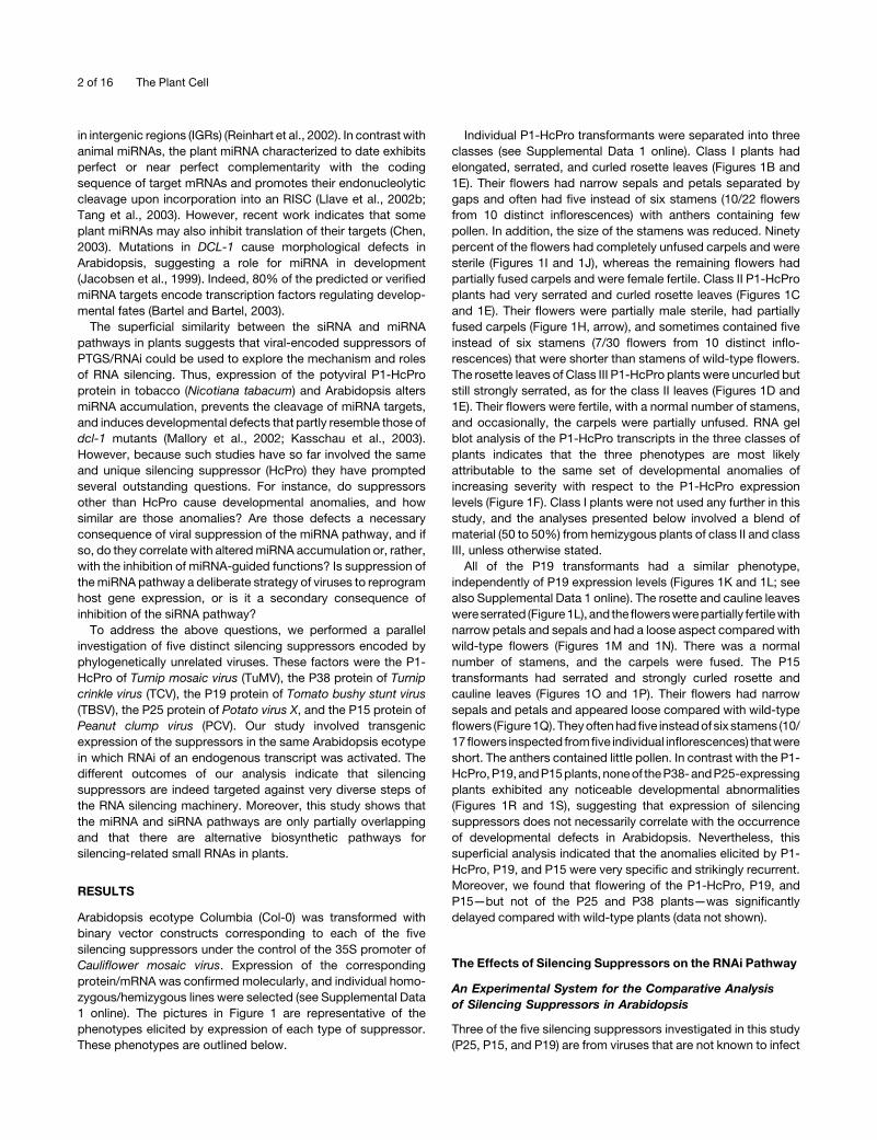
Figure 1. The P1-HcPro, P19, and P15 Proteins, but Not the P38 or P25 Proteins, Induce Developmental Defects in Arabidopsis.

RNA Silencing and Viral Suppressors 3 of 16

Arabidopsis naturally, whereas TCV and TuMV (encoding the

HcPro and P38 proteins, respectively) are infectious in this

species (Ren et al., 2000; Kasschau et al., 2003). Therefore, a first

step toward a comparative analysis of these proteins was to

ascertain that they indeed suppressed RNAi in this species. To

that aim, two representative lines of each suppressor were

crossed with a second Arabidopsis line referred to as line CHS-

RNAi. In this line, accumulation of the chalcone synthase (CHS)

mRNA is silenced by constitutive expression of an inverted-

repeat transgene designed to produce CHS dsRNAs (see

Supplemental Data 1 online). The dsRNA is processed by

a Dicer-like enzyme into siRNAs that guide degradation of the

CHS mRNA, presumably upon incorporation into an RISC. CHS

encodes an enzyme involved in synthesis of anthocyanins,

purple pigments whose accumulation provides an indirect

measurement of the CHS mRNA levels (see Supplemental Data

1 online). These levels are normally low in wild-type plants, but

CHS expression and anthocyanin accumulation are strongly

induced by exposing plants to intense light (Figure 2A, lane 1). By

contrast, the CHS mRNA and anthocyanins in line CHS-RNAi

remain below detection levels upon the same light treatment

(Figure 2A, lane 2).

To test the effect of silencing suppressors, expression of CHS

was light induced, and 1 d later, total RNA and anthocyanins

were extracted from leaves and quantified by RNA gel blot

analysis and by spectrophotometry, respectively. Anthocyanin

accumulation (as assessed in four independent extractions) was

restored to 82% 6 8% of the wild-type levels in the P1-HcPro-,

P38-, P25-, and P15-expressing plants, and accordingly, the

levels of the CHS transcript were high (Figure 2A, lanes 4 to 7).

The effect of P19 was less pronounced: anthocyanins accumu-

lated to 50%6 5% of the wild-type levels, and the increase of the

CHS mRNA was intermediate (Figure 2A, lane 3). Collectively,

these results indicate that all of the five viral proteins suppressed

silencing triggered by the same CHS-RNAi locus in the same

Arabidopsis ecotype, allowing a strict comparative study of their

effects.

Silencing Suppressors Exert Contrasted Effects upon

siRNA Accumulation and Processing of Long dsRNA

In plants, siRNAs are in two distinct size classes of 21 and 24

nucleotides (Hamilton et al., 2002). RNA gel blot analysis

confirmed that both CHS-specific siRNA species accumulated

in line CHS-RNAi (Figure 2B, lane 2). However, the 24-nucleotide

species was below detection limit in the P19 samples, whereas

the levels of the 21-nucleotide siRNA were reduced 1.5- to 2-fold

(Figure 2B, lane 3). P1-HcPro caused a 4.5- to 5-fold reduction of

the 21-nucleotide siRNA levels and had a less pronounced effect

on the 24-nucleotide siRNA (Figure 2B, lane 4). P38, P25, and

P15 caused a marked reduction (more than eightfold) of both

CHS siRNAs (Figure 2B, lanes 5 to 7). These figures were

reproduced when strand-specific probes were used, indicating

that the suppressors had a similar effect on both sense and

antisense strands of siRNAs (data not shown).

Because siRNAs are processed from dsRNA by Dicer-like

enzymes, we investigated if the reduced siRNA levels in the

suppressor-expressing lines could result from reduced process-

ing of the CHS dsRNA, which may increase the stability of this

molecule. To test this possibility, total RNA extracted from leaves

of line CHS-RNAi and from the crosses with silencing suppres-

sors was digested with RNase A, a single-stranded RNA–

degrading enzyme, and subsequently heat denatured. The levels

of CHS dsRNA were then assessed by detection of RNase A–

resistant high molecular weight RNA using a CHS cDNA probe.

This molecule was absent in samples from line CHS-RNAi,

presumably because it was processed into siRNAs (Figure 2C,

lane 2). It was also absent in the P19, P38, P15, and P25 samples

(Figure 2C, lane 3 and lanes 5 to 7). However, a high molecular

weight RNA was readily detected in the P1-HcPro samples

(Figure 2C, lane 4). It was not detected if total RNA had been heat

denatured before RNase A treatment (data not shown), suggest-

ing that the signal was from a genuine dsRNA. Therefore, we

conclude that, among the five proteins, only P1-HcPro directly

interferes with dsRNA processing. This interference is likely

Figure 1. (continued).

(A) Wild-type Arabidopsis ecotype Col-0.

(B) to (D) P1-HcPro–transgenic Arabidopsis of class I (B), class II (C), and class III (D).

(E) Compared leaf morphology among the class I, II, and III P1-HcPro plants.

(F) Compared levels of P1-HcPro transcripts in flowers of class I, class II, and class III P1-HcPro plants, as assessed by RNA gel blot analysis. rRNA,

ethidium bromide staining of rRNA.

(G) Flower from wild-type Arabidopsis ecotype Col-0. The sepals were opened to allow observation of the internal floral whorls.

(H) A flower from a P1-HcPro plant of class II. Note the loose flower structure, narrow sepals and petals, and partially unfused carpel (arrow).

(I) Flower from a P1-HcPro plant of class I. The petals were removed to allow observation of the carpel and narrow sepals.

(J) Close-up view of the unfused carpel in the flower depicted in (I).

(K) P19-transgenic Arabidopsis.

(L) Compared rosette leaf morphology in wild-type and P19 plants.

(M) and (N) Altered flower morphology in P19 plants (M) as compared with wild-type plants (N). Note the narrow sepals and petals and overall loose

structure of the flower.

(O) P15-transgenic Arabidopsis.

(P) Compared rosette leaf morphology in wild-type and P15 plants.

(Q) Flower defects in P15-expressing plants, as compared with wild-type plants. Note the narrow sepals and petals and overall loose aspect of the

flower.

(R) and (S) P38 transformants (R) and P25 transformants (S) do not exhibit a noticeable developmental phenotype.

4 of 16 The Plant Cell

partial because significant levels of CHS siRNAs were still

detected in the P1-HcPro samples (Figure 2B, lane 4).

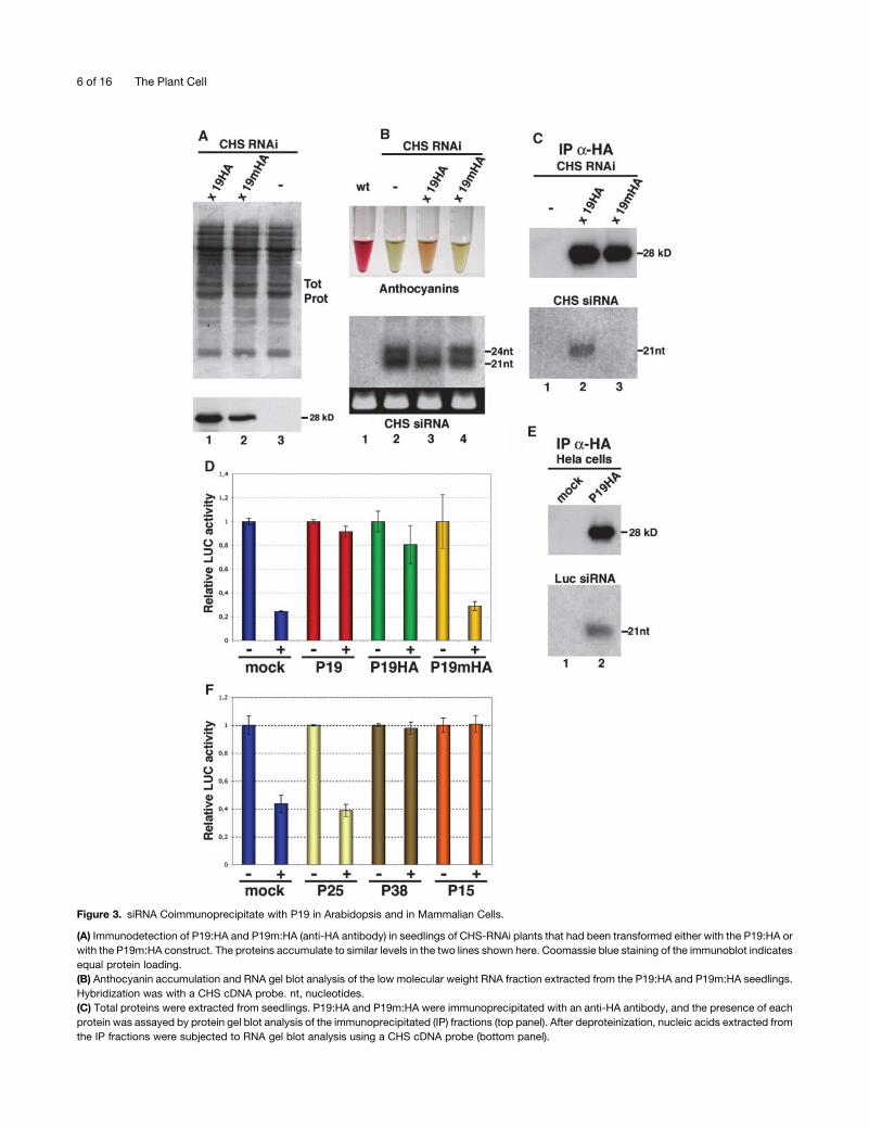
siRNA Binding Is Crucial for P19-Mediated Suppression

of RNAi in Vivo

Previous work has established that the 21- and 24-nucleotide

siRNAs are functionally distinct in plants, with the 21-nucleotide

species being sufficient to guide target cleavage (Hamilton et al.,

2002). Based on those findings, it was striking that in contrast

with the other silencing suppressors, P19 only exerted a very

modest effect on accumulation of the 21-nucleotide CHS siRNAs

(Figure 2B, lane 3) yet it promoted a substantial increase in the

CHS mRNA levels (Figure 2A, lane 3). The P19 protein of

Cymbidium ringspot virus, a relative of the TBSV P19 protein,

specifically binds to siRNAs in vitro (Silhavy et al., 2002), and two

recent reports actually show cocrystallization of P19 homo-

dimers with siRNAs (Vargason et al., 2003; Ye et al., 2003).

Consequently, it was proposed that sequestration of the

21-nucleotide siRNAs by P19 may contribute to its suppression

of silencing activity, a scenario consistent with the effect of the

protein in line CHS-RNAi. However, siRNA binding by P19 so far

only has been documented in vitro, and none of the above

reports has provided experimental evidence for a link between

siRNA binding and silencing suppression by the protein in vivo.

To address those important issues, the influenza virus

hemagglutinin (HA) epitope was fused at the C terminus of the

TBSV P19, leading to P19HA. This protein variant retained

;50% of its silencing suppression activity in N. benthamiana.

P19mHA, constructed in parallel, carries a single point mutation

that abolishes this function (see Supplemental Data 2 online).

Both constructs were transformed into Arabidopsis line CHS-

RNAi, and two lines were identified by protein gel blot analysis,

which produced similar levels of P19HA and P19mHA, re-

spectively (Figure 3A). Upon light induction, anthocyanins

accumulated in line P19HA to 25% 6 6% of the wild-type levels

(as assessed in four independent extractions), consistent with

the moderate silencing suppression in N. benthamina (Figure 3B,

lane 3; see also Supplemental Data 2 online). By contrast and as

expected, no anthocyanin accumulated in line P19mHA (Figure

3B, lane 4). The effect of P19HA on accumulation of CHS siRNAs

was similar to that of the wild-type P19 (Figure 2B, lane 3): the

levels of 24-nucleotide siRNAs were reduced, and those of

21-nucleotide siRNAs were slightly decreased (Figure 3B,

lane 3). P19mHA had no effect on CHS siRNAs of either class

(Figure 3B, lane 4).

P19HA and P19mHA were immunoprecipitated from total

seedling proteins, and the nucleic acids extracted from those

fractions were subjected to RNA gel blot analysis. Twenty-one-

nucleotide sense and antisense CHS siRNAs were readily

detected in the nucleic acid fraction of the P19HA immunopre-

cipitates (Figure 3C, lane 2). Analysis of the flow-through fraction

revealed the presence of nonimmunoprecipitated protein to-

gether with CHS siRNAs (see Supplemental Data 2 online). By

contrast, CHS siRNAs were below detection limit in the

immunoprecipitates of the nonfunctional P19mHA but were

found exclusively in the flow-through fraction, together with

unbound P19mHA (Figure 3C, lane 3; see also Supplemental

Data 2 online). siRNA binding by P19HA was not observed if the

HA antibody was omitted in the reactions (data not shown).

These results were consistent with the proposal that siRNA

binding may contribute significantly to the antisilencing effects of

P19 in vivo.

If this was the case, we predicted that the protein would inhibit

RNAi in a broad range of organisms because siRNAs are

ubiquitously involved in RNA silencing phenomena among

eukaryotes. P19, P19HA, and P19mHA were therefore mobilized

into the mammalian expression vector pSG5m. Human Hela cells

were first cotransfected with a plasmid encoding the firefly

luciferase (LUC) mRNA, together with P19-expressing vectors.

One day later, they were supertransfected with synthetic siRNAs

targeted against the LUC mRNA, whose levels were measured

24 h later using a standard dual-LUC reporter assay (Elbashir

et al., 2001a). As shown in Figure 3D, P19 inhibited degradation

of the LUC mRNA, indicating that it functions in human cells.

Figure 2. Suppression of CHS-RNAi by P1-HcPro, P19, P38, P25,

and P15.

(A) One day after light induction, total RNA was extracted from leaves of

wild-type plants or of line CHS-RNAi crossed or not with the silenc-

ing suppressor–expressing lines. Fifteen micrograms of this RNA

was subjected to RNA gel blot analysis using a CHS cDNA probe.

Anthocyanins were extracted in parallel and quantified by spectropho-

tometry.

(B) RNA gel blot analysis of low molecular weight RNA (15 mg) extracted

before light induction. The hybridization was with a CHS cDNA probe. nt,

nucleotides.

(C) Twenty-five micrograms of the RNA used in (B) was treated with

RNase A, deproteinized, heat denatured, and subjected to RNA gel blot

analysis using a CHS cDNA probe.

RNA Silencing and Viral Suppressors 5 of 16

Figure 3. siRNA Coimmunoprecipitate with P19 in Arabidopsis and in Mammalian Cells.

(A) Immunodetection of P19:HA and P19m:HA (anti-HA antibody) in seedlings of CHS-RNAi plants that had been transformed either with the P19:HA or

with the P19m:HA construct. The proteins accumulate to similar levels in the two lines shown here. Coomassie blue staining of the immunoblot indicates

equal protein loading.

(B) Anthocyanin accumulation and RNA gel blot analysis of the low molecular weight RNA fraction extracted from the P19:HA and P19m:HA seedlings.

Hybridization was with a CHS cDNA probe. nt, nucleotides.

(C) Total proteins were extracted from seedlings. P19:HA and P19m:HA were immunoprecipitated with an anti-HA antibody, and the presence of each

protein was assayed by protein gel blot analysis of the immunoprecipitated (IP) fractions (top panel). After deproteinization, nucleic acids extracted from

the IP fractions were subjected to RNA gel blot analysis using a CHS cDNA probe (bottom panel).

6 of 16 The Plant Cell

P19HA also suppressed RNAi, and as expected, P19mHA did

not (Figure 3D). As in Arabidopsis, it is likely that P19HA bound

the LUC siRNAs because they were found in the P19HA

immunoprecipitates (Figure 3E). Collectively, these results

support the proposal that binding of siRNAs by the P19 protein

is necessary for its suppression of silencing activity in vivo.

The Effects of Other Viral Suppressors in Hela Cells

Having established that P19 functions in Hela cells, we were

prompted to test the effects of the other proteins in this system.

Thus, P38, P25, P19, and P1-HcPro were mobilized in the

pSG5m expression vector. In vitro transcription/translation

analyses (see Methods) confirmed synthesis of the expected

proteins, except for the TuMV P1-HcPro, which was therefore

omitted in the subsequent tests. The values presented in Figure

3F are from at least three independent experiments that were

performed in triplicate for each suppressor. We found that P38

and P15, but not P25, significantly and consistently compro-

mised RNAi of the LUC mRNA, suggesting that P38 and P15 are

indeed functional as silencing suppressors in Hela cells, as was

shown for P19.

The Effects of Silencing Suppressors on the

miRNA Pathway

Accumulation of miRNAs Is Altered in the P1-HcPro and

P19 Plants but Not in the P15, P25, or P38 Plants

We then investigated the effect of silencing suppressors on

accumulation of a subset of 21-nucleotide miRNAs (Bartel and

Bartel, 2003). Although the results presented in Figure 4A are all

from RNAs of inflorescence tissues, similar conclusions were

drawn from analysis of RNAs extracted from stems and leaves of

the same plants (see also Figure 7A). We also refer the reader to

Supplemental Data 3 online for analysis of additional miRNAs

from the same tissues. We found that P38, P25, and P15 did not

exert any noticeable effect on miRNA accumulation (Figure 4A,

lanes 4, 6, and 7). By contrast, the level of these molecules was

consistently increased in the P1-HcPro plants (Figure 4A, lane 3),

in agreement with previous reports in Arabidopsis (Kasschau

et al., 2003).

As for P1-HcPro, P19 also caused alteration of miRNA

accumulation, but its effects were more complex. Thus, there

was a consistent shift in the mobility of all 21-nucleotide miRNAs

investigated (Figure 4A, lane 2). A detailed analysis, illustrated

here with miR156 and miR164 (Figure 4B), showed that this shift

affected approximately half of the original pool of 21-nucleotide

miRNA and corresponded to the accumulation of an RNA

species with the apparent mobility of a 19- to 20-nucleotide

synthetic RNA oligonucleotide (19- to 20-nucleotide-like miRNA

species). This increased mobility could have been caused by

addition of a phosphate group at the 39 end of the authentic

21-nucleotide miRNA, which normally bares a 59 terminal phos-

phate and a 39 terminal hydroxyl group. However, the results

of calf intestinal phosphatase (CIP) and polynucleotide kinase

(PNK) treatments of small RNAs from the P19-expressing plants

were not consistent with this hypothesis (see Supplemental

Data 4 online). It is likely, therefore, that the electrophoretic

mobility of the 19- to 20-nucleotide-like miRNA is caused by a

lack of one to two nucleotides.

Binding of P19 to miRNA/miRNA* Duplexes Is Coupled to

Enhanced Accumulation and Altered Electrophoretic

Mobility of miRNAs*

Because suppression of RNAi by P19 involves binding of small

RNA, we were prompted to test if this property could also explain

the effect of the protein upon miRNAs. To that aim, the nucleic

acid fractions from the immunoprecipitates of the P19HA- and

P19mHA-expressing plants (Figures 3A to 3C) were subjected to

RNA gel blot analysis with an oligonucleotide probe specific for

miR171. As shown in Figure 5A, miR171 was readily detected in

the nucleic acid fraction of the P19HA immunoprecipitates (lane

3) but not in the nucleic acid fraction of the mutant P19mHA

immunoprecipitates (lane 2). Similar results were obtained with

miR167 (data not shown), suggesting that P19 does indeed bind

miRNAs. However, mature miRNAs accumulate in plants as

single-stranded, 21-nucleotide species (Bartel and Bartel, 2003),

and it was shown that the P19 protein exhibits very poor affinity

for such molecules in vitro, whereas it efficiently binds

21-nucleotide dsRNA (Silhavy et al., 2002; Ye et al., 2003). To

reconcile our in vivo data with those results, we envisaged the

possibility that P19 could bind to the duplexes corresponding

to the primary cleavage products of DCL-1, which would

incorporate the fragment from the other arm of the miRNA

stem-loop precursor, known as miRNA* (Figure 5B; Hutvagner

Figure 3. (continued).

(D) Dual-LUC assay performed in human Hela cells transfected with pGL3-CMV (encoding the firefly LUC) and pRL-CMV (encoding the renilla LUC)

together with pSG5m (mock) or with pSGP19 (P19), psGP19HA (P19:HA), or psGP19mHA (P19m:HA). Twenty-four hours later, cells were

supertransfected with 0 (�) or 300 ng (1) of siRNAs directed against the firefly LUC mRNA. The renilla LUC mRNA is not targeted by these siRNAs and is

therefore used as a reference in this assay. For each treatment, the luminescence ratio firefly/renilla was calculated. This ratio was then normalized to

the luminescence values from a transfection experiment performed in parallel, in which anti-LUC siRNAs were omitted. This provided a relative LUC

activity. For each treatment, results of two independent assays are presented. Values from each assay were from duplicate independent transfections.

(E) Human Hela cells were transfected with pSGP19:HA together with anti-LUC siRNAs. Two days later, P19:HA was immunoprecipitated with an anti-

HA antibody, and the presence of the protein was assayed by protein gel blot analysis (top panel). After deproteinization, nucleic acids were extracted

from the immunoprecipitated fractions (IP) and subjected to RNA gel blot analysis using radiolabeled anti-LUC siRNAs as probe (bottom panel). nt,

nucleotides.

(F) Same experimental set up as in (D) but performed with pSG5m expression vectors for the P25, P38, and P15 proteins. The values in each bar are

from three independent experiments conducted in triplicate.

RNA Silencing and Viral Suppressors 7 of 16

and Zamore, 2002; Reinhart et al., 2002). The proposal that

P19 binds to miRNA/miRNA* duplexes had two testable implica-

tions.

The first implication was that, as well as miR171, miR171* also

would be detected in the P19HA immunoprecipitates. Computer

analysis predicts a unique stem-loop precursor transcript for

miR171, which is encoded in an IGR located on chromosome 3.

The putative position of miR171* in the stem-loop structure is

indicated in Figure 5B, in which the sequence of miR171 is

highlighted in red. To test that P19 binds to miR171*, the

membrane shown in Figure 5A was stripped and hybridized with

an oligonucleotide probe specific for miR171*. As shown in

Figure 5C, miRNA171* was detected in the nucleic acid fraction

from the P19HA immunoprecipitates but not from those of

P19mHA.

In wild-type Arabidopsis and other organisms, miRNAs*, as

opposed to miRNAs, are usually at or below detection limit of

RNA gel blot analysis, presumably because the miRNA* does

not incorporate into RISC and is therefore rapidly degraded

(Khvorova et al., 2003; Schwarz et al., 2003). Therefore, the

second implication of the proposed binding of miRNA/miRNA*

duplexes by P19 was an increased stability of miRNAs*. RNA gel

blot analysis of small RNAs extracted from inflorescences

revealed that the levels of miR171* were indeed dramatically

enhanced in the P19-expressing and P19HA-expressing plants

(Figures 5D and 5E). By contrast, they remained below detection

limit in the P19mHA samples, confirming that enhanced miR171*

accumulation was inherent to the small RNA binding capacity of

P19. Moreover, a detailed analysis of miR171* showed that its

electrophoretic mobility was altered in the P19 samples: it

migrated as a 19- to 20-nucleotide synthetic RNA oligonucleo-

tide (Figure 5F), as observed previously for the miRNAs

accumulating in the P19 plants (Figures 4A and 4B).

The most straightforward interpretation of those collective

results is that binding of miRNA/miRNA* duplexes by P19 is

coupled to a change that affects both RNA strands of the duplex

and causes their enhanced electrophoretic mobility.

P1-HcPro, P19, and P15, but Not P38 or P25, Prevent

Endonucleolytic Cleavage and Degradation of

miRNA Targets

In plants, several miRNAs are thought to mediate cleavage of

homologous transcripts upon incorporation into an RISC (Llave

et al., 2002b; Tang et al., 2003). Therefore, we investigated if

expression of the silencing suppressors prevented miRNA-

guided cleavage of specific cellular mRNAs. Based on previous

studies, miRNA targets can be divided into at least two classes

(Kasschau et al., 2003). The first class—illustrated here with the

CUC1 transcript targeted by miR164—comprises mRNAs whose

endonucleolytic cleavage is coupled to the subsequent degra-

dation of both 59 and 39 cleavage products (Figure 6A, case 1).

RNA gel blot analysis of total RNA extracted from inflorescences

of the suppressor lines indicated that the levels of the CUC1

mRNA were enhanced 8, 7.5, and 5 times in the presence of P15,

P1-HcPro, and P19, respectively. By contrast, degradation of the

CUC1 mRNA was unaffected by P25 and P38 (Figure 6B).

The second class of miRNA targets investigated includes

transcripts whose endonucleolytic cleavage leads to accumula-

tion of a stable 39 cleavage fragment (Figure 6A, case 2). The

SCL6-IV and ARF10 mRNAs, targeted respectively by mIR171

and miR160, belong to this category. RNA gel blot analysis of

Figure 4. The Effects of Silencing Suppressors on miRNA Accumulation.

(A) miRNA accumulation in flowers of wild-type, P19, P1-HcPro (HC),

P15, P38, and P25 transgenic plants, as assessed by RNA gel blot

analysis. The probes used were labeled oligonucleotides complementary

to the miRNA indicated at the top left corner of each filter. Two labeled

oligoribonucleotide standards were used as size markers (21 nucleotides

and 24 nucleotides). rRNA, ethidium bromide staining of rRNA. The filters

on the right and the left panels correspond to two separate RNA

preparations, hence the use of separate wild-type samples for internal

reference. See also Supplemental Data 3 online for analysis of additional

miRNAs. nt, nucleotides.

(B) High-resolution RNA gel blot analysis of miR156 and miR164 from the

P19 samples used in (A) reveals a mobility shift because of the

accumulation of a nucleic acid species with the apparent electrophoretic

mobility of a 19- to 20-nucleotide RNA, as assessed with a labeled

oligoribonucleotide.

8 of 16 The Plant Cell

RNA from inflorescences showed that P15, P1-HcPro, and P19

inhibited accumulation of the 39 cleavage fragment of the SCL6-

IV and ARF10 mRNAs, although some cleaved 39 products were

detected at low levels in the P19 samples (Figure 6C). By

contrast, accumulation of those fragments was the same in the

P38 and P25 samples as it was in control samples from wild-type

plants (Figure 6C).

The Effect of Silencing Suppressors on Silencing-Related

Small RNAs Other Than 21-Nucleotide miRNAs

A 24-Nucleotide miRNA Species Accumulates Preferentially

in Leaves, and Its Levels Are Altered in the P19 and

P1-HcPro Plants

In the course of this analysis, we found that miR156, miR160,

miR164, and miR165 occur in two distinct size classes of 21 and

24 nucleotides (Figure 7A, left panels; see also Supplemental

Data 3 online). The 24-nucleotide miRNA was by far more

abundant in leaves, as illustrated by the comparative analysis of

the data presented in Figures 7A and 4A and in Supplemental

Data 3 online, which were all generated from various tissues

of the same plants. By contrast, we could not detect a

24-nucleotide species for miRNAs 162, 167, 169, and 171, even

after long exposures (Figure 7A, right panels; see also Supple-

mental Data 3 online). Treatments with CIP and PNK indicated

that the 24-nucleotide species of miR156, like its 21-nucleotide

form, carries a 59 terminal phosphate and a 39 terminal OH (see

Supplemental Data 4 online), suggesting that it is a genuine

miRNA.

We tested the effect of the silencing suppressor on accu-

mulation of the 24-nucleotide miRNAs, as opposed to the

21-nucleotide miRNAs. As observed for the 21-nucleotide

miRNA species (Figure 4A, lane 3), the levels of 24-nucleotide

miRNAs were enhanced by P1-HcPro, and this effect was

successfully exploited to show that the 24-nucleotide species of

miR156, miR160, miR164, and miR165 were indeed present, but

at low levels, in flowers and stems (Figure 7B, left and middle

panels; data not shown). By contrast, similar analysis of miR171

and miR169 confirmed the lack of a 24-nucleotide species for

those miRNAs, as accumulation of a unique 21-nucleotide form

was enhanced by P1-HcPro in flowers and stems (Figure 7B,

right panel; data not shown). Strikingly, the 24-nucleotide

species of all miRNAs investigated (when applicable) were below

detection limit in P19-expressing tissues, whereas their levels

were not changed in the P38, P25, and P15 plants (Figure 7A;

data not shown). We conclude, from the specific effect of P19,

that the 24-nucleotide miRNA species may be biosynthetically

distinct from the 21-nucleotide species.

The Effects of Silencing Suppressors on Other

Silencing-Related Small RNAs

The small RNAs investigated so far are part of a group of

21-nucleotide, evolutionarily conserved miRNAs, which only

represent a fraction of the silencing-related small RNAs found

in Arabidopsis (Bartel and Bartel, 2003). Although originally

Figure 5. The P19 Protein Binds to miRNA/miRNA* Duplexes.

(A) Accumulation of miR171 in seedlings of line CHS-RNAi expressing or

not P19mHA and P19HA, as described in Figure 3. Hybridization was

with a labeled oligonucleotide complementary to miR171. Total proteins

were extracted from the same tissues, and P19:HA and P19m:HA were

immunoprecipitated (IP) with an anti-HA antibody (data not shown).

Upon deproteinization, nucleic acids extracted from the immunopreci-

pitated fractions were subjected to RNA gel blot analysis using a labeled

oligonucleotide complementary to miR171 as probe (bottom panel). nt,

nucleotides.

(B) Predicted secondary structure of the miR171 precursor transcript.

The sequence of miR171 is highlighted in red. The predicted cleavage

product of DCL-1 is the miRNA/miRNA* duplex, of which only one strand

(corresponding to miR171) is incorporated into the RISC for target

cleavage. The other strand (corresponding to miR171*) is unstable,

presumably because of rapid degradation.

(C) The membrane in (A) (bottom panel) was stripped and rehybridized

with a labeled oligonucleotide complementary to the sequence of the

predicted miR171*. This small RNA is present in the P19HA but not in the

P19mHA immunoprecipitates.

(D) and (E) The membrane in (A) (top panel) was stripped and

rehybridized with an oligonucleotide complementary to the sequence

of the predicted miR171*. There is strong enhancement of the

accumulation of miR171* in the P19HA samples, which also occurs in

inflorescences (E) of the P19-expressing plants depicted in Figure 1K.

(F) High-resolution RNA gel blot analysis of the RNAs extracted in (E)

reveals a migration shift for miR171* because of the accumulation of

a nucleic acid species with the apparent electrophoretic mobility of a 19-

to 20-nucleotide RNA, as assessed with a labeled oligoribonucleotide.

RNA Silencing and Viral Suppressors 9 of 16

annotated as a member of this group, miR163 differs in that

it does not have an ortholog in rice (Oryza sativa), and it

accumulates as a single 24-nucleotide species in all tested

tissues of Arabidopsis (Reinhart et al., 2002; Figure 7C). These

peculiarities, combined with the specific effect exerted by P19 on

the 24-nucleotide miRNAs evoked above (Figure 7A), incited us

to assay specifically for miR163 in the suppressor-transgenic

lines. As shown in Figure 7C, the effects of P1-HcPro, P15, and

P25 remained the same. However, the effects of P38 and P19 on

miR163 were distinctively different from their effects on the

miRNAs tested so far. Thus, P19 caused a fivefold reduction, but

did not eliminate, the accumulation of miR163, whereas its levels

were enhanced at least seven times in the P38 plants (Figure 7C).

We further assayed the levels of other small RNAs that also

accumulate exclusively as 24-nucleotide species. As shown in

Figure 7D, the levels of small RNA 96, which is specifically

expressed in flowers (Llave et al., 2002a), were unaffected by

the silencing suppressors. The same observation was made with

the 24-nucleotide small RNAs corresponding to the AtSN1

retroelement (Hamilton et al., 2002; Figure 7E). The silencing

suppressors also had no effect on accumulation of the flower-

specific, 21- to 22-nucleotide small RNA2 (Llave et al., 2002a;

Figure 7F). Therefore, only a fraction of the Arabidopsis silencing-

related small RNAs was affected by the suppressors, suggest-

ing the existence of varied biosynthetic pathways for these

molecules.

DISCUSSION

From the high diversity of viral-encoded suppressors, it was

originally anticipated that these proteins might be useful to

dissect various aspects of the siRNA pathway. More recently, the

emerging link between RNA silencing and development has

prompted the additional idea that silencing suppressors also

may be exploited to understand elements of the miRNA biology

in plants. This study was first aimed at addressing both of these

issues. It was also motivated by the necessity to set up

a consensus system whereby comparative biochemical and

genetic approaches of silencing suppression could be un-

dertaken. Indeed, most if not all studies had so far involved

model systems that varied greatly in terms of host plant, silencing

trigger, mode of protein delivery, and timing/pattern of expres-

sion. Consequently, it has been very difficult, or even impossible,

to conduct a side-by-side analysis of the suppressors. We have

now established such a comparative system in Arabidopsis, and

based on the contrasted effect of those factors on the

accumulation and/or the function of siRNAs, miRNAs, and other

types of endogenous small RNAs, this work provides experi-

mental support to the widely held belief that these proteins are

targeted against very diverse steps of the silencing machinery.

We outline below several important conclusions that could be

drawn from this system.

The first important finding was that only three of the five

silencing suppressors tested (P1-HcPro, P19, and P15) exerted

a significant effect on miRNA accumulation or miRNA-guided

functions, although all of them inhibited RNAi of the CHS

Figure 6. Effects of the Silencing Suppressors on miRNA-Mediated

Cleavage of Target mRNAs.

(A) Two possible outcomes of miRNA-guided cleavage of endogenous

transcripts in plants. In the first case (1), both the 59 and 39 cleavage

fragments are degraded, whereas in the second case (2), the 39 cleaved

fragment remains stable. The miRNA is indicated in red.

(B) and (C) Fifteen micrograms of total RNA extracted from inflores-

cences of the various suppressor-expressing lines were subjected to

RNA gel blot analysis using cDNA probes specific for the CUC1 (B) or the

SCL6-IV and ARF10 mRNAs (C). The size of the predicted 39 cleavage

products of SCL6-IV and ARF10 [SCL6-IV(b) and ARF10(b), respectively]

is indicated. nt, nucleotides.

10 of 16 The Plant Cell

transcript. This observation supports the emerging evidence

that, as seen in animals, the miRNA and siRNA pathways are only

partially overlapping in plants.

The second significant outcome of this work is that it

establishes a clear correlation between occurrence of morpho-

logical defects and alteration of the miRNA pathway in plants.

Hence, silencing suppressors that affected miRNA accumulation

and/or miRNA-guided cleavage (P1-HcPro, P19, and P15)

induced developmental anomalies, whereas suppressors with-

out such effects (P25 and P38) did not. We can therefore infer

that at least some of the observed developmental abnormalities

are attributable to perturbation of the miRNA biology.

Taking this inference into account, the strong similarity of the

morphological defects elicited by P1-HcPro, P15, and P19 is

remarkable because those proteins are so diverse in terms of

viral origin, amino acid sequence, and structure. Moreover,

TuMV, PCV, and TBSV cause dissimilar developmental symp-

toms, if any, in their respective hosts, and neither PCV nor TBSV

naturally infect Arabidopsis. Therefore, a third important point

emerging from this study is that it makes it unlikely that inhibition

of the miRNA pathway by suppressors reflects a deliberate viral

strategy to reprogram or alter host genome expression. Rather,

we propose that the developmental anomalies as a result

of suppression of RNA silencing are mostly secondary conse-

quences of inhibition of the siRNA pathway, at some steps

shared with the miRNA pathway.

Finally, this study identified several Arabidopsis small RNAs

that were resistant to the activity of all five silencing suppressors,

suggesting that there are distinct biosynthetic pathways for

those molecules. This indicates that silencing suppressors are

valuable probes of the diversity and complexity of the plant small

RNAs and that they may also provide a powerful handle toward

their functional classification.

The Position of Viral Suppressors in the Silencing

Pathways and Their Possible Mode of Action

In agreement with previous findings in tobacco (Mallory et al.,

2002), we observed that P1-HcPro reduced dsRNA processing

by Dicer, although this reduction was only partial because

Figure 7. Diversity and Complexity of the Arabidopsis Silencing-Related

Small RNAs.

(A) RNA gel blot analysis of RNAs extracted from leaves and stems of the

plants that were used to produce the inflorescence data presented in

Figure 4. RNA gel blot conditions were the same. See also Supplemental

Data 3 online for analysis of additional miRNAs. nt, nucleotides.

(B) Total RNA was extracted from inflorescences of wild-type, P1-HcPro

class I, and P1-HcPro class III plants and subjected to RNA gel blot

analysis. The probes used were labeled oligonucleotides complementary

to miR156, miR160, and miR171, respectively. rRNA, ethidium bromide

staining of rRNA.

(C) Accumulation of the 24-nucleotide miR163 in leaves and flowers of

the suppressor lines.

(D) and (E) Accumulation of small RNA 96 (D) and of small RNAs of the

AtSN1 retroelement (E) in flowers from the various suppressor-express-

ing lines.

(F) Flower-specific accumulation of the 21-nucleotide small RNA2 in

inflorescences of the various suppressor-expressing lines. The probes

used in (C) to (F) were labeled oligonucleotides complementary to the

corresponding small RNAs.

RNA Silencing and Viral Suppressors 11 of 16

substantial levels of CHS siRNAs were still detected (Figure 2).

Despite those residual siRNA levels, however, degradation of the

CHS mRNA was prevented, suggesting that in addition to its

effect on Dicer activity, P1-HcPro also inhibits the activity of

RISC, a proposal consistent with the effect of the protein on

miRNA-guided cleavage of endogenous transcripts (Figure 6).

Taking into account the idea that viral suppressors are not

primarily directed against the miRNA pathway, we propose, to

reconcile those observations, that P1-HcPro interacts with

a factor present in both the miRNA/siRNA-programmed RISC

and Dicer-like RNP complexes. Perhaps this factor is akin to rgs-

CaM, a calmodulin-related protein known to interact with and to

mimic the effect of P1-HcPro in tobacco (Anandalakshmi et al.,

2000). P1-HcPro also exerted a striking effect on all the miRNAs

investigated here, in that it enhanced their steady state levels

(Figures 4 and 7). As proposed previously (Xie et al., 2003), this

effect may be partly explained by the fact that expression of

DCL-1 is itself regulated by miR162 (also investigated here;

Figures 4 and 7). Thus, inhibition, by P1-HcPro, of miR162-

guided cleavage of the DCL-1 mRNA may cause enhanced

processing of miRNA precursors by the stabilized DCL-1 protein

(Xie et al., 2003).

Unlike P1-HcPro, P15 significantly reduced the levels of CHS

siRNA without interfering with dsRNA processing, suggesting

that the protein may act downstream of Dicer (Figure 2). The

reduced siRNA levels may result from a lack of incorporation of

these molecules into RISC, which may cause their instability.

Similarly, the failure of miRNA to incorporate into RISC could

explain the inhibitory effect of P15 on miRNA-guided cleavage

(Figure 6). As envisaged for siRNAs, this situation should lead to

miRNA instability. It is possible that this instability may be, in turn,

compensated by the increased DCL-1 levels because of

inhibition, by P15, of miR162-mediated cleavage of the DCL-1

transcript (Xie et al., 2003). This would explain why the overall

miRNA content appears unchanged in the P15 plants (Figures 4

and 7).

Both P25 and P38 caused a large reduction in siRNA levels and

did not stabilize the CHS dsRNA (Figure 2), suggesting that they

could act downstream of the Dicer processing step. However, it

is also possible that these proteins do interfere with Dicer

processing, but that the resulting dsRNA is metabolized through

another cellular pathway. Alternatively, P25 and P38 could either

prevent accumulation of the dsRNA trigger of RNAi or cause

siRNA instability. We can already assume that the two proteins

do not have the same target, based on their differential effect on

miR163. Their respective position in the siRNA pathway could be

clarified by crossing the P25 and P38 plants with the P1-HcPro

plants. For instance, if one of these factors precludes accumu-

lation of dsRNA, its effect should be epistatic upon the P1-

HcPro–mediated stabilization of this molecule (Figure 2C).

Conversely, a protein enhancing siRNA turnover will not have

this epistatic effect.

The coimmunoprecipitation of P19 with siRNA (Figure 3)

supports recent in vitro binding assays (Silhavy et al., 2002) and

crystallographic data (Vargason et al., 2003; Ye et al., 2003) and

strongly suggests that the protein binds these molecules in vivo.

Moreover, the lack of binding from a nonfunctional protein variant

indicates that binding is indeed necessary for silencing suppres-

sion by P19. Therefore, sequestration of siRNAs by P19 may

prevent their incorporation or may interfere with the activity of

RISC. Inhibition, by P19, of RNAi triggered by synthetic siRNAs in

Hela cells is also consistent with it affecting RISC (Figure 3).

However, although necessary, siRNA sequestration may not be

sufficient to explain entirely the effect of P19 because we

consistently found reduced siRNA levels in P19-expressing

Arabidopsis (Figures 2 and 3).

Interestingly, the effects of P19 on CHS siRNAs targeting the

CHS mRNA for cleavage (Figure 2) differed from its effects on

siRNAs directed against sequences of the nopaline synthase

promoter (NOSpro) and inducing its transcriptional gene silenc-

ing (TGS) via DNA methylation (Papp et al., 2003). In this case,

P19 did not alter accumulation of the 24-nucleotide NOSpro

siRNAs but caused the appearance of NOSpro siRNAs with

a one- to two-nucleotide mobility shift similar to that observed

here with miRNAs (Papp et al., 2003). This observation indicates

that the fate of siRNAs (i.e., TGS versus PTGS) may strongly

influence the way they are affected by suppressors. Consistent

with this idea, it was shown that P1-HcPro reduces accumulation

of siRNA targeted against mRNA but exerts no effect on the

NOSpro siRNAs (Mette et al., 2001). Our observation and those

of Papp and colleagues also suggest that 21-nucleotide TGS-

related siRNAs—as opposed to PTGS-related siRNAs—may

share common biosynthetic steps with miRNAs because they

are affected in a similar way by P19 (Papp et al., 2003).

We have shown that P19, but not the mutant protein,

coimmunoprecipitates with miR171 and miR171*, strongly

suggesting that, in addition to siRNA duplexes, the protein also

binds miRNA/miRNA* duplexes (Figure 5). Sequestration of this

duplex is in agreement with the strong stabilization of miRNA*

observed in the P19 plants, whereas this molecule is not

detectable in wild-type plants (Figure 5). The change in

electrophoretic mobility of miRNA caused by P19 is consistent

with previous observations made with miR159 (Papp et al.,

2003). The results presented here with many other miRNAs

indicate that this phenomenon is not peculiar to miR159 and is

most likely because of the removal of one to two nucleotides

(Figure 4; see also Supplemental Data 4 online). Because

miRNAs are produced by DCL-1, a Dicer-like enzyme, the

miRNA/miRNA* duplexes should harbor the two-nucleotide 39

overhangs that are characteristic of Dicer cleavage products

(Figure 5B). Thus, to explain the effect of P19, we propose that

the bound miRNA/miRNA* duplexes may become accessible to

an exonuclease that removes the two-nucleotide 39 overhangs

on each strand of the duplex, consistent with the finding that

miR171* also shows a shift in electrophoretic mobility in the P19

plants (Figure 5D). This modified pool of miRNAs may no longer

be incorporated into RISC, and this may form the basis for the

inhibition of miRNA-guided cleavage by P19.

Viral Suppressors, miRNAs, and Plant Development

We found that neither P38 nor P25 elicited developmental

anomalies in Arabidopsis. In the case of P25, we cannot rule out

the possibility that, because Potato virus X does not infect

Arabidopsis naturally, the protein has not sufficiently coevolved

12 of 16 The Plant Cell

with Arabidopsis factors that may be required to induce

developmental anomalies in this species. For instance, the

related P25 protein of white clover mosaic potexvirus was shown

to severely alter leaf morphology in N. benthamiana, which is

a systemic host for this virus (Foster et al., 2002).

Although variable in intensity, the morphological defects of the

P1-HcPro, P15, and P19 plants (Figure 1) were very specific and

strikingly similar, and their occurrence was positively correlated

with the altered biology of a subset of miRNAs. Moreover, some

of the defects in those lines were reminiscent of those observed

in partial mutants of dcl-1, most notably, the delayed onset of

flowering and the narrow sepals and petals in flowers, together

with the split carpels of the P1-HcPro plants (Kasschau et al.,

2003). Therefore, it is reasonable to propose that at least part of

the observed defects is caused by inhibition of the miRNA

pathway. However, very few miRNAs have been characterized in

Arabidopsis (Bartel and Bartel, 2003), and it remains to be

determined if the ones investigated in this study are indeed

representative of the entire population. In addition, miRNAs

acting at the level of translation, such as miR172 (Aukerman and

Sakai, 2003; Chen, 2003), were not tested here. A second limit in

the extent to which general conclusions can be drawn from our

observations is that we have not attempted to characterize the

expression pattern of the suppressors in meristems and organ

primordia, in which the impact of miRNA-mediated regulation of

developmental fates is likely to be important (Bartel and Bartel,

2003).

Despite those limits, it is nevertheless interesting to note that,

unlike P19 and P1-HcPro, P15 had little or no effect on miRNA

levels, yet the P15 plants exhibited developmental anomalies,

suggesting that altered miRNA accumulation per se is not

the primary cause of at least some of these defects (Figures 1

and 4). There also was no obvious correlation between the

occurrence of developmental anomalies and the effect exerted

by P1-HcPro, P19, and P15 on the levels of the full-length

SCL6-IV and ARF10 transcripts (Figure 6C). However, the

decrease in accumulation of their respective 39 cleaved

fragments—generated by the action of miR171 and miR160

(Kasschau et al., 2003)—paralleled the penetrance of the

developmental defects. Thus, the absence of cleavage frag-

ments in the P1-HcPro and P15 tissues correlated with a strong

phenotype, whereas the less pronounced effect of P19 was

correlated with a milder phenotype (Figures 1 and 6C). By

contrast, the enhanced levels of the full-length CUC1 mRNA—

caused by the same silencing suppressors—strictly correlated

with developmental defects (Figure 6A). However, the CUC1

transcript differs from those of SCL6-IV and ARF10 in that its

cleavage by miR164 does not lead to accumulation of a stable

39 fragment but, rather, promotes its complete degradation

(Kasschau et al., 2003).

Although the set of miRNA targets investigated in this study

was limited, these observations are intriguing because they

suggest that plant miRNAs may not only ensure clearance of

developmentally important mRNAs (Bartel and Bartel, 2003), as

observed here for the CUC1 transcript, but they also may be

involved in the generation of biologically active RNA molecules

upon endonucleolytic cleavage of some of their targets. Thus,

the stability of the 39 cleavage fragments of SCL6-IV, ARF10, and

probably of other mRNAs may have important biological

implications in plant development.

Viral Suppressors as Probes of the Complexity and

Diversity of Endogenous Silencing-Related Small RNAs

Some miRNAs occur in two size classes of 21 nucleotides and 24

nucleotides, as seen, for instance, with miR160 and miR164.

However, cleavage mediated by those two miRNAs was in-

vestigated in inflorescences, where the 24-nucleotide species

was normally below detection limit (Figure 4). This suggests that

21-nucleotide miRNAs are sufficient to guide mRNA cleavage. A

similar conclusion can be inferred from the effect of miR171,

which, unlike miR160 and miR164, accumulated as a single

21-nucleotide species in all tissues, including in flowers where

it mediated SCL6-IV cleavage (Figures 4 and 7B). siRNAs also

accumulate as 21-nucleotide and 24-nucleotide species, with the

24-nucleotide siRNA being dispensable for target cleavage

(Hamilton et al., 2002). In addition, the two siRNA species are

likely produced by distinct Dicers (Tang et al., 2003), and this also

maybe thecase for the21-nucleotide and24-nucleotidemiRNAs.

Indeed, the differential effect of P19 upon their accumulation

suggests that they have distinct biosynthetic pathways.

What could be the function of the 24-nucleotide miRNAs?

Based on the link between 24-nucleotide siRNAs and methylation

of homologous nuclear DNA (Hamilton et al., 2002; Zilberman

et al., 2003), it is possible that 24-nucleotide miRNAs signal

sequence-specific DNA methylation, directing, for instance, TGS

of their precursors. This negative feedback may be particularly

relevant in mature organs, such as leaves (where accumulation of

the 24-nucleotide miRNA is highest; Figures 4 and 7), in which

developmental regulation by miRNAs may no longer be required.

The effects of silencing suppressors on other silencing-related

small RNAs provide compelling evidence for a considerable

variety of biosynthetic and/or functional pathways for those

molecules. The 24-nucleotide miR163 clearly defines a novel

class of such RNAs because its accumulation pattern in the

silencing suppressor lines was unique (Figure 7C). This suggests

that the function of miR163 differs from that of the 21-nucleotide

and 24-nucleotide miRNAs. The sequence of miR163 is im-

perfectly complementary to the coding region of the mRNAs of

five related S-adenosyl-L-Met–dependent methyltransferases

(Bartel and Bartel, 2003), and it will be interesting to assess how

regulation of this family differs from that of the other miRNA

targets. Unlike miR163, the AtSN1 small RNAs and small RNA96

were unaffected by silencing suppressors (Figures 7D and 7E),

indicating that these 24-nucleotide RNAs also differ biosynthet-

ically and/or functionally from miRNAs (both 21-nucleotide and

24-nucleotide long), from miR163 and from the 24-nucleotide

siRNAs.

A similar inference can be made for small RNA2, which unlike

most silencing-related endogenous small RNAs that are tran-

scribed from IGRs, is processed from a pre-mRNA with an

unusual potential to form an extensive secondary structure (Llave

et al., 2002a). Small RNA2 accumulates as a 21- to 22-nucleotide

RNA with sense and antisense polarities, and based on these

features, it was suggested that it could be synthesized and could

RNA Silencing and Viral Suppressors 13 of 16

function like an endogenous siRNA (Llave et al., 2002a).

However, none of the viral suppressors had an effect on this

small RNA (Figure 7F), whereas they all altered accumulation of

the CHS siRNAs produced from long dsRNA hairpins (Figure 2).

To some extent, this overall complexity could be anticipated

from the diversity and apparent lack of redundancy of the various

members of the Dicer and Argonaute families in Arabidopsis

(Carmell et al., 2002; Morel et al., 2002; Finnegan et al., 2003).

Most likely, multiple combinatory interactions between those

factors form the basis for a complex network of biosynthetic and

functional pathways for small RNAs in plants. The challenge is

now to elucidate the biological role(s) of those molecules. We

anticipate that silencing suppressors will be useful in this task, as

diagnostic tools for the classification of endogenous small RNAs.

It should then become possible to identify the features that are

shared by small RNAs within each class and, thereby, apprehend

their function. A recent study in C. elegans indicates that 21- to

24-nucleotide endogenous small RNAs are much more heterog-

eneous in this organism than was initially anticipated (Ambros

et al., 2003), and it is probable that such diversity also applies to

human small RNAs. Having established that P19, and maybe

other plant virus suppressors (Figure 3F), inhibits RNAi in Hela

cells now opens the fascinating possibility to interfere with, and

maybe unravel, biological processes that are regulated by

different classes of small RNAs in mammals.

METHODS

DNA Constructs

The T-DNA expression cassettes for the P25, P19, P38, and P15 silencing

suppressors were described previously (Voinnet et al., 1999, 2000;

Dunoyer et al., 2002; Thomas et al., 2003). The cDNA of P1-HcPro was

amplified by RT-PCR (Superscript; Stratagene, La Jolla, CA; primer

sequences available upon request) from total RNA of TuMV-infected

Arabidopsis thaliana (viral strain CDN1) and inserted into the SmaI site of

pBin61 (Hamilton et al., 2002). Construction of P19HA and P19mHA is

detailed in Supplemental Data 2 online. Probes specific for the SCL6-IV,

CUC1, and ARF10 mRNAs were amplified from Arabidopsis genomic

DNA according to Kasschau et al. (2003). Probe specific for the CHS

mRNA and siRNAs was a 256-bp fragment amplified from Arabidopsis

genomic DNA (primer sequences available upon request). For mamma-

lian cell expression, all of the suppressor open reading frames were

excised from the corresponding binary vectors and inserted into theSmaI

site of the mammalian expression vector pSG5m, except for P15, which

was inserted as an EcoRV restriction fragment. Production of the

corresponding proteins from pSG5m was confirmed using the TnT quick

coupled transcription/translation system (Promega, Madison, WI), except

for the TuMV P1-HcPro, which was therefore not used in those studies.

Plasmid pGL3-CMV carrying the firefly LUC gene was constructed by

mobilization of the CMV promoter from pRL-CMV (Promega) into the

SmaI restriction site of pGL3-basic vector (Promega).

Protein and RNA Gel Blot Analysis

Total proteins were extracted from tissues of 6-week-old Arabidopsis and

were resolved on SDS-PAGE, electroblotted, and subjected to protein gel

blot analysis using either anti-TCV-P38 antiserum at a dilution of 1/500

(Thomas et al., 2003), anti-TBSV-P19 antiserum (a kind gift from H.

Sholthof, Texas A&M University) at a dilution of 1/5000, or anti-P15 at

a dilution of 1/5000 (Dunoyer et al., 2002). Total RNA was extracted from

Arabidopsis tissues (6-week-old plants) with Tri-Reagent (Sigma, St.

Louis, MO) according to the manufacturer’s instructions. RNA gel blot

analysis of high and low molecular weight RNA was on 15mg of total RNA,

unless otherwise stated, and was as described previously. Ethidium

bromide staining of total RNA before transfer was used to confirm

equal loading. Radiolabeled probes for detection of the P1-HcPro, CHS,

SCL6-IV, CUC1, and ARF10 transcripts were made by random priming

reactions in the presence of a-32P-dCTP. DNA oligonucleotides

complementary to miRNA sequences were end labeled with g-32P-ATP

using T4 PNK (New England Biolabs, Beverly, MA). Detection of the

AtSN1 small RNAs was as described (Hamilton et al., 2002). Densito-

metric analysis of at least two independent RNA gel blots exposed to

x-ray film was used to assess relative RNA levels.

CIP, PNK, and RNase A Analyses

High and low molecular weight RNA fractions were separated by

precipitation with 5% polyethylene glycol (molecular weight 8000) and

0.5 M NaCl (48C for 30 min). After ethanol precipitation, low molecular

weight RNA was resuspended in RNase-free water. Approximately 15 mg

of small RNAs were incubated with either 20 units of CIP (New England

Biolabs) or 10 units of T4 PNK according to Mallory et al. (2002). Samples

were resolved on high-resolution 20% polyacrylamide gels. For RNase A

analysis, 25 mg of total RNA was digested at 378C for 30 min with 2.5 mg/

mL of RNase A/T1 (Ambion, Austin, TX) in RNase buffer containing 10 mM

Tris-HCl, pH 7.5, 200 mM NaCl, 100 mM LiCl, and 1 mM EDTA. As

a control, total RNA was heat denatured for 5 min at 908C before RNase A/

T1 treatment. RNA was extracted with phenol/chloroform and ethanol

precipitated.

Immunoprecipitation

For immunoprecipitation experiments, 400 mg of seedlings were ground

in liquid nitrogen, thawed in two volumes of extraction buffer (50 mM Tris-

HCl, pH 7.5, 150 mM NaCl, 4-[2-aminoethyl]-benzenesulfonyl fluoride,

2 mg/mL of antipain, 2 mg/mL of leupeptin, and 2 mg/mL of aprotinin) for

20 min at 48C, and centrifuged at 12,000 rpm for 5 min at 48C. The anti-

body directed against the HA tag (Sigma) was coupled with protein A

Sepharose, and immune complexes were centrifuged and washed four

times in extraction buffer. The flow-through fractions were collected

before washes, and the efficiency of immunoprecipitation was assessed

by protein gel blot analysis. Proteins in the flow-through fractions were

precipitated with 10% trichloroacetic acid. For RNA analysis, immune

complexes, dissociated in 1% SDS, and flow-through fractions were

subjected to Tri-Reagent extraction (Sigma).

Hela Cell Transfection Experiments and Dual-LUC Assay

Human Hela cells (5 3 105) were transfected with 500 ng of pGL3-CMV,

500 ng of pRL-CMV, 1 mg of suppressor-expressing vector, and

Lipofectamin 2000 reagent (Invitrogen, Carlsbad, CA) as specified by

the manufacturer. One day later, 300 ng of siRNAs targeting firefly LUC

(Elbashir et al., 2001b) were transfected using the same procedure.

Twenty-four hours later, cells were lysed in passive lysis buffer for dual-

LUC assays (Promega). Each experiment was repeated at least three

times in triplicate.

ACKNOWLEDGMENTS

We thank P. Zamore for helpful comments and suggestions on the

manuscript. We are grateful to R.Wagner and his team for excellent

plant care. P.D. was supported by a FEBS long-term fellowship. Work in

14 of 16 The Plant Cell

our laboratory is supported by an ATIP Jeune chercheur from the Centre

National de la Recherche Scientifique.

Received January 6, 2004; accepted February 24, 2004.

REFERENCES

Ambros, V., Lee, R.C., Lavanway, A., Williams, P.T., and Jewell, D.

(2003). MicroRNAs and other tiny endogenous RNAs in C. elegans.

Curr. Biol. 13, 807–818.

Anandalakshmi, R., Marathe, R., Ge, X., Herr, J.M., Mau, C., Mallory,

A., Pruss, G., Bowman, L., and Vance, V.B. (2000). A calmodulin-

related protein that suppresses posttranscriptional gene silencing in

plants. Science 290, 142–144.

Aukerman, M.J., and Sakai, H. (2003). Regulation of flowering time and

floral organ identity by a microRNA and its APETALA2-like target

genes. Plant Cell 15, 2730–2741.

Bartel, B., and Bartel, D.P. (2003). MicroRNAs: At the root of plant

development? Plant Physiol. 132, 709–717.

Bernstein, E., Caudy, A.A., Hammond, S.M., and Hannon, G.J.

(2001). Role for a bidentate ribonuclease in the initiation step of RNA

interference. Nature 409, 363–366.

Brigneti, G., Voinnet, O., Li, W.X., Ji, L.H., Ding, S.W., and

Baulcombe, D.C. (1998). Viral pathogenicity determinants are

suppressors of transgene silencing in Nicotiana benthamiana. EMBO

J. 17, 6739–6746.

Carmell, M.A., Xuan, Z., Zhang, M.Q., and Hannon, G.J. (2002). The

Argonaute family: Tentacles that reach into RNAi, developmental

control, stem cell maintenance, and tumorigenesis. Genes Dev. 16,

2733–2742.

Chen, X. (2003). A microRNA as a translational repressor of APETALA2

in Arabidopsis flower development. Science 303, 2022–2025.

Dernburg, A.F., and Karpen, G.H. (2002). A chromosome RNAissance.

Cell 111, 159–162.

Dunoyer, P., Pfeffer, S., Fritsch, C., Hemmer, O., Voinnet, O., and

Richards, K.E. (2002). Identification, subcellular localization and

some properties of a cysteine-rich suppressor of gene silencing

encoded by peanut clump virus. Plant J. 29, 555–567.

Elbashir, S.M., Harborth, J., Lendeckel, W., Yalcin, A., Weber, K.,

and Tuschl, T. (2001b). Duplexes of 21-nucleotide RNAs mediate

RNA interference in cultured mammalian cells. Nature 411, 494–498.

Elbashir, S.M., Lendeckel, W., and Tuschl, T. (2001a). RNA interference

is mediated by 21- and 22-nucleotide RNAs. Genes Dev. 15, 188–200.

Finnegan, E.J., Margis, R., and Waterhouse, P.M. (2003). Post-

transcriptional gene silencing is not compromised in the Arabidopsis

CARPEL FACTORY (DICER-LIKE1) mutant, a homolog of Dicer-1 from

Drosophila. Curr. Biol. 13, 236–240.

Fire, A., Xu, S., Montgomery, M.K., Kostas, S.A., Driver, S.E., and

Mello, C.C. (1998). Potent and specific genetic interference by double-

stranded RNA in Caenorhabditis elegans. Nature 391, 806–811.

Foster, T.M., Lough, T.J., Emerson, S.J., Lee, R.H., Bowman, J.L.,

Forster, R.L., and Lucas, W.J.A. (2002). A surveillance system

regulates selective entry of RNA into the shoot apex. Plant Cell 14,

1497–1508.

Grishok, A., Pasquinelli, A.E., Conte, D., Li, N., Parrish, S., Ha, I.,

Baillie, D.L., Fire, A., Ruvkun, G., and Mello, C. (2001). Genes and

mechanisms related to RNA interference regulate expression of the

small temporal RNAs that control C. elegans developmental timing.

Cell 106, 23–34.

Hamilton, A.J., and Baulcombe, D.C. (1999). A species of small

antisense RNA in post-transcriptional gene silencing in plants.

Science 286, 950–952.

Hamilton, A.J., Voinnet, O., Chappell, L., and Baulcombe, D.C.

(2002). Two classes of short interfering RNA in RNA silencing. EMBO

J. 21, 4671–4679.

Hammond, S.M., Bernstein, E., Beach, D., and Hannon, G. (2000). An

RNA-directed nuclease mediates post-transcriptional gene silencing

in Drosophila cell extracts. Nature 404, 293–296.

Hutvagner, G., and Zamore, P.D. (2002). A microRNA in a multiple-

turnover RNAi enzyme complex. Science 297, 2056–2060.

Jacobsen, S.E., Running, M.P., and Meyerowitz, E.M. (1999).

Disruption of an RNA helicase/RNAse III gene in Arabidopsis causes

unregulated cell division in floral meristems. Development 126, 5231–

5243.

Kasschau, K.D., and Carrington, J.C. (1998). A counterdefensive

strategy of plant viruses: Suppression of post-transcriptional gene

silencing. Cell 95, 461–470.

Kasschau, K.D., Xie, Z., Allen, E., Llave, C., Chapman, E.J., Krizan,

K.A., and Carrington, J.C. (2003). P1/HC-Pro, a viral suppressor of

RNA silencing, interferes with Arabidopsis development and miRNA

function. Dev. Cell 4, 205–217.

Khvorova, A., Reynolds, A., and Jayasena, S.D. (2003). Functional

siRNAs and miRNAs exhibit strand bias. Cell 115, 209–216.

Llave, C., Kasschau, K.D., Rector, M.A., and Carrington, J.C.

(2002a). Endogenous and silencing-associated small RNAs in plants.

Plant Cell 14, 1605–1619.

Llave, C., Xie, Z., Kasschau, K.D., and Carrington, J.C. (2002b).

Cleavage of scarecrow-like mRNA targets directed by a class of

Arabidopsis miRNA. Science 297, 2053–2056.

Mallory, A.C., Reinhart, B.J., Bartel, D., Vance, V.B., and Bowman,

L.H. (2002). A viral suppressor of RNA silencing differentially regulates

the accumulation of short interfering RNAs and micro-RNAs in

tobacco. Proc. Natl. Acad. Sci. USA 99, 15228–15233.

Mette, M.F., Matzke, A.J.M., and Matzke, M.A. (2001). Resistance of

RNA-mediated TGS to HC-Pro, a viral suppressor of PTGS, suggests

alternative pathways for dsRNA processing. Curr. Biol. 11, 1119–1123.

Morel, J.-B., Godon, C., Mourrain, P., Beclin, C., Boutet, S.,

Feuerbach, F., Proux, F., and Vaucheret, H. (2002). Fertile

hypomorphic ARGONAUTE (ago1) mutants impaired in post-tran-

scriptional gene silencing and virus resistance. Plant Cell 14, 629–639.

Moss, E.G. (2002). MicroRNAs: Hidden in the genome. Curr. Biol. 12,

R138–R140.

Olsen, P.H., and Ambros, V. (1999). The lin-4 regulatory RNA controls

developmental timing in Caenorhabditis elegans by blocking LIN-14

protein synthesis after the initiation of translation. Dev. Biol. 216,

671–680.

Papp, I., Mette, M.F., Aufsatz, W., Daxinger, L., Schauer, S.E., Ray,

A., van der Winden, J., Matzke, M., and Matzke, A.J. (2003).

Evidence for nuclear processing of plant micro RNA and short

interfering RNA precursors. Plant Physiol. 132, 1382–1390.

Reinhart, B.J., Weinstein, E.G., Rhoades, M., Bartel, B., and Bartel,

D.P. (2002). MicroRNAs in plants. Genes Dev. 16, 1616–1626.

Ren, T., Qu, F., and Morris, T.J. (2000). HRT gene function requires

interaction between a NAC protein and viral capsid protein to confer

resistance to turnip crinkle virus. Plant Cell 12, 1917–1926.

Schwarz, D.S., Hutvagner, G., Du, T., Xu, Z., Aronin, N., and Zamore,

P.D. (2003). Asymmetry in the assembly of the RNAi enzyme complex.

Cell 115, 199–208.

Silhavy, D., Molnar, A., Lucioli, A., Szittya, G., Hornyik, C., Tavazza,

M., and Burgyan, J. (2002). A viral protein suppresses RNA silencing

and binds silencing-generated, 21- to 25-nucleotide double-stranded

RNAs. EMBO J. 21, 3070–3080.

Tang, G., Reinhart, B.J., Bartel, D.P., and Zamore, P.D. (2003). A

biochemical framework for RNA silencing in plants. Genes Dev. 17,

49–63.

RNA Silencing and Viral Suppressors 15 of 16

Thomas, C.L., Leh, V., Lederer, C., and Maule, A.J. (2003). Turnip

crinkle virus coat protein mediates suppression of RNA silencing in

Nicotiana benthamiana. Virology 306, 33–41.

Vargason, J.M., Szittya, G., Burgyan, J., and Tanaka Hall, T.M.

(2003). Size selective recognition of siRNA by an RNA silencing

suppressor. Cell 115, 799–811.

Voinnet, O. (2001). RNA silencing as a plant immune system against

viruses. Trends Genet. 17, 449–459.

Voinnet, O., Lederer, C., and Baulcombe, D.C. (2000). A viral

movement protein prevents systemic spread of the gene silencing

signal in Nicotiana benthamiana. Cell 103, 157–167.

Voinnet, O., Pinto, Y.M., and Baulcombe, D.C. (1999). Suppression of

gene silencing: A general strategy used by diverse DNA and RNA

viruses. Proc. Natl. Acad. Sci. USA 96, 14147–14152.

Xie, Z., Kasschau, K.D., and Carrington, J.C. (2003). Negative

feedback regulation of Dicer-Like1 in Arabidopsis by microRNA-

guided mRNA degradation. Curr. Biol. 13, 784–789.

Ye,K.,Malinina,L.,andPatel,D.J. (2003). Recognition of small interfering

RNA by a viral suppressor of RNA silencing. Nature 426, 874–878.

Zilberman, D., Cao, X., and Jacobsen, S.E. (2003). ARGONAUTE4

control of locus-specific siRNA accumulation and DNA and histone

methylation. Science 299, 716–719.

NOTE ADDED IN PROOF

While this manuscript was in press, a study of the genetic requirements

for Arabidopsis siRNA and miRNA synthesis provided compelling

evidence for the functional diversification of small RNA pathways in

plants. The citation is Xie, Z., Johansen, L.K., Gustafson, A.M.,

Kasschau, K.D., Lellis, A.D., Zilberman, D., Jacobsen, S.E., and

Carrington, J.C. (2004). Genetic and functional diversification of small

RNA pathways in plants. PLoS Biol. 2, E104.

16 of 16 The Plant Cell

DOI 10.1105/tpc.020719; originally published online April 14, 2004;Plant Cell

VoinnetPatrice Dunoyer, Charles-Henri Lecellier, Eneida Abreu Parizotto, Christophe Himber and Olivier

RNA SilencingProbing the MicroRNA and Small Interfering RNA Pathways with Virus-Encoded Suppressors of

 This information is current as of April 12, 2019


Permissions https://www.copyright.com/ccc/openurl.do?sid=pd_hw1532298X&issn=1532298X&WT.mc_id=pd_hw1532298X

eTOCs http://www.plantcell.org/cgi/alerts/ctmain

Sign up for eTOCs at:

CiteTrack Alerts http://www.plantcell.org/cgi/alerts/ctmain

Sign up for CiteTrack Alerts at:

Subscription Information http://www.aspb.org/publications/subscriptions.cfm

is available at:Plant Physiology and The Plant CellSubscription Information for

ADVANCING THE SCIENCE OF PLANT BIOLOGY © American Society of Plant Biologists


Recommended