J. Cell Sci. 3, 309-326 (1968) 309 Printed in Great Britain PROBLEMS OF ELECTRON STEREOSCOPY OF BIOLOGICAL TISSUE E. G. GRAY AND R. A. WILLIS Department of Anatomy, University College London, Gower Street, London, W.C. 1 SUMMARY A detailed study of stereoscopy of tissue sections with the transmission electron microscope has been carried out using different tilt angles and staining techniques, aided with models. Pictures with tilt angle difference of 20 0 or more can be fused to give pairs of micrographs showing stereodepth. Cytoplasmic filaments, glycogen and ribosomal granules give good stereodepth. Oblique membranes, especially when stained with lead or phosphotungstic acid, give good depth in stereo-pairs but membranes running more vertically in the section do not. A theory, based on the ' transparency factor' that is a feature of transmission electron micro- scopy, is given to account for this discrepancy. A learning process is involved in achieving depth perception when viewing stereo-pairs of electron micrographs of tissue sections. Complex structures often cannot be perceived in depth even after prolonged scrutiny through the stereo- scope. Where structures overlap in the thickness of the section, they can be 'separated out' with stereoscopy and in this way clarified, for depending on which micrograph of a stereo-pair is presented to which eye, a structure can be made to appear either in the front or the back of a section. In general, problems of depth perception in stereo-pairs of micrographs obtained with transmission electron microscopy arise because with our normal everyday vision using reflected light, we look at things, whereas with the electron microscope using a transmission electron beam we look through things. INTRODUCTION The depth of field (approximately 0-2 [i) of a light microscope is comparable with the resolution (0-2 /i). On the other hand, the depth of field of the electron microscope (~ 0-2 ft) is 200 times more than the resolution (10 A) and several times more than the normal section thickness (600 A). This means that organelles with dimensions below 600 A, e.g. ribosomes, neurofUaments, synaptic vesicles and membrane profiles, lie enclosed within the thickness of the section and so are viewed with the electron microscope not as sections but as whole structures. Thus more complete information can be gained by tilting the section in the electron beam to show the various aspects of these organelles, and with suitable tilt angles pairs of micrographs can be viewed in a stereoscope to give a three-dimensional representation of the object. Electron stereoscopy is, of course, not new. Yet in spite of its theoretical advantages it had gained surprisingly little popularity in the examination of plastic sections of biological material with the electron microscope (see, for example, Helmcke, 1954, 1965; Williams & Kallman, 1955; Valdr6, Coiley & Parsons, 1964; Shalla, Carroll & DeZoeten, 1964; Peachey, 1965; Willis & Gray, 1966; Kelly, 1966; Chandler & Willis, 1966; Willis, 1966). Here an attempt has been made, with the aid of models, 20 Cell Sci. 3
Transcript
J. Cell Sci. 3, 309-326 (1968) 309
Printed in Great Britain
PROBLEMS OF ELECTRON STEREOSCOPY
OF BIOLOGICAL TISSUE
E. G. GRAY AND R. A. WILLISDepartment of Anatomy, University College London, Gower Street, London, W.C. 1
SUMMARY
A detailed study of stereoscopy of tissue sections with the transmission electron microscopehas been carried out using different tilt angles and staining techniques, aided with models.Pictures with tilt angle difference of 200 or more can be fused to give pairs of micrographsshowing stereodepth. Cytoplasmic filaments, glycogen and ribosomal granules give goodstereodepth. Oblique membranes, especially when stained with lead or phosphotungstic acid,give good depth in stereo-pairs but membranes running more vertically in the section do not.A theory, based on the ' transparency factor' that is a feature of transmission electron micro-scopy, is given to account for this discrepancy. A learning process is involved in achieving depthperception when viewing stereo-pairs of electron micrographs of tissue sections. Complexstructures often cannot be perceived in depth even after prolonged scrutiny through the stereo-scope. Where structures overlap in the thickness of the section, they can be 'separated out'with stereoscopy and in this way clarified, for depending on which micrograph of a stereo-pair ispresented to which eye, a structure can be made to appear either in the front or the back of asection. In general, problems of depth perception in stereo-pairs of micrographs obtained withtransmission electron microscopy arise because with our normal everyday vision using reflectedlight, we look at things, whereas with the electron microscope using a transmission electronbeam we look through things.
INTRODUCTION
The depth of field (approximately 0-2 [i) of a light microscope is comparable withthe resolution (0-2 /i). On the other hand, the depth of field of the electron microscope(~ 0-2 ft) is 200 times more than the resolution (10 A) and several times more thanthe normal section thickness (600 A). This means that organelles with dimensionsbelow 600 A, e.g. ribosomes, neurofUaments, synaptic vesicles and membrane profiles,lie enclosed within the thickness of the section and so are viewed with the electronmicroscope not as sections but as whole structures. Thus more complete informationcan be gained by tilting the section in the electron beam to show the various aspects ofthese organelles, and with suitable tilt angles pairs of micrographs can be viewed in astereoscope to give a three-dimensional representation of the object.
Electron stereoscopy is, of course, not new. Yet in spite of its theoretical advantagesit had gained surprisingly little popularity in the examination of plastic sections ofbiological material with the electron microscope (see, for example, Helmcke, 1954,1965; Williams & Kallman, 1955; Valdr6, Coiley & Parsons, 1964; Shalla, Carroll &DeZoeten, 1964; Peachey, 1965; Willis & Gray, 1966; Kelly, 1966; Chandler &Willis, 1966; Willis, 1966). Here an attempt has been made, with the aid of models,
20 Cell Sci. 3
310 E.G. Gray and R. A. Willis
to study some of the factors and the limitations involved in obtaining a three-dimensional image with the electron microscope from plastic sections of biologicalmaterial. For general principles of binocular vision see Helmholtz (1909), Julesz(1962, 1963, 1964, 1965), Davson (1963), Ogle (1962), Gregory (1966) and Valyus(1966).
METHODS
Material was examined from numerous sources: that described here was takeneither from the cerebral cortex of the rat, spinal cord of the goldfish or optic nerve ofthe octopus.
The visual area of the rat cortex was fixed by aldehyde perfusion (Westrum & Lund,1966) post-fixed in osmium tetroxide, dehydrated in acetone and stained on the sectionwith lead citrate (Reynolds, 1963) (Figs. 6, 8). Goldfish spinal cord was fixed by per-fusion with a mixture of glutaraldehyde and formaldehyde (E. G. Gray, unpublished)and post-fixed with osmium tetroxide (1 % at pH 7-3). It was dehydrated withethanol and blockstained for 1-2 h with 1 % uranyl acetate at the 70% ethanol stage.The sections were further stained for 15 min with lead citrate (Figs. 7, 13, 14). Someof the material was fixed initially by immersing thin slices in 1 % osmium tetroxide insaline, buffered with veronal acetate. This material was dehydrated in ethanol andstained for 3 h in 1 % phosphotungstic acid at the absolute ethanol stage (Figs. 9-11).Small pieces of octopus optic nerve were fixed in o-6% potassium permanganatesolution buffered with veronal acetate (Luft, 1956). This material was dehydrated inethanol and remained unstained (Fig. 12).
The material was embedded in Araldite and silver or gold sections were examinedwith a Siemens Elmiskop 1 electron microscope equipped with a Valdre (1962)cartridge. This permits tilting through angles of up to 450 whilst the microscope is inoperation, without a given region of the specimen being lost from view, and so thesection can be photographed in its various aspects without difficulty. Because of theaxial rotation of the electron beam it is necessary to orientate the resulting stereo-pairsof micrographs to obtain maximum depth when viewed with the microscope. Theangle of orientation was determined by plotting the angular rotation observed on thefluorescent screen against magnification (Willis, 1966).
The Editors and the Cambridge University Press have kindly co-operated with thereproduction of the photographic stereopairs. These have been printed by 300-linecontact screen offset lithography to overcome the interference with the stereoscopiceffect caused by the less fine half-tone letterpress screen when the pictures are viewedthrough the magnifier.
RESULTS
Degree of tilt
Numerous stereo-pairs of electron micrographs were examined with tilt anglesvarying from 30 to 450. The difference factor, of course, increases with tilt with accom-panying increase in apparent depth ind it was found that micrographs with tilt angles
Electron stereoscopy 311
up to 200 could be effectively fused in the stereoscope. Beyond 200 the pictures becometoo dissimilar for easy fusion. Nevertheless, structures can and should be examinedeven with maximum tilt for the quite different aspects give added information. Thedramatic difference in appearance in section of a structure tilted through 45 ° has beendescribed elsewhere (Chandler & Willis, 1966).
Section thickness
As mentioned in the Introduction, the depth of field is not a limiting factor andsections in the grey and gold and even blue range are suitable for electron microscopy.The range of thickness corresponding to these interference colours is approximately200-2000 A (Peachey, 1958). To obtain optimum resolutions it is best, where possible,to cut sections just sufficiently thick to enclose completely a given structure within thethickness of the section, or much thicker if the relative three-dimensional location ofsuch structures is required. Apart from the slight loss of resolution involved in usingthicker sections, the main disadvantage is the difficulty of interpretation if structuresare closely packed so as to overlap in the thickness of the section.
Angle of orientation of stereo-pairs
Since the eyes, of course, lie in a horizontal plane, the final stereo-pair of micro-graphs must be orientated for viewing with the stereoscope so that the direction oftilt corresponds with the horizontal axis of the eyes. Calibration of the orientation anglewas mentioned in Methods.
In practice the angle of orientation may deviate as much as + 5° from the calculatedoptimum without any apparent loss in depth. Also it is relatively easy to find therequired angle simply by rotating the stereo-pair until the maximum depth is perceived.If, however, parallax measurements (see Boyde, 1967; Helmcke, 1965) are to be madewith a photogrammetric device, whereby the section thickness and the relative depthsand positions of structures within the section can be determined, then accurate orienta-tion and also degree of tilt need to be known. Observations of sections with parallaxmeasurements are in progress and will be described elsewhere.
Viewing the stereo-pairs
For adequate appeciation of detail, the micrographs reproduced here should beviewed with a stereoscope with lens magnification of about 5 dioptres. Greatermagnifications show up the texture of the paper which tends to intrude upon theclarity of the photo-stereoscopic effect. Alternatively, but less effectively, a simplestereoscope can be constructed using a gelatin prism (Julesz, 1965). A simpler versionof this method, designed by the present authors (Fig. 1) is made by mounting acoverslip at an angle of 10-150 on a glass microscope slide with plasticine, the cavitybeing filled with microscope immersion oil. Some people can fuse the stereopair bygazing at them from a distance of about 5-18 in. by first crossing the eyes and thenslowly uncrossing them. Only the use of a good stereoscope, however, will give clarityand the advantage of a magnified image.
312 E. G. Gray and R. A. Willis
Depth perception in tissue sections
Before considering the stereoscopic effect, it is important at this point to clarifythe differences seen in the conventional single electron micrographs from that ofnormal vision. Binocular vision of everyday surrounding objects gives the perceptionof depth partly because of a neural mechanism, which obtains information from theangular difference factor of two coded retinal images. This mechanism is far fromunderstood at present. In addition, binocular or monocular vision gives depthperception because of perspective and the distribution of light and shade that areusually present. Colour 'perspective' also plays a part in viewing very distant objects,
Immersion oilinjector
Plasticene
Microscope slide Cover slip
Fig. i. A method for making a simple stereoscope. To use hold it about 6 in. in frontof the right eye, thin edge towards the nose. Adjust the prism so that both stereo-scopic images can be seen through it. Both images should also be visible to the left eye.With little difficulty the images should rearrange themselves so that there appear tobe only 3 images, of which the centre one is the fused stereoscopic image. Oncebinocular fusion has occurred the image can be made sharper by moving the prismcloser to the right eye (after Julesz, 1965).
but this is not relevant here (see Ogle, 1962). These three features of angular differencefactor, perspective and shadowing, are, of course, well known and need not be des-cribed in detail here. Thus a single photograph of a static scene taken with an everydaycamera will contain perspective and shadowing, sufficient to give some illusion ofdepth. The addition of a second photograph of the scene taken at a slightly differentangle will provide all the information for the full illusion of depth. The single electronmicrograph of a tissue section, on the other hand, since it has been analysed by aparallel beam of electrons passing through it, shows important differences. It should beremembered (see Introduction) that all structures lying in different levels of the sectionare in focus at once (just as in an X-ray plate). However, perspective in the structuresis completely absent. For example, particles of uniform size scattered throughout the
Electron stereoscopy 313
thickness of the section will all appear to be the same size in the electron micrograph(or on the fluorescent screen) whether they lie in the back or the front of the section.Secondly, because of the parallel beam of electrons, shadowing is completely absent.Thirdly, most structures in the section are to some degree transparent since not allthe electrons passing through them are scattered. Very dense structures may, however,completely scatter the electrons and so appear opaque. In everyday vision we areaccustomed to viewing surrounding objects not by transmitted light, of course, but bylight reflected from opaque surfaces, although views through one or a series of trans-parent objects are occasionally encountered.
So in summary, depth information in electron micrographs in the form of perspec-tive and shadowing is absent and structures appear transparent (transparency factor)rather than opaque. Since these three features deviate from the 'reality' of everydayvision they are likely to conflict with the sensation of depth.
Thus it is only by production of micrographs with a suitable angular differencefactor (as described above) that a sensation of depth may be produced from electronmicrographs and it is this feature alone that has to be relied on and emphasized toachieve our objective.
Let us now examine some stereo-pairs of electron micrographs (preferably witha suitable stereoscope). Figure 6, for example, shows a section through a synapse of thecerebral cortex of the rat. The central profile is the presynaptic bag. It contains a singlemitochondrial profile and numerous ring-shaped profiles, the well-known synapticvesicles. The bag contacts a dendrite to the right and the membranes at this region(lining the synaptic cleft) have associated dense material (see Gray & Guillery, 1966).
When the pair are examined stereoscopically, structures, for the first few seconds,tend to appear flat, although most observers immediately notice the dense contamina-tion particle lying apparently above the picture (on the other side of the carbon supportfilm). Then the synaptic vesicles, in particular, can be seen in depth lying at differentlevels of the section. Those lying furthest away look no smaller than the nearest,illustrating the absence of perspective. Also the vesicles appear transparent, for it ispossible to see far vesicles through the near ones. Note that although the pictures arefused from the outset the effect of depth increases as one makes a ' mental effort' toappreciate the relative three-dimensional positions of the vesicles. Thus a learningfactor seems involved in this depth perception and will be referred to again below.Note, in addition, that the near vesicles tend to capture the attention of the viewer.
Now if attention is turned to the various membranes, although we know that theyare sheets of material running at various angles through the thickness of the section,those more vertically oriented (e.g. at the synaptic cleft and at the top) still appearrather as lines than sheets, just as when the pictures are examined separately, whereasthose membranes that run much more obliquely across the path of the electron beam,especially the membrane enclosing the presynaptic bag (away from the cleft), clearlyhave the three-dimensional appearance of sheets, although they are much less crisp andless contrasty. (Some observers require a much longer learning period than others toappreciate this effect.) Three-dimensional appreciation of membrane structures willbe considered in more detail below.
314 E. G. Gray and R. A. Willis
Granular structures are easily visualized in depth. For example intrasynapticglycogen granules can be seen lying at different depths in the section amongst thesynaptic vesicles. Ribosomes make an especially interesting study (to be describedin a separate paper). The polysomes show a remarkable three-dimensional positioningof the individual ribosomes that is quite incomprehensible in examination of singlemicrographs.
Filamentous structures give a dramatic three-dimensional picture. Figure 11 showsaxonal neurofilaments cut transversely and obliquely. Figure 13 shows filamentsmixed with tubules in two apposed neuroglial processes.
Various features of these observations will now be considered in more detail.
The problem of stereoscopy and membrane tilt
The problem of achieving depth perception on membrane structures will now beconsidered. Since the early days of electron microscopy it has been appreciated that inelectron micrographs, membranes oriented vertically in the section parallel to theelectron beam can be seen as crisp dense lines 60-80 A thick, whereas membraneslying in the section at an angle may either appear blurred and of low contrast, or if theangle is too acute, almost invisible. The reason is that the more the membrane slopes
10° ;
400 A -
•o 2 20
finVisibility level J Visible
Invisible
80 A
Fig. 2. Diagrammatic representation of the relationship between membrane tilt on theone hand and on the other the resulting width and contrast level of the resultant imageon the photographic plate.
the less electron-scattering capacity it has. To the present day the range of differentmembrane orientations in micrographs often causes much difficulty in determiningmembrane distribution and continuity. The theoretical expectation of electron-scattering properties of membranes at various degrees of tilt is illustrated in Fig. 2.The degree of electron scattering depends on the amount of membrane lying in thepath of the electrons and on the amount of heavy metal (e.g. osmium, uranium, leador phosphotungstic acid) that has been incorporated at the membrane site duringfixation and staining. Possible electron diffraction effects have been ignored here.The amount of scattering of a membrane must, of course, exceed the scattering factor
Electron stereoscopy 315
of the surrounding plastic by a certain critical amount before the membrane can bevisualized at all. Since the degree of scattering is proportional to the depth of membraneoriented across the beam, this factor has been used in Fig. 2 as an index of degreeof scattering. A membrane 80 A thick lying at 5 tilt angles within a section 400 Athick has been chosen arbitrarily for illustration.
It will be seen that the vertical membrane has maximum scattering (20) over its fullthickness, that is, 80 A. When the membrane lies at a tilt of io°, when the upper edgelies immediately over the lower diagonal edge, the area presented to the beam willincrease to a width of 180 A. However, only the central point of the shadow will havemaximum contrast (20) and this drops off sharply to zero on each side. For a 200 tiltthe theoretical shadow widens to 240 A, with a central portion showing a contrastindex of io-6; for 450, there is a 330 A shadow with a central portion of contrast 6, andfor a horizontal membrane, a shadow depending on the length it runs through thesection and scattering of 4 (i.e. equivalent to its thickness of 80 A). However, where theregion of the theoretical shadow does not exceed significantly the scattering of thesurrounding 400 A thick plastic, it will be invisible, as mentioned above. Now fromobservation we know that a horizontal membrane may be invisible and this has ascattering index of 4 and we know that two membranes lying within the section thick-ness are scarcely visible. So a broken line has been drawn across the lower diagram atindex 8 (the visibility level) and any thickness below this line, that is, less than 8, willtherefore be very faint or invisible.
Thus only the central 95 A of a io° tilt will be visible and even within this 95 Athe contrast will fall off sharply on each side. For the 200 tilt only 100 A of the theo-retical 240 A will be visible and this scarcely so since the central part exceeds thecritical index of 8 by only 2-6. The 45° tilt and horizontal are both effectively invisible.
The loss of contrast due to reduction in scattering capacity is of course well knownand was encountered in Fig. 6. The phenomenon is also clearly seen in the exampleof sectioned filaments (Fig. 11) in the filaments tilted most (those for the most part inthe right-hand of the stereo-pair). However, to see how far the theoretical considera-tions of Fig. 2 hold in practice a section of a pair of membranes was tilted and photo-graphed through 0-450 in 12 approximately equal steps (Fig. 14). (Not all of the stepsare shown—see caption.) Starting from the top it will be seen that the membranesfirst run obliquely then become vertical as the section tilt increases and again they runobliquely with further increase in tilt. Maximum density (i.e. contrast or electronscattering) can be seen where the membranes appear thinnest and hence vertical onthe specimen stage and parallel with the electron beam.
Measurements of membrane thickness (made opposite the arrow in Fig. 14) areplotted graphically in Fig. 3 (curve C). For comparison curve B is plotted to show thetheoretical curve expected in a 400 A thick section if the full face of the section couldbe visualized at various degrees of tilt. This is derived from Fig. 2 and measured alongthe base line, ignoring the ' visibility level'. Curve A is plotted for a 400 A thick sectionto indicate the actual expected change in the membrane width at various tilt angles(that is, measurements made along the 'visibility level' in Fig. 2). Surprisingly, itturns out that the actual measurements made (from Fig. 14) show (curve C) that the
316 E.G. Gray and R. A. Willis
changes in membrane width exceed both the theoretical expectations (curves A and B).Further consideration shows that it is necessary to take into account section thickness.If Fig. 2 is redrawn on the basis of an 800 A thick section (instead of 400 A—see thedotted line extension on the 10° and 200 membrane) and curves D (expected widthchange) and E (theoretical width change, not taking into account the visibility level)plotted, these curves will be seen to indicate a greater theoretical change in width thanexpected. Thus it is clear that comparisons between measured and theoretical valuescannot be made graphically in this manner unless one knows accurately the thicknessof the section from which the measured values have been made. As a corollary it showsthat the thicker the section the less will be the discrepancy between theoretical andobserved changes in membrane width as tilt increases.
220 r
200
180
.a.160
1 4 0
1 2 0
100
80
20° 15° 10° 5° 0
Tilt angle10° 15° 20°
Fig. 3. Graphs to show change in membrane thickness with section tilt, A and B,theoretical curves for 400 A thick section (derived from Fig. 2). C, actual measure-ments made from Fig. 14. D and E, theoretical curves for a 800 A thick section(derived from Fig. 2).
This means that in the thinner sections (less than 400 A) studies of membranes withtilt to give a stereo-pair will not show quite the depth that they might do, for theobserved increase in width in the more tilted profile will be less than that of the fullface presented to the beam. In other words, the visual clue of ' width increase' to givethe depth illusion will have diminished effect. Such studies with thicker sections (upto 800 A or more) would be expected to provide better stereodepth of membranessince the discrepancy between observed width and actual face presented to the beamis much less.
Electron stereoscopy 317
In everyday vision, when a black opaque panel (considered as a model of a mem-brane) is observed 'on edge' with binocular vision the essential clue is the angulardifference (ignoring slight differences in light reflexions and perspective which are notrelevant to the present argument). In other words, the eye that sees the panel in itsmore tilted aspect nevertheless sees its full reflected width from edge to edge. Inaddition there is no disparity in contrast of the two retinal images, for each will appearequally black.
In the stereo-pair of membranes in electron micrographs, however, contrast isreduced by tilting. So we must now consider whether the decreased density of theimage of the more tilted membrane, the tilt contrast disparity (let us call it), willinterfere significantly with depth perception.
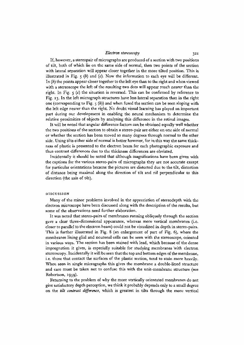
To investigate this problem a model was built out of card strips, wire and rods, allblack. The model was photographed and then tilted 150 and photographed again toproduce stereo-pairs (Figs. 15,16). The card strips represent membranes and the otherstructures cytoplasmic filaments. When photographing the model, the lighting wasarranged to eliminate shadows just as in electron micrographs (see above). Perspective,also absent in electron micrographs (see above), will occur in the model, but it isknown from studies of 'distorted' models (see Gregory, 1966) that this is not, byitself, a critical factor in depth perception. Stereoscopic examination of Fig. 15 con-firms the fact that the absence of shadows does not interfere with depth perception(although of course shadows may enhance the depth effect, no doubt as a learnedphenomenon).
To simulate tilt contrast disparity one of the vertical panels and two of the rods inthe right, more tilted, of the pair has been reduced in contrast, that is, changed fromblack to a shade of grey by underexposing it during photographic printing. Thecontrast has been reduced more in that of Fig. 15 than of Fig. 16. When these figuresare viewed stereoscopically it will be seen that the other panels stand out, because ofperfect fusion, crisply and in depth. The panel with contrast disparity, however, has ahazy appearance resulting from retinal rivalry of the contrast-differing pair. The left-hand black image tends to be dominant, but the right-hand grey image intrudes andthen disappears from time to time. Similar observations can be made on the rods.
But does this contrast disparity completely abolish stereodepth? Observers generallyagree that in spite of the contrast difference the panel and rods show stereodepth justlike the surrounding structures that have not been interfered with. (Clearly quantitativeassessment of stereo vision is required here and we are investigating methods atpresent.) However, we can assume that contrast disparity may decrease the clarity ofthe image but not seriously interfere with appreciation of stereodepth. This is bornout in Fig. 11, where the filaments show clear stereodepth, although the more tiltedprofiles (mostly in the right picture) have reduced contrast, that is, are paler.
Learned depth-appreciation in stereographs
When various observers are asked to view the electron stereographs with the stereo-scope, no difficulty is usually encountered in fusing the pair, but it may take severalseconds before depth perception is reported. Even after several minutes some observers
318 E.G. Gray and R. A. Willis
are unable to achieve any perception of depth, especially those who are unused to oruntrained in stereoscopy. Such a delay is especially marked when an unfamiliar objectis the subject of the pair. When the three-dimensional contours of the pair are des-cribed to the observer, however, he usually gradually acquires full depth perception.Similarly, if the subject is not too complex, the observer may gradually teach himselffull depth appreciation as he explores the contours through the stereoscope.
Since the images are fused at the outset, it can be assumed that the full informationfor depth perception has been transmitted through the optic nerves, but a period oflearning may be necessary before the neural mechanism for depth perception functions.
The above remarks can be illustrated in the following way. Figure 17 shows a stereo-pair of a familiar enough object, a human skull and cervical region. Most observerscan fuse the pair immediately and complete depth perception is also achieved immedi-ately or after a few seconds. The reader should now view the pair of Fig. 18 with thestereoscope and note carefully how long it takes (a) to fuse the pair, (b) what depth,if any, is appreciated, and (c) the subsequent time intervals at which any of thevarious structures appear in depth. This experiment should be carried out beforereading on.
The pair in Fig. 18 is, in fact, the pair of Fig. 17 reversed so that the left photographof Fig. 17 is now presented to the right eye in Fig. 18 and the right to the left. In otherwords Fig. 17 is the cameo version and Fig. 18 the intaglio (see, for example, Helmholtz,1909). The effect, of course, is to reverse completely the parallax so that structuresclosest in Fig. 17 should appear farthest away in Fig. 18 and vice versa. To put itanother way, if a thin replica is made of the surfaces in Fig. 17 and is then removedand looked at from behind the three-dimensional appearance should coincide withthat of Fig. 18.
Most observers viewing Fig. 18 with a stereoscope report almost immediate fusionand the result is a single picture showing no apparent depth. The flat image maypersist for several minutes to the naive observer. Now if the observer is prompted asto what he should see, for example, the upper incisor teeth should appear farthest away,the bony orbits should project out towards the observer as two cones set at an angle,the turbinal bones of the nasal cavities should project towards the observer, the fore-head should appear concave (not convex) and so on. Most observers, with or withoutprompting, learn to appreciate the reversed depth after various time intervals. Seeingthe forehead as concave usually presents some difficulty.
Now it could be argued that the cameo version (Fig. 17) of the skull is so familiarto the observer that the memory of it tends to suppress the induction of an intaglioversion by the neural mechanism in spite of the fact that the optic nerves are supplyingit with all the necessary information, and that such situations do not occur in electronmicrographs. However, this is not so. Most electron microscopists are very familiarwith the structures that they see flat in single micrographs, so that when presentedwith a stereo-pair of a structure known previously only as a two-dimensional entitya relearning has to take place and the more complex the structure, the more difficultthis new three-dimensional interpretation is likely to be (see Discussion).
Electron stereoscopy 319
Three-dimensional perception of superimposed structures
When we examine with normal binocular vision two opaque objects, one of which ispartly superimposed on the other, naturally it is the nearer of the two objects thatpartly obscures the farther one. As Helmholtz (1909) pointed out, this clue, learnedduring the childhood development of vision, helps to give the illusion of depth infar distant objects, such as the profile of one mountain running down and partlyobscuring another one (Fig. 4). At such distances (perhaps tens of miles), the angulardifference between the two retinal images is too small to be detected and utilized bythe neuronal mechanism.
This point is illustrated if Figs. 17 and 18 are viewed stereoscopically. In the cameoversion (Fig. 17) the clavicle appears near and the upper ribs lie behind it. Nowtheoretically in the intaglio version (Fig. 18) the situation should be completely changedfor the reversed angular disparity should now show the clavicle lying behind the ribs(or rather the parts of the ribs not obscured by the clavicle). The reader will no doubtagree, however, that even in the intaglio the clavicle still appears in front of the ribs,although the picture in this region appears flat. Thus the neural mechanism, althoughpresented with the full angular difference factor, which demands that the ribs shouldappear in front of the clavicle, has been inhibited in favour of the common-sense (i.e.learned) interpretation. This demands that when A obscures B, A must be in front of B.
Fig. 4. We have no difficulty in deciding which mountain is in front of which, forcommon sense dictates that what obscures must be in front.
It is important to understand this phenomenon, for precisely similar situations areencountered in electron micrographs. In some situations in electron stereographs thiseffect will tend to abolish three-dimensional perception and in others can be utilizedto advantage.
For example when a very dense structure overlies or underlies a relatively trans-parent structure within the thickness of the section, the former will always tend toobscure the latter since (as explained above, p. 312) we are dealing with a transmittedand not a reflected luminant. This is illustrated in Fig. 9. If viewed stereoscopically,it will be seen that a cluster of carbon particles lies in a bubble of the transparentsupporting carbon film, which is superimposed upon a section of brain containingmembranes and myelin sheaths. Now if the intaglio version of this pair is viewedstereoscopically (Fig. 10) the carbon particles should theoretically lie behind themembranes and sheaths. This can be seen to be so in places, but those particlescoinciding with part of the myelin sheath (right) still appear in front of the sheath.Thus the common-sense view that that which obscures must be in front has prevailedand the depth conception has been suppressed in this part of the section.
320 E. G. Gray and R. A. Willis
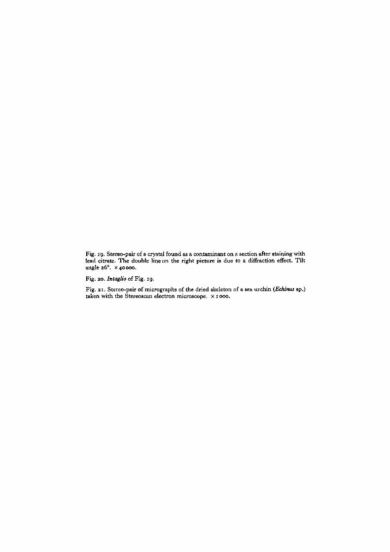
On the other hand, where in the electron microscope one structure overlies anotherand there is not too great a difference in their relative contrasts (i.e. densities),stereoscopy can be especially advantageous. For example, if the crystal (Fig. 19) isviewed stereoscopically it will be seen to have four mottled arms with a fifth armpointing upwards towards the viewer. Now if these same micrographs are reversedin position to give the intaglio version (Fig. 20) most viewers (but sometimes onlyafter prolonged scrutiny) are now able to see the mottled undersurface of the crystalwith the fifth arm, now apparently much more obscured, extending away from theundersurface of the crystal. Thus the contrast of the fifth arm is not sufficiently greatfor the common-sense view to prevail and one can apparently perceive either the topsurface or the bottom surface of the crystal at will simply by reversing the presentationof the same pair of micrographs to the eyes. The nearer surface will always appear mostclearly, since now common sense has come to our aid and the detail of that surfacelying behind will tend to be suppressed, since one expects to see things in front moreclearly than things behind them.
Note on orientation of the section in the beam
It was noted that when some stereo-pairs were examined the fused image of thesection appeared to slope across the field of vision with either the left (for exampleFig. 13) or right-hand edge nearest, while in other pairs the section appeared quiteflat. This can be explained as follows. When the section appears flat the micrographscorrespond to a section that has first been photographed tilted to one side of normal
(b)
Fig. 5. Diagram to be viewed with stereoscope to illustrate the effects of differentinformation to each eye (see text for details).
and then to obtain the second photograph tilted through normal and then to anapproximately equal degree of tilt in the opposite direction. This is illustrated inFig. 5 (a). Consider any two structures in the section (with lateral separation) illustratedhere as two spots (left two of Fig. 5 a). Now if the section is tilted as described, thetwo dots (right of Fig. 5 a) will now be exactly the same distance apart and if thesetwo pairs of dots are fused with a stereoscope the result will be two dots lying at thesame level, i.e. the section as a whole will appear untilted, since each eye receives thesame information in this respect.
Electron stereoscopy 321
If, however, a stereopair of micrographs are produced of a section with two positionsof tilt, both of which lie on the same side of normal, then two points of the sectionwith lateral separation will appear closer together in the more tilted position. This isillustrated in Fig. 5 (b) and (c). Now the information to each eye will be different.In (b) the points appear closer together to the left eye than to the right and when viewedwith a stereoscope the left of the resulting two dots will appear much nearer than theright. In Fig. 5 (c) the situation is reversed. This can be confirmed by reference toFig. 13. In the left micrograph structures have less lateral separation than in the rightone (corresponding to Fig. 5 (b)) and when fused the section can be seen sloping withthe left edge nearer than the right. No doubt visual learning has played an importantpart during our development in enabling the neural mechanism to determine therelative proximities of objects by analysing this difference in the retinal images.
It will be noted that angular difference factors can be obtained equally well whetherthe two positions of the section to obtain a stereo-pair are either on one side of normalor whether the section has been moved so many degrees through normal to the otherside. Using tilts either side of normal is better however, for in this way the same thick-ness of plastic is presented to the electron beam for each photographic exposure andthus contrast differences due to the thickness differences are obviated.
Incidentally it should be noted that although magnifications have been given withthe captions for the various stereo-pairs of micrographs they are not accurate exceptfor particular orientations because the pictures are distorted due to the tilt, distortionof distance being maximal along the direction of tilt and nil perpendicular to thisdirection (the axis of tilt).
DISCUSSION
Many of the minor problems involved in the appreciation of stereodepth with theelectron microscope have been discussed along with the description of the results, butsome of the observations need further elaboration.
It was noted that stereo-pairs of membranes running obliquely through the sectiongave a clear three-dimensional appearance, whereas more vertical membranes (i.e.closer to parallel to the electron beam) could not be visualized in depth in stereo-pairs.This is further illustrated in Fig. 8 (an enlargement of part of Fig. 6), where themembranes lining glial and neuronal cells can be seen with the stereoscope, orientedin various ways. The section has been stained with lead, which because of the denseimpregnation it gives, is especially suitable for studying membranes with electronstereoscopy. Incidentally it will be seen that the top and bottom edges of the membrane,i.e. those that contact the surfaces of the plastic section, tend to stain more heavily.When seen in single micrographs this gives the membrane a double-lined structureand care must be taken not to confuse this with the unit-membrane structure (seeRobertson, 1959).
Returning to the problem of why the more vertically orientated membranes do notgive satisfactory depth perception, we think it probably depends only to a small degreeon the tilt contrast difference, which is greatest in tilts through the more vertical
322 E. G. Gray and R. A. Willis
positions. As pointed out (p. 317) this could cause some degree of retinal rivalry. Theanswer is really to be found in the transparency factor (p. 312). When one sees on amicrograph a thin dense line representing a section of the membrane running downnear vertically (say 5° to the beam) through the plastic the density one sees is reallythe summation due to superimposition of all the dense and less dense patches thatoccur through the structure of the membrane as it extends perhaps 600 A or moredown through the section. These patches of greater and less density can be seen in thelead-stained preparations of Fig. 8 where the membranes run obliquely. They have amottled or reticulated appearance and are not homogeneously dense. Thus the thinline of the vertical membrane has no reality in the sense that a solid sheet of materialhas when seen edge-on or not quite so with the eyes under the ordinary conditions ofreflected light. Here each eye is presented, except for the angular difference, withvirtually the same picture of the topography of the surface of the sheet. However theimage of the membrane in the plastic section in the more tilted of the pair of stereo-graphs (tilting now from 5° to 150) is not simply another more angular view of thesheet of material constituting the membrane in the less tilted picture. It now appearsas a slightly wider and less dense 'line', but the density is now the sum of the densitiesin the membrane now superimposed in a different combination due to the differenttilt angle (see Fig. 2). Thus in the (5°+ 150) 'stereo'-pair one is not seeing the sameset of densities at different angles but two different sets of densities. The images aredisparate and cannot be related by the neural mechanism to give a stereoscopic effect.On the other hand, the texture in the oblique membrane when tilted further forstereoscopy remains visible in the second micrograph for the relations of superimposedmaterial alters only slightly at these wide angles (Fig. 2). (It is, of course, importantto determine to what extent the texture revealed in these lead-stained membranes isrelated to their in vivo architecture, but the question is not relevant here.)
For further elucidation of this problem membranes fixed in potassium permanganatewere studied with electron stereoscopy. It is well known that when fixed by this methodmembranes exhibit a marked granularity (Fig. 12). This is a 'stereo'-pair and thethree-lined tight junctions can be seen in the left, the less tilted of the pair. The indivi-dual granules are approximately 25 A in diameter and one would assume that theylie scattered one above the other through the thickness of the section, which is abouttwenty times their diameter. Now if the granules are real in the sense that they doexist as fixation products within the plastic section then tilting and photographingshould show these same granules but from another angle and the granules should beseen with the stereoscope in three dimensions. The right picture of Fig. 12 shows atilt of 15° and it can in fact be seen that a completely new set of granules has becomevisible, (some smaller and less dense, especially in the region of the tight junctions).These cannot be related to those in the left picture, nor can the pictures be fused togive a stereoscopic effect. Thus the granules that make up the membranes are reallyillusory. They are simply points of high density where scattering material (perhapsvery small granules) lies vertically superimposed in the electron beam. (The fact thatthe granules are illusory in no way invalidates Robertson's (1959) unit membranetheory, of course.)
Electron stereoscopy 323
Filaments cut in any plane show clear depth in stereo-pairs. This is because theyhave a relatively greater scattering power and can easily be seen when running hori-zontally through the section. Also, at the magnifications we have used so far theyappear as homogeneously dense structures and so although visualized with a trans-mitted illuminant the result is similar to the appearance that would be given using areflected illuminant (suitable to our mode of vision). Glycogen granules also can beseen as discrete structures located at different positions and levels within the structure.Attempts to visualize with high resolution and stereoscopy substructure within anindividual neurofilament or glycogen granule however would meet the same difficultiesas those presented by membranes.
So we can now begin to understand what is perhaps the main problem involvedin stereoscopy of tissue sections. Our eyes and brain mechanisms have evolved toutilize a luminant to which most everyday structures are opaque and we analyse lightpatterns reflected off their various surfaces (either with monocular vision or binocularstereo vision). The electron microscope, on the other hand, uses a transmitted illumi-nant, which passes through the sectioned structures that we are attempting to visualize(either with single exposures or stereo-pairs of micrographs). A given structure withinthe section is not seen by its surface contours and texture but as a series of densitydistributions corresponding to degrees of electron scattering. Stereoscopy of sectionswith the transmission electron microscope must thus be limited and an under-standing of this limitation is important if one is to persevere and get the bestresults.
The scanning electron microscope is now coming into general use and here theilluminant is reflected or back-scattered electrons. In this way it is possible to seesurfaces just as in everyday vision, either in single micrographs or stereo-pairs.Figure 21 shows a stereo-pair of the dried skeleton of the sea urchin (Echinus sp.)(see Lester & Boyde (1967) for method of preparation). Viewed with the stereoscopethe three-dimensional effect is immediate and dramatic for most observers whetherthe material is familiar or not. Unfortunately the resolution at present is an order ofmagnitude less than in the transmission microscope. Perhaps one day it will be possibleto remove the plastic from a section without seriously interfering with the structureswithin it. This would then be suitable for reflecting stereo-electron microscopy, andfully comprehensible by visual systems that have evolved to look at things rather thanthrough them.
We have also seen that learning plays an important part in depth appreciation instereo-pairs taken by transmission electron microscopy and we find that complexstructures not understood at the onset (e.g. the intaglio of the skull, Fig. 18) aredifficult to visualize in depth without prolonged observation. It is often necessary togaze perhaps for periods of up to 30 min through the stereoscope, gradually teachingoneself how different parts of a given structure lie in front of or behind each other.
Also we have noted that in a stereo-pair where several structures lie superimposedsimply by reversing the pictures presented to the eyes a structure can either be seenlying at the back of or in front of the section. Here we illustrated this point withreference to a crystal, but a similar effect was observed on tissue sections and negatively
324 E. G. Gray and R. A. Willis
stained material. By this 'placing1 the structure in the front of the section it is seennearest through the stereoscope and its morphology is far better understood becauseone is not looking at it 'through' other structures. Thus it is possible to separate outstructures for visual attention which when seen in single micrographs are superimposedand confused.
As far as we know this is the first paper dealing in detail with electron stereoscopyof tissue sections. We know of several electron microscopists, and there are no doubtmany more, who have attempted stereoscopy on their sections at one time or anotherbut who discontinued the method because of disappointing initial results. We hopethat our observations and remarks will stimulate a new interest in this technique as aninvaluable and really indispensable method of getting more information from tissuesections.
We are indebted to Professor J. Z. Young, F.R.S., for his interest and support in this work:also to Professor A. F. Huxley, F.R.S., Dr A. Boyde and Dr J. Y. Lettvin for helpful advice.Miss T. Charlton gave technical assistance and Mr S. Waterman made the photographic prints.The diagrams were drawn by Mrs J. Astafiev.
REFERENCES
BOYDE, A. & WILLIS, R. A. (1966). Calibration of tilt cartridges for Siemens Elmiskop I.J. Anat. 100, 706.
BOYDE, A. (1967). A single-stage carbon-replica method and some related techniques for theanalysis of the electron microscope image. Jl R. nricrosc. Soc. 86, 359-370.
CHANDLER, R. L. & WILLIS, R. A. (1966). An intranuclear fibrillar lattice in neurons. J. Cell Sci.1, 283-286.
DAVSON, H. (1963). The Eye (2nd edition). London: Churchill.GRAY, E. G. & GUILLERY, R. W. (1966). Synaptic morphology in the normal and degenerating
nervous system. Int. Rev. Cytol. 19, 111-182.GREGORY, R. L. (1966). Eye and Brain—the Psychology of Seeing. London: Weidenfeld and
Nicholson.HELMCKE, J. G. (1954). Theorie und Praxis der elektronenmikroskopischen Stereoaufnahmen.
Optik. Stuttg. 11, 201-205.HELMCKE, J. G. (1965). Determination of the third dimension of objects by stereoscopy. Lab.
Invest. 14, 195-200.HELMHOLTZ, H. von (1909). Handbuch der Physiological Optics, vol. 3 (ed. J. P. Southall,
trans. Optical Society of America, 1925). Menasha, Wisconsin.JULESZ, B. (1962). Towards the automation of binocular depth perception. In Information
Processing (Proc. of I.F.I.P. Congress). London: North Holland Publ. Co.JULESZ, B. (1963). Stereopsis and binocular rivalry of contours. J. opt. Soc. Am. 53, 994-999.JULESZ, B. (1964). Binocular depth perception without familiarity clues. Science, N. Y. 145, 3630.JULESZ, B. (1965). Texture and visual perception. Scient. Am. 212, No. 2, 38-48.KELLY, D. E. (1966). Fine structure of desmosomes, hemidesmosomes and an adepidermal
globular layer in developing newt epidermis. J. Cell Biol. 28, 51^72.LESTER, K. S. & BOYDE, A. (1967). Electron microscopy of predentinal surfaces. Calc. Tiss. Res.
!, 44-54-LUFT, J. H. (1956). Permanganate—a new fixative for electron microscopy. J. biophys. biochem.
Cytol. 2, 799-801.OGLE, K. N. (1962). The optical space sense. In The Eye, vol. 4, Visual Optics and the Optical
Space Sense (ed. H. Davson). New York: Academy Press.PEACHEY, L. D. (1958). Section thickness and compression. In 4/A Int. Conf. Electron Microsc,
Berlin, vol. 2 (ed. W. Bargmann et al.). Berlin: Springer.
Electron stereoscopy 325
PEACHEY, L. D. (1965). Electron microscopy of tilted biological sections. R.C.A. Scient. Instrum.Neics 10, 7-12.
REYNOLDS, E. S. (1963). The use of lead citrate at high pH as an electron-opaque stain inelectron microscopy, jf. Cell Biol. 17, 208-212.
ROBERTSON, J. D. (1959). The ultrastructure of cell membranes and their derivatives. Biochem.Soc. Symp. 16, 3-43.
SHALLA, T. A., CARROLL, T. W. & DE ZOETEN, G. A. (1964). Penetration of stain in ultrathinsections of TMV. Stain Technol. 39, 257-65.
VALDRE, U. (1962). A simple goniometer stage for the Siemens electron microscope. J. Sci.Instrum. 39, 278-281.
VALDRE, U., COILEY, J. & PARSONS, D. E. (1964). The use of goniometer stages for the examina-tion of non-metallic and biological specimens. Proc. -yrd European Reg. Conf. on ElectronMiscrosc. vol. B, 21-22. Prague: Czechoslovak Academy of Sciences.
VALYUS, N. A. (1966). Stereoscopy. Trans, from Russian by Department of Education andScience. London and New York: Focal Press.
WESTRUM, L. E. & LUND, R. D. (1966). Formalin perfusion for correlative light and electron-microscopical studies of the nervous system. J. Cell Set. 1, 229—238.
WILLIAMS, R. C. & KALLMAN, F. (1955). Interpretations of electron micrographs of single andserial sections. J. biophys. biochem. Cytol. 1, 301-314.
WILLIS, R. A. (1966). Stereo electron microscopy of biological thin sections. Proc. R. microsc.Soc. 1, 167.
WILLIS R. A. & GRAY, E. G. (1966). Electron stereoscopy of tissue sections. J. Anat. 100, 690.
(Received 25 October 1967)
Cell Sci. 3
326 E. G. Gray and R. A. Willis
Fig. 6. Stereo-pair of a synapse of the cerebral cortex of the rat (material kindly fixedby Dr L. E. Westrum). Tilt angle i6°. x 30000.
Fig. 7. Stereo-pair of a synapse of the goldfish spinal cord containing glycogen. Tiltangle 200. x 35000.
Fig. 8. Part of the preparation of Fig. 6 enlarged. Tilt angle 160. x 60000.
Journal of Cell Science, Vol. 3, No. 2
E. G. GRAY AND R. A. WILLIS {Facing p. 326)
Fig. 9. Stereo-pair of myelinated axons in rat brain. A cluster of carbon contaminationparticles is present. Tilt angle 200. x 32000.
Fig. 10. The 'intaglio' of Fig. 9.
Fig. 11. Stereo-pair of sectioned neurofilaments in a myelinated axon of the fish spinalcord. Tilt angle 10°. x 50000.
Journal of Cell Science, Vol. 3, No. 2
E. G. GRAY AND R. A. WILLIS
Fig. 12. Stereo-pair of membranes apposed in places to form tight junctions fromoptic nerve of the octopus fixed in permanganate. Tilt angle 45°. x 00000.
Fig. 13. Stereo-pair of micrographs selected from the series shown in Fig. 14. Tiltangle 120. x 40000.
12
Journal of Cell Science, Vol. 3, No. 2
«*
E. G. GRAY ANP R. A. WILLIS
Fig. 14. Series of micrographs of the apposed membranes of two neuroglial processesin the spinal cord of the goldfish. The membranes have a desmosome (left). Thesection has been tilted from o to 45° in approximately 40 steps (not all the micrographsare included), x 70000.
Journal of Cell Science, Vol. 3, No. 2
E. G. GRAY AND R. A. WILLIS
Fig. 15. Stereo-pair of photographs of a model made from black card, pieces of blackcarbon rod and wire. The contrast level has been reduced on some of the structures(right-hand picture). Tilt angle 15°.
Fig. 16. As Fig. 15, but with greater reduction in the contrast of the structures.
Journal of Cell Science, Vol. 3, No. 2
15
I
I *1
16
I
l * L1
E. G. GRAY AND R. A. WILLIS
Fig. 17. Stereo-pair of photographs of a human skull. Tilt angle about 120.
Fig. 18. Intaglio version of Fig. 17.
Journal of Cell Science, Vol. 3, No. 2
E. G. GRAY AND R. A. WILLIS
Fig. 19. Stereo-pair of a crystal found as a contaminant on a section after staining withlead citrate. The double line on the right picture is due to a diffraction effect. Tiltangle 26°. x 40000.
Fig. 20. Intaglio of Fig. 19.
Fig. 21. Stereo-pair of micrographs of the dried skeleton of a sea urchin {Echinus sp.)taken with the Stereoscan electron microscope, x 1000.


![Preparation Physicochemical Characterization and Catalytic ... · modified polymeric Catalysts [18] (Table 2). 3.3. High Resolution Scanning Electron Micrograph SEM at various stages](https://static.documents.pub/doc/80x56/5f1fdc0fc7f36e47270b0f24/preparation-physicochemical-characterization-and-catalytic-modified-polymeric.jpg)


