Digital immunohistochemistry platform for thestaining variation monitoring based onintegration of image and statistical analyses withlaboratory information systemAida Laurinaviciene1,2, Benoit Plancoulaine3, Indra Baltrusaityte1,2, Raimundas Meskauskas1,2, Justinas Besusparis1,2,Daiva Lesciute-Krilaviciene2, Darius Raudeliunas2, Yasir Iqbal1, Paulette Herlin2, Arvydas Laurinavicius1,2*
From 12th European Congress on Digital PathologyParis, France. 18-21 June 2014
Abstract
Background: Digital immunohistochemistry (IHC) is one of the most promising applications brought by newgeneration image analysis (IA). While conventional IHC staining quality is monitored by semi-quantitative visualevaluation of tissue controls, IA may require more sensitive measurement. We designed an automated system todigitally monitor IHC multi-tissue controls, based on SQL-level integration of laboratory information system withimage and statistical analysis tools.
Methods: Consecutive sections of TMA containing 10 cores of breast cancer tissue were used as tissue controls inroutine Ki67 IHC testing. Ventana slide label barcode ID was sent to the LIS to register the serial section sequence.The slides were stained and scanned (Aperio ScanScope XT), IA was performed by the Aperio/Leica Colocalizationand Genie Classifier/Nuclear algorithms. SQL-based integration ensured automated statistical analysis of the IA databy the SAS Enterprise Guide project. Factor analysis and plot visualizations were performed to explore slide-to-slidevariation of the Ki67 IHC staining results in the control tissue.
Results: Slide-to-slide intra-core IHC staining analysis revealed rather significant variation of the variables reflecting thesample size, while Brown and Blue Intensity were relatively stable. To further investigate this variation, the IA resultsfrom the 10 cores were aggregated to minimize tissue-related variance. Factor analysis revealed association betweenthe variables reflecting the sample size detected by IA and Blue Intensity. Since the main feature to be extracted fromthe tissue controls was staining intensity, we further explored the variation of the intensity variables in the individualcores. MeanBrownBlue Intensity ((Brown+Blue)/2) and DiffBrownBlue Intensity (Brown-Blue) were introduced to bettercontrast the absolute intensity and the colour balance variation in each core; relevant factor scores were extracted.Finally, tissue-related factors of IHC staining variance were explored in the individual tissue cores.
Conclusions: Our solution enabled to monitor staining of IHC multi-tissue controls by the means of IA, followedby automated statistical analysis, integrated into the laboratory workflow. We found that, even in consecutive serialtissue sections, tissue-related factors affected the IHC IA results; meanwhile, less intense blue counterstain wasassociated with less amount of tissue, detected by the IA tools.
* Correspondence: [email protected] of Pathology, Forensic Medicine and Pharmacology, Faculty ofMedicine, Vilnius University, Vilnius, LithuaniaFull list of author information is available at the end of the article
Laurinaviciene et al. Diagnostic Pathology 2014, 9(Suppl 1):S10http://www.diagnosticpathology.org/content/9/S1/S10
BackgroundDigital immunohistochemistry (IHC) is one of the mostpromising applications brought by digital pathology,enabling new generation image analysis (IA) tools [1-3].Robust and efficient digital IHC systems are expected toenable high throughput, accurate, and reproduciblemeasurement of tissue markers, along with their spatialdistribution. Conventional IHC routine is mostly basedon qualitative and semi-quantitative visual evaluation ofthe tissue tested as well as tissue controls, to monitorthe IHC staining quality. Multi-tissue controls on thesame IHC slide can further improve the staining qualitycontrol [4]. While visual quality monitor is deemed suf-ficient for the conventional IHC, IA-based approachmay require more sensitive monitoring in the digitalIHC [5]. Although quantitative IA has been referred asvaluable way to quantify staining intensity and assureday-to-day consistency of control tissue reactivity [6],we are not aware of published work on this aspect. Wehave previously shown that HER2 IHC multi-tissue con-trols, monitored by IA, reveal the staining intensitydrifts and unexpected deviations undetected by routineslide-by-slide review by a pathologist [7]. Furthermore,data reduction by factor analysis has been helpful inretrieving hidden variation sources in IHC IA data [8]and could be useful in exploring quality indicators fordigital IHC.On the other hand, successful implementation of digi-
tal IHC depends on seamless integration of the IA andstatistical analysis tools into pathology diagnosis andresearch workflow. Implementation, validation, calibra-tion, continuous quality monitoring - all require swiftquantitative feedback from the IA results. Digital IHCtissue control is a particular case, representing this effi-ciency need and possible solution scenarios.We herewith present an automated system to monitor
digital IHC multi-tissue controls, based on SQL-levelintegration of laboratory information system (LIS) withimage and statistical analysis tools. The platform enablesto explore hidden IHC staining variation factors in theserial sections of multi-tissue controls used in diagnosticIHC routine, based on multivariate analyses and visualrepresentation of the IA results.
MethodsIHC multi-tissue controls were constructed from paraffinblocks of breast cancer tissue with a broad range of Ki67IHC positivity. Tissue microarrays (TMA) containing 10tissue cores of 1 millimeter diameter were produced andconsecutive serial sections were cut and stored at +4°C.Upon demand, when Ki67 IHC on breast cancer tissuewere ordered by pathologist in a diagnostic routine, theslides with the multi-tissue control sections were usedto add a section of a diagnostic sample. A unique and
ascending barcode ID number was sent to the LIS by theVentana Ultra machine when the slide label was printedat the microtome workstation, thus allowing retrieval ofthe serial section sequence from the LIS for further datamanagement and integration with the IA results. Afterthe IHC slides were routinely stained (Ultraview DABdetection kit on Ventana Ultra staining system (VentanaMedical Systems, Tucson, Arizona, USA; counterstainedwith Meyer’s hematoxylin prepared in house), they werescanned (Aperio ScanScope XT, 20× objective magnifica-tion), TMA multi-tissue was assigned an appropriateAperio TMALAB design, and IA algorithms were run onthe control TMA spot images as well as on the test tissuewhole slide images (WSI). SQL-based data integrationensured automated analysis of the TMA and WSI IAdata by the SAS Enterprise Guide project, constructed tomanage and analyze the IA data. Factor analysis and plotvisualizations were performed to extract and monitorslide-to-slide variation of the Ki67 IHC staining charac-teristics, based on the sequence of serial multi-tissuesections identified by the Ventana label barcode ID in theLIS. Three TMA blocks were used in the study conse-quently until exhausted, to produce serial multi-tissuecontrol sections (84, 31, and 69, respectively). Separatestatistical analyses were carried out for each block; resultsfrom the third block are presented in the Results section.The control tissue samples (TMA cores) were labeled asrepresented in the Ki67 IHC spot images of the thirdTMA block (Figure 1); IA-detected variance between dif-ferent tissue cores on the same slide is expected to reflect“intra-slide inter-tissue” variation, while IA-detected var-iance between consecutive sections of the same tissuecore - “inter-slide intra-tissue” variance, expected toreflect the variation of IHC staining properties overtime.The latter, if established, would then serve as “digitalIHC control” for the test tissue on the same slide.The IA was performed by the Aperio/Leica Colocali-
zation v.9 algorithm, tuned to extract brown and bluecolors, as well as the Genie Classifier v.1/Nuclear v.9algorithm, calibrated to enumerate Ki67-positive andnegative tumour nuclear profiles in the breast cancertissue [9]. Automated data management and statisticalanalysis workflow were achieved in the SAS EnterpriseGuide v.5.1. Summary and variation statistics of the IAoutput variables were performed (Table 1). Factor analy-sis was carried out using factoring method of principalcomponent analysis: factors were retained based on thethreshold of the smallest eigenvalue of 1.0. Orthogonalvarimax rotation of the initial factors was performed.A level of statistical significance was not set in thisexploratory experiment. The LIS is a SQL and WEB-based system PathIS®, developed and maintained by theNational Center of Pathology and the Baltic InformationTechnologies Institute, Vilnius, Lithuania.
Laurinaviciene et al. Diagnostic Pathology 2014, 9(Suppl 1):S10http://www.diagnosticpathology.org/content/9/S1/S10
Page 2 of 13
Figure 1 The design of the TMA used as the IHC multi-tissue controls. The cores are labeled with their sample identifiers used in this study.IA-detected variance between different tissue cores on the same slide is expected to reflect “intra-slide inter-tissue"variation, while IA-detectedvariance between consecutive sections of the same tissue core - “inter-slide intra-tissue” variance expected to reflect the variation of IHC stainingproperties overtime. The latter, if established, would then serve as “digital IHC control” to measure the test tissue “intra-slide inter-tissue” varianceon the same slide.
Table 1 Slide-to-slide IHC staining variation of the 10 multi-tissue control samples, based on IA output variables
Laurinaviciene et al. Diagnostic Pathology 2014, 9(Suppl 1):S10http://www.diagnosticpathology.org/content/9/S1/S10
Page 3 of 13
ResultsSlide-to-slide IHC staining variation of the 10 multi-tis-sue control samples, represented by selected IA outputvariables, is presented in the Table 1 and Figure 2 (A, B,C, D and E, F, G, H plots represent data obtained by theGenie/Nuclear and Colocalization algorithms,respectively).Firstly, rather significant intra-core variation can be
noted in the variables reflecting the sample size of thespots (Total nuclei, Total stained area, Figure 2A, E):while continuous drift of these variables is likely to reflecttissue variability in the consecutive sections, the irregula-rities, often parallel in majority of the spots, may reflecttissue artefacts and/or staining variation. Indeed, inspec-tion of the spot images with major abnormalities revealedpresence of tissue artefacts.Secondly, the variation of Ki67-positive nuclei detected in
the consecutive sections was rather significant (Figure 2B,C); it was relatively more notable at the low end of scale(where main clinical interest is), also represented by higherrelative error values in the cores with less Ki67 positivity(Table 1). To avoid potential impact of misdetection of
negative tumour nuclei on the Ki67 positivity estimation,we calculated the “Positive Density” variable as the ratio ofKi67-positive nuclei to the Area of Analysis to be used infurther analyses. Remarkably, the variation of the PositiveDensity appeared less aberrant at the low end of scale(Figure 2D), although this was not necessarily reflected bythe Relative Error values compared to the Ki67-positivepercent (Table 1).Thirdly, the variation of the Brown and Blue Intensity
as well as their ratio (Figure 2F, G, H) reflected inter-core variation dependent on the Ki67 positivity of thetumours sampled; however, the range of Blue Intensityinter-core variation was lower than that of the BrownIntensity. Intra-core variation of both Brown and BlueIntensity was rather low, while aberrant spot imagesrevealed mostly tissue artefacts affecting the IA results.Finally, since the multi-controls represent tissue samples
from tumours with different Ki67 positivity, it is expectedthat the IA results on individual spots would reflect this;however, slide-to-slide variation of the same core wouldreveal continuous change due to some unavoidable tissuevariation in the serial sections. Importantly, one can note
Table 1 Slide-to-slide IHC staining variation of the 10 multi-tissue control samples, based on IA output variables(Continued)
Blue Area 0.10 0.12 0.13 0.18 0.18 0.18 0.20 0.14 0.12 0.09
Brown Area 0.36 0.22 0.07 0.09 0.05 0.05 0.05 0.04 0.61 0.02
Blue Area 0.27 0.15 0.27 0.22 0.25 0.22 0.13 0.22 0.17 0.88
Brown Area 0.33 0.39 0.31 0.25 0.41 0.38 0.32 0.35 0.16 0.65
Laurinaviciene et al. Diagnostic Pathology 2014, 9(Suppl 1):S10http://www.diagnosticpathology.org/content/9/S1/S10
Page 4 of 13
Figure 2 Line plots representing slide-to-slide IHC staining variation of the TMA multicontrols. The sequence of Ventana slide label ID isplotted as Barcode on the x axis to represent consecutive serial sections of TMA blocks of the 10 multi-tissue control cores (labelled asSampleID), based on image analysis results of: A. Total Nuclei; B. Positive Nuclei; C. Percent Positive Nuclei; D. Positive Density; E. Total StainedArea (mm2); F. Brown Intensity; G. Blue Intensity; H. Brown/Blue Intensity ratio.
Laurinaviciene et al. Diagnostic Pathology 2014, 9(Suppl 1):S10http://www.diagnosticpathology.org/content/9/S1/S10
Page 5 of 13
the pattern that in some slides this variation appears paral-lel in most spots, while on other occasions it appears unre-lated (Figure 1).To further investigate potential sources of this varia-
tion, we have aggregated the IA results from the 10 coresas appropriate to represent them as one sample. Sincethe tissue-related variation in all of the 10 cores isexpected to be random (except possible variation of thetissue section thickness and the slide scanning regime),aggregation of the data would represent a “super-sample”
were tissue-related impact on the IA variance would bereduced. Therefore, variables like Median Blue Intensity,Total Stained area, Total Nuclei, would summarize paral-lel but disregard random variation of the individual coreIA data. Factor analysis of the aggregated variables(Figure 3) revealed that the major source of variation(Factor 1) was characterized by positive loadings of thevariables reflecting “sample size” detected by the IA algo-rithms: Blue Area and Brown Area by the Colocalization,and Area of Analysis, Positive Nuclei, Negative Nuclei by
Figure 3 Factor pattern representing parallel variance of the Colocalization and Genie/Nuclear algorithm output variables inaggregated image analysis data from the 10 TMA cores. The variable loading plots: A. Factor-1 versus Factor-2; B. Factor-1 versus Factor-3.
Laurinaviciene et al. Diagnostic Pathology 2014, 9(Suppl 1):S10http://www.diagnosticpathology.org/content/9/S1/S10
Page 6 of 13
the Genie/Nuclear. Remarkably, the Factor 1 alsorevealed strong negative loading of Blue Intensity values(more intense blue correlated with more tissue detectedby both algorithms). Meanwhile, the Factor 2 was repre-sented by positive loadings of the Percent of PositiveNuclei and negative loadings of Brown Intensity(more intense brown correlated with higher Percent ofPositive Nuclei). The factor pattern implies possibleimpact of tissue staining intensity variation on IA perfor-mance in terms of tissue detection, however, the percen-tage of positive nuclei is relatively independent of thiseffect (by definition, Factors 1 and 2 are linearly indepen-dent). To further demonstrate the relationships, the plotsof the Factor 1 and 2 scores in the consecutive sectionsare presented in the Figure 4: while the Factor 2 scoresreveal aberrant variation, the Factor 1 scores presentnotable drift with several peaks, potentially pointing tothe IHC counterstain intensity changes, although impactof tissue-related factors cannot be ruled out. The peculiarrelationship between the variables is also illustrated by
the plot of Area of Analysis (detected by the Genie) andBlue Intensity (Figure 5).Since the main feature to be extracted from the IHC
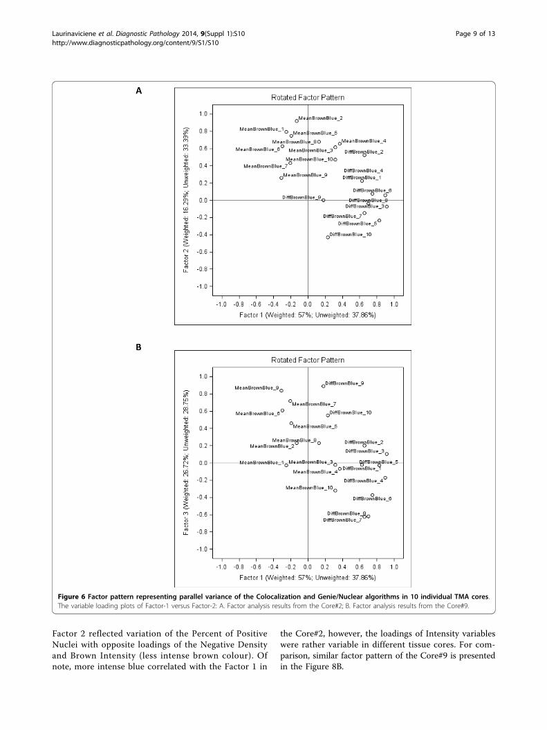
tissue controls is Brown and Blue staining intensity (thevariation is expected to be parallel to that of a test sam-ple), we further concentrated on exploring the variationsources of the intensity variables in the individual cores,as presented in Figure 2F, G. The data were transformedto enable factor analysis on Brown and Blue intensity foreach spot; furthermore, MeanBrownBlue Intensity((Brown+Blue)/2) and DiffBrownBlue Intensity (Brown-Blue) were introduced to better contrast the absoluteintensity and the colour balance variation. Indeed, factoranalysis (Figure 6A) extracted Factor-1 characterized bypositive loadings of DiffBrownBlue Intensity and Factor-2 characterized by positive loadings of MeanBrownBlueIntensity of the majority of the 10 cores. Since by defini-tion these factors are independent, Factor-1 is expectedto reflect Brown-Blue Intensity variation in oppositedirections but parallel in the majority of the spots and
Figure 4 Line plots representing slide-to-slide IHC staining variation of the Factor scores. The factor scores generated from the analysispresented in Fig. 3 are plotted against the sequence of Ventana slide label ID (labelled as Barcode) on the x axis
Laurinaviciene et al. Diagnostic Pathology 2014, 9(Suppl 1):S10http://www.diagnosticpathology.org/content/9/S1/S10
Page 7 of 13
represents the colour balance per se, mostly independentof the tissue-related variation. Factor-2 characterizesabsolute intensity variation of both colours in the samedirection, parallel in most spots, and therefore is likelyto be dependent on tissue and/or scanning variations(section thickness, scanning regime, etc.). The pattern ofthe Factor-3 (Figure 6B) is somewhat peculiar: it is char-acterized by parallel variance of the MeanBrownBlueand DiffBrownBlue for the Core#9 and opposite var-iance of these variables for the Core#7. In other words,when Core#9 becomes darker it is because of deeperBrown, and vice versa, when Core#7 becomes darker itis because of deeper Blue. Importantly, the Factor-3does reveal variable loading pattern for other cores,therefore, it is likely to express core-specific behaviourof the colour balance (with 2 extreme examples Core#7and Core#9), thus can be interpreted as tissue-relatedvariation which has been extracted as “noise” from the
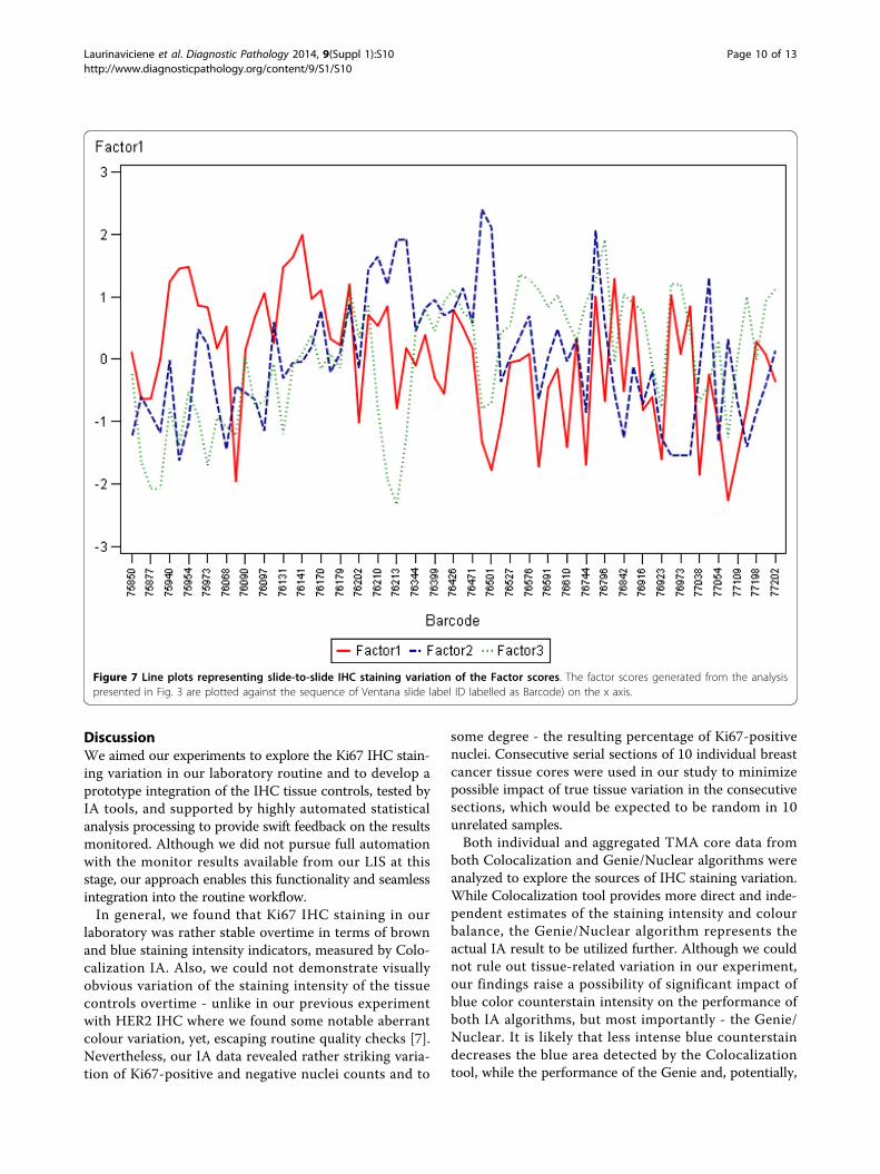
Factors 1 and 2. We therefore suggest that the Factor-1scores provide a quantitative measure of Brown andBlue Intensity balance “purified” from the impact of tis-sue-related variation removed into the Factors 2 and 3.Consequently, slide-to-slide variation of the Factorscores can be monitored as depicted on Figure 7 and 7further explored for quality assurance of digital IHC.Interdependencies between the Genie/Nuclear and
Colocalization variables were further investigated by fac-tor analyses performed for each individual tissue core.Although the factor patterns revealed some peculiaritiesfor individual tissue cores, some common variance pat-terns could be generalized from the majority of thecores. As an example, a rather representative factor pat-tern of the Core#2 is plotted in the Figure 8A. Factor 1was mainly represented by positive loadings of the vari-ables expressing the epithelial cancer compartment size(analysis area, counts of positive and negative nuclei).
Figure 5 Line plots representing slide-to-slide IHC staining variation of selected image analysis variables from aggregated TMA data.Aggregated data (Median Blue Intensity and Area of Analysis) from image analysis of 10 TMA cores are plotted against the sequence of Ventanaslide label ID (labelled as Barcode) on the x axis.
Laurinaviciene et al. Diagnostic Pathology 2014, 9(Suppl 1):S10http://www.diagnosticpathology.org/content/9/S1/S10
Page 8 of 13
Factor 2 reflected variation of the Percent of PositiveNuclei with opposite loadings of the Negative Densityand Brown Intensity (less intense brown colour). Ofnote, more intense blue correlated with the Factor 1 in
the Core#2, however, the loadings of Intensity variableswere rather variable in different tissue cores. For com-parison, similar factor pattern of the Core#9 is presentedin the Figure 8B.
Figure 6 Factor pattern representing parallel variance of the Colocalization and Genie/Nuclear algorithms in 10 individual TMA cores.The variable loading plots of Factor-1 versus Factor-2: A. Factor analysis results from the Core#2; B. Factor analysis results from the Core#9.
Laurinaviciene et al. Diagnostic Pathology 2014, 9(Suppl 1):S10http://www.diagnosticpathology.org/content/9/S1/S10
Page 9 of 13
DiscussionWe aimed our experiments to explore the Ki67 IHC stain-ing variation in our laboratory routine and to develop aprototype integration of the IHC tissue controls, tested byIA tools, and supported by highly automated statisticalanalysis processing to provide swift feedback on the resultsmonitored. Although we did not pursue full automationwith the monitor results available from our LIS at thisstage, our approach enables this functionality and seamlessintegration into the routine workflow.In general, we found that Ki67 IHC staining in our
laboratory was rather stable overtime in terms of brownand blue staining intensity indicators, measured by Colo-calization IA. Also, we could not demonstrate visuallyobvious variation of the staining intensity of the tissuecontrols overtime - unlike in our previous experimentwith HER2 IHC where we found some notable aberrantcolour variation, yet, escaping routine quality checks [7].Nevertheless, our IA data revealed rather striking varia-tion of Ki67-positive and negative nuclei counts and to
some degree - the resulting percentage of Ki67-positivenuclei. Consecutive serial sections of 10 individual breastcancer tissue cores were used in our study to minimizepossible impact of true tissue variation in the consecutivesections, which would be expected to be random in 10unrelated samples.Both individual and aggregated TMA core data from
both Colocalization and Genie/Nuclear algorithms wereanalyzed to explore the sources of IHC staining variation.While Colocalization tool provides more direct and inde-pendent estimates of the staining intensity and colourbalance, the Genie/Nuclear algorithm represents theactual IA result to be utilized further. Although we couldnot rule out tissue-related variation in our experiment,our findings raise a possibility of significant impact ofblue color counterstain intensity on the performance ofboth IA algorithms, but most importantly - the Genie/Nuclear. It is likely that less intense blue counterstaindecreases the blue area detected by the Colocalizationtool, while the performance of the Genie and, potentially,
Figure 7 Line plots representing slide-to-slide IHC staining variation of the Factor scores. The factor scores generated from the analysispresented in Fig. 3 are plotted against the sequence of Ventana slide label ID labelled as Barcode) on the x axis.
Laurinaviciene et al. Diagnostic Pathology 2014, 9(Suppl 1):S10http://www.diagnosticpathology.org/content/9/S1/S10
Page 10 of 13
the Nuclear algorithms is not sufficiently robust to detectproper amount of epithelial tissue mask and cell nucleiwithin the mask. Importantly, the range of blue intensityvariation in our data was relatively low compared to that
of the amount of tissue and cell numbers detected. Ofnote, our data reveal an association rather than a causalrelationship between the variables; one needs to designmore targeted experiments to obtain direct evidence.
Figure 8 Factor pattern representing parallel variance of the Colocalization and Genie/Nuclear algorithms in 10 individual TMA cores.The variable loading plots of Factor-1 versus Factor-2: A. Factor analysis results from the Core#2; B. Factor analysis results from the Core#9.
Laurinaviciene et al. Diagnostic Pathology 2014, 9(Suppl 1):S10http://www.diagnosticpathology.org/content/9/S1/S10
Page 11 of 13
The IA issues with hematoxylin counterstain, used rou-tinely in IHC, have been highlighted, alternative counter-staining and IA techniques have been proposed [10-14].Our experiment provides new data supporting the impor-tance of further optimization and standardization of IHCprocedures to achieve reliable processes and results withadoption of digital IHC. Interestingly, our data shed thelight on how reproducible the Ki67 index would be in theconsecutive sections of one 1 mm diameter core, if per-formed for research or clinical use. While variation of thepercentage of Ki67-positive nuclei (the IA result) wassatisfactory (standard deviation in all 10 cores rangedfrom 3 to 8, and relative error was within the range of0.07 to 0.39, Table 1), the variation of cell numbersdetected (the process) was higher. One may argue thatthe process variability needs to be dealt with, to achieverobust results by digital IHC techniques.Inter-laboratory IHC staining variation is likely to be
more significant and may impact visual estimation of Ki67index: a recent international Ki67 reproducibility study[15] revealed unsatisfactory results of visual estimationwhich was even worse when the slides were stained locally.This implies significant inter-laboratory Ki67 IHC stainingvariability which should be considered when applying IAtools with unknown sensitivity to the staining characteris-tics. Although we have recently reported [9] on a metho-dology enabling accurate Ki67 index measurement inTMA samples by IA, the issue of IA calibration to possibleinter-laboratory IHC staining variation and comparabilityof the Ki67 data between pathology labs remains open.One approach could be measuring signal-to-noise ratio ofthe images to evaluate quality before IA [16], however,adjustment of the images and/or analyses may requireanother effort. Ideally, IA tools should be robust and resis-tant to the IHC staining and scanning variations; however,this property requires further analysis and developmentefforts. As our study shows, one particular approach couldbe replacing the measurement of Ki67 index by the esti-mate of density of Ki67-positive cells in the tumour area:these two variables closely correlate; however, the Ki67-positive density does not rely on accurate detection ofnegative nuclei. This latter aspect may be especially rele-vant in the tumours with low Ki67 positivity.
ConclusionsOur study presents a case for digital pathology solution tomonitor staining of IHC multi-tissue controls by themeans of digital IA, followed by automated statistical ana-lysis procedures and integrated into the laboratory routine.We found that, even in consecutive serial tissue sections,tissue-related factors affected the IHC IA results; mean-while, less intense blue counterstain was associated withless amount of tissue, detected by the IA tools.
List of abbreviationsDiffBrownBlue: difference of Brown and Blue Intensity (Brown-Blue); IA:image analysis; IHC: immunohistochemistry; LIS: laboratory informationsystem; MeanBrownBlue: mean of Brown and Blue Intensity (Brown+Blue)/2);TMA: tissue microarray; WSI: whole slide image.
Competing interestsThe authors declare that they have no competing interests.
Authors’ contributionsAiL, IB, JB, RM, and DLK designed and carried out the TMA IHC controls anddigital image analysis workflow, edited the manuscript. AL, BP and PHperformed statistical analyses and drafted essential parts of the manuscript.AL, DR, YI designed and constructed SQL-based integration of LIS, IA andstatistical analysis systems. All authors participated in conception and designof the study, reviewing the analysis results, critically revised and approvedthe final manuscript.
Authors’ informationNone.
AcknowledgementsThis research is funded by European Social Fund under the Global Grantmeasure.Publication of this supplement has been funded by 12th European Congresson Digital Pathology. This article has been published as part of DiagnosticPathology Volume 9 Supplement 1, 2014: Selected articles from the 12thEuropean Congress on Digital Pathology. The full contents of thesupplement are available online at http://www.diagnosticpathology.org/supplements/9/S1
Authors’ details1Department of Pathology, Forensic Medicine and Pharmacology, Faculty ofMedicine, Vilnius University, Vilnius, Lithuania. 2National Center of Pathology,affiliate of Vilnius University Hospital Santariskiu Clinics, Vilnius, Lithuania.3Path-Image/BioTiCla, University of Normandy, Unicaen, Caen, France.
image analysis in histopathology: a valuable tool in medical diagnostics.ExpertRevMolDiagn 2008, 8(6):707-725.
2. Laurinavicius A, Laurinaviciene A, Dasevicius D, Elie N, Plancoulaine B, Bor C,Herlin P: Digital image analysis in pathology: benefits and obligation.Anal Cell Pathol (Amst) 2012, 35(2):75-78.
3. Al-Janabi S, Huisman A, Van Diest PJ: Digital pathology: current status andfuture perspectives. Histopathology 2012, 61(1):1-9.
4. Packeisen J, Buerger H, Krech R, Boecker W: Tissue microarrays: a newapproach for quality control in immunohistochemistry. J Clin Pathol 2002,55(8):613-615.
5. Dowsett M, Nielsen TO, A’Hern R, Bartlett J, Coombes RC, Cuzick J, Ellis M,Henry NL, Hugh JC, Lively T, et al: Assessment of Ki67 in breast cancer:recommendations from the International Ki67 in Breast Cancer workinggroup. J Natl Cancer Inst 2011, 103(22):1656-1664.
6. Hammond ME, Hayes DF, Dowsett M, Allred DC, Hagerty KL, Badve S,Fitzgibbons PL, Francis G, Goldstein NS, Hayes M, et al: American Societyof Clinical Oncology/College of American Pathologists guidelinerecommendations for immunohistochemical testing of estrogen andprogesterone receptors in breast cancer. Arch Pathol Lab Med 2010,134(6):907-922.
7. Laurinavicius A, Besusparis J, Didziapetryte J, Radziuviene G, Meskauskas R,Laurinaviciene A: Digital immunohistochemistry: new horizons andpractical solutions in breast cancer pathology. Diagnostic Pathology 2013,8(Suppl 1):S15.
8. Laurinavicius A, Laurinaviciene A, Ostapenko V, Dasevicius D, Jarmalaite S,Lazutka J: Immunohistochemistry profiles of breast ductal carcinoma:factor analysis of digital image analysis data. Diagn Pathol 2012, 7(1):27.
9. Laurinavicius A PB, Laurinaviciene A, Herlin P, Meskauskas R, Baltrusaityte I,Besusparis J, Dasevičius D, Elie N, Iqbal Y, Bor C, Ellis JO: A methodology to
Laurinaviciene et al. Diagnostic Pathology 2014, 9(Suppl 1):S10http://www.diagnosticpathology.org/content/9/S1/S10
ensure and improve accuracy of Ki67 labelling index estimation byautomated digital image analysis in breast cancer tissue. Breast CancerResearch.
10. Brey EM, Lalani Z, Johnston C, Wong M, McIntire LV, Duke PJ, Patrick CW Jr:Automated selection of DAB-labeled tissue for immunohistochemicalquantification. J Histochem Cytochem 2003, 51(5):575-584.
11. Pham NA, Morrison A, Schwock J, Aviel-Ronen S, Iakovlev V, Tsao MS, Ho J,Hedley DW: Quantitative image analysis of immunohistochemical stainsusing a CMYK color model. Diagn Pathol 2007, 2:8.
12. Stefanovic D, Stefanovic M, Nikin Z: Romanowsky-Giemsa as acounterstain for immunohistochemistry: optimizing a traditional reagent.Biotechnic & histochemistry: official publication of the Biological StainCommission 2013, 88(6):329-335.
13. Zehntner SP, Chakravarty MM, Bolovan RJ, Chan C, Bedell BJ: Synergistictissue counterstaining and image segmentation techniques for accurate,quantitative immunohistochemistry. J Histochem Cytochem 2008,56(10):873-880.
14. Bernardo V, Lourenco SQ, Cruz R, Monteiro-Leal LH, Silva LE, Camisasca DR,Farina M, Lins U: Reproducibility of immunostaining quantification anddescription of a new digital image processing procedure for quantitativeevaluation of immunohistochemistry in pathology. Microsc Microanal2009, 15(4):353-365.
16. Ali HR, Irwin M, Morris L, Dawson SJ, Blows FM, Provenzano E, Mahler-Araujo B, Pharoah PD, Walton NA, Brenton JD, et al: Astronomicalalgorithms for automated analysis of tissue protein expression in breastcancer. Br J Cancer 2013, 108(3):602-612.
doi:10.1186/1746-1596-9-S1-S10Cite this article as: Laurinaviciene et al.: Digital immunohistochemistryplatform for the staining variation monitoring based on integration ofimage and statistical analyses with laboratory information system.Diagnostic Pathology 2014 9(Suppl 1):S10.
Submit your next manuscript to BioMed Centraland take full advantage of:
• Convenient online submission
• Thorough peer review
• No space constraints or color figure charges
• Immediate publication on acceptance
• Inclusion in PubMed, CAS, Scopus and Google Scholar
• Research which is freely available for redistribution
Submit your manuscript at www.biomedcentral.com/submit
Laurinaviciene et al. Diagnostic Pathology 2014, 9(Suppl 1):S10http://www.diagnosticpathology.org/content/9/S1/S10