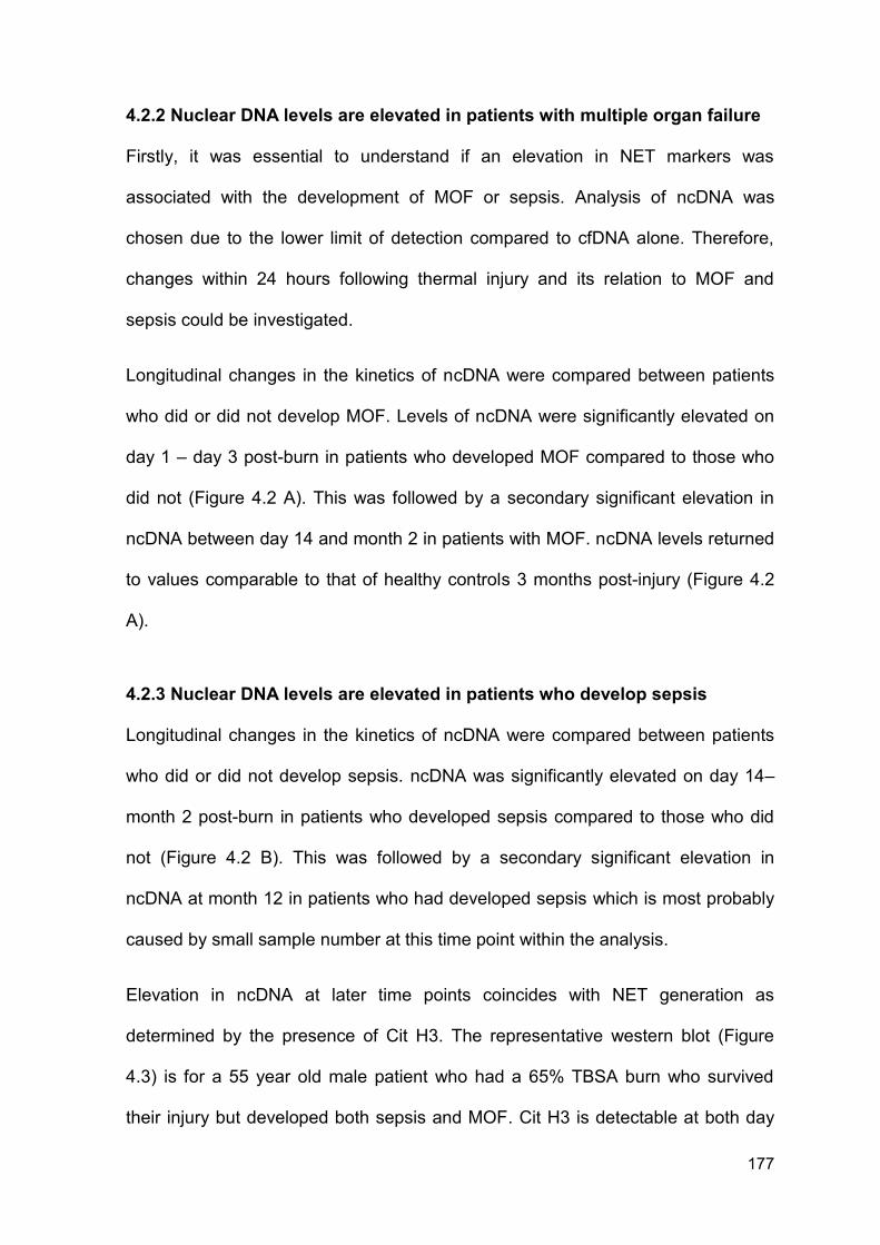
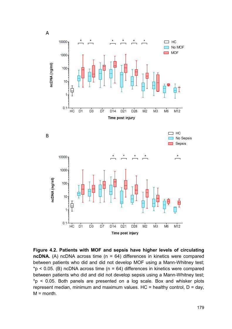
PRODUCTION AND IMPAIRED REGULATION OF NEUTROPHIL EXTRACELLULAR TRAPS FOLLOWING SEVERE THERMAL INJURY, IMPLICATIONS FOR SEPSIS AND MULTIPLE ORGAN FAILURE Robert Johnathon Dinsdale A thesis submitted to the University of Birmingham for the degree of DOCTOR OF PHILOSOPHY Supervisors Dr Paul Harrison Professor Steve Watson Institute of Inflammation and Ageing College of Medical and Dental Sciences University of Birmingham August 2017
Transcript
PRODUCTION AND IMPAIRED REGULATION
OF NEUTROPHIL EXTRACELLULAR TRAPS
FOLLOWING SEVERE THERMAL INJURY,
IMPLICATIONS FOR SEPSIS AND MULTIPLE
ORGAN FAILURE
Robert Johnathon Dinsdale
A thesis submitted to the University of Birmingham for the
degree of
DOCTOR OF PHILOSOPHY
Supervisors
Dr Paul Harrison
Professor Steve Watson
Institute of Inflammation and Ageing
College of Medical and Dental Sciences
University of Birmingham
August 2017
University of Birmingham Research Archive
e-theses repository This unpublished thesis/dissertation is copyright of the author and/or third parties. The intellectual property rights of the author or third parties in respect of this work are as defined by The Copyright Designs and Patents Act 1988 or as modified by any successor legislation. Any use made of information contained in this thesis/dissertation must be in accordance with that legislation and must be properly acknowledged. Further distribution or reproduction in any format is prohibited without the permission of the copyright holder.
Abstract
Advancements in burn care have improved immediate outcome, however, the
prevalence of sepsis and multiple organ failure (MOF) remain significant. Although
well characterised the mechanisms responsible for the pathogenesis of MOF and
increased propensity to infection are poorly understood. Neutrophil extracellular
traps (NETs) provide protection against invading pathogens but also contribute to
thrombosis.
Sepsis is required for NET generation following severe thermal injury.
Quantification of circulating NET biomarkers shows good discriminatory power for
diagnosis of sepsis. Interestingly, neutrophils isolated from 24 patients with severe
thermal injuries, ≥ 15% total body surface area, had a significantly reduced ability
to form NETs ex vivo, potentially mediated by phenotypical changes of neutrophils
and inhibitory effects of formyl peptides.
This thesis identified a major biological mechanism driving MOF after severe
thermal injury, namely the compromise to the actin scavenging system which
leads to reduced DNAse activity and a build-up of circulating DNA. Preliminary
analysis suggests that DNAse activity can be restored by prehospital use of fresh
frozen plasma following major trauma. Thus, administration of blood products or
manipulation of the actin scavenging system is a potential therapeutic target.
This thesis has identified a number of novel mechanisms responsible for the
regulation of NETs following severe thermal injuries and their implications for
sepsis and MOF.
Acknowledgements
Firstly, I must thank my supervisor Dr Paul Harrison for all support,
encouragement and opportunities provided. I will be forever grateful for his
supervision and friendship (even if he is a Blue).
I am grateful to Professor Steve Watson, Professor Janet Lord and Professor
Naiem Moiemen for their advice, discussion and mentorship. I would like to thank
all of the Lord Group members for making my time within the lab enjoyable. A
special thank you must go to Dr Jon Hazeldine and Dr Peter Hampson who have
been amazing colleagues and good friends. Although at times we disagree on
everything from food to physical activity, I am truly grateful for advice, jokes and
weekends smashing golf balls into the rough.
I would like to thank my funders the Scar Free Foundation, burns clinical team and
burns nurses at the Queen Elizabeth Hospital and all collaborating sites for their
contribution to this work. Special thank you must go to Amy Bamford and Dr
Khaled Altarrah for their help throughout my studies. Most importantly, thank you
to all patients and families who have been part of this work. Without you this could
never have happened.
To my friends, The Booze Hound Gang, Trauma Boyz and The Marangas, thank
you for always being there, the scrambled eggs, nights out in fancy dress and
unconditional friendship you have all shown me. A special thank you must be
extended to Georgia Walton who is responsible for my love of junk food and late
nights. You have always been and continue to be a source of laughter,
companionship and unwavering support. Here is to kicking Louise out of her seat.
Thank you to my greatest teacher the black dog, your intermittent barking has
fuelled my determination and passion.
I would like to thank my whole family for always being there for me and supporting
me throughout this whole process. Dad and lovely mummy, thank you for all of the
support, love and advice throughout everything. Thank you for everything you
have ever given me from the countless opportunities to the reassurance on top of
the landing. I hope this work makes you proud. Grace, Maggie and Elsie, thank
you for your companionship. To the members of my family who will never be able
to read this, I hope it would have made you proud.
Thank you.
You’ll never walk alone
Disclosures
All work presented in this thesis was performed as part of The Scientific
Investigation of the Biological Pathways Following Thermal Injury in Adults and
Children (SIFTI Study) and is therefore part of a collaboration between a number
of investigators. Where applicable, credit has been given to persons responsible
for data generation or collaboration. Importantly, all data presented in this thesis
was analysed and presented by Robert J Dinsdale.
All patients recruited within Camp Bastion, Afghanistan, were recruited by
Professor Midwinter, Dr Kirkman, Professor Woolley, Dr Watts and Dr Dalle
Lucca. Again, all laboratory experiments and data analysis was performed by
Robert J Dinsdale.
Publications arising from this thesis
1. Dinsdale RJ, Devi A, Hampson P, Wearn CM, Bamford AL, Hazeldine J,
Bishop J, Ahmed S, Watson C, Lord JM, Moiemen N, Harrison P. Changes in
novel haematological parameters following thermal injury. A prospective
minimum and maximum values. Pooled serum from 9 healthy volunteers was used
to calibrate 100% degradation.
193
4.2.9 Thermal injury results in a decrease in circulating vitamin d binding
protein and gelsolin levels
Levels of VDBP were quantified by Dr Khaled Altarrah (University of Birmingham,
UK) as part of the SIFTI Trial. All data was analysed by Robert J Dinsdale.
The actin scavenging system is controlled by two proteins; VDBP and GSN.
Levels of VDBP were quantified in patients with burns ≥ 20% TBSA (n = 50) to
establish the effect thermal injury has on the actin scavenging system.
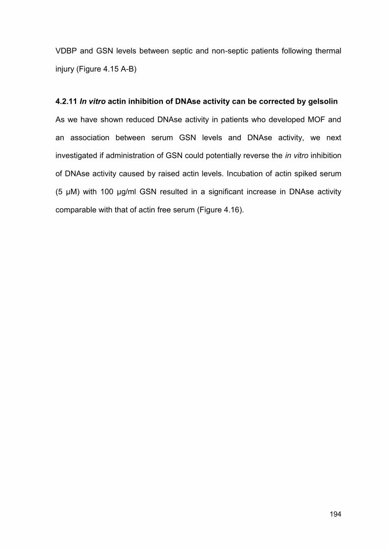
Thermal injury resulted in a rapid and significant reduction in VDBP from day 1 -
day 3 post injury compared to levels in healthy controls (Figure 4.13 A). There was
a significant increase in VDBP levels at month 3 post thermal injury relative to
healthy individuals (Figure 4.13 A).
Levels of VDBP weakly correlated with DNAse activity across all time points (r =
0.15, p = 0.013). However, there was no significant difference in VDBP kinetics
between patients with and without MOF (Figure 4.13 B).
4.2.10 Thermal injury results in a decrease in circulating gelsolin levels
Levels of GSN were quantified to fully investigate the effect severe thermal injury
has on the actin scavenging system. Levels of GSN were measured in all 64
patients.
Thermal injury resulted in a significant reduction in GSN from day 1 - day 14 post
injury (n = 64) compared to levels in healthy control (Figure 4.14 A). Additionally,
kinetics of GSN was comparable between patients with and without MOF (Figure
4.14 B). Levels of GSN also weakly correlated with DNAse activity across all time
points (r = 0.1331, p = 0.0058). There were no significant differences in circulating
194
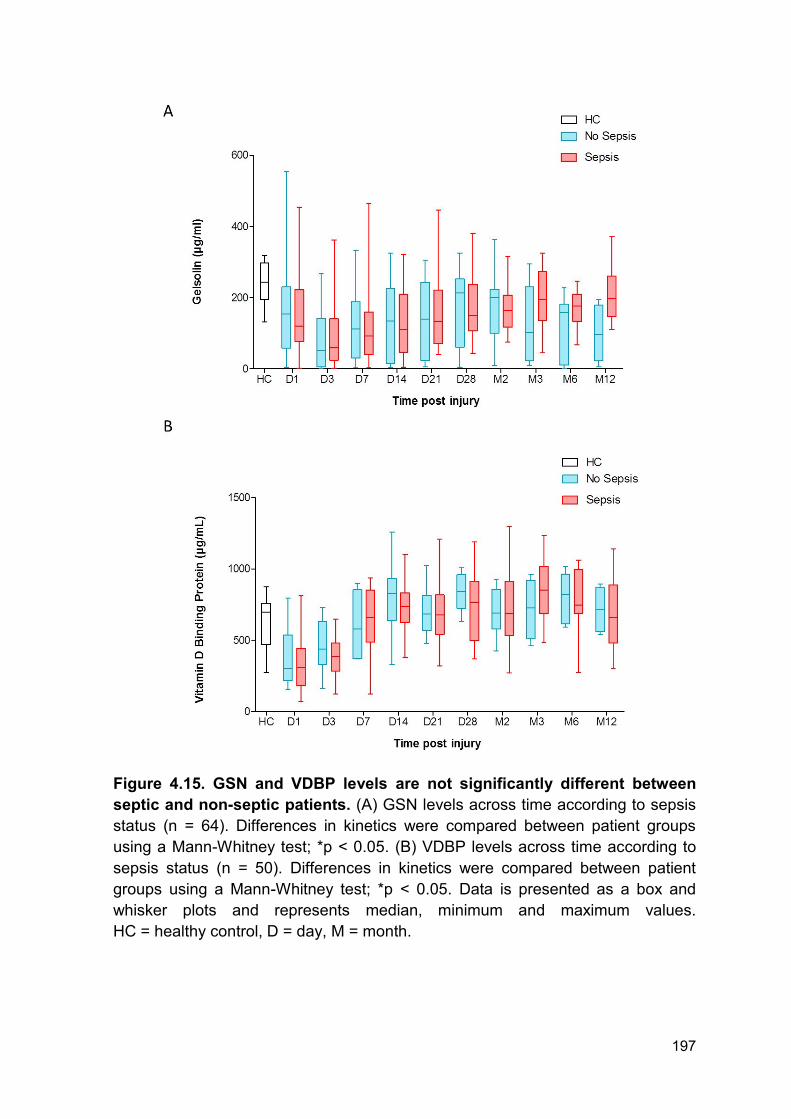
VDBP and GSN levels between septic and non-septic patients following thermal
injury (Figure 4.15 A-B)
4.2.11 In vitro actin inhibition of DNAse activity can be corrected by gelsolin
As we have shown reduced DNAse activity in patients who developed MOF and
an association between serum GSN levels and DNAse activity, we next
investigated if administration of GSN could potentially reverse the in vitro inhibition
of DNAse activity caused by raised actin levels. Incubation of actin spiked serum
(5 µM) with 100 µg/ml GSN resulted in a significant increase in DNAse activity
comparable with that of actin free serum (Figure 4.16).
195
Figure 4.13. Levels of VDBP are reduced following thermal injury. (A) VDBP
across time (n = 50). Differences in kinetics were compared to data from 10
healthy volunteers using a Mann-Whitney test; *p < 0.005. (B) VDBP across time
(n = 50) between patients with (n = 18) and without MOF (n = 32) using a Mann-
Whitney test; *p < 0.05. Data is presented as a box and whisker plots and
represents median, minimum and maximum values. HC = healthy control,
D = day, M = month.
196
Figure 4.14. Levels of GSN are reduced following thermal injury. (A) GSN
levels across time (n = 64) compared to healthy controls (n = 10). Data was
analysed by Mann-Whitney test compared to healthy controls *p < 0.005. (B) GSN
levels across time comparing those who did (n = 24) and those who did not
(n = 40) develop MOF. Differences in kinetics were compared between patient
groups using a Mann-Whitney test; *p < 0.05. Data is presented as a box and
whisker plots and represents median, minimum and maximum values. HC =
healthy control, D = day, M = month.
197
Figure 4.15. GSN and VDBP levels are not significantly different between
septic and non-septic patients. (A) GSN levels across time according to sepsis
status (n = 64). Differences in kinetics were compared between patient groups
using a Mann-Whitney test; *p < 0.05. (B) VDBP levels across time according to
sepsis status (n = 50). Differences in kinetics were compared between patient
groups using a Mann-Whitney test; *p < 0.05. Data is presented as a box and
whisker plots and represents median, minimum and maximum values.
HC = healthy control, D = day, M = month.
198
Figure 4.16. GSN recovers actin inhibition of DNAse activity in vitro. DNAse
activity following incubation of actin spiked serum with vehicle control or 100 µg/ml
GSN (n = 8). Data was analysed using a paired t test (*p < 0.05). Data is
presented as before and after to display changes within independent experiments.
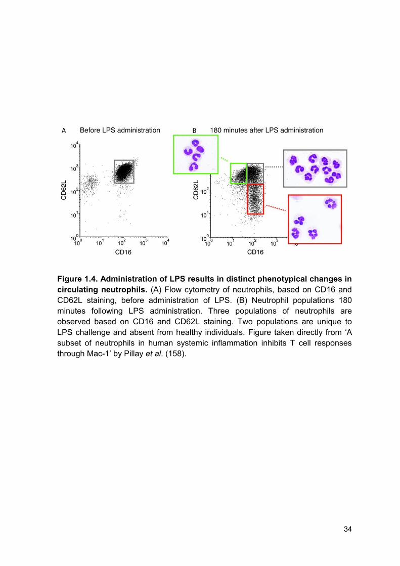
199
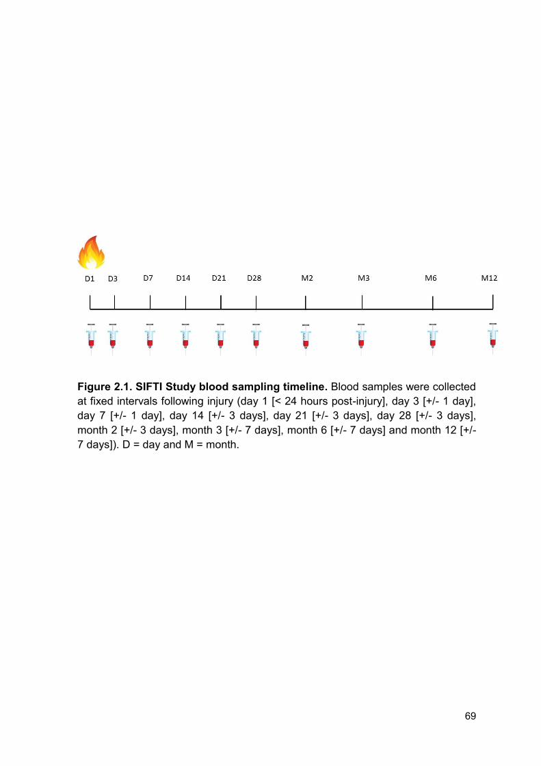
4.2.12 Patient demographics for patients with severe injuries caused by
explosions
To investigate if blood products, namely fresh frozen plasma (FFP), can potentially
increase DNAse activity by correcting the blood based actin scavenging system,
levels of GSN, VDBP and DNAse activity were quantified in plasma from patients
following severe traumatic injury caused by explosions. This cohort was split into
patients who had (n = 6) or had not received FFP (n = 6) prior to hospital
admission and blood sampling. There was no significant difference in ISS, NISS
and time to admission following injury between the 2 groups. Full patient
demographics can be found in Table 4.2. On average patients received 3 units of
blood products before admission to hospital. Due to the nature of this work and
the cohort it was not possible to obtain clinical data on outcomes or secondary
complications.
4.2.13 Fresh frozen plasma increases gelsolin levels and DNAse activity
following severe injury caused by explosion but has no effect on vitamin d
binding protein levels
All patients in this analysis were matched for clinical scores of severity and
admission times. In the total cohort (n = 12), DNAse activity and VDBP levels were
comparable between healthy controls and patients with severe injuries caused by
explosions (Figure 4.17 A, C). However, GSN levels were significantly lower in
patients with severe injuries caused by explosions when compared to healthy
individuals (Figure 4.17 B).
Patients who did not receive blood products before hospital admission had
significantly lower DNAse activity and plasma GSN levels compared to healthy
controls (Figure 4.18 A-B). However, there was no difference when comparing
200
plasma VDBP levels (Figure 4.18 C). DNAse activity, GSN and VDBP were
comparable between patients who received blood products and healthy controls
(Figure 4.18 A-C).
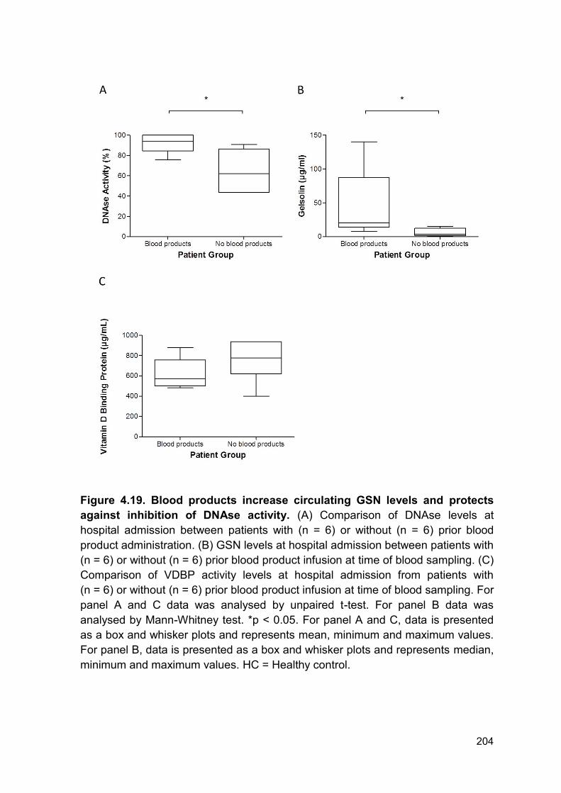
Administration of blood products and FFP before admission to hospital
significantly increased DNAse activity compared to patients who had not received
blood products upon admission to hospital (Figure 4.19 A). Furthermore,
administration of blood products and FFP before admission to hospital significantly
increased circulating GSN levels compared to patients who had not received blood
products and FFP (Figure 4.19 B). Finally, administration of blood products and
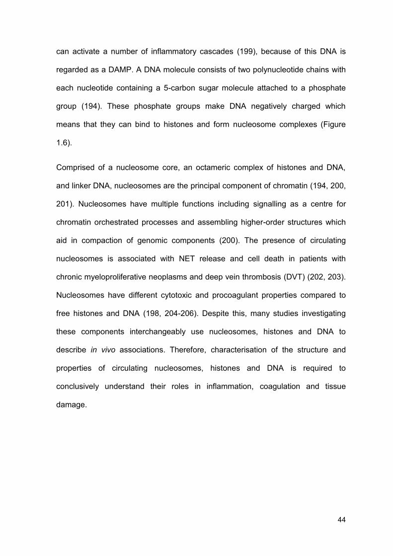
FFP did not affect circulating levels of VDBP in both patient groups (Figure 4.19
C).
201
Characteristic No FFP pre admission (n = 6)
FFP pre admission (n = 6)
p
ISS (min-max) 27 (17-59) 22 (16-42) ns
NISS (min-max) 36 (18-75) 35 (16-66) ns
Minutes to admission (min-max) 75 (30-135) 83 (50-130) ns
Table 4.2. Patient demographics for patients with severe injuries caused by
explosions (Chapter 4). FFP post or pre admission variables were analysed by
Mann-Whitney (continuous variables) or Chi-squared test (categorical variables).
202
Figure 4.17. Severe injury caused by explosion caused a significant
reduction in circulating GSN levels compared to healthy controls. (A) Plasma
DNAse activity in healthy individuals (n = 10) and patients with severe injuries
caused by explosions (n = 12). (B) Circulating plasma GSN levels in healthy
individuals (n = 10) and patients with severe injuries caused by explosions
(n = 12). (C) Circulating plasma VDBP levels in healthy individuals (n = 10) and
patients with severe injuries caused by explosions (n = 12). All data was
compared by Mann-Whitney test *p < 0.05. Data is presented as a box and
whisker plots and represents median, minimum and maximum values.
HC = Healthy control.
203
Figure 4.18. Patients who do not receive blood products before admission to
hospital have significantly lower DNAse and GSN levels compared to
healthy individuals. (A) Plasma DNAse activity in healthy individuals (n = 10) and
patients who did and did not receive blood products before admission (n = 6). (B)
Circulating plasma GSN levels in healthy individuals (n = 10) and patients who did
and did not receive blood products before admission (n = 6). (C) Circulating
plasma VDBP levels in healthy individuals (n = 10) and patients who did and did
not receive blood products before admission (n = 6). All data was compared by
one way ANOVA and Dunn's multiple comparison test *p < 0.05. Data is
presented as a box and whisker plots and represents median, minimum and
maximum values. HC = Healthy control.
204
Figure 4.19. Blood products increase circulating GSN levels and protects
against inhibition of DNAse activity. (A) Comparison of DNAse levels at
hospital admission between patients with (n = 6) or without (n = 6) prior blood
product administration. (B) GSN levels at hospital admission between patients with
(n = 6) or without (n = 6) prior blood product infusion at time of blood sampling. (C)
Comparison of VDBP activity levels at hospital admission from patients with
(n = 6) or without (n = 6) prior blood product infusion at time of blood sampling. For
panel A and C data was analysed by unpaired t-test. For panel B data was
analysed by Mann-Whitney test. *p < 0.05. For panel A and C, data is presented
as a box and whisker plots and represents mean, minimum and maximum values.
For panel B, data is presented as a box and whisker plots and represents median,
minimum and maximum values. HC = Healthy control.
205
4.3 Discussion
Circulating levels of cfDNA have been described as a novel biomarker of
secondary complications and mortality with studies reporting positive prognostic
and diagnostic utility (109, 180, 220, 304-306). In a cohort of 67 patients with
severe sepsis, quantification of ncDNA showed positive prognostic utility for
predicting 24 hours mortality in patients who presented to the emergency
department (354).
Circulating DNA can arise from a number of sources including tissue damage,
apoptosis, necrosis and NETosis (114). In our analysis levels of ncDNA within 24
hours of injury correlated with measurements of burn size and severity. Therefore,
we propose that the initial elevation in ncDNA is originating from tissue damage
caused by the burn injury. Due to the cytotoxic nature of DNA (355, 356), the initial
elevation in circulating ncDNA may be contributing to the immediate host tissue
and organ damage which occurs in patients with burn injuries. Consistent with
previous literature (354), ncDNA levels are significantly higher in septic patients
compared to non-septic individuals. We propose that tissue damage, surgery and
NETosis may also be contributing to this secondary increase. Interestingly, levels
of ncDNA are significantly higher at 12 months following injury in patients who
developed sepsis compared to those who didn’t. This elevation is either caused by
lower sample numbers in the analysis for 12 month samples, a reactive response
by the body in which DNA cannot be cleared or further release from tissue
remodelling and scar formation.
NETs and histones form key components of host defence and innate immunity
(118, 355, 357). Engelmann and Massberg described an innate immune response
206
in which coagulation is activated in an attempt to ensnare, recognise and remove
bacteria (296, 358, 359). This process is termed immunothrombosis and is in part
mediated by the production of NETs (296). NETs are capable of providing both a
stimulus to and scaffold for thrombus formation. Uncontrolled or excessive NET
generation and immunothrombosis can result in increased risk of thrombosis, host
tissue damage and the accumulation of pathogenic components, including DNA,
in the microvasculature (226).
Fuchs and colleagues demonstrated that NETs perfused with blood caused
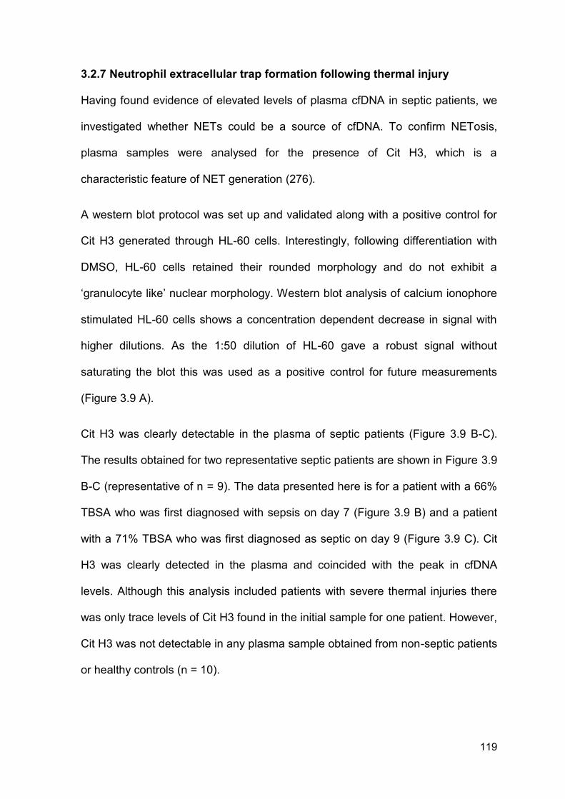
recruitment of red cells, increased fibrin deposition, platelet adhesion and
activation. Importantly, this could be prevented by incubation with DNAse which
promotes NET breakdown (232). In a murine model of DVT, Cit H3 was found in
close proximity to thrombi. Furthermore, DNA and histones form components of
the scaffold required for the pathogenesis of DVT (233). In addition to promoting
thrombosis, elevated levels of NET components are cytotoxic and can result in
host tissue damage. For example, incubation of activated endothelial cells with
NETs results in cell damage which, again, can be prevented by degrading NETs
with DNAse (360).
Here, we report that ncDNA was elevated from day 1 to day 3 and again at day 14
to 2 months post-injury in patients who developed MOF compared to those who
did not. Here we report an accumulation of circulating ncDNA for up to 28 days
following injury. We hypothesised that clearance of host and NET derived DNA
was dysregulated. Multiple groups have shown that degradation of NETs with
DNAse can protect against tissue damage, thrombosis and procoagulant
interactions (232, 360). Furthermore, elevated levels of circulating DNA are also
associated with and contribute to the progression of autoimmune diseases,
207
notably SLE (237, 238). This build-up of circulating DNA can be explained in part
to the impaired DNAse activity caused by the presence of ‘DNAse inhibitors’ or
inhibitory antibodies in these patients (237).
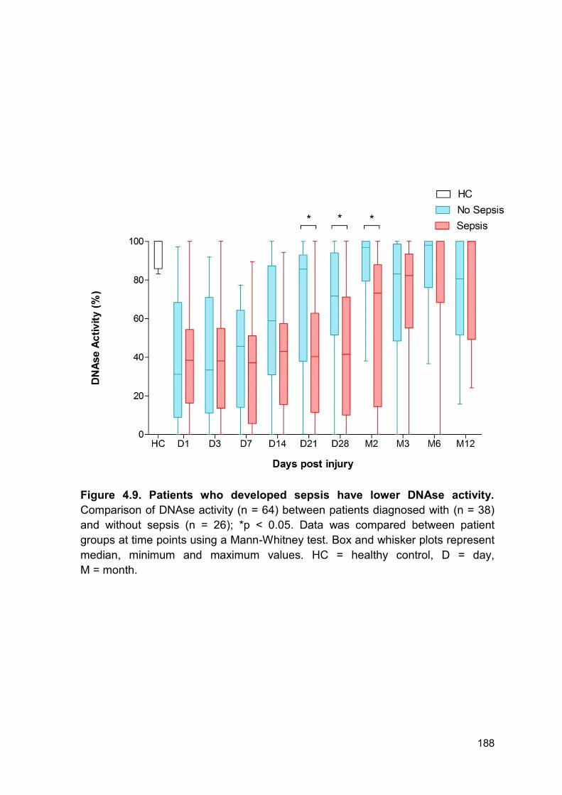
As we report a build-up of circulating DNA and reduced ex vivo NET formation we
next investigated DNAse activity in patient samples. Here we report a reduction in
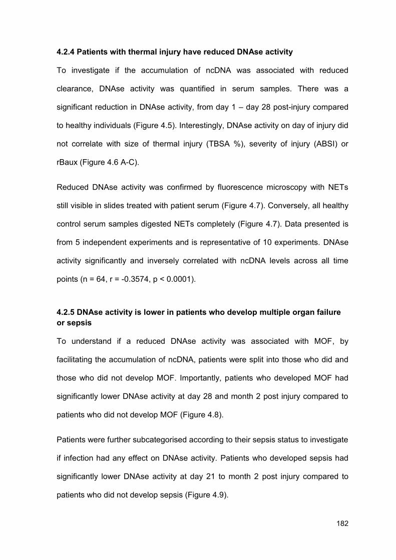
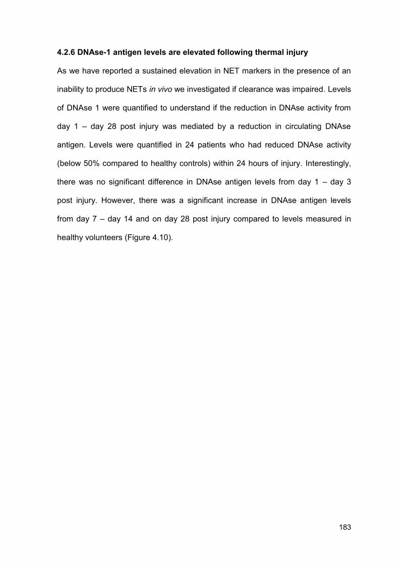
DNAse activity from day 1 – day 28 post injury in the total cohort. Of note, there
was large variation in DNAse activity within the whole cohort at all time points.
Furthermore, levels of DNAse activity did not correlate with injury severity. This
may be explained by timing of sample, clinical intervention or heterogeneity within
the patient cohort. Importantly, DNAse activity was lower in patients who
developed MOF or sepsis compared to those who did not develop either. Both
groups had comparable reduction in DNAse activity for the first 14 days following
thermal injury. Therefore, one might hypothesise that the initial injury causes the
immediate reduction in activity reported in both groups and the later reduction may
be caused by further tissue damage or surgical procedures. Within this cohort the
average amount of individual septic episodes was 2.5 episodes per patient. The
average time to first episode was 5 days following injury (range 3 – 70 days) and
the time to last episode was 23 days following injury (range 3 – 130 days).
Furthermore, whilst most patients underwent surgery within the first 5 days for
debridement and immediate treatment of burn wounds, patients did have further
surgical procedures if required, e.g. skin graft rejection. Thus, the variation within
DNAse activity at the later time points following thermal injury may be explained by
the ongoing tissue damage from surgical procedures and sporadic infectious
episodes.
208
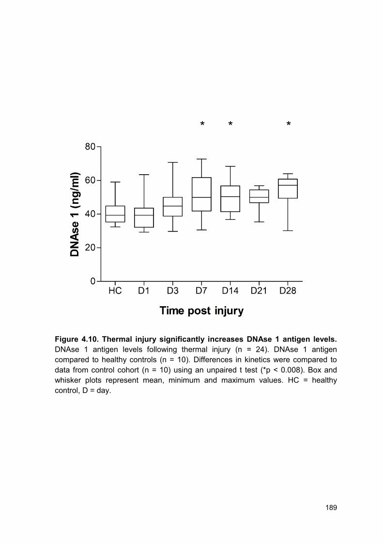
It is important to consider the mechanism by which DNAse activity inhibition
occurs. Here, we report that levels of total DNAse antigen are increased post
thermal injury, which is consistent with existing literature (239). Therefore, the
apparent reduction in DNAse activity is indicative of inhibition of enzymatic activity.
In this thesis, only levels of DNAse 1 antigen were quantified as this is the
predominant enzyme responsible for the degradation of circulating DNA. However,
there are different isoforms of DNAse, including DNAse-γ, therefore we cannot
exclude the possibility that the reduction in DNAse activity may be mediated in
part by reduced DNAse-γ antigen levels. Thus, further study of DNAse isoforms
and their contribution to reduced DNAse activity following injury is required.
A naturally occurring inhibitor of DNAse activity is actin, which is an abundant
protein in mammalian cells (240). Actin exists in a balance between monomeric
and filamentous actin, which is essential for cellular function (241, 242). However,
this predisposition of actin to rapidly polymerise is extremely dangerous if it occurs
in the circulation. Actin is recognised as a DAMP due to its rapid release,
immunostimulatory actions and conserved structure (361). Polymerised actin, F-
actin, binds to the DNGR-1 receptor (CLEC9A) and primes cytotoxic T-cells
against dead cell antigens (362-364). In addition to its DAMP properties,
extracellular actin can cause direct damage to the microvasculature, impair clot
lysis and activate platelets (240, 243, 244).
Actin can bind to DNAse forming a stoichiometric 1:1 complex which inhibits
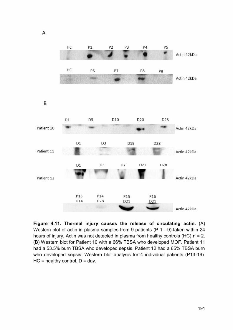
enzymatic activity (245, 365). In this analysis circulating actin is detected in
patients immediately following and for up to 28 days post-injury. The initial release
of actin most likely originates from the extensive tissue damage occurring
following severe thermal injury. This will not only inhibit DNAse activity but may
209
also be responsible for the immediate stimulation of the immune system and host
tissue damage that occurs following thermal injury due to its DAMP properties and
cytotoxic nature (240, 243, 244, 361-364).
Here we show that actin is detectable for up to 28 days post injury. In Patient 10
(Figure 4.11 B) actin is cleared at day 10 and is then detectable at day 20 - day
23. Therefore, this secondary appearance of actin is not from the initial tissue
damage and may be caused by further host tissue damage during surgery,
infection or MOF. Patient 10 has been chosen as a representative of this study
due to quality of blot. However, this appearance of actin at the later time points is
present in multiple patients. This is consistent with previous literature reporting
elevated levels of actin in septic individuals (246). This prolonged appearance of
actin may be in part responsible for the prolonged inhibition of DNAse activity and
may also be responsible for host tissue damage and a pro-thrombotic phenotype
associated with actin toxicity. One limitation of these data is the inability to quantify
circulating actin by an ELISA based method. Thus, within this analysis we make
no attempt to relate circulating levels of actin with DNAse activity, nor do we
suggest there is an increase in circulating levels at different time points tested.
Further studies are warranted to investigate the relationship between exact actin
concentrations and secondary complications in order to study the dynamic kinetic
changes and interactions. Furthermore, performing western blot analysis on
plasma is extremely difficult and challenging due to the increased concentration of
high molecular weight proteins which distort gels and cause high background
signal. This has affected the quality of western blot produced and prevents further
analysis of blots by densitometry. Therefore, novel and more accurate
210
methodologies are required to quantify levels of actin in blood products from
patients.
Many of the detrimental effects of circulating levels of actin are attributed to
saturation of the actin scavenging system. Control of the actin scavenging system
is mediated by two key proteins; GSN and VDBP (240). GSN is normally found in
high quantities in healthy individuals and can bind to both monomeric and
filamentous actin (246, 247). GSN functions to clear circulating actin via 2 distinct
processes; preventing further polymerisation or severing existing filamentous actin
(240, 249-251). It is therefore not surprising that studies have reported a decrease
in circulating levels of GSN in a number of disease pathologies connected with
actin release (252-259). Of note, Huang et al reported in 95 patients with thermal
injuries that plasma GSN levels were reduced and associated with mortality,
development of sepsis and MODS (254). In a rat burn model, plasma GSN levels
decreased within 12 hours to 6 days post-injury. This was accompanied by
increased pulmonary microvasculature permeability, which was corrected by
administration of recombinant plasma GSN prior to burn injury (261). Moreover,
administration of GSN can also reverse actin inhibition of lung macrophage
binding and uptake of bacteria (260). As GSN functions may extend beyond
simply actin scavenging, GSN levels may not only serve as a biomarker of poor
outcomes but could also be a potential therapeutic target to reduce or prevent
secondary complications.
VDBP is an abundant circulating protein that is a key component of vitamin D
transport (262, 263). Unlike GSN, VDBP binds to monomeric actin only (264, 265).
Binding of VDBP to actin prevents further polymerisation by rapidly clearing
residual monomeric actin from the circulation (266, 267). Levels of VDBP have
211
been suggested as good prognostic markers of outcome and organ damage
following severe trauma (268-271).
Following severe thermal injury there is an immediate and transient decrease in
both VDBP, up to 3 days post injury, and GSN, up to 14 days post injury,
compared to levels quantified in healthy volunteers. Thus, severe thermal injury
results in dysregulation of the actin scavenging system which will facilitate the
accumulation of monomeric and polymerised circulating actin which can then
cause host damage and inhibit DNAse activity (240, 243, 244, 361-364). Of note,
patients with thermal injuries have significantly higher levels of VDBP at 3 months
following injury compared to healthy individuals. This overproduction is consistent
with previous publications (271) and suggests a reactive response mediated
potentially by the injury, recovery, medications or diet.
The decrease in both VDBP and GSN may be caused by a number of
mechanisms which include; loss of protein through endothelium dysfunction,
dilution during fluid resuscitation or saturation through elevated levels of actin.
Severe thermal injury results in systemic endothelial dysfunction and capillary leak
(366). Like albumin, it is possible that VDBP and GSN may be lost by this
mechanism. However, it is reported that endothelial dysfunction and capillary leak
are only present for up to 5 hours post-injury (367). Therefore, this would not
explain the decrease seen at 48 hours post-injury in this analysis as capillary leak
and endothelial dysfunction will have been corrected. It is also possible that fluid
resuscitation may cause the immediate decrease in both GSN and VDBP. All
patients received a standardised burn resuscitation protocol as per the Parkland
Formula thus this would not fully explain the difference in the later kinetics of GSN
and VDBP. Of note, the VDBP ELISA does not bind to VDBP complexed with
212
actin. Hence, this may explain the initial reduction in VDBP reported within this
thesis.
In addition to their mechanistic functions, GSN and VDBP have been proposed as
biomarkers of poor outcomes following trauma and burns (254, 268-271). In our
cohort, there are no differences in VDBP and GSN levels or kinetics between
patients who did and did not develop MOF or sepsis. This is surprising and
contradictory to existing literature; however, there are a number of potential
explanations. The significant reduction in VDBP and GSN occurred before the
onset of sepsis and MOF in this cohort. Therefore, the decrease may solely be
caused by immediate actin release following injury due to the severe nature of the
injuries included within this analysis. Furthermore, differences may not have been
seen due to the severity of injuries included within this analysis, low numbers of
patients or differences in timing of sample between studies.
From this analysis, we have developed a hypothesis model depicting how patients
are predisposed to the pathogenesis of MOF following severe thermal injury
(Figure 4.20). We hypothesise that following severe thermal injury there is the
initial release of both monomeric and polymeric actin which results in the rapid
consumption of circulating GSN and VDBP, potentially due to extensive tissue
damage and actin release. This reduction facilitates the rapid accumulation of
circulating actin and binding to DNAse and inhibition of enzymatic activity. All
patients are then predisposed to the development of MOF following severe
thermal injury but that a second stimulus is required to elevate DNA and reveal
this susceptibility. This could arise through repeated surgery with resultant tissue
damage or through NETosis in septic patients. This is supported by the growing
amount of evidence reporting the involvement of NETs in immunothrombosis (273,
213
368). This phenotype will facilitate the build-up of circulating DNA and NET
components which further the pathogenesis of MOF by promoting impaired
fibrinolysis, thrombotic complications, tissue damage and occlusion of the
microvasculature (195, 224, 235, 236). This hypothesis model provides a novel
mechanistic link between the initial traumatic injury and subsequent infection in
potentially mediating the pathogenesis of MOF.
214
Figure 4.20. Hypothesis model: The link between severe thermal injury and
pathogenesis of MOF through disruption of the actin scavenging system.
Following severe thermal injury polymerised and monomeric actin is released
which immediately reduces VDBP and GSN levels. The polymerised actin can
then bind to DNAse and inhibit its activity. Following infection, NETs are released
and accumulate due to reduced degradation. This increases the pathogenesis of
MOF.
215
In 2011, Cohen and colleagues demonstrated the therapeutic potential of GSN in
a rat model of sepsis caused by double puncture of the cecum. Administration of 1
mg/ml recombinant human GSN, only once, resulted in significantly improved
survival and reduced tissue damage compared to sham animals (369). The group
concluded that administration of GSN is a potential therapy to reduce the severity
of illness caused by infection and sepsis. They propose that GSN may have an
effect on expression and regulation of pro-inflammatory cytokines. However, we
would suggest that GSN is having a multifaceted role by regulating cytokine
release (369), inflammatory cell function (260) and readdressing the overwhelmed
actin scavenging system. We show that administration of GSN in vitro can restore
DNAse activity in actin spiked serum, providing further evidence that GSN is a
potential therapy. However, this experiment is performed in vitro with no
contribution from VDBP or any other cellular interactions. Therefore, further
experiments are required to investigate the potential of GSN and VDBP combined
in vivo.
To investigate this possibility we performed a preliminary retrospective analysis in
samples from military patients following severe injury caused by explosions
receiving FFP prior to first blood sampling and hospital admission. Severe trauma
caused by explosions is becoming an increasingly common form of injury due to
conflicts and terrorism. Injuries can be classified into primary to quaternary injuries
depending upon severity and mechanism of blast injury (370). Burn injuries are
also common among patients with injuries caused by explosions and therefore
clinical treatment is comparable.
Patients with severe injury caused by explosions who are hemodynamically
unstable receive packed red blood cells and FFP in a 1:1 ratio immediately
216
following their injuries. By definition, FFP contains high levels of GSN and VDBP
which may explain some of the known therapeutic potential in the context of
traumatic injury by boosting the depleted levels of the actin scavenging proteins
(371). In our preliminary analysis, we have split our cohort into two groups who did
or did not receive blood products before admission to hospital. The decision to
give blood products was based upon resources available during transportation
from the battlefield and not injury severity or mechanism of injury. Importantly,
subgroups within this analysis are matched for ISS, NISS and time to admission
following injury. Theoretically, the only difference between the two groups is the
infusion of blood products.
Here we provide preliminary data showing that early administration of blood
products significantly increases GSN levels immediately post severe trauma
caused by explosion. This increase in GSN was also accompanied by a significant
increase in DNAse activity. However, VDBP levels were not affected. This data
would suggest that an early increase in circulating GSN is able to improve DNAse
activity rapidly following injury independently of VDBP. In 2005, Chhabra et al
showed that the N-terminal fragment of GSN could bind to and disrupt actin-
DNAse complexes, in turn, restoring enzymatic activity (372). Therefore, this may
be a potential mechanism by which GSN can rapidly restore or enhance DNAse
activity independently of VDBP. However, it is important to note that blood
products will contain many other soluble factors which may enhance DNAse
activity, including DNAse 1 itself. Therefore, the restoration of DNAse activity may
not be solely attributed to the increases in GSN. Thus, further studies are required
to investigate all components within blood products to fully understand if they are
contributing any therapeutic benefit.
217
Although not common, patients recruited within our primary analysis did receive
blood products but only when clinically required. In total 21 patients received at
least 1 unit (220 ml) FFP following injury which may be an important confounder in
this study. This may also contribute to the variation and lack of difference between
patients who did and did not develop MOF. The median time to first unit received
was 3 days post injury (range 1 – 57 days post injury) with 4 patients received FFP
during the first 24 hours post injury. However, as not all patients received FFP,
coupled with the sporadic nature and lack of sustained administration we cannot
study any differences between patients who did and did not receive FFP following
thermal injury.
These data provide preliminary evidence that early administration of FFP to
patients with severe injuries may offer a simple way of boosting the depletion of
actin scavenging system and therefore improve DNAse activity. Larger studies are
therefore required to fully investigate the therapeutic potential of both FFP and
GSN alone post major trauma and severe thermal injury. Given the extensive
literature and ongoing debate on the utility of FFP in trauma and resuscitation it
may be more applicable to utilise GSN in isolation to scavenge excess actin (373-
375). GSN is unlikely to be immunogenic due to the high concentrations found in
healthy individuals. Therefore, we propose that administration of FFP (containing
GSN) or GSN alone immediately to patients with severe thermal injuries may be a
potential safe therapeutic which addresses dysregulation of the actin scavenging
system. In turn, reducing the accumulation of DNA and NET components and
potentially protecting against the pathogenesis of MOF.
In summary, this chapter presents data of several novel findings and a model of
post-injury complications in which DNAse activity is reduced following thermal
218
injury, driven most likely by raised circulating actin and acute reductions in the
actin scavengers GSN and VDBP. The reduced DNAse activity may be
contributing to the pathogenesis of MOF by mediating the sustained elevation of
circulating DNA from the injury and secondary complications. In addition, two
potential therapeutic agents, FFP and GSN, been identified which have the
potential to restore DNAse activity and thus protect against host tissue damage
associated with MOF.
219
Chapter 5
General Discussion
220
General Discussion
5.1 Limitations
Despite showing for the first time the association between and potential
mechanisms responsible for dysregulated NET release following severe thermal
injury, there are a number of limitations which must be considered.
This study was designed to be exploratory and hypothesis generating in nature
and not confirmatory. Hence we cannot estimate a specific outcome or obtain a
pre-determined level of precision from our results. Data generated should be
regarded as a pilot to obtain data to inform a potential larger confirmatory study.
Furthermore, in this thesis, clinical samples were obtained and analysed at fixed
time points. Whilst this remains a strength of the current study it limits the ability to
study the full kinetics of many in vivo markers and their relation to secondary
complications or clinical treatments. A second study has been established in which
blood samples are taken daily over the first 14 days which will allow us to
investigate and characterise the daily kinetics of neutrophil function, NET release
and diagnostic utility of described biomarkers.
Whilst we have shown the protective role of immediate administration of blood
products in patients with severe injuries caused by explosions it remains extremely
preliminary data in a small population of patients. Due to the nature of injuries and
sensitivity of data it has not been possible to correlate increases in DNAse activity
and GSN with improved outcome in this patient cohort. In addition, in this analysis
only GSN, VDBP and DNAse activity have been quantified and investigated.
Blood products contain many soluble mediators, including DNAse, and therefore
221
the protection against reduced DNAse activity cannot be solely attributed to
increases in GSN levels. Therefore, further characterisation of blood products or
the use of GSN and/or VDBP in isolation is required.
5.2 Future Work
Severe thermal injuries constitute a major form of traumatic injury (1).
Advancements in the initial care of patients with burns have dramatically improved
immediate survival (18, 19, 31). However, delayed mortality associated with sepsis
and MOF remain a significant health care problem (38, 47). Although sepsis and
MOF are well characterised the mechanisms mediating the increased
susceptibility to infection and the pathogenesis of MOF are poorly understood.
This thesis has investigated and identified a number of potential mechanisms,
therapeutic targets and biomarkers which have the potential to improve patient
outcome following severe thermal injury.
Infection and sepsis remain a major clinical burden in patients with severe thermal
injuries. Whilst this has been reported, the mechanisms responsible for the
increased susceptibility and incidence are poorly understood. Data from this thesis
reports a reduction in ex vivo NET production and intracellular ROS formation
which may underlie the inability to clear pathogens and be in part responsible for
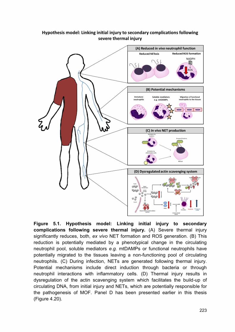
the increased incidence of infections (Figure 5.1 A). The exact mechanism by
which this occurs is yet to be described but possible factors include; abnormal
neutrophil maturity phenotype or release of soluble mediators (e.g. mtDAMPs)
(Figure 5.1 B). Thus, understanding the mechanisms responsible for reduced
neutrophil function may aid in both the early identification of at risk patients and
222
development of novel therapeutics which reduce infection and secondary
complication rates following severe thermal injury.
Furthermore, data within this thesis identifies a major biological mechanism driving
MOF after major trauma, namely the compromise to the actin scavenging system
which leads to reduced DNAse activity and a build-up of cfDNA. NETs are
generated during sepsis in patients with thermal injuries. The exact mechanism of
NETosis reamins to be identified, however, potential mechanisms include direct
interactions with bacteria or inflammatory cells (Figure 5.1 C). Included in this
thesis is a model of post-injury complications in which DNAse activity is reduced
following thermal injury caused by actin release from host tissue damage and
dysregulation of the actin scavenging system (Figure 5.1 D). Our data provide a
novel mechanistic link between the initial traumatic injury and subsequent infection
in potentially mediating the pathogenesis of MOF (Figure 4.20). Although our
study is based in burns patients it is likely to have relevance to all major trauma
and to support this, pilot observational trial data from patients with severe injuries
caused by explosions suggests that DNAse activity can be restored by the
prehospital administration of FFP. MOF is a leading cause of mortality following
severe thermal injury and data within this thesis identifies several potential novel
therapies to overcome the suppression of DNAse activity and improve outcomes
after trauma.
Future work investigating potential therapeutics to modulate abnormal neutrophil
function and the actin scavenging system is described below in priority order.
223
Figure 5.1. Hypothesis model: Linking initial injury to secondary
complications following severe thermal injury. (A) Severe thermal injury
significantly reduces, both, ex vivo NET formation and ROS generation. (B) This
reduction is potentially mediated by a phenotypical change in the circulating
neutrophil pool, soluble mediators e.g. mtDAMPs or functional neutrophils have
potentially migrated to the tissues leaving a non-functioning pool of circulating
neutrophils. (C) During infection, NETs are generated following thermal injury.
Potential mechanisms include direct induction through bacteria or through
neutrophil interactions with inflammatory cells. (D) Thermal injury results in
dysregulation of the actin scavenging system which facilitates the build-up of
circulating DNA, from initial injury and NETs, which are potentially responsible for
the pathogenesis of MOF. Panel D has been presented earlier in this thesis
(Figure 4.20).
224
5.2.1 In vivo characterisation of NETosis
Levels of cfDNA are associated with cellular damage and secondary complications
in a number of disease pathologies (108, 376). However, it is important to consider
the nature and structure of circulating DNA as it is released, complexed with
histones, in nucleosomes following NETosis. Whilst this is known, the broad
effects of free histones, free DNA and nucleosomes are described interchangeably
whereas there is a clear difference in their biological activity (377).
Free histones are cationic, highly conserved between species and orchestrate
gene transcription. Intravenous administration of free histones results in rapid
mortality of animals which can be prevented using an anti-histone antibody (198).
Histones can also mediate cytotoxic damage to endothelial and epithelial cells via
activation of TLRs (197). Whilst the mechanism by which histones are cytotoxic
remains unclear, studies have proposed that their cationic nature facilitates the
direct binding of histones to cells resulting in perforation, cell death and damage
caused by calcium influx (195, 196).
One potential therapeutic to reduce histone cytotoxicity is the administration of
negatively charged molecules such as heparins (378, 379). The non-anticoagulant
form of unfractionated heparin, for example, has been trialled as a novel
therapeutic in sepsis (378). Whilst this study did not report an improvement in 28
day mortality, Wildhagen and colleagues report a beneficial effect of this novel
therapy in mediating the inhibition of histone cytotoxicity (378). During sepsis there
is an abnormal consumption coagulopathy and thus an increased risk of bleeding.
Hence, administration of a non-anticoagulated form of heparin may have
promising effects on reducing tissue damage mediated by histones during sepsis
without potentiating bleeding.
225
DNA is regarded as a DAMP due to its rapid release and stimulation of immune
processes (199). The origin of DNA, nuclear, mitochondrial or bacterial, affects its
biological activity. In 2015, Bhagirath and colleagues isolated DNA from septic
patients and determined the different function of nuclear, mitochondrial and
bacterial DNA on coagulation and inflammation. All three sources of DNA were
capable of inducing thrombin formation, by an intrinsic dependent pathway
mechanism, and activation of platelets by platelet integrin αIIbβ3. Although, all
three have similar procoagulant properties, their functions in initiating inflammation
differ. Whilst, ncDNA and mtDNA prolonged in vitro neutrophil life, bacterial DNA
did not. However, only bacterial DNA promoted in vitro secretion of IL-6 (206).
Due to their opposing charges, DNA and histones will bind together and form a
nucleosome complex with an overall neutral charge. Like free histones and DNA,
nucleosomes can activate neutrophils (380). However, nucleosomes have
different cytotoxic properties compared to free histones and DNA. Unlike free
histones, administration of nucleosomes does not result in a significant increase in
mortality (204, 205). This may be explained by the inability of nucleosomes, unlike
histones, to cause direct damage or cell death to cultured endothelial cells.
However, subsequent physical or enzymatic degradation of nucleosomes then
resulted in damage to endothelial cells (381). Thus components of nucleosomes,
histones and DNA, are then responsible for the cellular damage and this effect is
nullified when components are complexed. In this thesis, we have investigated
three potential sources of DNA; nuclear, mitochondrial, and NET derived.
However, the status by which they exist, free or complexed, has not been fully
investigated. Thus, further study is required to investigate the relationship between
226
free histones, DNA and nucleosome complexes with secondary complications and
host tissue damage.
In 2013, Abrams et al reported the potential therapeutic benefit of CRP in
neutralising in vivo and in vitro histone induced endothelial damage, enhanced
coagulation and increased vascular permeability (381). Supported by earlier work
(382, 383), CRP is able to compete with phospholipid-containing liposomes and
form CRP-histone complexes, confirmed using immunofluorescence staining and
a gel overlay assay. The formation of CRP-histone complexes reduced integration
of histones into the cellular wall of endothelial cells and in turn cellular damage.
The exact mechanism of binding between CRP and histones remains to be
identified, however, the authors propose that the positively charged histones are
likely binding the negatively charged central pore of the CRP pentamer (381).
Interestingly, it has yet to be established if CRP-histone complexes have
increased clearance. Thus, Abrams and colleagues demonstrated, for the first
time, that CRP is a conserved mechanism which protects the body against histone
toxicity in the acute phase following trauma and illness (381). Hence, it may be
possible to use CRP, or a therapeutic mimicking CRPs mechanism of action, as
an intervention to neutralise the cytotoxic effect of circulating histones following
severe thermal injury.
As described above cfDNA can originate from a number of sources (114). In 2016,
Lehmann-Werman and colleagues described a novel technique by which the
tissue specific origin of cfDNA could be elucidated (384). It is likely that following
thermal injury circulating DNA is heterogeneous in origin given the severe nature
of injury, surgical procedures, infection, host tissue damage and NET release.
Despite the shared homology in nucleotide sequences, DNA also has unique
227
methylation patterns depending upon the cell or tissue of origin (385). Thus, it is
possible to infer cell death within specific organs and its contribution to the
circulating cfDNA. As proof of principle, Lehmann-Werman and colleagues
detected pancreatic β-cell DNA in patients with diagnosed type-1 diabetes and
islet-graft recipients, neuronal or glial DNA in patients with traumatic brain injury or
cardiac arrest and exocrine pancreas DNA in pancreatic cancer or pancreatitis
patients (384). Given the exploratory nature of this study, however, they were
unable to conclusively evaluate the diagnostic or prognostic utility of methylation
patterns. However, investigation of methylation patterns in patients following
thermal injury may have the potential to aid in the accurate diagnosis of specific
organ dysfunction and responses to clinical treatment.
5.2.2 DNAse isoforms and their functions
In 1997, Rodriguez and colleagues described three members of a DNAse 1 family.
All three displayed similarity in their nucleotide, amino acid sequences (386) and
biochemical properties, dependency on Ca2+ and Mg2+ (387). DNAse 1 is the
major endonuclease responsible for the breakdown of circulating nucleic acids.
However, in recent years groups have begun investigating the physiological roles
of DNAse-γ (386-388). Like our analysis, many assays established to measure
DNAse activity, therefore, quantify total DNAse. Furthermore, most groups focus
upon the role of DNAse 1 alone and thus do not investigate the role of DNAse-γ
(237, 353, 389).
As it functions internucleosomally and degrades nuclear chromatin, DNAse-γ was
originally reported to be involved in apoptotic DNA fragmentation (390-392).
However, cells transfected with DNAse-γ can secrete this enzyme thus suggesting
228
a mechanism by which exocytosis can occur (393). Consequently, DNAse-γ,
potentially from monocytes (394), may contribute to total circulating DNAse
activity.
DNAse 1 and –γ function synergistically to clear total circulating DNA through
distinct substrate consumption and degradation. DNAse 1 has a higher affinity and
accelerated clearance of free DNA compared to DNAse-γ which is more efficient
at degrading chromatin (387, 395).
Recent studies report a clear link between genetic mutations of DNAse-γ and
development multiple autoimmune diseases (396-398). Highlighted in DNAse-γ
mice which develop an SLE phenotype associated with the presence of
autoantigens against chromatin (399). Although DNAse-γ has been related to
autoimmunity, there have there have been no investigations into its function and
potential role in secondary complications following trauma or severe burn. Thus,
further work could investigate the combined and individual functions of DNAse-1
and –γ activity in the degradation of chromatin and DNA release following severe
thermal injury. This may reveal additional mechanistic information and/or novel
therapeutics to reduce secondary complications driven by elevated circulating
DNA or chromatin.
5.2.3 Targeting the build-up of toxic and pro-thrombotic DNA following
thermal injury
Following thermal injury, there is the disruption of the actin scavenging system
which predisposes patients to the accumulation of tissue derived and NET derived
DNA (Figure 4.20). This can then facilitate further host tissue damage, obstruct
blood flow and induce thrombosis (233, 236, 360). Modulation of this system by
229
targeting DNAse, GSN or VDBP has the potential to protect against this pro-
thrombotic and host cytotoxic phenotype.
5.2.4 DNAse as a therapy
DNAse is the major extracellular endonuclease found in a number of bodily fluids
and is responsible for the breakdown and clearance of circulating chromatin and
DNA. Severe thermal injury results in the immediate release of DNA which is
followed by a secondary release during infection. Both populations of DNA are
implicated in the generation of thrombosis, host tissue damage, endothelial
damage and secondary complications in trauma patients and animal models of
thrombosis (232, 233, 356, 400). Thus, targeting total DNA accumulation by
administration of DNAse may provide therapeutic benefit.
Using a murine model of DVT, extracellular chromatin formed a scaffold upon
which venous thrombosis could occur and was involved in the pathogenesis of the
DVT. Administration of DNAse 1 was capable of protecting animals from DVT
(233). Furthermore, PAD4 deficient mice form fewer thrombi after inferior vena
cava stenosis compared to wild-type animals (401). Thus confirming the role of
NET derived DNA in the propagation of venous thrombus formation. In addition to
their procoagulant activity, DNA, derived from NETs, can also cause direct
damage to endothelial cells (360). Interestingly, coculture of neutrophils with
activated endothelial cells results in NETosis which caused direct cellular damage
thus it appears to be self-propagating. This could also be abrogated by the
degradation of NETs using DNAse (360). These data provide evidence that
administration of DNAse to target host derived and NET derived DNA may have
potential to alleviate host tissue damage and reduce the pro-thrombotic
230
phenotype. Importantly, administration of DNAse 1 is a licenced therapeutic in
patients with cystic fibrosis and used to reduce the viscosity of sputum through
direct degradation of NET derived DNA.
It is also very important to consider the potential complications associated with
DNAse. Administration of DNAse in a murine model of sepsis resulted in an
increased pro-inflammatory response along with increased colonisation of bacteria
and mortality (221). This was further confirmed by Mai et al who highlighted the
importance of timing when targeting NETs. Early administration of DNAse resulted
in increased mortality, whereas later administration reduced tissue damage and
decreased circulating DNA levels resulting in improved survival (402). These data
provide evidence that DNA should only be broken down when in excess and
contributing an enhanced pro-inflammatory and thrombotic phenotype. Given the
clinical complexity and heterogeneous nature of patients following severe burns
this may be challenging. Furthermore, as actin is released following severe
thermal injury this may inhibit any administered DNAse, rendering the therapy
potentially non-effective. Hence, a more targeted approach towards NETs or
modulation of the actin scavenging system may be more applicable.
5.2.5 Inhibition of PAD4
NETosis can also be prevented by inhibition of PAD4. Indeed, PAD4 knockout
mice are totally protected against venous thrombosis formation and tissue damage
following liver injury (225, 275). However, these studies do not take into account
the pre-existing immunosuppression which occurs following thermal injury.
Therefore, elimination of NETs completely could leave, an already
immunocompromised patient, susceptible to further infection. Moreover, PAD4
231
deficient mice are more susceptible to necrotizing fasciitis infection than wild-type
mice and this susceptibility is attributed to the inability to form NETs (276).
5.2.6 Modulation of actin scavenging system
The actin scavenging system is comprised of two key proteins; VDBP and GSN.
Following severe thermal injury, there are reductions in circulating levels of both
proteins which predispose patients to reduce DNAse activity and the build-up of
circulating DNA (Figure 4.13 - 4.14). As both VDBP and GSN are extremely
abundant within the blood they are unlikely to be toxic if used as direct
therapeutics.
One potential mechanism to replenish depleted VDBP and GSN is through
administration of blood products, namely FFP. Although not known, replenishment
of the actin scavenging system may play a role in a number of the reported
therapeutic benefits of using blood products following major trauma (371). In this
thesis, administration of FFP during transportation from site of injury to hospital in
major trauma victims improved DNAse activity in addition to increasing circulating
GSN levels but without increasing circulating VDBP levels (Figure 4.19). Whilst
this data suggests a role of GSN in recovery of DNAse activity any improvement
by FFP will be multifactorial and caused by a number of soluble factors which may
enhance DNAse activity, including DNAse 1 itself. Therefore, the restoration of
DNAse activity cannot be solely attributed to the increase in GSN. Thus, further
studies are required to investigate whether other components within blood
products are contributing.
Whilst VDBP remains a potential therapy, to date, no group has investigated the
effect of administration of VDBP on outcome following trauma or burns. Despite
232
this, large quantities of protein can be produced from plasma fractions (403).
Following thermal injury GSN and VDBP both become saturated, most likely
caused by the release of monomeric and polymeric actin. Administration of GSN
and VDBP as a combination therapy may therefore aid in the rapid recovery of the
actin scavenging system.
There is growing evidence for the use of GSN in the treatment of infection, trauma
and burns (261, 404-406). In 2007, Lee and colleagues reported a reduction in
mortality and a dynamic change in cytokine release in GSN depleted mice that
were given 20 mg/mL recombinant human plasma GSN after challenge with
endotoxin. Although the mechanism responsible remains unclear, improvements
in outcome were associated with depolymerisation of actin in these animals (404).
Interestingly, levels of total circulating actin did not decrease which suggests that
GSN can modulate the activity of actin independent of VDBP which is consistent
with previous literature (407, 408).
A clinical trial has been established to assess the pharmacokinetics and safety of
recombinant plasma GSN given to patients admitted to an ICU, including patients
with trauma, infection and burns. The hypothesis of the study states that
administration of GSN will increase circulating GSN levels and decrease the
incidence of secondary complications, e.g. MOF or death (Trial Number:
NCT00671307). Results for this study are not currently available, however, a
second study may be warranted to focus upon patients admitted to an ICU with a
dysregulated actin scavenging system.
Whilst the beneficial effects of GSN alone have been described (261, 404-406),
the cumulative benefit of GSN with VDBP or DNAse have not. Given the
233
saturation and dysregulation of the actin scavenging system, one might
hypothesise that a combination therapy may be more appropriate. Furthermore, it
may be possible to produce novel therapies which contain the active N-terminal
fragment of GSN which is responsible for its ability to restore DNAse activity
independently of VDBP (372). Therefore, one might propose the development of a
novel compound which mimics the N-terminal fragment of GSN for its use in a
clinical trial investigating early and sustained administration of GSN and its effects
on DNAse activity and outcome following severe thermal injury. Of note, GSN
function also extends beyond control of actin homeostasis and as a potential use
as a biomarker of poor outcome as it also exhibits various anti-inflammatory
activities (260, 408). Thus, understanding the mechanism(s) by which GSN
provides benefit is fundamental to its development as a therapy.
5.2.7 Targeting reduced neutrophil function following thermal injury
The association between severe thermal injury and abnormal neutrophil function
has been well reported (156, 159-161, 173, 174, 177). Furthermore, this reduction
or dysregulation in neutrophil function may underlie the increased incidence of
nosocomial infections reported within these patient cohorts. In this thesis, it has
been shown that measurements of abnormal neutrophil function are potential
biomarkers of sepsis, in addition to being novel therapeutic targets.
5.2.8 Granulocyte colony-stimulating factor and neutrophil maturity
Traumatic injury induces marked alterations in haematopoiesis which is
characterised by a repolarisation towards a myeloid lineage (409, 410). Production
and maturation of neutrophils is multifaceted with the principle regulator of
granulopoiesis being G-CSF (162, 163) which causes activation of signal
234
transducer and activator of transcription 3 (STAT3), via MEK-1/-2 and ERK-1/-2
(411, 412).
Severe thermal injury results in the release of IGs/neutrophils which are
associated with sepsis status and reduced neutrophil function (Chapter 3, Figure
3.14, 3.17 and 3.18). One limitation of the work presented in this thesis is the lack
of mechanistic insight responsible for the release of IGs following thermal injury.
This release may be induced by emergency granulopoiesis caused by excessive
stress from immediate injury and consequent infectious episodes. In a study which
included 83 severely injured patients, sustained elevation in G-CSF was
associated with the release of immature cells and increased risk of infection (157).
In addition, Ertel and colleagues report an inhibition of neutrophil apoptosis
caused by local G-CSF which propagates damage caused by neutrophils at sites
of injury (413). Therefore, one might propose that inhibition of G-CSF can
potentially reduce circulating IGs and protect against, neutrophil mediated,
immunosuppression.
Conversely, administration of G-CSF has positive effects in mice (168, 414).
Namely, an increase in neutrophil chemotaxis in animals who received
recombinant G-CSF following burn injury which was attributed to a combination of
myeloid expansion and direct enhancement of neutrophil function (414). Gardner
et al report a key role for G-CSF/STAT3 axis in providing protection against post-
traumatic infection which was consistent with human gene analysis. Paradoxically,
thermal injury resulted in protection from a lethal Klebsiella pneumoniae
pulmonary challenge. Inhibition of G-CSF reduced STAT3 activation and
diminished this protective response by preventing myeloid differentiation and
neutrophil release (168). Thus, caution must be advised when targeting G-CSF
235
following severe thermal injury as it potentially exhibits multiple roles in host
protection and innate immunity (157, 168, 413, 414). Therefore a more targeted
approach modulating neutrophil maturation may be more applicable.
5.2.9 Resolvins
Acute inflammation is necessary for the host to respond to initial tissue damage or
invading pathogens and is required to maintain host protection and homeostasis.
However, if inflammation is prolonged or uncontrolled it can lead to host tissue
damage and secondary complications (415). Resolution of inflammation was long
characterised as an eventual reduction of pro-inflammatory mediators by a
passive process. As reviewed by Serhan and colleagues, resolution of
inflammation is now considered an active and responsive process controlled by
the release of a number of negative regulators (416). Resolvins, protectins and
lipoxins are three examples of anti-inflammatory mediators which induce
resolution and containment of acute inflammation (417, 418).
Resolvins are named after their role in the resolution of inflammation and
produced from eicosapentaenoic or docosahexaenoic acid, the most abundant
omega-3 polyunsaturated fatty acids (417, 419). Administration of resolvin D2
(RvD2) can enhance both neutrophil phagocytosis and killing of engulfed E.Coli
(420). In a cecal ligation and puncture model of sepsis, treatment with RvD2
causes a reduction in leukocyte-endothelial interactions, cytokine production, and
a reduction in local and systemic bacterial burden (420). Furthermore, Kurihara et
al reported a restoration of neutrophil chemotaxis following burn injury when
animals were administered RvD2 which was attributed to the direct action of RvD2
on neutrophils (421). Whilst it is known that resolvin D1 can act through ALX/FPR2
236
and GPR32 G-protein coupled receptors (422), the receptors responsible for RvD2
actions are currently unknown. Hence, further characterisation of RvD2 and its
receptors is essential to understand the mechanisms by which immunoregulation
and enhancement of neutrophil function occur. Nevertheless, RvD2 may be a
potential therapeutic to reduce both the uncontrolled inflammation and reduced
neutrophil function which occurs following severe thermal injury.
5.2.10 Haemoperfusion therapy
Direct haemoperfusion therapy can filter cells or soluble mediators from the blood
of patients. This results in reduced uncontrolled activation of the immune system
and a reduction in host tissue damage (423, 424). Whilst direct haemoperfusion
therapy is a recognised treatment for septic shock in Japan, only parts of Western
Europe currently use this technology (425). Direct haemoperfusion therapy using a
polymixin-B immobilized fibre cartridge (PMX-DHP) primarily works by removal of
endotoxin from the blood. However, recent evidence has shown a broader
mechanistic role for PMX-DHP in the removal of activated neutrophils which can
cause endothelial damage (424). Whilst the mechanism responsible for selective
removal of activated cells remains unclear the authors propose that it is caused by
a direct interaction between activated neutrophils and polymixin-B located on the
fibres of the filter (424). Thus, depletion of activated neutrophils by PMX-DHP may
help to control inflammation in patients with septic shock.
More recently, Lee and colleagues developed a microfibre mesh decorated in
polythylenimine and polyamidoamine dendrimers which can capture DAMPs,
including extracellular DNA. In vitro immobilisation of DAMPs located within blood
from trauma patients results in reduced activation of toll-like receptor (TLR) -2, 3, 4
237
and 9 compared to unfiltered serum in a monocyte derived reporter cell line.
Furthermore, immobilisation of DAMPs in hearts transplanted in vivo abrogated
thrombus formation (423). In this thesis, DAMPs cause dysregulation of neutrophil
function in vitro which mimics our in vivo findings in patients with severe thermal
injuries (Figure 3.21 - 3.25). Hence, immobilisation of DAMPs using the microfibre
mesh decorated in polythylenimine and polyamidoamine dendrimers has the
potential to protect against dysregulation of neutrophil function following thermal
injury and reduce thrombotic complications associated with elevated DNA and
histones. However, direct haemoperfusion therapy using polythylenimine and
polyamidoamine dendrimers remains experimental with limited translation to
clinical practice due to difficulty in integrating these microfibre meshes into
available technology.
238
Bibliography
1. Department for the Management of Noncommunicable Diseases D, Violence and Injury Prevention. World Health Organisation Injuries and Violence: The Facts (2014) 2014 [cited 2017 28/07/2017]. Available from: http://apps.who.int/iris/bitstream/10665/149798/1/9789241508018_eng.pdf?ua=1&ua=1&ua=1. 2. Haagsma JA, Graetz N, Bolliger I, Naghavi M, Higashi H, Mullany EC, et al. The global burden of injury: incidence, mortality, disability-adjusted life years and time trends from the Global Burden of Disease study 2013. Inj Prev. 2016;22(1):3-18. 3. Sanchez JL, Pereperez SB, Bastida JL, Martinez MM. Cost-utility analysis applied to the treatment of burn patients in a specialized center. Arch Surg. 2007;142(1):50-7; discussion 7. 4. Stylianou N, Buchan I, Dunn KW. A review of the international Burn Injury Database (iBID) for England and Wales: descriptive analysis of burn injuries 2003-2011. BMJ Open. 2015;5(2):e006184. 5. Santaniello JM, Luchette FA, Esposito TJ, Gunawan H, Reed RL, Davis KA, et al. Ten year experience of burn, trauma, and combined burn/trauma injuries comparing outcomes. J Trauma. 2004;57(4):696-700; dicussion -1. 6. Wilson D, Jackson T, Sapey E, Lord JM. Frailty and sarcopenia: The potential role of an aged immune system. Ageing Res Rev. 2017;36:1-10. 7. Kling KM, Lopez-Rodriguez E, Pfarrer C, Muhlfeld C, Brandenberger C. Aging exacerbates acute lung injury-induced changes of the air-blood barrier, lung function, and inflammation in the mouse. Am J Physiol Lung Cell Mol Physiol. 2017;312(1):L1-L12. 8. Hazeldine J, Lord JM. The impact of ageing on natural killer cell function and potential consequences for health in older adults. Ageing Res Rev. 2013;12(4):1069-78. 9. Hazeldine J, Harris P, Chapple IL, Grant M, Greenwood H, Livesey A, et al. Impaired neutrophil extracellular trap formation: a novel defect in the innate immune system of aged individuals. Aging Cell. 2014;13(4):690-8. 10. Pham TN, Kramer CB, Wang J, Rivara FP, Heimbach DM, Gibran NS, et al. Epidemiology and outcomes of older adults with burn injury: an analysis of the National Burn Repository. J Burn Care Res. 2009;30(1):30-6. 11. El-Helbawy RH, Ghareeb FM. Inhalation injury as a prognostic factor for mortality in burn patients. Ann Burns Fire Disasters. 2011;24(2):82-8. 12. Jackson DM. [The diagnosis of the depth of burning]. Br J Surg. 1953;40(164):588-96. 13. Hettiaratchy S, Dziewulski P. ABC of burns: pathophysiology and types of burns. BMJ. 2004;328(7453):1427-9. 14. Wolf SE, Rose JK, Desai MH, Mileski JP, Barrow RE, Herndon DN. Mortality determinants in massive pediatric burns. An analysis of 103 children with > or = 80% TBSA burns (> or = 70% full-thickness). Ann Surg. 1997;225(5):554-65; discussion 65-9. 15. Du Y, Wang L, Shi H, Gao M. Comparison of clinical effect of dopamine and norepinephrine in the treatment of septic shock. Pak J Pharm Sci. 2015;28(4 Suppl):1461-4. 16. Nielson CB, Duethman NC, Howard JM, Moncure M, Wood JG. Burns: Pathophysiology of Systemic Complications and Current Management. J Burn Care Res. 2017;38(1):e469-e81. 17. Ahrns KS. Trends in burn resuscitation: shifting the focus from fluids to adequate endpoint monitoring, edema control, and adjuvant therapies. Crit Care Nurs Clin North Am. 2004;16(1):75-98. 18. Mann R, Heimbach D. Prognosis and treatment of burns. West J Med. 1996;165(4):215-20.
19. Jackson PC, Hardwicke J, Bamford A, Nightingale P, Wilson Y, Papini R, et al. Revised estimates of mortality from the Birmingham Burn Centre, 2001-2010: a continuing analysis over 65 years. Ann Surg. 2014;259(5):979-84. 20. Monafo WW. Initial management of burns. N Engl J Med. 1996;335(21):1581-6. 21. Barrow RE, Jeschke MG, Herndon DN. Early fluid resuscitation improves outcomes in severely burned children. Resuscitation. 2000;45(2):91-6. 22. Snell JA, Loh NH, Mahambrey T, Shokrollahi K. Clinical review: the critical care management of the burn patient. Crit Care. 2013;17(5):241. 23. Tan A, Smailes S, Friebel T, Magdum A, Frew Q, El-Muttardi N, et al. Smoke inhalation increases intensive care requirements and morbidity in paediatric burns. Burns. 2016;42(5):1111-5. 24. Sakallioglu AE, Haberal M. Current approach to burn critical care. Minerva Med. 2007;98(5):569-73. 25. Sheridan RL. Fire-Related Inhalation Injury. N Engl J Med. 2016;375(5):464-9. 26. Rehberg S, Maybauer MO, Enkhbaatar P, Maybauer DM, Yamamoto Y, Traber DL. Pathophysiology, management and treatment of smoke inhalation injury. Expert Rev Respir Med. 2009;3(3):283-97. 27. Dries DJ, Endorf FW. Inhalation injury: epidemiology, pathology, treatment strategies. Scand J Trauma Resusc Emerg Med. 2013;21:31. 28. Taylor FW, Gumbert JL. Cause of Death from Burns: Role of Respiratory Damage. Ann Surg. 1965;161:497-501. 29. Walker PF, Buehner MF, Wood LA, Boyer NL, Driscoll IR, Lundy JB, et al. Diagnosis and management of inhalation injury: an updated review. Crit Care. 2015;19:351. 30. Schwacha MG, Thobe BM, Daniel T, Hubbard WJ. Impact of thermal injury on wound infiltration and the dermal inflammatory response. J Surg Res. 2010;158(1):112-20. 31. Kasten KR, Makley AT, Kagan RJ. Update on the critical care management of severe burns. J Intensive Care Med. 2011;26(4):223-36. 32. Xiao-Wu W, Herndon DN, Spies M, Sanford AP, Wolf SE. Effects of delayed wound excision and grafting in severely burned children. Arch Surg. 2002;137(9):1049-54. 33. Toussaint J, Chung WT, Osman N, McClain SA, Raut V, Singer AJ. Topical antibiotic ointment versus silver-containing foam dressing for second-degree burns in swine. Acad Emerg Med. 2015;22(8):927-33. 34. Sevgi M, Toklu A, Vecchio D, Hamblin MR. Topical antimicrobials for burn infections - an update. Recent Pat Antiinfect Drug Discov. 2013;8(3):161-97. 35. Farina JA, Jr., Rosique MJ, Rosique RG. Curbing inflammation in burn patients. Int J Inflam. 2013;2013:715645. 36. Balk RA. Systemic inflammatory response syndrome (SIRS): where did it come from and is it still relevant today? Virulence. 2014;5(1):20-6. 37. Xiao W, Mindrinos MN, Seok J, Cuschieri J, Cuenca AG, Gao H, et al. A genomic storm in critically injured humans. J Exp Med. 2011;208(13):2581-90. 38. Mann EA, Baun MM, Meininger JC, Wade CE. Comparison of mortality associated with sepsis in the burn, trauma, and general intensive care unit patient: a systematic review of the literature. Shock. 2012;37(1):4-16. 39. Binkowska AM, Michalak G, Slotwinski R. Current views on the mechanisms of immune responses to trauma and infection. Cent Eur J Immunol. 2015;40(2):206-16. 40. Sharma SK, Naidu G. The role of danger-associated molecular patterns (DAMPs) in trauma and infections. J Thorac Dis. 2016;8(7):1406-9. 41. Lam NY, Rainer TH, Chiu RW, Joynt GM, Lo YM. Plasma mitochondrial DNA concentrations after trauma. Clin Chem. 2004;50(1):213-6. 42. Zhang Q, Raoof M, Chen Y, Sumi Y, Sursal T, Junger W, et al. Circulating mitochondrial DAMPs cause inflammatory responses to injury. Nature. 2010;464(7285):104-7.
240
43. Hazeldine J, Hampson P, Opoku FA, Foster M, Lord JM. N-Formyl peptides drive mitochondrial damage associated molecular pattern induced neutrophil activation through ERK1/2 and P38 MAP kinase signalling pathways. Injury. 2015;46(6):975-84. 44. Simmons JD, Lee YL, Mulekar S, Kuck JL, Brevard SB, Gonzalez RP, et al. Elevated levels of plasma mitochondrial DNA DAMPs are linked to clinical outcome in severely injured human subjects. Ann Surg. 2013;258(4):591-6; discussion 6-8. 45. Fernandez-Ruiz I, Arnalich F, Cubillos-Zapata C, Hernandez-Jimenez E, Moreno-Gonzalez R, Toledano V, et al. Mitochondrial DAMPs induce endotoxin tolerance in human monocytes: an observation in patients with myocardial infarction. PLoS One. 2014;9(5):e95073. 46. Angus DC, van der Poll T. Severe sepsis and septic shock. N Engl J Med. 2013;369(21):2063. 47. Paratz JD, Lipman J, Boots RJ, Muller MJ, Paterson DL. A new marker of sepsis post burn injury?*. Crit Care Med. 2014;42(9):2029-36. 48. Baue AE. Multiple, progressive, or sequential systems failure. A syndrome of the 1970s. Arch Surg. 1975;110(7):779-81. 49. Penner JA. Disseminated intravascular coagulation in patients with multiple organ failure of non-septic origin. Semin Thromb Hemost. 1998;24(1):45-52. 50. Kidokoro A, Iba T, Hong J. Role of DIC in multiple organ failure. Int J Surg Investig. 2000;2(1):73-80. 51. Vincent JL, De Backer D. Does disseminated intravascular coagulation lead to multiple organ failure? Crit Care Clin. 2005;21(3):469-77. 52. Toh CH, Alhamdi Y, Abrams ST. Current Pathological and Laboratory Considerations in the Diagnosis of Disseminated Intravascular Coagulation. Ann Lab Med. 2016;36(6):505-12. 53. Levi M, Toh CH, Thachil J, Watson HG. Guidelines for the diagnosis and management of disseminated intravascular coagulation. British Committee for Standards in Haematology. Br J Haematol. 2009;145(1):24-33. 54. Bick RL. Disseminated intravascular coagulation: a review of etiology, pathophysiology, diagnosis, and management: guidelines for care. Clin Appl Thromb Hemost. 2002;8(1):1-31. 55. Wada H, Hasegawa K, Watanabe M. DIC: an update on diagnosis and treatment. Rinsho Ketsueki. 2017;58(5):523-9. 56. Yanagida Y, Gando S, Sawamura A, Hayakawa M, Uegaki S, Kubota N, et al. Normal prothrombinase activity, increased systemic thrombin activity, and lower antithrombin levels in patients with disseminated intravascular coagulation at an early phase of trauma: comparison with acute coagulopathy of trauma-shock. Surgery. 2013;154(1):48-57. 57. Dobson GP, Letson HL, Sharma R, Sheppard FR, Cap AP. Mechanisms of early trauma-induced coagulopathy: The clot thickens or not? J Trauma Acute Care Surg. 2015;79(2):301-9. 58. Kono M, Saigo K, Takagi Y, Takahashi T, Kawauchi S, Wada A, et al. Heme-related molecules induce rapid production of neutrophil extracellular traps. Transfusion. 2014;54(11):2811-9. 59. Nguyen LN, Nguyen TG. Characteristics and outcomes of multiple organ dysfunction syndrome among severe-burn patients. Burns. 2009;35(7):937-41. 60. Dellinger RP, Levy MM, Rhodes A, Annane D, Gerlach H, Opal SM, et al. Surviving sepsis campaign: international guidelines for management of severe sepsis and septic shock: 2012. Crit Care Med. 2013;41(2):580-637. 61. Castellanos-Ortega A, Suberviola B, Garcia-Astudillo LA, Holanda MS, Ortiz F, Llorca J, et al. Impact of the Surviving Sepsis Campaign protocols on hospital length of stay and mortality in septic shock patients: results of a three-year follow-up quasi-experimental study. Crit Care Med. 2010;38(4):1036-43. 62. Levy MM, Dellinger RP, Townsend SR, Linde-Zwirble WT, Marshall JC, Bion J, et al. The Surviving Sepsis Campaign: results of an international guideline-based
241
performance improvement program targeting severe sepsis. Crit Care Med. 2010;38(2):367-74. 63. Ferrer R, Artigas A, Suarez D, Palencia E, Levy MM, Arenzana A, et al. Effectiveness of treatments for severe sepsis: a prospective, multicenter, observational study. Am J Respir Crit Care Med. 2009;180(9):861-6. 64. Orban C. Diagnostic criteria for sepsis in burn patients. Chirurgia (Bucur). 2012;107(6):697-700. 65. Greenhalgh DG, Saffle JR, Holmes JHt, Gamelli RL, Palmieri TL, Horton JW, et al. American Burn Association consensus conference to define sepsis and infection in burns. J Burn Care Res. 2007;28(6):776-90. 66. Singer M, Deutschman CS, Seymour CW, Shankar-Hari M, Annane D, Bauer M, et al. The Third International Consensus Definitions for Sepsis and Septic Shock (Sepsis-3). JAMA. 2016;315(8):801-10. 67. Blair JM, Webber MA, Baylay AJ, Ogbolu DO, Piddock LJ. Molecular mechanisms of antibiotic resistance. Nat Rev Microbiol. 2015;13(1):42-51. 68. van Langeveld I, Gagnon RC, Conrad PF, Gamelli RL, Martin B, Choudhry MA, et al. Multiple-Drug Resistance in Burn Patients: A Retrospective Study on the Impact of Antibiotic Resistance on Survival and Length of Stay. J Burn Care Res. 2017;38(2):99-105. 69. Assicot M, Gendrel D, Carsin H, Raymond J, Guilbaud J, Bohuon C. High serum procalcitonin concentrations in patients with sepsis and infection. Lancet. 1993;341(8844):515-8. 70. Mehanic S, Baljic R. The importance of serum procalcitonin in diagnosis and treatment of serious bacterial infections and sepsis. Mater Sociomed. 2013;25(4):277-81. 71. von Heimburg D, Stieghorst W, Khorram-Sefat R, Pallua N. Procalcitonin--a sepsis parameter in severe burn injuries. Burns. 1998;24(8):745-50. 72. Sachse C, Machens HG, Felmerer G, Berger A, Henkel E. Procalcitonin as a marker for the early diagnosis of severe infection after thermal injury. J Burn Care Rehabil. 1999;20(5):354-60. 73. Uzzan B, Cohen R, Nicolas P, Cucherat M, Perret GY. Procalcitonin as a diagnostic test for sepsis in critically ill adults and after surgery or trauma: a systematic review and meta-analysis. Crit Care Med. 2006;34(7):1996-2003. 74. Schuetz P, Raad I, Amin DN. Using procalcitonin-guided algorithms to improve antimicrobial therapy in ICU patients with respiratory infections and sepsis. Curr Opin Crit Care. 2013;19(5):453-60. 75. Cabral L, Afreixo V, Almeida L, Paiva JA. The Use of Procalcitonin (PCT) for Diagnosis of Sepsis in Burn Patients: A Meta-Analysis. PLoS One. 2016;11(12):e0168475. 76. Seoane L, Pertega S, Galeiras R, Astola I, Bouza T. Procalcitonin in the burn unit and the diagnosis of infection. Burns. 2014;40(2):223-9. 77. Lavrentieva A, Kontakiotis T, Lazaridis L, Tsotsolis N, Koumis J, Kyriazis G, et al. Inflammatory markers in patients with severe burn injury. What is the best indicator of sepsis? Burns. 2007;33(2):189-94. 78. Minami E, Ito S, Sugiura T, Fujita Y, Sasano H, Sobue K. Markedly elevated procalcitonin in early postoperative period in pediatric open heart surgery: a prospective cohort study. J Intensive Care. 2014;2(1):38. 79. Pepys MB, Hirschfield GM. C-reactive protein: a critical update. J Clin Invest. 2003;111(12):1805-12. 80. Kim H, Hur M, Moon HW, Yun YM, Di Somma S, Network G. Multi-marker approach using procalcitonin, presepsin, galectin-3, and soluble suppression of tumorigenicity 2 for the prediction of mortality in sepsis. Ann Intensive Care. 2017;7(1):27. 81. Sapin F, Biston P, Piagnerelli M. Predictive value of C-reactive protein in critically ill patients after abdominal surgery. Clinics (Sao Paulo). 2017;72(1):23-9.
242
82. Luzzani A, Polati E, Dorizzi R, Rungatscher A, Pavan R, Merlini A. Comparison of procalcitonin and C-reactive protein as markers of sepsis. Crit Care Med. 2003;31(6):1737-41. 83. Lobo SM, Lobo FR, Bota DP, Lopes-Ferreira F, Soliman HM, Melot C, et al. C-reactive protein levels correlate with mortality and organ failure in critically ill patients. Chest. 2003;123(6):2043-9. 84. Su L, Han B, Liu C, Liang L, Jiang Z, Deng J, et al. Value of soluble TREM-1, procalcitonin, and C-reactive protein serum levels as biomarkers for detecting bacteremia among sepsis patients with new fever in intensive care units: a prospective cohort study. BMC Infect Dis. 2012;12:157. 85. Pileri D, Accardo Palombo A, D'Amelio L, D'Arpa N, Amato G, Masellis A, et al. Concentrations of cytokines IL-6 and IL-10 in plasma of burn patients: their relationship to sepsis and outcome. Ann Burns Fire Disasters. 2008;21(4):182-5. 86. Finnerty CC, Herndon DN, Przkora R, Pereira CT, Oliveira HM, Queiroz DM, et al. Cytokine expression profile over time in severely burned pediatric patients. Shock. 2006;26(1):13-9. 87. Xia ZF, Coolbaugh MI, He F, Herndon DN, Papaconstantinou J. The effects of burn injury on the acute phase response. J Trauma. 1992;32(2):245-50; discussion 50-1. 88. Jeschke MG, Mlcak RP, Finnerty CC, Norbury WB, Gauglitz GG, Kulp GA, et al. Burn size determines the inflammatory and hypermetabolic response. Crit Care. 2007;11(4):R90. 89. Gauglitz GG, Finnerty CC, Herndon DN, Mlcak RP, Jeschke MG. Are serum cytokines early predictors for the outcome of burn patients with inhalation injuries who do not survive? Crit Care. 2008;12(3):R81. 90. Jeschke MG, Patsouris D, Stanojcic M, Abdullahi A, Rehou S, Pinto R, et al. Pathophysiologic Response to Burns in the Elderly. EBioMedicine. 2015;2(10):1536-48. 91. Kraft R, Herndon DN, Finnerty CC, Cox RA, Song J, Jeschke MG. Predictive Value of IL-8 for Sepsis and Severe Infections After Burn Injury: A Clinical Study. Shock. 2015;43(3):222-7. 92. Gouel-Cheron A, Allaouchiche B, Guignant C, Davin F, Floccard B, Monneret G, et al. Early interleukin-6 and slope of monocyte human leukocyte antigen-DR: a powerful association to predict the development of sepsis after major trauma. PLoS One. 2012;7(3):e33095. 93. Marck RE, Montagne HL, Tuinebreijer WE, Breederveld RS. Time course of thrombocytes in burn patients and its predictive value for outcome. Burns. 2013;39(4):714-22. 94. Ha SO, Park SH, Park SH, Park JS, Huh JW, Lim CM, et al. Fraction of immature granulocytes reflects severity but not mortality in sepsis. Scand J Clin Lab Invest. 2015;75(1):36-43. 95. Simon TL, Curreri PW, Harker LA. Kinetic characterization of hemostasis in thermal injury. J Lab Clin Med. 1977;89(4):702-11. 96. Nierhaus A, Klatte S, Linssen J, Eismann NM, Wichmann D, Hedke J, et al. Revisiting the white blood cell count: immature granulocytes count as a diagnostic marker to discriminate between SIRS and sepsis--a prospective, observational study. BMC Immunol. 2013;14:8. 97. Nigro KG, O'Riordan M, Molloy EJ, Walsh MC, Sandhaus LM. Performance of an automated immature granulocyte count as a predictor of neonatal sepsis. Am J Clin Pathol. 2005;123(4):618-24. 98. Wagner DD, Burger PC. Platelets in inflammation and thrombosis. Arterioscler Thromb Vasc Biol. 2003;23(12):2131-7. 99. Rossaint J, Zarbock A. Platelets in leucocyte recruitment and function. Cardiovasc Res. 2015;107(3):386-95. 100. Clark SR, Ma AC, Tavener SA, McDonald B, Goodarzi Z, Kelly MM, et al. Platelet TLR4 activates neutrophil extracellular traps to ensnare bacteria in septic blood. Nat Med. 2007;13(4):463-9.
243
101. Harrison P, Ault KA, Chapman S, Charie L, Davis B, Fujimoto K, et al. An interlaboratory study of a candidate reference method for platelet counting. Am J Clin Pathol. 2001;115(3):448-59. 102. Briggs C, Harrison P, Machin SJ. Continuing developments with the automated platelet count. Int J Lab Hematol. 2007;29(2):77-91. 103. Zini G, d'Onofrio G, Briggs C, Erber W, Jou JM, Lee SH, et al. ICSH recommendations for identification, diagnostic value, and quantitation of schistocytes. Int J Lab Hematol. 2012;34(2):107-16. 104. Tanaka Y, Tanaka Y, Gondo K, Maruki Y, Kondo T, Asai S, et al. Performance evaluation of platelet counting by novel fluorescent dye staining in the XN-series automated hematology analyzers. J Clin Lab Anal. 2014;28(5):341-8. 105. Lesesve JF, Martin M, Banasiak C, Andre-Kerneis E, Bardet V, Lusina D, et al. Schistocytes in disseminated intravascular coagulation. Int J Lab Hematol. 2014;36(4):439-43. 106. Lesesve JF, Salignac S, Lecompte T. Laboratory measurement of schistocytes. Int J Lab Hematol. 2007;29(2):149-51. 107. Saukkonen K, Lakkisto P, Pettila V, Varpula M, Karlsson S, Ruokonen E, et al. Cell-free plasma DNA as a predictor of outcome in severe sepsis and septic shock. Clin Chem. 2008;54(6):1000-7. 108. Rhodes A, Wort SJ, Thomas H, Collinson P, Bennett ED. Plasma DNA concentration as a predictor of mortality and sepsis in critically ill patients. Crit Care. 2006;10(2):R60. 109. Dwivedi DJ, Toltl LJ, Swystun LL, Pogue J, Liaw KL, Weitz JI, et al. Prognostic utility and characterization of cell-free DNA in patients with severe sepsis. Crit Care. 2012;16(4):R151. 110. Lo YM, Rainer TH, Chan LY, Hjelm NM, Cocks RA. Plasma DNA as a prognostic marker in trauma patients. Clin Chem. 2000;46(3):319-23. 111. Shoham Y, Krieger Y, Perry ZH, Shaked G, Bogdanov-Berezovsky A, Silberstein E, et al. Admission cell free DNA as a prognostic factor in burns: quantification by use of a direct rapid fluorometric technique. Biomed Res Int. 2014;2014:306580. 112. Yoon KA, Park S, Lee SH, Kim JH, Lee JS. Comparison of circulating plasma DNA levels between lung cancer patients and healthy controls. J Mol Diagn. 2009;11(3):182-5. 113. Papadopoulou E, Davilas E, Sotiriou V, Georgakopoulos E, Georgakopoulou S, Koliopanos A, et al. Cell-free DNA and RNA in plasma as a new molecular marker for prostate and breast cancer. Ann N Y Acad Sci. 2006;1075:235-43. 114. van der Vaart M, Pretorius PJ. Circulating DNA. Its origin and fluctuation. Ann N Y Acad Sci. 2008;1137:18-26. 115. van der Vaart M, Pretorius PJ. The origin of circulating free DNA. Clin Chem. 2007;53(12):2215. 116. Summers C, Rankin SM, Condliffe AM, Singh N, Peters AM, Chilvers ER. Neutrophil kinetics in health and disease. Trends Immunol. 2010;31(8):318-24. 117. Segal AW. How neutrophils kill microbes. Annu Rev Immunol. 2005;23:197-223. 118. Brinkmann V, Reichard U, Goosmann C, Fauler B, Uhlemann Y, Weiss DS, et al. Neutrophil extracellular traps kill bacteria. Science. 2004;303(5663):1532-5. 119. Menegazzi R, Decleva E, Dri P. Killing by neutrophil extracellular traps: fact or folklore? Blood. 2012;119(5):1214-6. 120. Tecchio C, Micheletti A, Cassatella MA. Neutrophil-derived cytokines: facts beyond expression. Front Immunol. 2014;5:508. 121. Scapini P, Lapinet-Vera JA, Gasperini S, Calzetti F, Bazzoni F, Cassatella MA. The neutrophil as a cellular source of chemokines. Immunol Rev. 2000;177:195-203. 122. Tecchio C, Cassatella MA. Neutrophil-derived cytokines involved in physiological and pathological angiogenesis. Chem Immunol Allergy. 2014;99:123-37. 123. Tecchio C, Scapini P, Pizzolo G, Cassatella MA. On the cytokines produced by human neutrophils in tumors. Semin Cancer Biol. 2013;23(3):159-70.
244
124. Dinauer MC, Lekstrom-Himes JA, Dale DC. Inherited Neutrophil Disorders: Molecular Basis and New Therapies. Hematology Am Soc Hematol Educ Program. 2000:303-18. 125. Mocsai A. Diverse novel functions of neutrophils in immunity, inflammation, and beyond. J Exp Med. 2013;210(7):1283-99. 126. Jones DA, Abbassi O, McIntire LV, McEver RP, Smith CW. P-selectin mediates neutrophil rolling on histamine-stimulated endothelial cells. Biophys J. 1993;65(4):1560-9. 127. Ley K, Laudanna C, Cybulsky MI, Nourshargh S. Getting to the site of inflammation: the leukocyte adhesion cascade updated. Nat Rev Immunol. 2007;7(9):678-89. 128. Muller WA. Leukocyte-endothelial-cell interactions in leukocyte transmigration and the inflammatory response. Trends Immunol. 2003;24(6):327-34. 129. Anderson DC, Springer TA. Leukocyte adhesion deficiency: an inherited defect in the Mac-1, LFA-1, and p150,95 glycoproteins. Annu Rev Med. 1987;38:175-94. 130. Nuzzi PA, Lokuta MA, Huttenlocher A. Analysis of neutrophil chemotaxis. Methods Mol Biol. 2007;370:23-36. 131. Lin F, Nguyen CM, Wang SJ, Saadi W, Gross SP, Jeon NL. Neutrophil migration in opposing chemoattractant gradients using microfluidic chemotaxis devices. Ann Biomed Eng. 2005;33(4):475-82. 132. Boner A, Zeligs BJ, Bellanti JA. Chemotactic responses of various differentiational stages of neutrophils from human cord and adult blood. Infect Immun. 1982;35(3):921-8. 133. Hatano Y, Taniuchi S, Masuda M, Tsuji S, Ito T, Hasui M, et al. Phagocytosis of heat-killed Staphylococcus aureus by eosinophils: comparison with neutrophils. APMIS. 2009;117(2):115-23. 134. Leliefeld PH, Wessels CM, Leenen LP, Koenderman L, Pillay J. The role of neutrophils in immune dysfunction during severe inflammation. Crit Care. 2016;20:73. 135. Babior BM. NADPH oxidase: an update. Blood. 1999;93(5):1464-76. 136. Bedard K, Krause KH. The NOX family of ROS-generating NADPH oxidases: physiology and pathophysiology. Physiol Rev. 2007;87(1):245-313. 137. Liao Y, Liu P, Guo F, Zhang ZY, Zhang Z. Oxidative burst of circulating neutrophils following traumatic brain injury in human. PLoS One. 2013;8(7):e68963. 138. Dong YL, Abdullah K, Yan TZ, Rutan T, Broemeling L, Robson M, et al. Effect of thermal injury and sepsis on neutrophil function. J Trauma. 1993;34(3):417-21. 139. Spicer SS, Hardin JH. Ultrastructure, cytochemistry, and function of neutrophil leukocyte granules. A review. Lab Invest. 1969;20(5):488-97. 140. Belaaouaj A, Kim KS, Shapiro SD. Degradation of outer membrane protein A in Escherichia coli killing by neutrophil elastase. Science. 2000;289(5482):1185-8. 141. Fox S, Leitch AE, Duffin R, Haslett C, Rossi AG. Neutrophil apoptosis: relevance to the innate immune response and inflammatory disease. J Innate Immun. 2010;2(3):216-27. 142. Luo HR, Loison F. Constitutive neutrophil apoptosis: mechanisms and regulation. Am J Hematol. 2008;83(4):288-95. 143. Edwards SW, Derouet M, Howse M, Moots RJ. Regulation of neutrophil apoptosis by Mcl-1. Biochem Soc Trans. 2004;32(Pt3):489-92. 144. Akgul C, Moulding DA, White MR, Edwards SW. In vivo localisation and stability of human Mcl-1 using green fluorescent protein (GFP) fusion proteins. FEBS Lett. 2000;478(1-2):72-6. 145. Lovell JF, Billen LP, Bindner S, Shamas-Din A, Fradin C, Leber B, et al. Membrane binding by tBid initiates an ordered series of events culminating in membrane permeabilization by Bax. Cell. 2008;135(6):1074-84. 146. Scatizzi JC, Hutcheson J, Bickel E, Haines GK, 3rd, Perlman H. Pro-apoptotic Bid is required for the resolution of the effector phase of inflammatory arthritis. Arthritis Res Ther. 2007;9(3):R49.
245
147. Andina N, Conus S, Schneider EM, Fey MF, Simon HU. Induction of Bim limits cytokine-mediated prolonged survival of neutrophils. Cell Death Differ. 2009;16(9):1248-55. 148. Nolan B, Collette H, Baker S, Duffy A, De M, Miller C, et al. Inhibition of neutrophil apoptosis after severe trauma is NFkappabeta dependent. J Trauma. 2000;48(4):599-604; discussion -5. 149. Turina M, Hoth JJ, Turpen RM, Scott MJ, Cheadle WG. Alveolar interleukin-10 regulates neutrophil apoptosis in severely traumatized patients. J Trauma. 2007;63(4):733-9. 150. Paunel-Gorgulu A, Kirichevska T, Logters T, Windolf J, Flohe S. Molecular mechanisms underlying delayed apoptosis in neutrophils from multiple trauma patients with and without sepsis. Mol Med. 2012;18:325-35. 151. Matsuda T, Saito H, Fukatsu K, Han I, Inoue T, Furukawa S, et al. Cytokine-modulated inhibition of neutrophil apoptosis at local site augments exudative neutrophil functions and reflects inflammatory response after surgery. Surgery. 2001;129(1):76-85. 152. Scheel-Toellner D, Wang K, Assi LK, Webb PR, Craddock RM, Salmon M, et al. Clustering of death receptors in lipid rafts initiates neutrophil spontaneous apoptosis. Biochem Soc Trans. 2004;32(Pt 5):679-81. 153. Dunogue B, Pilmis B, Mahlaoui N, Elie C, Coignard-Biehler H, Amazzough K, et al. Chronic Granulomatous Disease in Patients Reaching Adulthood: A Nationwide Study in France. Clin Infect Dis. 2017;64(6):767-75. 154. Introne WJ, Westbroek W, Golas GA, Adams D. Chediak-Higashi Syndrome. In: Pagon RA, Adam MP, Ardinger HH, Wallace SE, Amemiya A, Bean LJH, et al., editors. GeneReviews(R). Seattle (WA)1993. 155. Robertson CM, Perrone EE, McConnell KW, Dunne WM, Boody B, Brahmbhatt T, et al. Neutrophil depletion causes a fatal defect in murine pulmonary Staphylococcus aureus clearance. J Surg Res. 2008;150(2):278-85. 156. Hazeldine J, Hampson P, Lord JM. The impact of trauma on neutrophil function. Injury. 2014;45(12):1824-33. 157. Cook KM, Sifri ZC, Baranski GM, Mohr AM, Livingston DH. The role of plasma granulocyte colony stimulating factor and bone marrow dysfunction after severe trauma. J Am Coll Surg. 2013;216(1):57-64. 158. Pillay J, Kamp VM, van Hoffen E, Visser T, Tak T, Lammers JW, et al. A subset of neutrophils in human systemic inflammation inhibits T cell responses through Mac-1. J Clin Invest. 2012;122(1):327-36. 159. Warden GD, Mason AD, Jr., Pruitt BA, Jr. Suppression of leukocyte chemotaxis in vitro by chemotherapeutic agents used in the management of thermal injuries. Ann Surg. 1975;181(3):363-9. 160. Kim Y, Goldstein E, Lippert W, Brofeldt T, Donovan R. Polymorphonuclear leucocyte motility in patients with severe burns. Burns. 1989;15(2):93-7. 161. Warden GD, Mason AD, Jr., Pruitt BA, Jr. Evaluation of leukocyte chemotaxis in vitro in thermally injured patients. J Clin Invest. 1974;54(4):1001-4. 162. Richards MK, Liu F, Iwasaki H, Akashi K, Link DC. Pivotal role of granulocyte colony-stimulating factor in the development of progenitors in the common myeloid pathway. Blood. 2003;102(10):3562-8. 163. Lord BI, Bronchud MH, Owens S, Chang J, Howell A, Souza L, et al. The kinetics of human granulopoiesis following treatment with granulocyte colony-stimulating factor in vivo. Proc Natl Acad Sci U S A. 1989;86(23):9499-503. 164. Liu F, Poursine-Laurent J, Wu HY, Link DC. Interleukin-6 and the granulocyte colony-stimulating factor receptor are major independent regulators of granulopoiesis in vivo but are not required for lineage commitment or terminal differentiation. Blood. 1997;90(7):2583-90. 165. Sugimoto K, Mori KJ. Cascade regulation of cytokine production in granulopoiesis. Leukemia. 1997;11 Suppl 3:464-7.
246
166. Tsuji T, Sugimoto K, Yanai T, Takashita E, Mori KJ. Induction of granulocyte-macrophage colony-stimulating factor (GM-CSF) and granulocyte colony-stimulating factor (G-CSF) expression in bone marrow and fractionated marrow cell populations by interleukin 3 (IL-3): IL-3-mediated positive feedback mechanisms of granulopoiesis. Growth Factors. 1994;11(1):71-9. 167. Manz MG, Boettcher S. Emergency granulopoiesis. Nat Rev Immunol. 2014;14(5):302-14. 168. Gardner JC, Noel JG, Nikolaidis NM, Karns R, Aronow BJ, Ogle CK, et al. G-CSF drives a posttraumatic immune program that protects the host from infection. J Immunol. 2014;192(5):2405-17. 169. Drifte G, Dunn-Siegrist I, Tissieres P, Pugin J. Innate immune functions of immature neutrophils in patients with sepsis and severe systemic inflammatory response syndrome. Crit Care Med. 2013;41(3):820-32. 170. Tak T, Wijten P, Heeres M, Pickkers P, Scholten A, Heck AJR, et al. Human CD62Ldim neutrophils identified as a separate subset by proteome profiling and in vivo pulse-chase labeling. Blood. 2017;129(26):3476-85. 171. Butler KL, Ambravaneswaran V, Agrawal N, Bilodeau M, Toner M, Tompkins RG, et al. Burn injury reduces neutrophil directional migration speed in microfluidic devices. PLoS One. 2010;5(7):e11921. 172. Jones CN, Moore M, Dimisko L, Alexander A, Ibrahim A, Hassell BA, et al. Spontaneous neutrophil migration patterns during sepsis after major burns. PLoS One. 2014;9(12):e114509. 173. Sheng ZY, Tung YL. Neutrophil chemiluminescence in burned patients. J Trauma. 1987;27(6):587-95. 174. Alexander JW, Ogle CK, Stinnett JD, Macmillan BG. A sequential, prospective analysis of immunologic abnormalities and infection following severe thermal injury. Ann Surg. 1978;188(6):809-16. 175. Arturson G. Neutrophil granulocyte functions in severely burned patients. Burns Incl Therm Inj. 1985;11(5):309-19. 176. Bjerknes R, Vindenes H, Laerum OD. Altered neutrophil functions in patients with large burns. Blood Cells. 1990;16(1):127-41; discussion 42-3. 177. Parment K, Zetterberg A, Ernerudh J, Bakteman K, Steinwall I, Sjoberg F. Long-term immunosuppression in burned patients assessed by in vitro neutrophil oxidative burst (Phagoburst). Burns. 2007;33(7):865-71. 178. Yipp BG, Kubes P. NETosis: how vital is it? Blood. 2013;122(16):2784-94. 179. Logters T, Margraf S, Altrichter J, Cinatl J, Mitzner S, Windolf J, et al. The clinical value of neutrophil extracellular traps. Med Microbiol Immunol. 2009;198(4):211-9. 180. Hampson P, Dinsdale RJ, Wearn CM, Bamford AL, Bishop JRB, Hazeldine J, et al. Neutrophil Dysfunction, Immature Granulocytes, and Cell-free DNA are Early Biomarkers of Sepsis in Burn-injured Patients: A Prospective Observational Cohort Study. Ann Surg. 2017;265(6):1241-9. 181. Hirose T, Hamaguchi S, Matsumoto N, Irisawa T, Seki M, Tasaki O, et al. Presence of neutrophil extracellular traps and citrullinated histone H3 in the bloodstream of critically ill patients. PLoS One. 2014;9(11):e111755. 182. Takei H, Araki A, Watanabe H, Ichinose A, Sendo F. Rapid killing of human neutrophils by the potent activator phorbol 12-myristate 13-acetate (PMA) accompanied by changes different from typical apoptosis or necrosis. J Leukoc Biol. 1996;59(2):229-40. 183. Steinberg BE, Grinstein S. Unconventional roles of the NADPH oxidase: signaling, ion homeostasis, and cell death. Sci STKE. 2007;2007(379):pe11. 184. Fuchs TA, Abed U, Goosmann C, Hurwitz R, Schulze I, Wahn V, et al. Novel cell death program leads to neutrophil extracellular traps. J Cell Biol. 2007;176(2):231-41. 185. Papayannopoulos V, Metzler KD, Hakkim A, Zychlinsky A. Neutrophil elastase and myeloperoxidase regulate the formation of neutrophil extracellular traps. J Cell Biol. 2010;191(3):677-91.
247
186. Pilsczek FH, Salina D, Poon KK, Fahey C, Yipp BG, Sibley CD, et al. A novel mechanism of rapid nuclear neutrophil extracellular trap formation in response to Staphylococcus aureus. J Immunol. 2010;185(12):7413-25. 187. Rossaint J, Herter JM, Van Aken H, Napirei M, Doring Y, Weber C, et al. Synchronized integrin engagement and chemokine activation is crucial in neutrophil extracellular trap-mediated sterile inflammation. Blood. 2014;123(16):2573-84. 188. Branzk N, Lubojemska A, Hardison SE, Wang Q, Gutierrez MG, Brown GD, et al. Neutrophils sense microbe size and selectively release neutrophil extracellular traps in response to large pathogens. Nat Immunol. 2014;15(11):1017-25. 189. Wang Y, Li M, Stadler S, Correll S, Li P, Wang D, et al. Histone hypercitrullination mediates chromatin decondensation and neutrophil extracellular trap formation. J Cell Biol. 2009;184(2):205-13. 190. Nakashima K, Hagiwara T, Yamada M. Nuclear localization of peptidylarginine deiminase V and histone deimination in granulocytes. J Biol Chem. 2002;277(51):49562-8. 191. Vossenaar ER, Zendman AJ, van Venrooij WJ, Pruijn GJ. PAD, a growing family of citrullinating enzymes: genes, features and involvement in disease. Bioessays. 2003;25(11):1106-18. 192. Li Y, Liu B, Fukudome EY, Lu J, Chong W, Jin G, et al. Identification of citrullinated histone H3 as a potential serum protein biomarker in a lethal model of lipopolysaccharide-induced shock. Surgery. 2011;150(3):442-51. 193. Marino-Ramirez L, Kann MG, Shoemaker BA, Landsman D. Histone structure and nucleosome stability. Expert Rev Proteomics. 2005;2(5):719-29. 194. Venkatesh S, Workman JL. Histone exchange, chromatin structure and the regulation of transcription. Nat Rev Mol Cell Biol. 2015;16(3):178-89. 195. Abrams ST, Zhang N, Manson J, Liu T, Dart C, Baluwa F, et al. Circulating histones are mediators of trauma-associated lung injury. Am J Respir Crit Care Med. 2013;187(2):160-9. 196. Gillrie MR, Lee K, Gowda DC, Davis SP, Monestier M, Cui L, et al. Plasmodium falciparum histones induce endothelial proinflammatory response and barrier dysfunction. Am J Pathol. 2012;180(3):1028-39. 197. Allam R, Scherbaum CR, Darisipudi MN, Mulay SR, Hagele H, Lichtnekert J, et al. Histones from dying renal cells aggravate kidney injury via TLR2 and TLR4. J Am Soc Nephrol. 2012;23(8):1375-88. 198. Xu J, Zhang X, Pelayo R, Monestier M, Ammollo CT, Semeraro F, et al. Extracellular histones are major mediators of death in sepsis. Nat Med. 2009;15(11):1318-21. 199. Pisetsky DS. The origin and properties of extracellular DNA: from PAMP to DAMP. Clin Immunol. 2012;144(1):32-40. 200. McGinty RK, Tan S. Nucleosome structure and function. Chem Rev. 2015;115(6):2255-73. 201. McGhee JD, Felsenfeld G. Nucleosome structure. Annu Rev Biochem. 1980;49:1115-56. 202. Marin Oyarzun CP, Carestia A, Lev PR, Glembotsky AC, Castro Rios MA, Moiraghi B, et al. Neutrophil extracellular trap formation and circulating nucleosomes in patients with chronic myeloproliferative neoplasms. Sci Rep. 2016;6:38738. 203. van Montfoort ML, Stephan F, Lauw MN, Hutten BA, Van Mierlo GJ, Solati S, et al. Circulating nucleosomes and neutrophil activation as risk factors for deep vein thrombosis. Arterioscler Thromb Vasc Biol. 2013;33(1):147-51. 204. Gauthier VJ, Tyler LN, Mannik M. Blood clearance kinetics and liver uptake of mononucleosomes in mice. J Immunol. 1996;156(3):1151-6. 205. Decker P, Wolburg H, Rammensee HG. Nucleosomes induce lymphocyte necrosis. Eur J Immunol. 2003;33(7):1978-87.
248
206. Bhagirath VC, Dwivedi DJ, Liaw PC. Comparison of the Proinflammatory and Procoagulant Properties of Nuclear, Mitochondrial, and Bacterial DNA. Shock. 2015;44(3):265-71. 207. Levine B, Kroemer G. Autophagy in the pathogenesis of disease. Cell. 2008;132(1):27-42. 208. Remijsen Q, Vanden Berghe T, Wirawan E, Asselbergh B, Parthoens E, De Rycke R, et al. Neutrophil extracellular trap cell death requires both autophagy and superoxide generation. Cell Res. 2011;21(2):290-304. 209. Nakagawa I, Amano A, Mizushima N, Yamamoto A, Yamaguchi H, Kamimoto T, et al. Autophagy defends cells against invading group A Streptococcus. Science. 2004;306(5698):1037-40. 210. Gutierrez MG, Master SS, Singh SB, Taylor GA, Colombo MI, Deretic V. Autophagy is a defense mechanism inhibiting BCG and Mycobacterium tuberculosis survival in infected macrophages. Cell. 2004;119(6):753-66. 211. Birmingham CL, Brumell JH. Autophagy recognizes intracellular Salmonella enterica serovar Typhimurium in damaged vacuoles. Autophagy. 2006;2(3):156-8. 212. Kenno S, Perito S, Mosci P, Vecchiarelli A, Monari C. Autophagy and Reactive Oxygen Species Are Involved in Neutrophil Extracellular Traps Release Induced by C. albicans Morphotypes. Front Microbiol. 2016;7:879. 213. Hashiba M, Huq A, Tomino A, Hirakawa A, Hattori T, Miyabe H, et al. Neutrophil extracellular traps in patients with sepsis. J Surg Res. 2015;194(1):248-54. 214. Czaikoski PG, Mota JM, Nascimento DC, Sonego F, Castanheira FV, Melo PH, et al. Neutrophil Extracellular Traps Induce Organ Damage during Experimental and Clinical Sepsis. PLoS One. 2016;11(2):e0148142. 215. Margraf S, Logters T, Reipen J, Altrichter J, Scholz M, Windolf J. Neutrophil-derived circulating free DNA (cf-DNA/NETs): a potential prognostic marker for posttraumatic development of inflammatory second hit and sepsis. Shock. 2008;30(4):352-8. 216. Yang J, Selvaganapathy PR, Gould TJ, Dwivedi DJ, Liu D, Fox-Robichaud AE, et al. A microfluidic device for rapid quantification of cell-free DNA in patients with severe sepsis. Lab Chip. 2015;15(19):3925-33. 217. Gupta S, Kaplan MJ. The role of neutrophils and NETosis in autoimmune and renal diseases. Nat Rev Nephrol. 2016;12(7):402-13. 218. Demers M, Wagner DD. NETosis: a new factor in tumor progression and cancer-associated thrombosis. Semin Thromb Hemost. 2014;40(3):277-83. 219. Jorch SK, Kubes P. An emerging role for neutrophil extracellular traps in noninfectious disease. Nat Med. 2017;23(3):279-87. 220. Altrichter J, Zedler S, Kraft R, Faist E, Mitzner SR, Sauer M, et al. Neutrophil-derived circulating free DNA (cf-DNA/NETs), a potential prognostic marker for mortality in patients with severe burn injury. Eur J Trauma Emerg Surg. 2010;36(6):551-7. 221. Meng W, Paunel-Gorgulu A, Flohe S, Hoffmann A, Witte I, MacKenzie C, et al. Depletion of neutrophil extracellular traps in vivo results in hypersusceptibility to polymicrobial sepsis in mice. Crit Care. 2012;16(4):R137. 222. Tanaka K, Koike Y, Shimura T, Okigami M, Ide S, Toiyama Y, et al. In vivo characterization of neutrophil extracellular traps in various organs of a murine sepsis model. PLoS One. 2014;9(11):e111888. 223. Alhamdi Y, Abrams ST, Cheng Z, Jing S, Su D, Liu Z, et al. Circulating Histones Are Major Mediators of Cardiac Injury in Patients With Sepsis. Crit Care Med. 2015;43(10):2094-103. 224. Caudrillier A, Kessenbrock K, Gilliss BM, Nguyen JX, Marques MB, Monestier M, et al. Platelets induce neutrophil extracellular traps in transfusion-related acute lung injury. J Clin Invest. 2012;122(7):2661-71. 225. Kolaczkowska E, Jenne CN, Surewaard BG, Thanabalasuriar A, Lee WY, Sanz MJ, et al. Molecular mechanisms of NET formation and degradation revealed by intravital imaging in the liver vasculature. Nat Commun. 2015;6:6673.
249
226. Kaplan MJ, Radic M. Neutrophil extracellular traps: double-edged swords of innate immunity. J Immunol. 2012;189(6):2689-95. 227. Urban CF, Ermert D, Schmid M, Abu-Abed U, Goosmann C, Nacken W, et al. Neutrophil extracellular traps contain calprotectin, a cytosolic protein complex involved in host defense against Candida albicans. PLoS Pathog. 2009;5(10):e1000639. 228. Kawai C, Kotani H, Miyao M, Ishida T, Jemail L, Abiru H, et al. Circulating Extracellular Histones Are Clinically Relevant Mediators of Multiple Organ Injury. Am J Pathol. 2016;186(4):829-43. 229. Andrews RK, Arthur JF, Gardiner EE. Neutrophil extracellular traps (NETs) and the role of platelets in infection. Thromb Haemost. 2014;112(4):659-65. 230. Fuchs TA, Brill A, Wagner DD. Neutrophil extracellular trap (NET) impact on deep vein thrombosis. Arterioscler Thromb Vasc Biol. 2012;32(8):1777-83. 231. Levi M, Schultz M, van der Poll T. Sepsis and thrombosis. Semin Thromb Hemost. 2013;39(5):559-66. 232. Fuchs TA, Brill A, Duerschmied D, Schatzberg D, Monestier M, Myers DD, Jr., et al. Extracellular DNA traps promote thrombosis. Proc Natl Acad Sci U S A. 2010;107(36):15880-5. 233. Brill A, Fuchs TA, Savchenko AS, Thomas GM, Martinod K, De Meyer SF, et al. Neutrophil extracellular traps promote deep vein thrombosis in mice. J Thromb Haemost. 2012;10(1):136-44. 234. Gould TJ, Vu TT, Swystun LL, Dwivedi DJ, Mai SH, Weitz JI, et al. Neutrophil extracellular traps promote thrombin generation through platelet-dependent and platelet-independent mechanisms. Arterioscler Thromb Vasc Biol. 2014;34(9):1977-84. 235. Gould TJ, Vu TT, Stafford AR, Dwivedi DJ, Kim PY, Fox-Robichaud AE, et al. Cell-Free DNA Modulates Clot Structure and Impairs Fibrinolysis in Sepsis. Arterioscler Thromb Vasc Biol. 2015;35(12):2544-53. 236. Boneschansker L, Inoue Y, Oklu R, Irimia D. Capillary plexuses are vulnerable to neutrophil extracellular traps. Integr Biol (Camb). 2016;8(2):149-55. 237. Hakkim A, Furnrohr BG, Amann K, Laube B, Abed UA, Brinkmann V, et al. Impairment of neutrophil extracellular trap degradation is associated with lupus nephritis. Proc Natl Acad Sci U S A. 2010;107(21):9813-8. 238. Kotzin BL. Systemic lupus erythematosus. Cell. 1996;85(3):303-6. 239. Meng W, Paunel-Gorgulu A, Flohe S, Witte I, Schadel-Hopfner M, Windolf J, et al. Deoxyribonuclease is a potential counter regulator of aberrant neutrophil extracellular traps formation after major trauma. Mediators Inflamm. 2012;2012:149560. 240. Lee WM, Galbraith RM. The extracellular actin-scavenger system and actin toxicity. N Engl J Med. 1992;326(20):1335-41. 241. Ananthakrishnan R, Ehrlicher A. The forces behind cell movement. Int J Biol Sci. 2007;3(5):303-17. 242. Mogilner A, Oster G. Cell motility driven by actin polymerization. Biophys J. 1996;71(6):3030-45. 243. Haddad JG, Harper KD, Guoth M, Pietra GG, Sanger JW. Angiopathic consequences of saturating the plasma scavenger system for actin. Proc Natl Acad Sci U S A. 1990;87(4):1381-5. 244. Lind SE, Smith CJ. Actin stimulates plasmin generation by tissue and urokinase-type plasminogen activators. Arch Biochem Biophys. 1993;307(1):138-45. 245. Lazarides E, Lindberg U. Actin is the naturally occurring inhibitor of deoxyribonuclease I. Proc Natl Acad Sci U S A. 1974;71(12):4742-6. 246. Lee PS, Patel SR, Christiani DC, Bajwa E, Stossel TP, Waxman AB. Plasma gelsolin depletion and circulating actin in sepsis: a pilot study. PLoS One. 2008;3(11):e3712. 247. Smith DB, Janmey PA, Herbert TJ, Lind SE. Quantitative measurement of plasma gelsolin and its incorporation into fibrin clots. J Lab Clin Med. 1987;110(2):189-95. 248. Yin HL, Kwiatkowski DJ, Mole JE, Cole FS. Structure and biosynthesis of cytoplasmic and secreted variants of gelsolin. J Biol Chem. 1984;259(8):5271-6.
250
249. Kwiatkowski DJ, Janmey PA, Yin HL. Identification of critical functional and regulatory domains in gelsolin. J Cell Biol. 1989;108(5):1717-26. 250. Yin HL, Zaner KS, Stossel TP. Ca2+ control of actin gelation. Interaction of gelsolin with actin filaments and regulation of actin gelation. J Biol Chem. 1980;255(19):9494-500. 251. Yin HL, Iida K, Janmey PA. Identification of a polyphosphoinositide-modulated domain in gelsolin which binds to the sides of actin filaments. J Cell Biol. 1988;106(3):805-12. 252. Lind SE, Smith DB, Janmey PA, Stossel TP. Depression of gelsolin levels and detection of gelsolin-actin complexes in plasma of patients with acute lung injury. Am Rev Respir Dis. 1988;138(2):429-34. 253. Ito H, Kambe H, Kimura Y, Nakamura H, Hayashi E, Kishimoto T, et al. Depression of plasma gelsolin level during acute liver injury. Gastroenterology. 1992;102(5):1686-92. 254. Huang LF, Yao YM, Li JF, Dong N, Liu C, Yu Y, et al. Reduction of plasma gelsolin levels correlates with development of multiple organ dysfunction syndrome and fatal outcome in burn patients. PLoS One. 2011;6(11):e25748. 255. Suhler E, Lin W, Yin HL, Lee WM. Decreased plasma gelsolin concentrations in acute liver failure, myocardial infarction, septic shock, and myonecrosis. Crit Care Med. 1997;25(4):594-8. 256. Kulakowska A, Ciccarelli NJ, Wen Q, Mroczko B, Drozdowski W, Szmitkowski M, et al. Hypogelsolinemia, a disorder of the extracellular actin scavenger system, in patients with multiple sclerosis. BMC Neurol. 2010;10:107. 257. Dahl B, Schiodt FV, Ott P, Gvozdenovic R, Yin HL, Lee WM. Plasma gelsolin is reduced in trauma patients. Shock. 1999;12(2):102-4. 258. Osborn TM, Verdrengh M, Stossel TP, Tarkowski A, Bokarewa M. Decreased levels of the gelsolin plasma isoform in patients with rheumatoid arthritis. Arthritis Res Ther. 2008;10(5):R117. 259. Hu Y, Li H, Li WH, Meng HX, Fan YZ, Li WJ, et al. The value of decreased plasma gelsolin levels in patients with systemic lupus erythematosus and rheumatoid arthritis in diagnosis and disease activity evaluation. Lupus. 2013;22(14):1455-61. 260. Ordija CM, Chiou TT, Yang Z, Deloid GM, de Oliveira Valdo M, Wang Z, et al. Free actin impairs macrophage bacterial defenses via scavenger receptor MARCO interaction with reversal by plasma gelsolin. Am J Physiol Lung Cell Mol Physiol. 2017;312(6):L1018-L28. 261. Rothenbach PA, Dahl B, Schwartz JJ, O'Keefe GE, Yamamoto M, Lee WM, et al. Recombinant plasma gelsolin infusion attenuates burn-induced pulmonary microvascular dysfunction. J Appl Physiol (1985). 2004;96(1):25-31. 262. Bikle DD, Gee E, Halloran B, Kowalski MA, Ryzen E, Haddad JG. Assessment of the free fraction of 25-hydroxyvitamin D in serum and its regulation by albumin and the vitamin D-binding protein. J Clin Endocrinol Metab. 1986;63(4):954-9. 263. Cooke NE, Haddad JG. Vitamin D binding protein (Gc-globulin). Endocr Rev. 1989;10(3):294-307. 264. Van Baelen H, Bouillon R, De Moor P. Vitamin D-binding protein (Gc-globulin) binds actin. J Biol Chem. 1980;255(6):2270-2. 265. Janmey PA, Lind SE. Capacity of human serum to depolymerize actin filaments. Blood. 1987;70(2):524-30. 266. Dueland S, Nenseter MS, Drevon CA. Uptake and degradation of filamentous actin and vitamin D-binding protein in the rat. Biochem J. 1991;274 ( Pt 1):237-41. 267. Herrmannsdoerfer AJ, Heeb GT, Feustel PJ, Estes JE, Keenan CJ, Minnear FL, et al. Vascular clearance and organ uptake of G- and F-actin in the rat. Am J Physiol. 1993;265(6 Pt 1):G1071-81. 268. Dahl B, Schiodt FV, Nielsen M, Kiaer T, Williams JG, Ott P. Admission level of Gc-globulin predicts outcome after multiple trauma. Injury. 1999;30(4):275-81.
251
269. Dahl B, Schiodt FV, Ott P, Wians F, Lee WM, Balko J, et al. Plasma concentration of Gc-globulin is associated with organ dysfunction and sepsis after injury. Crit Care Med. 2003;31(1):152-6. 270. Dahl B, Schiodt FV, Kiaer T, Ott P, Bondesen S, Tygstrup N. Serum Gc-globulin in the early course of multiple trauma. Crit Care Med. 1998;26(2):285-9. 271. Dahl B, Schiodt FV, Rudolph S, Ott P, Kiaer T, Heslet L. Trauma stimulates the synthesis of Gc-globulin. Intensive Care Med. 2001;27(2):394-9. 272. Wang X, Qin W, Sun B. New strategy for sepsis: Targeting a key role of platelet-neutrophil interaction. Burns Trauma. 2014;2(3):114-20. 273. Kimball AS, Obi AT, Diaz JA, Henke PK. The Emerging Role of NETs in Venous Thrombosis and Immunothrombosis. Front Immunol. 2016;7:236. 274. Yang S, Qi H, Kan K, Chen J, Xie H, Guo X, et al. Neutrophil Extracellular Traps Promote Hypercoagulability in Patients With Sepsis. Shock. 2017;47(2):132-9. 275. Martinod K, Fuchs TA, Zitomersky NL, Wong SL, Demers M, Gallant M, et al. PAD4-deficiency does not affect bacteremia in polymicrobial sepsis and ameliorates endotoxemic shock. Blood. 2015;125(12):1948-56. 276. Li P, Li M, Lindberg MR, Kennett MJ, Xiong N, Wang Y. PAD4 is essential for antibacterial innate immunity mediated by neutrophil extracellular traps. J Exp Med. 2010;207(9):1853-62. 277. Kraft R, Herndon DN, Finnerty CC, Shahrokhi S, Jeschke MG. Occurrence of multiorgan dysfunction in pediatric burn patients: incidence and clinical outcome. Ann Surg. 2014;259(2):381-7. 278. Knaus WA, Draper EA, Wagner DP, Zimmerman JE. APACHE II: a severity of disease classification system. Crit Care Med. 1985;13(10):818-29. 279. Vincent JL, Moreno R, Takala J, Willatts S, De Mendonca A, Bruining H, et al. The SOFA (Sepsis-related Organ Failure Assessment) score to describe organ dysfunction/failure. On behalf of the Working Group on Sepsis-Related Problems of the European Society of Intensive Care Medicine. Intensive Care Med. 1996;22(7):707-10. 280. Tobiasen J, Hiebert JM, Edlich RF. The abbreviated burn severity index. Ann Emerg Med. 1982;11(5):260-2. 281. Osler T, Glance LG, Hosmer DW. Simplified estimates of the probability of death after burn injuries: extending and updating the baux score. J Trauma. 2010;68(3):690-7. 282. Dinsdale RJ, Devi A, Hampson P, Wearn CM, Bamford AL, Hazeldine J, et al. Changes in novel haematological parameters following thermal injury: A prospective observational cohort study. Sci Rep. 2017;7(1):3211. 283. Brinkmann V, Laube B, Abu Abed U, Goosmann C, Zychlinsky A. Neutrophil extracellular traps: how to generate and visualize them. J Vis Exp. 2010(36). 284. Baxter C. Fluid resuscitation, burn percentage, and physiologic age. J Trauma. 1979;19(11 Suppl):864-5. 285. Baxter CR, Shires T. Physiological response to crystalloid resuscitation of severe burns. Ann N Y Acad Sci. 1968;150(3):874-94. 286. Goldshtein H, Hausmann MJ, Douvdevani A. A rapid direct fluorescent assay for cell-free DNA quantification in biological fluids. Ann Clin Biochem. 2009;46(Pt 6):488-94. 287. Gogenur M, Burcharth J, Gogenur I. The role of total cell-free DNA in predicting outcomes among trauma patients in the intensive care unit: a systematic review. Crit Care. 2017;21(1):14. 288. Hoppenbrouwers T, Autar ASA, Sultan AR, Abraham TE, van Cappellen WA, Houtsmuller AB, et al. In vitro induction of NETosis: Comprehensive live imaging comparison and systematic review. PLoS One. 2017;12(5):e0176472. 289. Leshner M, Wang S, Lewis C, Zheng H, Chen XA, Santy L, et al. PAD4 mediated histone hypercitrullination induces heterochromatin decondensation and chromatin unfolding to form neutrophil extracellular trap-like structures. Front Immunol. 2012;3:307. 290. Ahmed AI, Soliman RA, Samir S. Cell Free DNA and Procalcitonin as Early Markers of Complications in ICU Patients with Multiple Trauma and Major Surgery. Clin Lab. 2016;62(12):2395-404.
252
291. Hazeldine J, Naumann DN, Toman E, Davies D, Bishop JRB, Su Z, et al. Prehospital immune responses and development of multiple organ dysfunction syndrome following traumatic injury: A prospective cohort study. PLoS Med. 2017;14(7):e1002338. 292. Thalin C, Daleskog M, Goransson SP, Schatzberg D, Lasselin J, Laska AC, et al. Validation of an enzyme-linked immunosorbent assay for the quantification of citrullinated histone H3 as a marker for neutrophil extracellular traps in human plasma. Immunol Res. 2017;65(3):706-12. 293. Liu S, Su X, Pan P, Zhang L, Hu Y, Tan H, et al. Neutrophil extracellular traps are indirectly triggered by lipopolysaccharide and contribute to acute lung injury. Sci Rep. 2016;6:37252. 294. Nakazawa D, Kumar S, Desai J, Anders HJ. Neutrophil extracellular traps in tissue pathology. Histol Histopathol. 2017;32(3):203-13. 295. Slaba I, Wang J, Kolaczkowska E, McDonald B, Lee WY, Kubes P. Imaging the dynamic platelet-neutrophil response in sterile liver injury and repair in mice. Hepatology. 2015;62(5):1593-605. 296. Engelmann B, Massberg S. Thrombosis as an intravascular effector of innate immunity. Nat Rev Immunol. 2013;13(1):34-45. 297. de Stoppelaar SF, van 't Veer C, van der Poll T. The role of platelets in sepsis. Thromb Haemost. 2014;112(4):666-77. 298. Chen G, Zhang D, Fuchs TA, Manwani D, Wagner DD, Frenette PS. Heme-induced neutrophil extracellular traps contribute to the pathogenesis of sickle cell disease. Blood. 2014;123(24):3818-27. 299. Dellinger RP, Levy MM, Rhodes A, Annane D, Gerlach H, Opal SM, et al. Surviving Sepsis Campaign: international guidelines for management of severe sepsis and septic shock, 2012. Intensive Care Med. 2013;39(2):165-228. 300. Kumar A, Roberts D, Wood KE, Light B, Parrillo JE, Sharma S, et al. Duration of hypotension before initiation of effective antimicrobial therapy is the critical determinant of survival in human septic shock. Crit Care Med. 2006;34(6):1589-96. 301. Rhodes A, Evans LE, Alhazzani W, Levy MM, Antonelli M, Ferrer R, et al. Surviving Sepsis Campaign: International Guidelines for Management of Sepsis and Septic Shock: 2016. Intensive Care Med. 2017;43(3):304-77. 302. Washington JA, 2nd, Ilstrup DM. Blood cultures: issues and controversies. Rev Infect Dis. 1986;8(5):792-802. 303. Weinstein MP. Blood culture contamination: persisting problems and partial progress. J Clin Microbiol. 2003;41(6):2275-8. 304. Avriel A, Paryente Wiessman M, Almog Y, Perl Y, Novack V, Galante O, et al. Admission cell free DNA levels predict 28-day mortality in patients with severe sepsis in intensive care. PLoS One. 2014;9(6):e100514. 305. Garnacho-Montero J, Huici-Moreno MJ, Gutierrez-Pizarraya A, Lopez I, Marquez-Vacaro JA, Macher H, et al. Prognostic and diagnostic value of eosinopenia, C-reactive protein, procalcitonin, and circulating cell-free DNA in critically ill patients admitted with suspicion of sepsis. Crit Care. 2014;18(3):R116. 306. Rhodes A, Cecconi M. Cell-free DNA and outcome in sepsis. Crit Care. 2012;16(6):170. 307. Lo YM, Zhang J, Leung TN, Lau TK, Chang AM, Hjelm NM. Rapid clearance of fetal DNA from maternal plasma. Am J Hum Genet. 1999;64(1):218-24. 308. Kolialexi A, Tsangaris GT, Antsaklis A, Mavroua A. Rapid clearance of fetal cells from maternal circulation after delivery. Ann N Y Acad Sci. 2004;1022:113-8. 309. Velders M, Treff G, Machus K, Bosnyak E, Steinacker J, Schumann U. Exercise is a potent stimulus for enhancing circulating DNase activity. Clin Biochem. 2014;47(6):471-4. 310. Sur Chowdhury C, Giaglis S, Walker UA, Buser A, Hahn S, Hasler P. Enhanced neutrophil extracellular trap generation in rheumatoid arthritis: analysis of underlying signal transduction pathways and potential diagnostic utility. Arthritis Res Ther. 2014;16(3):R122.
253
311. Hamaguchi S, Akeda Y, Yamamoto N, Seki M, Yamamoto K, Oishi K, et al. Origin of Circulating Free DNA in Sepsis: Analysis of the CLP Mouse Model. Mediators Inflamm. 2015;2015:614518. 312. Bossola M, Sanguinetti M, Scribano D, Zuppi C, Giungi S, Luciani G, et al. Circulating bacterial-derived DNA fragments and markers of inflammation in chronic hemodialysis patients. Clin J Am Soc Nephrol. 2009;4(2):379-85. 313. Szeto CC, Kwan BC, Chow KM, Kwok JS, Lai KB, Cheng PM, et al. Circulating bacterial-derived DNA fragment level is a strong predictor of cardiovascular disease in peritoneal dialysis patients. PLoS One. 2015;10(5):e0125162. 314. Simmons JD, Lee YL, Pastukh VM, Capley G, Muscat CA, Muscat DC, et al. Potential contribution of mitochondrial DNA damage associated molecular patterns in transfusion products to the development of acute respiratory distress syndrome after multiple transfusions. J Trauma Acute Care Surg. 2017;82(6):1023-9. 315. Itagaki K, Kaczmarek E, Lee YT, Tang IT, Isal B, Adibnia Y, et al. Mitochondrial DNA released by trauma induces neutrophil extracellular traps. PLoS One. 2015;10(3):e0120549. 316. Magna M, Pisetsky DS. The Alarmin Properties of DNA and DNA-associated Nuclear Proteins. Clin Ther. 2016;38(5):1029-41. 317. Timmermans K, Kox M, Vaneker M, van den Berg M, John A, van Laarhoven A, et al. Plasma levels of danger-associated molecular patterns are associated with immune suppression in trauma patients. Intensive Care Med. 2016;42(4):551-61. 318. Gu X, Yao Y, Wu G, Lv T, Luo L, Song Y. The plasma mitochondrial DNA is an independent predictor for post-traumatic systemic inflammatory response syndrome. PLoS One. 2013;8(8):e72834. 319. Huang H, Tohme S, Al-Khafaji AB, Tai S, Loughran P, Chen L, et al. Damage-associated molecular pattern-activated neutrophil extracellular trap exacerbates sterile inflammatory liver injury. Hepatology. 2015;62(2):600-14. 320. Boudreau LH, Duchez AC, Cloutier N, Soulet D, Martin N, Bollinger J, et al. Platelets release mitochondria serving as substrate for bactericidal group IIA-secreted phospholipase A2 to promote inflammation. Blood. 2014;124(14):2173-83. 321. Demaret J, Venet F, Friggeri A, Cazalis MA, Plassais J, Jallades L, et al. Marked alterations of neutrophil functions during sepsis-induced immunosuppression. J Leukoc Biol. 2015;98(6):1081-90. 322. Bhatia R, Dent C, Topley N, Pallister I. Neutrophil priming for elastase release in adult blunt trauma patients. J Trauma. 2006;60(3):590-6. 323. Partrick DA, Moore FA, Moore EE, Barnett CC, Jr., Silliman CC. Neutrophil priming and activation in the pathogenesis of postinjury multiple organ failure. New Horiz. 1996;4(2):194-210. 324. Raoof M, Zhang Q, Itagaki K, Hauser CJ. Mitochondrial peptides are potent immune activators that activate human neutrophils via FPR-1. J Trauma. 2010;68(6):1328-32; discussion 32-4. 325. Chen Y, Corriden R, Inoue Y, Yip L, Hashiguchi N, Zinkernagel A, et al. ATP release guides neutrophil chemotaxis via P2Y2 and A3 receptors. Science. 2006;314(5806):1792-5. 326. Li H, Itagaki K, Sandler N, Gallo D, Galenkamp A, Kaczmarek E, et al. Mitochondrial damage-associated molecular patterns from fractures suppress pulmonary immune responses via formyl peptide receptors 1 and 2. J Trauma Acute Care Surg. 2015;78(2):272-9; discussion 9-81. 327. Laskay T, van Zandbergen G, Solbach W. Neutrophil granulocytes--Trojan horses for Leishmania major and other intracellular microbes? Trends Microbiol. 2003;11(5):210-4. 328. Cornet E, Boubaya M, Troussard X. Contribution of the new XN-1000 parameters NEUT-RI and NEUT-WY for managing patients with immature granulocytes. Int J Lab Hematol. 2015;37(5):e123-6.
254
329. Luo Y, Lin J, Chen H, Zhang J, Peng S, Kuang M. Utility of neut-X, neut-Y and neut-Z parameters for rapidly assessing sepsis in tumor patients. Clin Chim Acta. 2013;422:5-9. 330. Stiel L, Delabranche X, Galoisy AC, Severac F, Toti F, Mauvieux L, et al. Neutrophil Fluorescence: A New Indicator of Cell Activation During Septic Shock-Induced Disseminated Intravascular Coagulation. Crit Care Med. 2016;44(11):e1132-e6. 331. Krysko DV, Agostinis P, Krysko O, Garg AD, Bachert C, Lambrecht BN, et al. Emerging role of damage-associated molecular patterns derived from mitochondria in inflammation. Trends Immunol. 2011;32(4):157-64. 332. Hernandez-Jimenez E, Gutierrez-Fernandez M, Cubillos-Zapata C, Otero-Ortega L, Rodriguez-Frutos B, Toledano V, et al. Circulating Monocytes Exhibit an Endotoxin Tolerance Status after Acute Ischemic Stroke: Mitochondrial DNA as a Putative Explanation for Poststroke Infections. J Immunol. 2017;198(5):2038-46. 333. Zhao C, Itagaki K, Gupta A, Odom S, Sandler N, Hauser CJ. Mitochondrial damage-associated molecular patterns released by abdominal trauma suppress pulmonary immune responses. J Trauma Acute Care Surg. 2014;76(5):1222-7. 334. Wang H, Bloom O, Zhang M, Vishnubhakat JM, Ombrellino M, Che J, et al. HMG-1 as a late mediator of endotoxin lethality in mice. Science. 1999;285(5425):248-51. 335. Hatada T, Wada H, Nobori T, Okabayashi K, Maruyama K, Abe Y, et al. Plasma concentrations and importance of High Mobility Group Box protein in the prognosis of organ failure in patients with disseminated intravascular coagulation. Thromb Haemost. 2005;94(5):975-9. 336. Schmausser B, Josenhans C, Endrich S, Suerbaum S, Sitaru C, Andrulis M, et al. Downregulation of CXCR1 and CXCR2 expression on human neutrophils by Helicobacter pylori: a new pathomechanism in H. pylori infection? Infect Immun. 2004;72(12):6773-9. 337. Nasser MW, Raghuwanshi SK, Malloy KM, Gangavarapu P, Shim JY, Rajarathnam K, et al. CXCR1 and CXCR2 activation and regulation. Role of aspartate 199 of the second extracellular loop of CXCR2 in CXCL8-mediated rapid receptor internalization. J Biol Chem. 2007;282(9):6906-15. 338. Healy LD, Puy C, Fernandez JA, Mitrugno A, Keshari RS, Taku NA, et al. Activated protein C inhibits neutrophil extracellular trap formation in vitro and activation in vivo. J Biol Chem. 2017;292(21):8616-29. 339. Hosseinzadeh A, Thompson PR, Segal BH, Urban CF. Nicotine induces neutrophil extracellular traps. J Leukoc Biol. 2016;100(5):1105-12. 340. Domingo-Gonzalez R, Martinez-Colon GJ, Smith AJ, Smith CK, Ballinger MN, Xia M, et al. Inhibition of Neutrophil Extracellular Trap Formation after Stem Cell Transplant by Prostaglandin E2. Am J Respir Crit Care Med. 2016;193(2):186-97. 341. Okubo K, Kamiya M, Urano Y, Nishi H, Herter JM, Mayadas T, et al. Lactoferrin Suppresses Neutrophil Extracellular Traps Release in Inflammation. EBioMedicine. 2016;10:204-15. 342. Rohm M, Grimm MJ, D'Auria AC, Almyroudis NG, Segal BH, Urban CF. NADPH oxidase promotes neutrophil extracellular trap formation in pulmonary aspergillosis. Infect Immun. 2014;82(5):1766-77. 343. Rohrbach AS, Slade DJ, Thompson PR, Mowen KA. Activation of PAD4 in NET formation. Front Immunol. 2012;3:360. 344. Itakura A, McCarty OJ. Pivotal role for the mTOR pathway in the formation of neutrophil extracellular traps via regulation of autophagy. Am J Physiol Cell Physiol. 2013;305(3):C348-54. 345. Tarlowe MH, Duffy A, Kannan KB, Itagaki K, Lavery RF, Livingston DH, et al. Prospective study of neutrophil chemokine responses in trauma patients at risk for pneumonia. Am J Respir Crit Care Med. 2005;171(7):753-9. 346. Campbell JJ, Foxman EF, Butcher EC. Chemoattractant receptor cross talk as a regulatory mechanism in leukocyte adhesion and migration. Eur J Immunol. 1997;27(10):2571-8. 347. Sagan L. On the origin of mitosing cells. J Theor Biol. 1967;14(3):255-74.
255
348. Taanman JW. The mitochondrial genome: structure, transcription, translation and replication. Biochim Biophys Acta. 1999;1410(2):103-23. 349. Admyre C, Axelsson LG, von Stein O, Zargari A. Immunomodulatory oligonucleotides inhibit neutrophil migration by decreasing the surface expression of interleukin-8 and leukotriene B4 receptors. Immunology. 2015;144(2):206-17. 350. Camicia G, Pozner R, de Larranaga G. Neutrophil extracellular traps in sepsis. Shock. 2014;42(4):286-94. 351. McDonald B, Davis RP, Kim SJ, Tse M, Esmon CT, Kolaczkowska E, et al. Platelets and neutrophil extracellular traps collaborate to promote intravascular coagulation during sepsis in mice. Blood. 2017;129(10):1357-67. 352. Cohen J, Vincent JL, Adhikari NK, Machado FR, Angus DC, Calandra T, et al. Sepsis: a roadmap for future research. Lancet Infect Dis. 2015;15(5):581-614. 353. Jimenez-Alcazar M, Napirei M, Panda R, Kohler EC, Kremer Hovinga JA, Mannherz HG, et al. Impaired DNase1-mediated degradation of neutrophil extracellular traps is associated with acute thrombotic microangiopathies. J Thromb Haemost. 2015;13(5):732-42. 354. Kung CT, Hsiao SY, Tsai TC, Su CM, Chang WN, Huang CR, et al. Plasma nuclear and mitochondrial DNA levels as predictors of outcome in severe sepsis patients in the emergency room. J Transl Med. 2012;10:130. 355. Kawasaki H, Iwamuro S. Potential roles of histones in host defense as antimicrobial agents. Infect Disord Drug Targets. 2008;8(3):195-205. 356. Silk E, Zhao H, Weng H, Ma D. The role of extracellular histone in organ injury. Cell Death Dis. 2017;8(5):e2812. 357. Pavia KE, Spinella SA, Elmore DE. Novel histone-derived antimicrobial peptides use different antimicrobial mechanisms. Biochim Biophys Acta. 2012;1818(3):869-76. 358. Luo D, Szaba FM, Kummer LW, Plow EF, Mackman N, Gailani D, et al. Protective roles for fibrin, tissue factor, plasminogen activator inhibitor-1, and thrombin activatable fibrinolysis inhibitor, but not factor XI, during defense against the gram-negative bacterium Yersinia enterocolitica. J Immunol. 2011;187(4):1866-76. 359. Loike JD, Sodeik B, Cao L, Leucona S, Weitz JI, Detmers PA, et al. CD11c/CD18 on neutrophils recognizes a domain at the N terminus of the A alpha chain of fibrinogen. Proc Natl Acad Sci U S A. 1991;88(3):1044-8. 360. Gupta AK, Joshi MB, Philippova M, Erne P, Hasler P, Hahn S, et al. Activated endothelial cells induce neutrophil extracellular traps and are susceptible to NETosis-mediated cell death. FEBS Lett. 2010;584(14):3193-7. 361. Srinivasan N, Gordon O, Ahrens S, Franz A, Deddouche S, Chakravarty P, et al. Actin is an evolutionarily-conserved damage-associated molecular pattern that signals tissue injury in Drosophila melanogaster. Elife. 2016;5. 362. Ahrens S, Zelenay S, Sancho D, Hanc P, Kjaer S, Feest C, et al. F-actin is an evolutionarily conserved damage-associated molecular pattern recognized by DNGR-1, a receptor for dead cells. Immunity. 2012;36(4):635-45. 363. Hanc P, Fujii T, Iborra S, Yamada Y, Huotari J, Schulz O, et al. Structure of the Complex of F-Actin and DNGR-1, a C-Type Lectin Receptor Involved in Dendritic Cell Cross-Presentation of Dead Cell-Associated Antigens. Immunity. 2015;42(5):839-49. 364. Zhang JG, Czabotar PE, Policheni AN, Caminschi I, Wan SS, Kitsoulis S, et al. The dendritic cell receptor Clec9A binds damaged cells via exposed actin filaments. Immunity. 2012;36(4):646-57. 365. Mannherz HG. Crystallization of actin in complex with actin-binding proteins. J Biol Chem. 1992;267(17):11661-4. 366. Bert JL, Bowen BD, Reed RK, Onarheim H. Microvascular exchange during burn injury: IV. Fluid resuscitation model. Circ Shock. 1991;34(3):285-97. 367. Vlachou E, Gosling P, Moiemen NS. Microalbuminuria: a marker of endothelial dysfunction in thermal injury. Burns. 2006;32(8):1009-16. 368. Gould TJ, Lysov Z, Liaw PC. Extracellular DNA and histones: double-edged swords in immunothrombosis. J Thromb Haemost. 2015;13 Suppl 1:S82-91.
256
369. Cohen TS, Bucki R, Byfield FJ, Ciccarelli NJ, Rosenberg B, DiNubile MJ, et al. Therapeutic potential of plasma gelsolin administration in a rat model of sepsis. Cytokine. 2011;54(3):235-8. 370. Mathews ZR, Koyfman A. Blast Injuries. J Emerg Med. 2015;49(4):573-87. 371. Mitra B, Mori A, Cameron PA, Fitzgerald M, Paul E, Street A. Fresh frozen plasma (FFP) use during massive blood transfusion in trauma resuscitation. Injury. 2010;41(1):35-9. 372. Chhabra D, Nosworthy NJ, dos Remedios CG. The N-terminal fragment of gelsolin inhibits the interaction of DNase I with isolated actin, but not with the cofilin-actin complex. Proteomics. 2005;5(12):3131-6. 373. Zhang LM, Li R, Zhao XC, Zhang Q, Luo XL. Increased Transfusion of Fresh Frozen Plasma is Associated with Mortality or Worse Functional Outcomes After Severe Traumatic Brain Injury: A Retrospective Study. World Neurosurg. 2017;104:381-9. 374. Mesar T, Larentzakis A, Dzik W, Chang Y, Velmahos G, Yeh DD. Association Between Ratio of Fresh Frozen Plasma to Red Blood Cells During Massive Transfusion and Survival Among Patients Without Traumatic Injury. JAMA Surg. 2017;152(6):574-80. 375. Hagiwara A, Kushimoto S, Kato H, Sasaki J, Ogura H, Matsuoka T, et al. Can Early Aggressive Administration of Fresh Frozen Plasma Improve Outcomes in Patients with Severe Blunt Trauma?--A Report by the Japanese Association for the Surgery of Trauma. Shock. 2016;45(5):495-501. 376. Chen Q, Ye L, Jin Y, Zhang N, Lou T, Qiu Z, et al. Circulating nucleosomes as a predictor of sepsis and organ dysfunction in critically ill patients. Int J Infect Dis. 2012;16(7):e558-64. 377. Marsman G, Zeerleder S, Luken BM. Extracellular histones, cell-free DNA, or nucleosomes: differences in immunostimulation. Cell Death Dis. 2016;7(12):e2518. 378. Wildhagen KC, Garcia de Frutos P, Reutelingsperger CP, Schrijver R, Areste C, Ortega-Gomez A, et al. Nonanticoagulant heparin prevents histone-mediated cytotoxicity in vitro and improves survival in sepsis. Blood. 2014;123(7):1098-101. 379. Demidenko O, Tsvetkova S. Action of heparin on protein fractions of isolated nuclei and on their DNA content. Histochemistry. 1978;57(3):265-71. 380. Ronnefarth VM, Erbacher AI, Lamkemeyer T, Madlung J, Nordheim A, Rammensee HG, et al. TLR2/TLR4-independent neutrophil activation and recruitment upon endocytosis of nucleosomes reveals a new pathway of innate immunity in systemic lupus erythematosus. J Immunol. 2006;177(11):7740-9. 381. Abrams ST, Zhang N, Dart C, Wang SS, Thachil J, Guan Y, et al. Human CRP defends against the toxicity of circulating histones. J Immunol. 2013;191(5):2495-502. 382. Du Clos TW, Zlock LT, Rubin RL. Analysis of the binding of C-reactive protein to histones and chromatin. J Immunol. 1988;141(12):4266-70. 383. Du Clos TW, Marnell L, Zlock LR, Burlingame RW. Analysis of the binding of C-reactive protein to chromatin subunits. J Immunol. 1991;146(4):1220-5. 384. Lehmann-Werman R, Neiman D, Zemmour H, Moss J, Magenheim J, Vaknin-Dembinsky A, et al. Identification of tissue-specific cell death using methylation patterns of circulating DNA. Proc Natl Acad Sci U S A. 2016;113(13):E1826-34. 385. Bergman Y, Cedar H. DNA methylation dynamics in health and disease. Nat Struct Mol Biol. 2013;20(3):274-81. 386. Rodriguez AM, Rodin D, Nomura H, Morton CC, Weremowicz S, Schneider MC. Identification, localization, and expression of two novel human genes similar to deoxyribonuclease I. Genomics. 1997;42(3):507-13. 387. Napirei M, Wulf S, Eulitz D, Mannherz HG, Kloeckl T. Comparative characterization of rat deoxyribonuclease 1 (Dnase1) and murine deoxyribonuclease 1-like 3 (Dnase1l3). Biochem J. 2005;389(Pt 2):355-64. 388. Koyama R, Arai T, Kijima M, Sato S, Miura S, Yuasa M, et al. DNase gamma, DNase I and caspase-activated DNase cooperate to degrade dead cells. Genes Cells. 2016;21(11):1150-63.
257
389. Xu XY, Yang WF, Zhang SG, Zhao Q, Linag LJ, Wang X, et al. [Correlation of DNase I in serum and synovial fluid with inflammatory activity in patients with rheumatoid arthritis]. Nan Fang Yi Ke Da Xue Xue Bao. 2016;36(9):1204-8. 390. Shiokawa D, Ohyama H, Yamada T, Takahashi K, Tanuma S. Identification of an endonuclease responsible for apoptosis in rat thymocytes. Eur J Biochem. 1994;226(1):23-30. 391. Shiokawa D, Kobayashi T, Tanuma S. Involvement of DNase gamma in apoptosis associated with myogenic differentiation of C2C12 cells. J Biol Chem. 2002;277(34):31031-7. 392. Nishimura K, Tanuma S. Presence of DNase gamma-like endonuclease in nuclei of neuronal differentiated PC12 cells. Apoptosis. 1998;3(2):97-103. 393. Baron WF, Pan CQ, Spencer SA, Ryan AM, Lazarus RA, Baker KP. Cloning and characterization of an actin-resistant DNase I-like endonuclease secreted by macrophages. Gene. 1998;215(2):291-301. 394. Wilber A, Lu M, Schneider MC. Deoxyribonuclease I-like III is an inducible macrophage barrier to liposomal transfection. Mol Ther. 2002;6(1):35-42. 395. Napirei M, Ludwig S, Mezrhab J, Klockl T, Mannherz HG. Murine serum nucleases--contrasting effects of plasmin and heparin on the activities of DNase1 and DNase1-like 3 (DNase1l3). FEBS J. 2009;276(4):1059-73. 396. Carbonella A, Mancano G, Gremese E, Alkuraya FS, Patel N, Gurrieri F, et al. An autosomal recessive DNASE1L3-related autoimmune disease with unusual clinical presentation mimicking systemic lupus erythematosus. Lupus. 2017;26(7):768-72. 397. Al-Mayouf SM, Sunker A, Abdwani R, Abrawi SA, Almurshedi F, Alhashmi N, et al. Loss-of-function variant in DNASE1L3 causes a familial form of systemic lupus erythematosus. Nat Genet. 2011;43(12):1186-8. 398. Zhao Q, Yang C, Wang J, Li Y, Yang P. Serum level of DNase1l3 in patients with dermatomyositis/polymyositis, systemic lupus erythematosus and rheumatoid arthritis, and its association with disease activity. Clin Exp Med. 2016. 399. Sisirak V, Sally B, D'Agati V, Martinez-Ortiz W, Ozcakar ZB, David J, et al. Digestion of Chromatin in Apoptotic Cell Microparticles Prevents Autoimmunity. Cell. 2016;166(1):88-101. 400. Russell RT, Christiaans SC, Nice T, Banks M, Mortellaro V, Morgan C, et al. Histone-Complexed DNA Fragments Levels are Associated with Coagulopathy, Endothelial Cell Damage, and Increased Mortality after Severe Pediatric Trauma. Shock. 2017. 401. Martinod K, Demers M, Fuchs TA, Wong SL, Brill A, Gallant M, et al. Neutrophil histone modification by peptidylarginine deiminase 4 is critical for deep vein thrombosis in mice. Proc Natl Acad Sci U S A. 2013;110(21):8674-9. 402. Mai SH, Khan M, Dwivedi DJ, Ross CA, Zhou J, Gould TJ, et al. Delayed but not Early Treatment with DNase Reduces Organ Damage and Improves Outcome in a Murine Model of Sepsis. Shock. 2015;44(2):166-72. 403. Jorgensen CS, Christiansen M, Laursen I, Krogsoe LB, Hojrup P, Blou L, et al. Large-scale purification and characterization of non-glycosylated Gc globulin (vitamin D-binding protein) from plasma fraction IV. Biotechnol Appl Biochem. 2006;44(Pt 1):35-44. 404. Lee PS, Waxman AB, Cotich KL, Chung SW, Perrella MA, Stossel TP. Plasma gelsolin is a marker and therapeutic agent in animal sepsis. Crit Care Med. 2007;35(3):849-55. 405. Lee PS, Drager LR, Stossel TP, Moore FD, Rogers SO. Relationship of plasma gelsolin levels to outcomes in critically ill surgical patients. Ann Surg. 2006;243(3):399-403. 406. Christofidou-Solomidou M, Scherpereel A, Solomides CC, Christie JD, Stossel TP, Goelz S, et al. Recombinant plasma gelsolin diminishes the acute inflammatory response to hyperoxia in mice. J Investig Med. 2002;50(1):54-60.
258
407. Goldschmidt-Clermont PJ, Van Baelen H, Bouillon R, Shook TE, Williams MH, Nel AE, et al. Role of group-specific component (vitamin D binding protein) in clearance of actin from the circulation in the rabbit. J Clin Invest. 1988;81(5):1519-27. 408. Lind SE, Smith DB, Janmey PA, Stossel TP. Role of plasma gelsolin and the vitamin D-binding protein in clearing actin from the circulation. J Clin Invest. 1986;78(3):736-42. 409. Livingston DH, Anjaria D, Wu J, Hauser CJ, Chang V, Deitch EA, et al. Bone marrow failure following severe injury in humans. Ann Surg. 2003;238(5):748-53. 410. Posluszny JA, Jr., Muthumalaiappan K, Kini AR, Szilagyi A, He LK, Li Y, et al. Burn injury dampens erythroid cell production through reprioritizing bone marrow hematopoietic response. J Trauma. 2011;71(5):1288-96. 411. Miranda MB, Xu H, Torchia JA, Johnson DE. Cytokine-induced myeloid differentiation is dependent on activation of the MEK/ERK pathway. Leuk Res. 2005;29(11):1293-306. 412. Chakraborty A, Tweardy DJ. Stat3 and G-CSF-induced myeloid differentiation. Leuk Lymphoma. 1998;30(5-6):433-42. 413. Ertel W, Keel M, Buergi U, Hartung T, Imhof HG, Trentz O. Granulocyte colony-stimulating factor inhibits neutrophil apoptosis at the local site after severe head and thoracic injury. J Trauma. 1999;46(5):784-92; discussion 92-3. 414. Sartorelli KH, Silver GM, Gamelli RL. The effect of granulocyte colony-stimulating factor (G-CSF) upon burn-induced defective neutrophil chemotaxis. J Trauma. 1991;31(4):523-9; discussion 9-30. 415. Nathan C. Points of control in inflammation. Nature. 2002;420(6917):846-52. 416. Serhan CN, Gotlinger K, Hong S, Arita M. Resolvins, docosatrienes, and neuroprotectins, novel omega-3-derived mediators, and their aspirin-triggered endogenous epimers: an overview of their protective roles in catabasis. Prostaglandins Other Lipid Mediat. 2004;73(3-4):155-72. 417. Serhan CN, Clish CB, Brannon J, Colgan SP, Chiang N, Gronert K. Novel functional sets of lipid-derived mediators with antiinflammatory actions generated from omega-3 fatty acids via cyclooxygenase 2-nonsteroidal antiinflammatory drugs and transcellular processing. J Exp Med. 2000;192(8):1197-204. 418. Levy BD, Clish CB, Schmidt B, Gronert K, Serhan CN. Lipid mediator class switching during acute inflammation: signals in resolution. Nat Immunol. 2001;2(7):612-9. 419. Serhan CN, Hong S, Gronert K, Colgan SP, Devchand PR, Mirick G, et al. Resolvins: a family of bioactive products of omega-3 fatty acid transformation circuits initiated by aspirin treatment that counter proinflammation signals. J Exp Med. 2002;196(8):1025-37. 420. Spite M, Norling LV, Summers L, Yang R, Cooper D, Petasis NA, et al. Resolvin D2 is a potent regulator of leukocytes and controls microbial sepsis. Nature. 2009;461(7268):1287-91. 421. Kurihara T, Jones CN, Yu YM, Fischman AJ, Watada S, Tompkins RG, et al. Resolvin D2 restores neutrophil directionality and improves survival after burns. FASEB J. 2013;27(6):2270-81. 422. Krishnamoorthy S, Recchiuti A, Chiang N, Fredman G, Serhan CN. Resolvin D1 receptor stereoselectivity and regulation of inflammation and proresolving microRNAs. Am J Pathol. 2012;180(5):2018-27. 423. Lee J, Jackman JG, Kwun J, Manook M, Moreno A, Elster EA, et al. Nucleic acid scavenging microfiber mesh inhibits trauma-induced inflammation and thrombosis. Biomaterials. 2017;120:94-102. 424. Kumagai T, Takeyama N, Yabuki T, Harada M, Miki Y, Kanou H, et al. Apheresis of activated leukocytes with an immobilized polymyxin B filter in patients with septic shock. Shock. 2010;34(5):461-6. 425. Ronco C, Klein DJ. Polymyxin B hemoperfusion: a mechanistic perspective. Crit Care. 2014;18(3):309.