5/22/2014 1 Prognostic factors in endometrial adenocarcinoma: FIGO staging and the CAP template Richard J. Zaino, MD Hershey Medical Center Penn State University Hershey, PA [email protected]Disclosure Consultant for Becker (NSF International) for cervical cancer screening Objectives 1) Examine the changes and utility of the 2008 FIGO staging scheme for endometrial cancer 2) Examine the application of and significance of the CAP template for endometrial cancer 3) Review the biology of the major types of endometrial adenocarcinoma 4) Examine prognostic factors in endometrial carcinoma Surgical Staging of Corpus Cancer (FIGO,1988) Stage Characteristics IA G123 tumor limited to endometrium IB G123 invasion to inner half of myometrium IC G123 invasion to outer half of myometrium IIA endocervical gland involvement IIB cervical stromal invasion IIIA tumor invades serosa, adnexa, or + peritoneal cyto IIIB vaginal metastases IIIC pelvic or para-aortic lymph node metastases IVA tumor invades bladder or bowel mucosa IVB distant, intraabdominal or inguinal node metastases
Transcript
5/22/2014
1
Prognostic factors in endometrial adenocarcinoma:
FIGO staging and the CAP template
Richard J. Zaino, MDHershey Medical CenterPenn State University
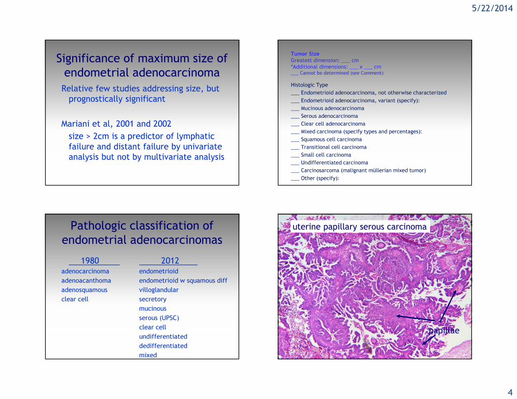
Significance of maximum size of endometrial adenocarcinomaRelative few studies addressing size, but prognostically significant
Mariani et al, 2001 and 2002 size > 2cm is a predictor of lymphatic failure and distant failure by univariate analysis but not by multivariate analysis
Tumor SizeGreatest dimension: ___ cm*Additional dimensions: ___ x ___ cm___ Cannot be determined (see Comment)
FIGO 1988 Stage I Corpus Cancer1) Is the distinction of non-invasive from inner half invasion reliable?
2) Should invasion be assessed in thirds or halves of myometrial thickness?
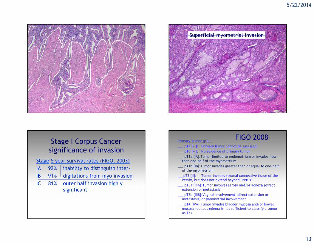
Superficial myometrial invasion
endometriummyometrium
5/22/2014
13
Superficial myometrial invasion
Stage I Corpus Cancersignificance of invasion
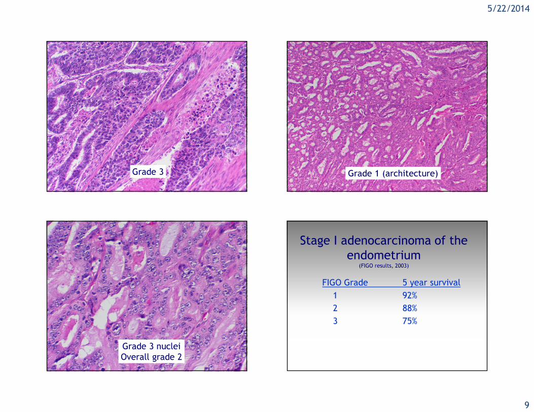
Stage 5 year survival rates (FIGO, 2003)IA 92% inability to distinguish inter-IB 91% digitations from myo invasionIC 81% outer half invasion highly
significant
Primary Tumor (pT)___ pTX [--]: Primary tumor cannot be assessed___ pT0 [--]: No evidence of primary tumor___ pT1a [IA]:Tumor limited to endometrium or invades less
than one-half of the myometrium ___ pT1b [IB]:Tumor invades greater than or equal to one-half
of the myometrium___pT2 [II]: Tumor invades stromal connective tissue of the
cervix, but does not extend beyond uterus___ pT3a [IIIA]:Tumor involves serosa and/or adnexa (direct
extension or metastasis) ___ pT3b [IIIB]:Vaginal involvement (direct extension or
mucosa (bullous edema is not sufficient to classify a tumor as T4)
FIGO 2008
5/22/2014
14
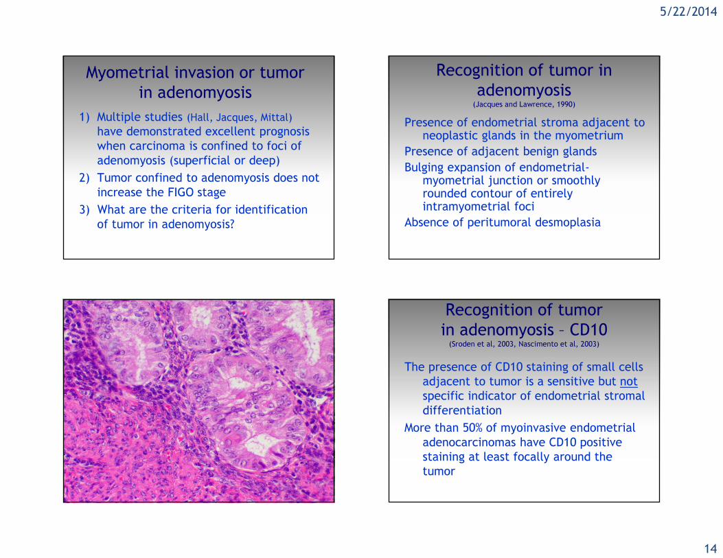
Myometrial invasion or tumor in adenomyosis
1) Multiple studies (Hall, Jacques, Mittal)have demonstrated excellent prognosis when carcinoma is confined to foci of adenomyosis (superficial or deep)
2) Tumor confined to adenomyosis does not increase the FIGO stage
3) What are the criteria for identification of tumor in adenomyosis?
Recognition of tumor in adenomyosis (Jacques and Lawrence, 1990)
Presence of endometrial stroma adjacent to neoplastic glands in the myometrium
Presence of adjacent benign glandsBulging expansion of endometrial-
myometrial junction or smoothly rounded contour of entirely intramyometrial foci
Absence of peritumoral desmoplasia
Recognition of tumorin adenomyosis – CD10(Sroden et al, 2003, Nascimento et al, 2003)
The presence of CD10 staining of small cells adjacent to tumor is a sensitive but notspecific indicator of endometrial stromal differentiation
More than 50% of myoinvasive endometrial adenocarcinomas have CD10 positive staining at least focally around the tumor
5/22/2014
15
5/22/2014
16
Difficulties in assessing the depth of myometrial invasion in endometrial carcinoma
Ali, Black and Soslow, (IJGP, 2007)Depth of invasion – disagreement in 29% of cases
sources of disagreementirregular endo-myometrial interfaceexophytic tumorsmooth muscle metaplasiatumor in adenomyosis
Most frequently - pathologists overestimate invasion
FIGO 1988Stage CharacteristicsIA G123 tumor limited to endometriumIB G123 invasion to inner half of myometriumIC G123 invasion to outer half of myometriumIIA endocervical gland involvementIIB cervical stromal invasionIIIA tumor invades serosa, adnexa, or + peritoneal cytoIIIB vaginal metastasesIIIC pelvic or para-aortic lymph node metastasesIVA tumor invades bladder or bowel mucosaIVB distant, intraabdominal or inguinal node
metastases
Primary Tumor (pT)___ pTX [--]: Primary tumor cannot be assessed___ pT0 [--]: No evidence of primary tumor___ pT1a [IA]:Tumor limited to endometrium or invades less
than one-half of the myometrium ___ pT1b [IB]:Tumor invades greater than or equal to one-half
of the myometrium___pT2 [II]: Tumor invades stromal connective tissue of the
cervix, but does not extend beyond uterus___ pT3a [IIIA]:Tumor involves serosa and/or adnexa (direct
extension or metastasis) ___ pT3b [IIIB]:Vaginal involvement (direct extension or
Stage III Corpus CancerStage III C – pelvic/paraaortic nodal mets
(Mariani et al, 2002)Stage IIIC often are also Stage IIIA/IIIB5 year DFS – 33% Stage IIIC with IIIA/B mostly extranodal failures
5 year DFS – 68% Stage IIIC without IIIA/B mostly nodal failures
Stage III Corpus CancerStage III C – pelvic/paraaortic nodal mets5 year DFS – about 65-80% + pelvic node5 year DFS – about 30% + paraaortic node
Significant survival differences between microscopic and grossly positive nodes, resected vs non-resected disease, radiated vs non-irradiated nodal beds, and capsular invasion and desmoplasia
Nodes with isolated tumor cellsVery few studiesUse of sentinel node examination currently undefined
Immunohistochemistry discloses isolated histiocyte-like cells (esp. with MELF)
Significance uncertainStaging rule is undefined
5/22/2014
21
Tentative staging conclusionsStage IA can reliably be distinguished from Stage IB pathologically
Stage II is poorly defined pathologically and may not be prognostically significant
Stage III disease is heterogeneousStage IIIA alone is heterogeneous + adnexal spread diminishes survival (70%)+ uterine serosa carries a worse prognosis (30%)
Tentative staging conclusionsStage IIIB (parametrium/vaginal mets) is rare, with a poor prognosis (25%)
Stage IIIC (good to have split)IIIC1 + pelvic nodes significant (70%)IIIC2 + paraaortic nodes significantly worse (30%)(Stage IIIC limited to nodes usually fails in nodal area)
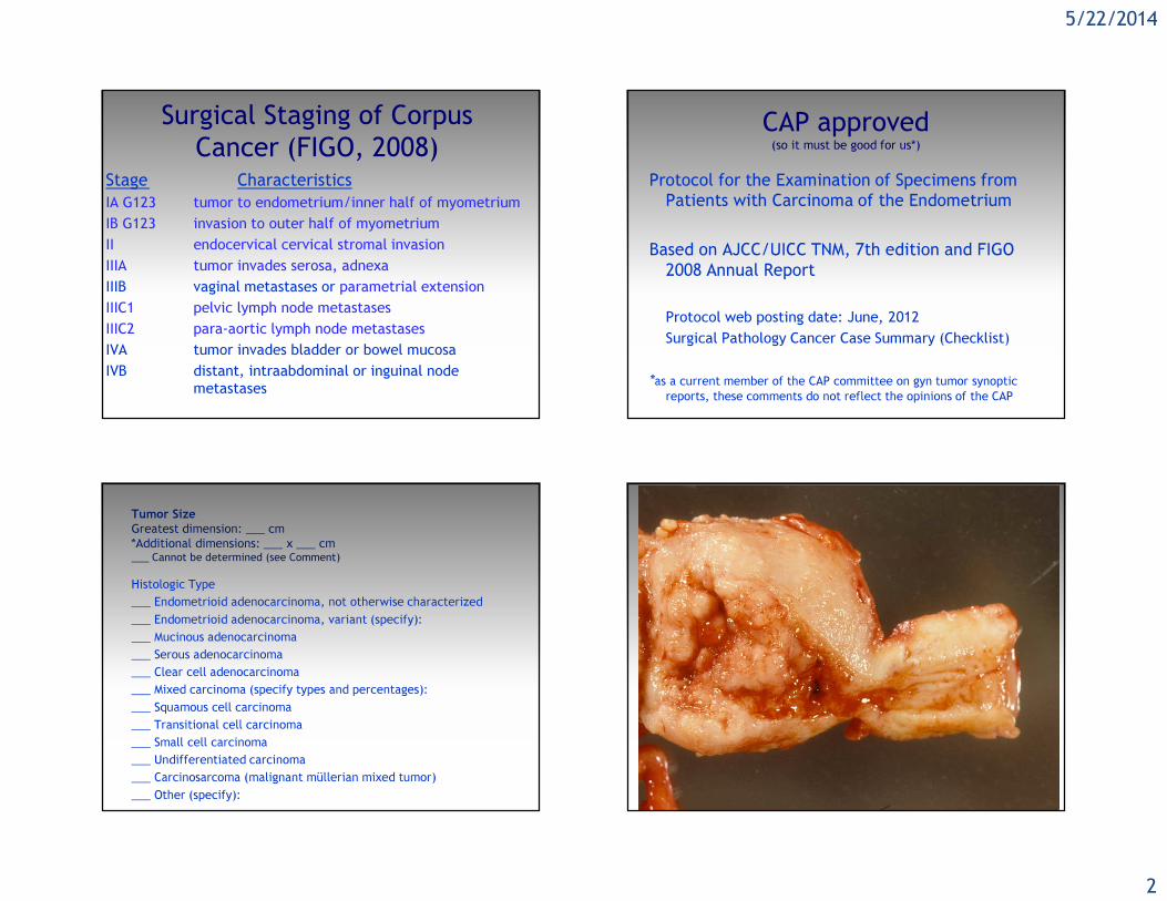
Surgical Staging of Corpus Cancer (FIGO, 2008)
Stage CharacteristicsIA G123 tumor to endometrium/inner half of myometriumIB G123 invasion to outer half of myometriumII endocervical cervical stromal invasionIIIA tumor invades serosa, adnexa IIIB vaginal metastases or parametrial extensionIIIC1 pelvic lymph node metastasesIIIC2 para-aortic lymph node metastasesIVA tumor invades bladder or bowel mucosaIVB distant, intraabdominal or inguinal node
metastases
The future?1) Pathologists need to be outspoken in the
identification of characteristics that relate to prognosis and response to therapy.
2) Future refinements are needed in identification of cell types.
3) Pathologists need to identify features that can help guide individualized treatment for endometrial carcinoma
5/22/2014
22
Objectives1) Examine the changes and utility of the
2008 FIGO staging scheme for endometrial cancer
2) Examine the application of and significance of the CAP template for endometrial cancer
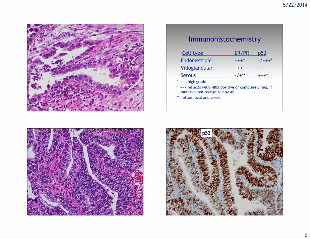
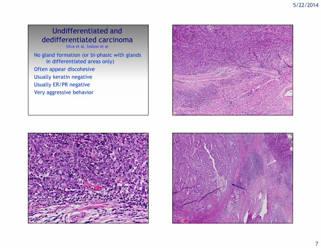
3) Review the biology of the major types of endometrial adenocarcinoma
4) Examine prognostic factors in endometrial carcinoma